Embed Size (px)
Citation preview
16 SEER Summary Staging Manual - 2000
DEFINITION OF ANATOMIC SITES WITHIN THE HEAD AND NECKadapted from the Summary Staging Guide 1977 published by the SEER Program,
and the AJCC Cancer Staging Manual Fifth Edition published bythe American Joint Committee on Cancer Staging.
Note: Not all sites in the lip, oral cavity, pharynx and salivary glands are listed below.All sites to which a Summary Stage scheme applies are listed at the begining of the scheme.
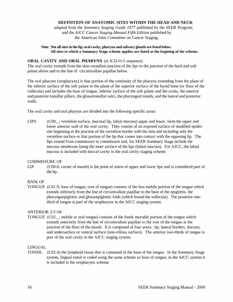
ORAL CAVITY AND ORAL PHARYNX (in ICD-O-3 sequence)The oral cavity extends from the skin-vermilion junction of the lips to the junction of the hard and softpalate above and to the line of circumvallate papillae below.
The oral pharynx (oropharynx) is that portion of the continuity of the pharynx extending from the plane ofthe inferior surface of the soft palate to the plane of the superior surface of the hyoid bone (or floor of thevallecula) and includes the base of tongue, inferior surface of the soft palate and the uvula, the anteriorand posterior tonsillar pillars, the glossotonsillar sulci, the pharyngeal tonsils, and the lateral and posteriorwalls.
The oral cavity and oral pharynx are divided into the following specific areas:
LIPS (C00._; vermilion surface, mucosal lip, labial mucosa) upper and lower, form the upper andlower anterior wall of the oral cavity. They consist of an exposed surface of modified epider-mis beginning at the junction of the vermilion border with the skin and including only thevermilion surface or that portion of the lip that comes into contact with the opposing lip. Thelips extend from commissure to commissure and, for SEER Summary Stage include themucous membrane lining the inner surface of the lips (labial mucosa). For AJCC, the labialmucosa is included with buccal cavity in the oral cavity staging scheme.
COMMISSURE OFLIP (C00.6; corner of mouth) is the point of union of upper and lower lips and is considered part of
the lip.
BASE OFTONGUE (C01.9; base of tongue, root of tongue) consists of the less mobile portion of the tongue which
extends inferiorly from the line of circumvallate papillae to the base of the epiglottis, thepharyngoepiglottic and glossoepiglottic folds (which bound the vallecula). The posterior one-third of tongue is part of the oropharynx in the AJCC staging system.
ANTERIOR 2/3 OFTONGUE (C02._; mobile or oral tongue) consists of the freely movable portion of the tongue which
extends anteriorly from the line of circumvallate papillae to the root of the tongue at thejunction of the floor of the mouth. It is composed of four areas: tip, lateral borders, dorsum,and undersurface or ventral surface (non-villous surface). The anterior two-thirds of tongue ispart of the oral cavity in the AJCC staging system.
LINGUALTONSIL (C02.4) the lymphoid tissue that is contained in the base of the tongue. In the Summary Stage
system, lingual tonsil is coded using the same scheme as base of tongue; in the AJCC system itis included in the oropharynx scheme.
17SEER Summary Staging Manual - 2000
UPPERGUM (C03.0; upper alveolar ridge) is the covering mucosa of the alveolar process of the maxilla,
extending from the line of attachment of mucosa in the upper gingival buccal gutter to thejunction of the hard palate. Its posterior margin is the upper end of the pterygopalatine arch.The gingiva is part of the oral cavity in the AJCC staging system.
LOWERGUM (C03.1; lower alveolar ridge) includes the alveolar process of the mandible and its covering
mucosa, which extends from the line of attachment of mucosa in the buccal gutter to the lineof free mucosa of the floor of the mouth. Posteriorly it extends to the ascending ramus of themandible (see retromolar trigone). The gingiva is part of the AJCC oral cavity staging system.
FLOOR OFMOUTH (C04._) consists of a semilunar shaped space over the mylohyoid and hypoglossus muscles,
extending from the inner surface of the lower alveolar ridge to the undersurface of the tongue.Its posterior boundary is the base of the anterior pillar of the tonsil. It is divided into two sidesby the frenulum of the tongue and contains the ostia of the submaxillary and lingual salivaryglands. The floor of mouth is part of the AJCC oral cavity staging system.
HARDPALATE (C05.0) consists of the semilunar area between the upper alveolar ridges and the mucous
membrane covering the palatine process of maxillary palatine bones. It extends from the innersurface of the superior alveolar ridge to the posterior edge of the palatine bone. In the TNMsystem, the hard palate is part of the oral cavity staging scheme.
SOFTPALATE (C05.1) consists of mucosa covering the oral cavity side of the palatine muscles and extends
from the posterior edge of the hard palate to the free border of the soft palate and includes theuvula. Its superior lateral margin is the pterygomandibular raphe. The inferior lateral margincompletes the faucial arch (glossopalatine arch) and includes the anterior surface of the of theanterior tonsillar pillar. In the AJCC system, the soft palate is part of the oropharynx stagingsystem.
UVULA (C05.2) is a soft tissue projection on the free border of the soft palate in the midline of thebody. In Summary Stage, the uvula is coded using the same scheme as the soft palate. In theAJCC system, the uvula is part of the oropharynx staging system.
OTHERMOUTH (C05.8-C05.9, C06.8-C06.9) includes overlapping lesions of the palate, overlapping lesions of
other and unspecified parts of mouth, and non-specific terms roof of mouth (palate, NOS);mouth, NOS (oral cavity, oral mucosa, buccal cavity); and minor salivary gland, NOS. All ofthese non-specific sites are included in the oral cavity scheme of the AJCC staging system.
CHEEKMUCOSA (C06.0) includes all the mucous membrane lining the inner surface of the cheek. In ICD-O-3
and the Summary Stage system, buccal mucosa includes the inner surface of the cheeks butnot the inner mucosal surface of the lips. In the AJCC staging system, the inner mucosa of thelips is included with the buccal mucosa in the oral cavity scheme.
18 SEER Summary Staging Manual - 2000
VESTIBULE OFMOUTH (C06.1; buccal sulcus, alveolar sulcus, labial sulcus) the space between the teeth and the lips
or cheeks and the mucosa that covers it. In the Summary Stage system, the vestibule ofmouth is included in the coding scheme for cheek (buccal) mucosa; in the AJCC stagingsystem, it is included in the oral cavity scheme.
RETROMOLARAREA (C06.2; retromolar triangle, retromolar gingiva, retromolar area) the attached mucosa overly-
ing the ascending ramus of the mandible from the level of the posterior surface of the lastmolar tooth to the apex superiorly. The retromolar trigone is coded using the same SummaryStage scheme as the gingiva or gums. It is part of the oral cavity staging scheme in the AJCCsystem.
TONSILS are the mucosa-covered lymphoid tissues lying between the palatoglossal and palatopharngealarches on the sidewalls of the oropharynx (palatine tonsils, C09.9), on the posterior wall of thenasopharynx (pharyngeal tonsils or adenoids (C11.1) and embedded in the base of the tongue(lingual tonsil, C02.4; described above). These three areas appear to form a ring of lymphoidtissue around the pharynx, which is referred to as Waldeyer’s ring (C14.2).
PAROTID GLAND AND OTHER MAJOR SALIVARY GLANDSThe parotid glands (C07.9) and the other major salivary glands, submandibular (submaxillary) (C08.0)and sublingual/submental (C08.1) are paired glands lying along the mandible and beneath the floor of themouth which produce serous or mucous secretions to moisten the mouth and begin the process of diges-tion.
OROPHARYNXANTERIORWALL consists of the pharyngoepiglottic and glossoepiglottic folds which bound the vallecula
(C10.0), and the lingual (anterior) surface of the epiglottis (C10.1). The vallecula is thehollow or sulcus formed at the junction of the base of the tongue and the epiglottis.
LATERALWALL (C10.2) includes the tonsillar pillars (C09.1), tonsillar fossae (C09.0), and tonsils (C09.9) of
the oropharynx.
POSTERIORWALL (C10.3) extends from the free borders of the soft palate to the tip of the epiglottis in the
oropharynx.
19SEER Summary Staging Manual - 2000
NASOPHARYNXThe nasopharynx begins anteriorly at the posterior choana and extends along the plane of the airway tothe level of the free border of the soft palate. It includes the vault, floor (superior surface of soft palate),posterior wall, lateral walls including the fossae of Rosenmuller and the mucosa covering the torustubarious forming the eustachian tube orifice. According to the AJCC, the posterior margins of thechoanal orifices and of the nasal septum are included in the nasal fossa (which has no TNM scheme), andare excluded from the nasopharynx staging system. However, all subsites listed above (except nasalfossa) are included in the nasopharynx Summary Stage scheme. Specific anatomic descriptions of majornasopharyngeal subsites include:
SUPERIOR, POSTERIORWALL (C11.0— superior, C11.1— posterior; vault) extends from the superior border of the choana to
the level of the free border of the soft palate. The lateral limit is the groove between thelateral wall and the base of the skull.
LATERALWALL (C11.2) extends from the base of the skull on each side to the level of the free border of the
soft palate. It includes Rosenmuller fossae (pharyngeal recesses).
HYPOPHARYNXThe hypopharynx is that portion of the pharyx extending from the plane of the superior border of the hyoidbone (or floor of the vallecula) to the plane corresponding to the lower border of the cricoid cartilage andincludes the pyriform fossae, the lateral and posterior hypopharyngeal walls and the postcricoid region.
PYRIFORMSINUS (C12.9; pyriform fossa) extends from the pharyngoepiglottic fold to the upper edge of the
esophagus at the lower border of the cricoid cartilage and is bounded laterally by the innersurface of the thyroid cartilage and medially by the hypopharyngeal surface of the aryepiglotticfold, posterior lateral surface of the arytenoid and cricoid cartilages.
POSTCRICOIDAREA (C13.0; postcricoid region, cricopharynx) extends from the posterior surface of the arytenoid
cartilages and their connecting folds to the inferior surface of the cricoid cartilage and con-nects the two pyriform sinuses. The lateral margin is the anterior part of the pyriform sinus.
POSTERIOR PHARYNGEALWALL (C13.2) extends from the superior level of the hyoid bone (or floor of the vallecula) to the
inferior margin of the cricoid cartilage, and from the apex of one pyriform sinus to the other.
20 SEER Summary Staging Manual - 2000
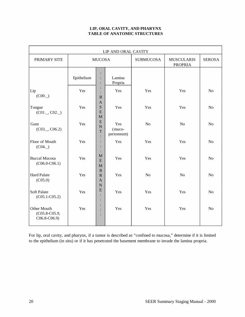
LIP, ORAL CAVITY, AND PHARYNXTABLE OF ANATOMIC STRUCTURES
LIP AND ORAL CAVITY
PRIMARY SITE MUCOSA SUBMUCOSA MUSCULARISPROPRIA
SEROSA
Epithelium:::::BASEMENT::::
MEMBRANE:::::
LaminaPropria
Lip (C00._)
Yes Yes Yes Yes No
Tongue (C01._, C02._)
Yes Yes Yes Yes No
Gum (C03._, C06.2)
Yes Yes(muco-
periosteum)
No No No
Floor of Mouth (C04._)
Yes Yes Yes Yes No
Buccal Mucosa (C06.0-C06.1)
Yes Yes Yes Yes No
Hard Palate (C05.0)
Yes Yes No No No
Soft Palate (C05.1-C05.2)
Yes Yes Yes Yes No
Other Mouth (C05.8-C05.9,
C06.8-C06.9)
Yes Yes Yes Yes No
For lip, oral cavity, and pharynx, if a tumor is described as “confined to mucosa,” determine if it is limitedto the epithelium (in situ) or if it has penetrated the basement membrane to invade the lamina propria.
21SEER Summary Staging Manual - 2000
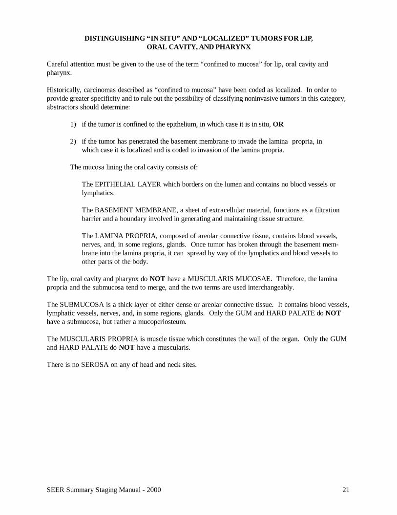
DISTINGUISHING “IN SITU” AND “LOCALIZED” TUMORS FOR LIP,ORAL CAVITY, AND PHARYNX
Careful attention must be given to the use of the term “confined to mucosa” for lip, oral cavity andpharynx.
Historically, carcinomas described as “confined to mucosa” have been coded as localized. In order toprovide greater specificity and to rule out the possibility of classifying noninvasive tumors in this category,abstractors should determine:
1) if the tumor is confined to the epithelium, in which case it is in situ, OR
2) if the tumor has penetrated the basement membrane to invade the lamina propria, inwhich case it is localized and is coded to invasion of the lamina propria.
The mucosa lining the oral cavity consists of:
The EPITHELIAL LAYER which borders on the lumen and contains no blood vessels orlymphatics.
The BASEMENT MEMBRANE, a sheet of extracellular material, functions as a filtrationbarrier and a boundary involved in generating and maintaining tissue structure.
The LAMINA PROPRIA, composed of areolar connective tissue, contains blood vessels,nerves, and, in some regions, glands. Once tumor has broken through the basement mem-brane into the lamina propria, it can spread by way of the lymphatics and blood vessels toother parts of the body.
The lip, oral cavity and pharynx do NOT have a MUSCULARIS MUCOSAE. Therefore, the laminapropria and the submucosa tend to merge, and the two terms are used interchangeably.
The SUBMUCOSA is a thick layer of either dense or areolar connective tissue. It contains blood vessels,lymphatic vessels, nerves, and, in some regions, glands. Only the GUM and HARD PALATE do NOThave a submucosa, but rather a mucoperiosteum.
The MUSCULARIS PROPRIA is muscle tissue which constitutes the wall of the organ. Only the GUMand HARD PALATE do NOT have a muscularis.
There is no SEROSA on any of head and neck sites.
22 SEER Summary Staging Manual - 2000
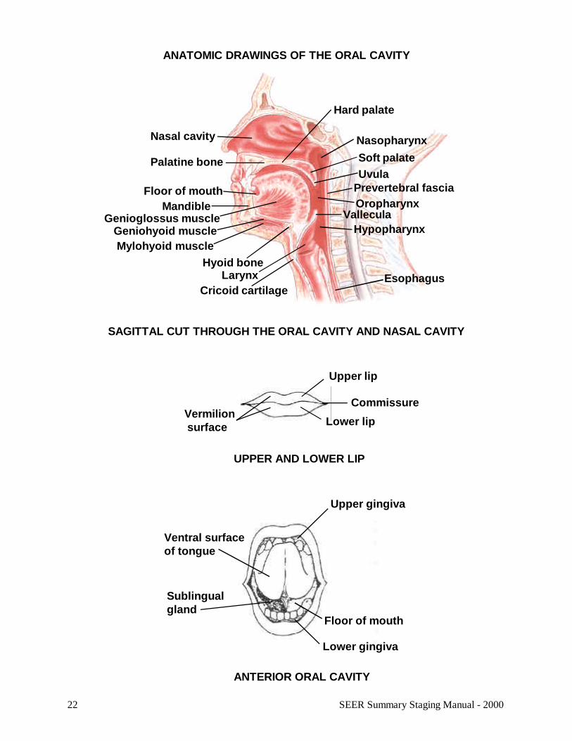
ANATOMIC DRAWINGS OF THE ORAL CAVITY
UPPER AND LOWER LIP
Vermilion surface
Upper lip
Lower lip
Commissure
ANTERIOR ORAL CAVITY
Upper gingiva
Ventral surfaceof tongue
Sublingualgland
Floor of mouth
Lower gingiva
SAGITTAL CUT THROUGH THE ORAL CAVITY AND NASAL CAVITY
Soft palate
Nasal cavity
Palatine bone
Floor of mouthMandible
Nasopharynx
Hard palate
Uvula
LarynxCricoid cartilage
Oropharynx
HypopharynxGenioglossus muscle
Geniohyoid muscleMylohyoid muscle
Hyoid bone
Vallecula
Esophagus
Prevertebral fascia
23SEER Summary Staging Manual - 2000
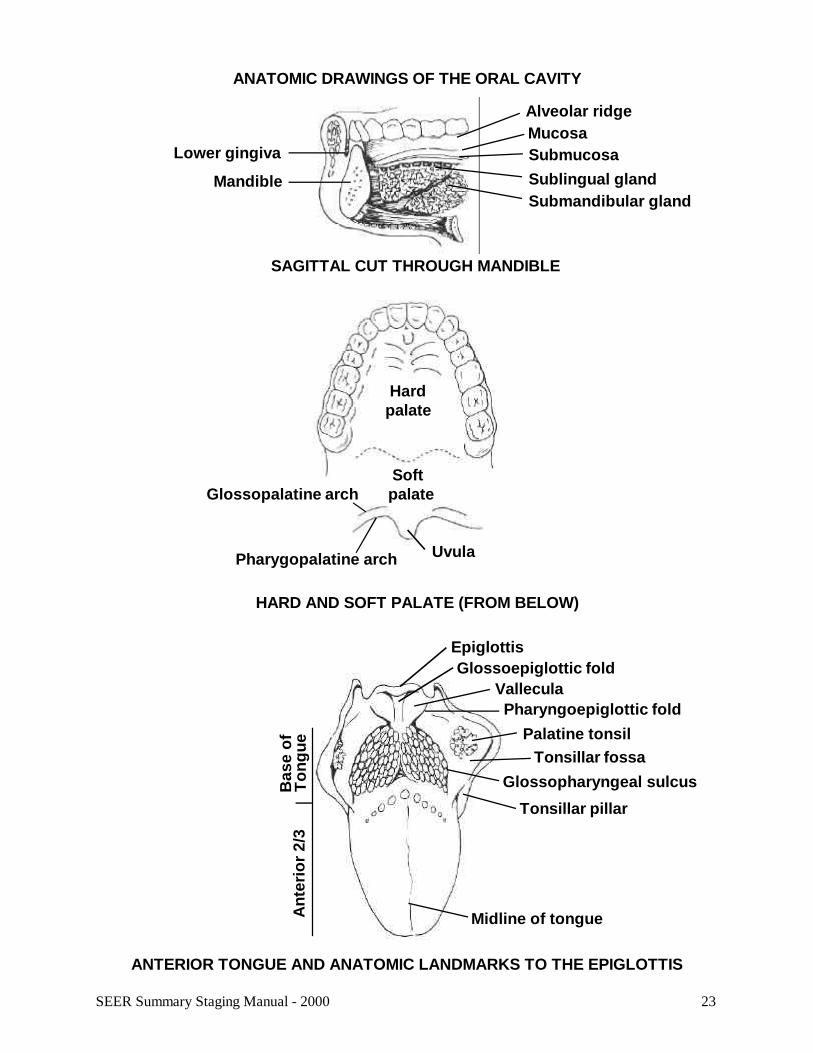
ANATOMIC DRAWINGS OF THE ORAL CAVITY
Glossopharyngeal sulcus
HARD AND SOFT PALATE (FROM BELOW)
Hardpalate
Softpalate
Uvula
Glossopalatine arch
Pharygopalatine arch
SAGITTAL CUT THROUGH MANDIBLE
Lower gingiva
Mandible
Alveolar ridgeMucosaSubmucosaSublingual glandSubmandibular gland
EpiglottisGlossoepiglottic fold
ValleculaPharyngoepiglottic fold
Palatine tonsilTonsillar fossa
Tonsillar pillar
Midline of tongueAnt
erio
r 2/
3B
ase
ofT
ongu
e
ANTERIOR TONGUE AND ANATOMIC LANDMARKS TO THE EPIGLOTTIS
24 SEER Summary Staging Manual - 2000
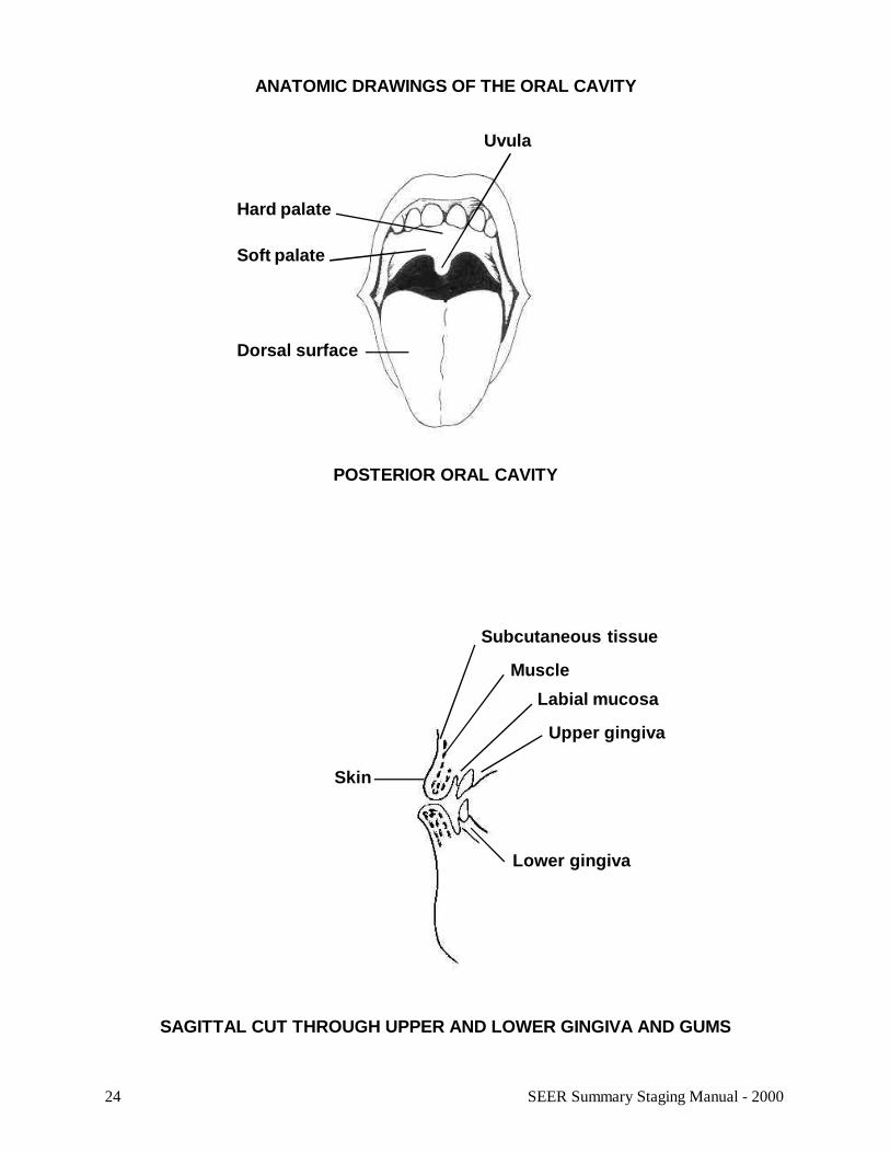
ANATOMIC DRAWINGS OF THE ORAL CAVITY
SAGITTAL CUT THROUGH UPPER AND LOWER GINGIVA AND GUMS
Subcutaneous tissue
Skin
Muscle
Labial mucosa
Upper gingiva
Lower gingiva
POSTERIOR ORAL CAVITY
Uvula
Hard palate
Soft palate
Dorsal surface

25SEER Summary Staging Manual - 2000
26 SEER Summary Staging Manual - 2000
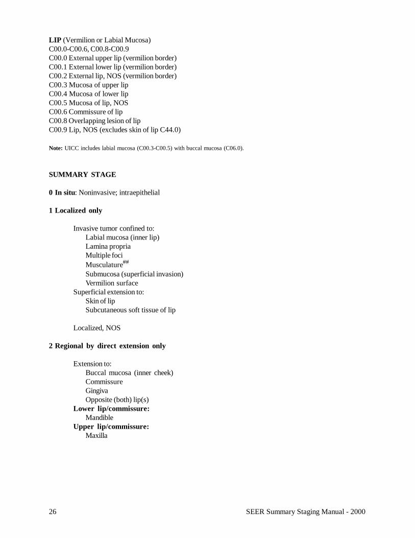
LIP (Vermilion or Labial Mucosa)C00.0-C00.6, C00.8-C00.9C00.0 External upper lip (vermilion border)C00.1 External lower lip (vermilion border)C00.2 External lip, NOS (vermilion border)C00.3 Mucosa of upper lipC00.4 Mucosa of lower lipC00.5 Mucosa of lip, NOSC00.6 Commissure of lipC00.8 Overlapping lesion of lipC00.9 Lip, NOS (excludes skin of lip C44.0)
Note: UICC includes labial mucosa (C00.3-C00.5) with buccal mucosa (C06.0).
SUMMARY STAGE
0 In situ: Noninvasive; intraepithelial
1 Localized only
Invasive tumor confined to:Labial mucosa (inner lip)Lamina propriaMultiple fociMusculature##
Submucosa (superficial invasion)Vermilion surface
Superficial extension to:Skin of lipSubcutaneous soft tissue of lip
Localized, NOS
2 Regional by direct extension only
Extension to:Buccal mucosa (inner cheek)CommissureGingivaOpposite (both) lip(s)
Lower lip/commissure:Mandible
Upper lip/commissure:Maxilla
27SEER Summary Staging Manual - 2000
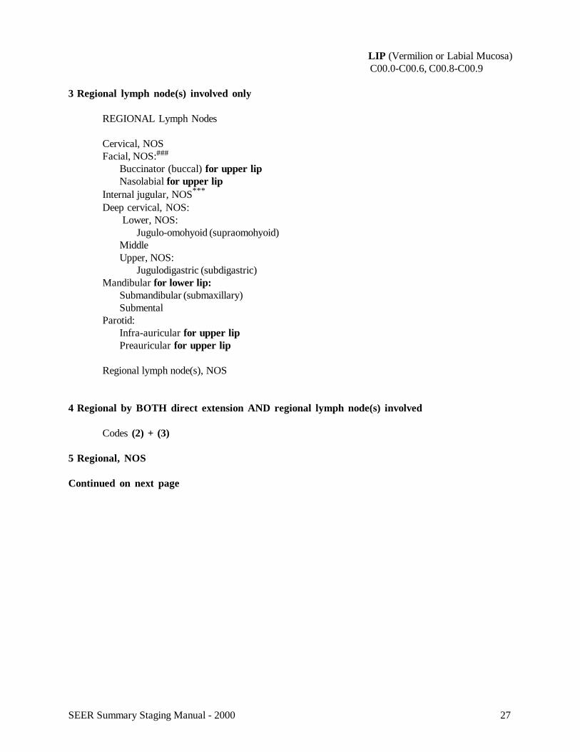
LIP (Vermilion or Labial Mucosa)C00.0-C00.6, C00.8-C00.9
3 Regional lymph node(s) involved only
REGIONAL Lymph Nodes
Cervical, NOSFacial, NOS:###
Buccinator (buccal) for upper lipNasolabial for upper lip
Internal jugular, NOS***
Deep cervical, NOS: Lower, NOS:
Jugulo-omohyoid (supraomohyoid)MiddleUpper, NOS:
Jugulodigastric (subdigastric)Mandibular for lower lip:
Submandibular (submaxillary)Submental
Parotid:Infra-auricular for upper lipPreauricular for upper lip
Regional lymph node(s), NOS
4 Regional by BOTH direct extension AND regional lymph node(s) involved
Codes (2) + (3)
5 Regional, NOS
Continued on next page
28 SEER Summary Staging Manual - 2000
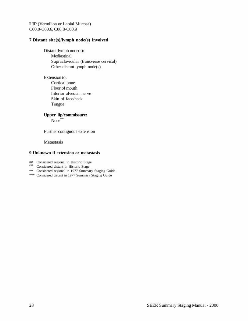
LIP (Vermilion or Labial Mucosa)C00.0-C00.6, C00.8-C00.9
7 Distant site(s)/lymph node(s) involved
Distant lymph node(s):MediastinalSupraclavicular (transverse cervical)Other distant lymph node(s)
Extension to:Cortical boneFloor of mouthInferior alveolar nerveSkin of face/neckTongue
Upper lip/commissure:Nose**
Further contiguous extension
Metastasis
9 Unknown if extension or metastasis
## Considered regional in Historic Stage### Considered distant in Historic Stage** Considered regional in 1977 Summary Staging Guide*** Considered distant in 1977 Summary Staging Guide

29SEER Summary Staging Manual - 2000
30 SEER Summary Staging Manual - 2000
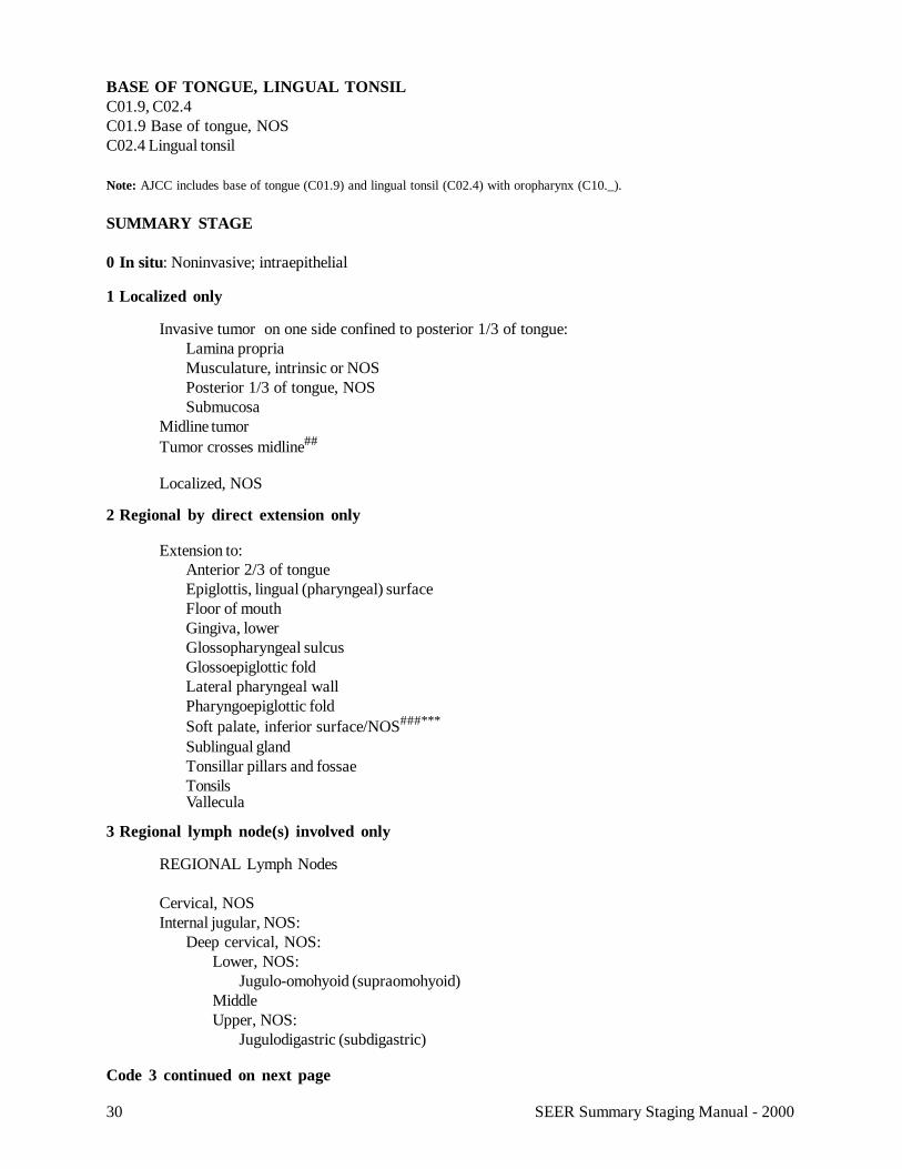
BASE OF TONGUE, LINGUAL TONSILC01.9, C02.4C01.9 Base of tongue, NOSC02.4 Lingual tonsil
Note: AJCC includes base of tongue (C01.9) and lingual tonsil (C02.4) with oropharynx (C10._).
SUMMARY STAGE
0 In situ: Noninvasive; intraepithelial
1 Localized only
Invasive tumor on one side confined to posterior 1/3 of tongue:Lamina propriaMusculature, intrinsic or NOSPosterior 1/3 of tongue, NOSSubmucosa
Midline tumorTumor crosses midline##
Localized, NOS
2 Regional by direct extension only
Extension to:Anterior 2/3 of tongueEpiglottis, lingual (pharyngeal) surfaceFloor of mouthGingiva, lowerGlossopharyngeal sulcusGlossoepiglottic foldLateral pharyngeal wallPharyngoepiglottic foldSoft palate, inferior surface/NOS###***
Sublingual glandTonsillar pillars and fossaeTonsilsVallecula
3 Regional lymph node(s) involved only
REGIONAL Lymph Nodes
Cervical, NOSInternal jugular, NOS:
Deep cervical, NOS:Lower, NOS:
Jugulo-omohyoid (supraomohyoid)MiddleUpper, NOS:
Jugulodigastric (subdigastric)
Code 3 continued on next page
31SEER Summary Staging Manual - 2000
BASE OF TONGUE, LINGUAL TONSILC01.9, C02.4
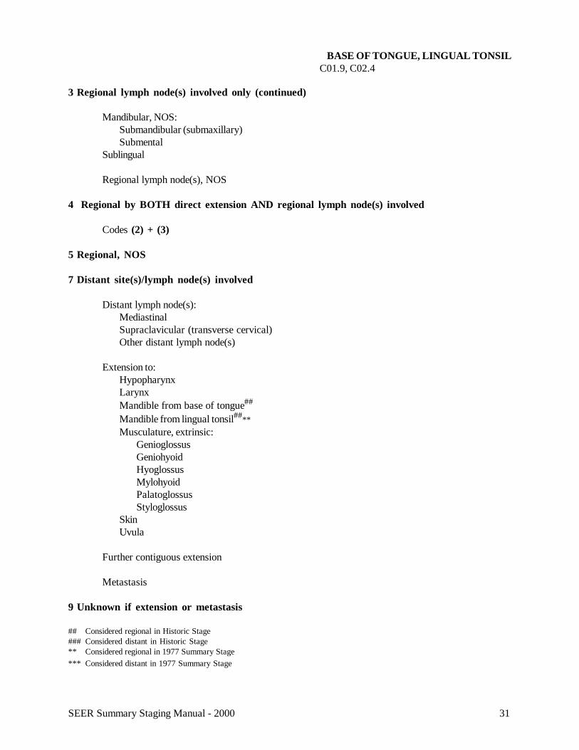
3 Regional lymph node(s) involved only (continued)
Mandibular, NOS:Submandibular (submaxillary)Submental
Sublingual
Regional lymph node(s), NOS
4 Regional by BOTH direct extension AND regional lymph node(s) involved
Codes (2) + (3)
5 Regional, NOS
7 Distant site(s)/lymph node(s) involved
Distant lymph node(s):MediastinalSupraclavicular (transverse cervical)Other distant lymph node(s)
Extension to:HypopharynxLarynxMandible from base of tongue##
Mandible from lingual tonsil##**Musculature, extrinsic:
GenioglossusGeniohyoidHyoglossusMylohyoidPalatoglossusStyloglossus
SkinUvula
Further contiguous extension
Metastasis
9 Unknown if extension or metastasis
## Considered regional in Historic Stage### Considered distant in Historic Stage** Considered regional in 1977 Summary Stage*** Considered distant in 1977 Summary Stage
32 SEER Summary Staging Manual - 2000
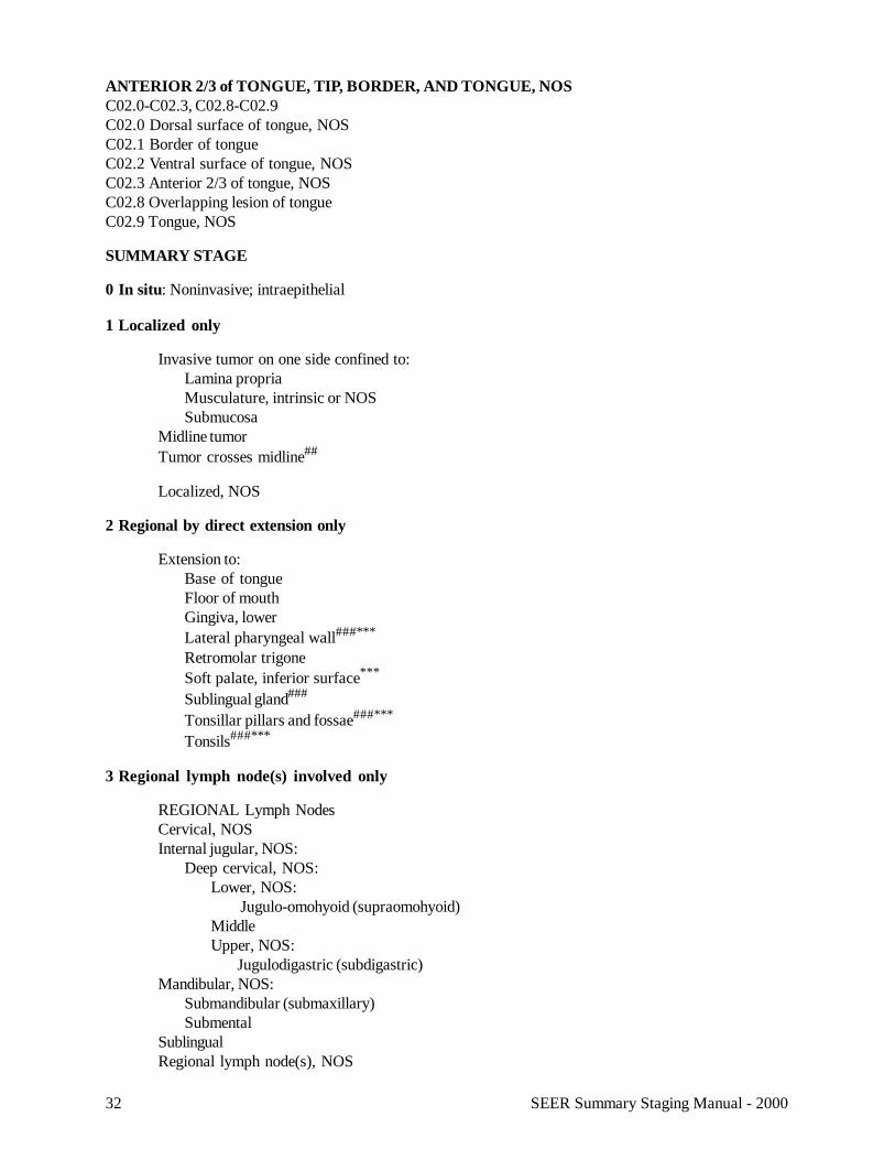
ANTERIOR 2/3 of TONGUE, TIP, BORDER, AND TONGUE, NOSC02.0-C02.3, C02.8-C02.9C02.0 Dorsal surface of tongue, NOSC02.1 Border of tongueC02.2 Ventral surface of tongue, NOSC02.3 Anterior 2/3 of tongue, NOSC02.8 Overlapping lesion of tongueC02.9 Tongue, NOS
SUMMARY STAGE
0 In situ: Noninvasive; intraepithelial
1 Localized only
Invasive tumor on one side confined to:Lamina propriaMusculature, intrinsic or NOSSubmucosa
Midline tumorTumor crosses midline##
Localized, NOS
2 Regional by direct extension only
Extension to:Base of tongueFloor of mouthGingiva, lowerLateral pharyngeal wall###***
Retromolar trigoneSoft palate, inferior surface***
Sublingual gland###
Tonsillar pillars and fossae###***
Tonsils###***
3 Regional lymph node(s) involved only
REGIONAL Lymph NodesCervical, NOSInternal jugular, NOS:
Deep cervical, NOS:Lower, NOS:
Jugulo-omohyoid (supraomohyoid)MiddleUpper, NOS:
Jugulodigastric (subdigastric)Mandibular, NOS:
Submandibular (submaxillary)Submental
SublingualRegional lymph node(s), NOS
33SEER Summary Staging Manual - 2000
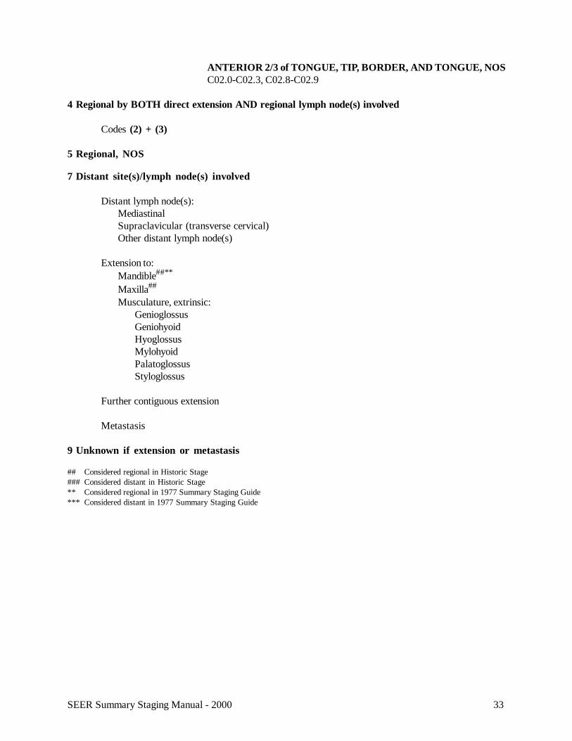
ANTERIOR 2/3 of TONGUE, TIP, BORDER, AND TONGUE, NOSC02.0-C02.3, C02.8-C02.9
4 Regional by BOTH direct extension AND regional lymph node(s) involved
Codes (2) + (3)
5 Regional, NOS
7 Distant site(s)/lymph node(s) involved
Distant lymph node(s):MediastinalSupraclavicular (transverse cervical)Other distant lymph node(s)
Extension to:Mandible##**
Maxilla##
Musculature, extrinsic:GenioglossusGeniohyoidHyoglossusMylohyoidPalatoglossusStyloglossus
Further contiguous extension
Metastasis
9 Unknown if extension or metastasis
## Considered regional in Historic Stage### Considered distant in Historic Stage** Considered regional in 1977 Summary Staging Guide*** Considered distant in 1977 Summary Staging Guide
34 SEER Summary Staging Manual - 2000
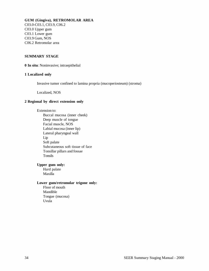
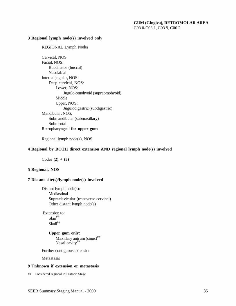
GUM (Gingiva), RETROMOLAR AREAC03.0-C03.1, C03.9, C06.2C03.0 Upper gumC03.1 Lower gumC03.9 Gum, NOSC06.2 Retromolar area
SUMMARY STAGE
0 In situ: Noninvasive; intraepithelial
1 Localized only
Invasive tumor confined to lamina propria (mucoperiosteum) (stroma)
Localized, NOS
2 Regional by direct extension only
Extension to:Buccal mucosa (inner cheek)Deep muscle of tongueFacial muscle, NOSLabial mucosa (inner lip)Lateral pharyngeal wallLipSoft palateSubcutaneous soft tissue of faceTonsillar pillars and fossaeTonsils
Upper gum only:Hard palateMaxilla
Lower gum/retromolar trigone only:Floor of mouthMandibleTongue (mucosa)Uvula
35SEER Summary Staging Manual - 2000
GUM (Gingiva), RETROMOLAR AREAC03.0-C03.1, C03.9, C06.2
3 Regional lymph node(s) involved only
REGIONAL Lymph Nodes
Cervical, NOSFacial, NOS:
Buccinator (buccal)Nasolabial
Internal jugular, NOS:Deep cervical, NOS:
Lower, NOS:Jugulo-omohyoid (supraomohyoid)
MiddleUpper, NOS:
Jugulodigastric (subdigastric)Mandibular, NOS:
Submandibular (submaxillary)Submental
Retropharyngeal for upper gum
Regional lymph node(s), NOS
4 Regional by BOTH direct extension AND regional lymph node(s) involved
Codes (2) + (3)
5 Regional, NOS
7 Distant site(s)/lymph node(s) involved
Distant lymph node(s):MediastinalSupraclavicular (transverse cervical)Other distant lymph node(s)
Extension to:Skin##
Skull##
Upper gum only:Maxillary antrum (sinus)##
Nasal cavity##
Further contiguous extension
Metastasis
9 Unknown if extension or metastasis
## Considered regional in Historic Stage
36 SEER Summary Staging Manual - 2000
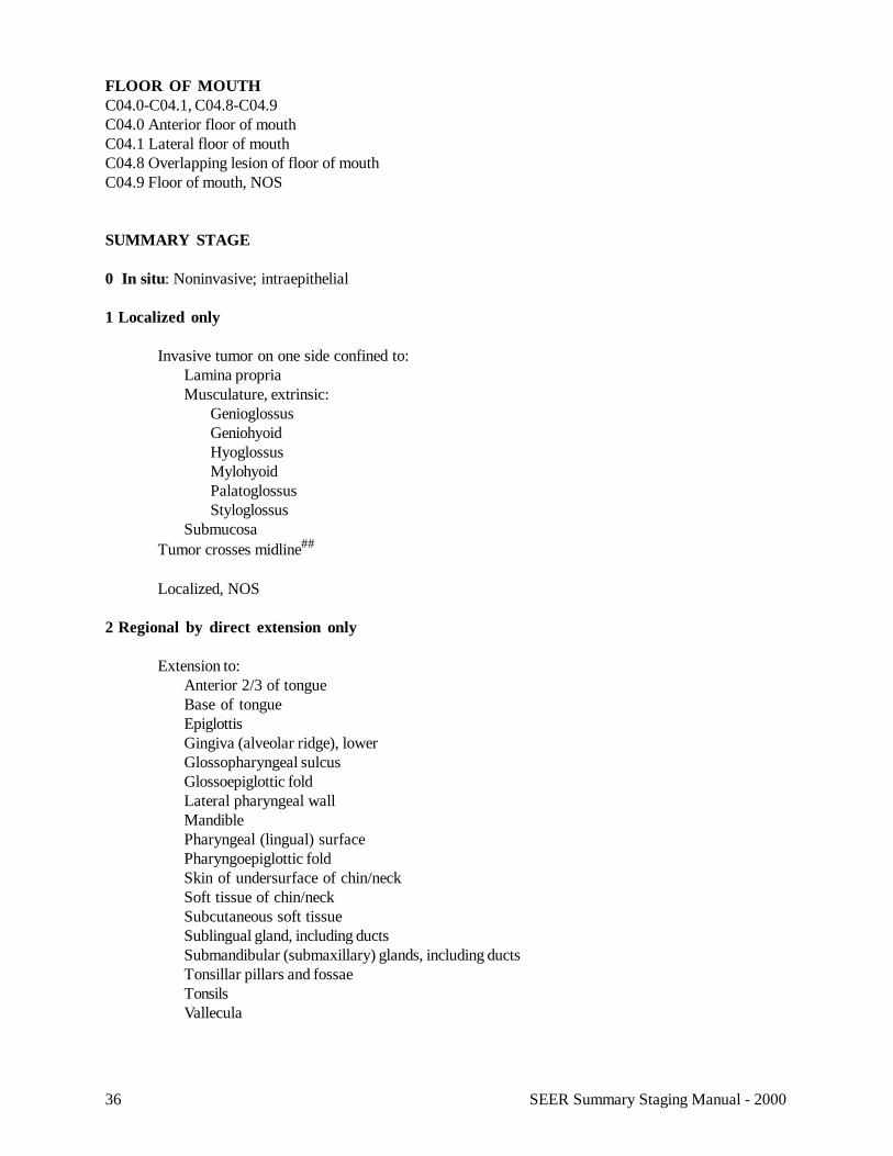
FLOOR OF MOUTHC04.0-C04.1, C04.8-C04.9C04.0 Anterior floor of mouthC04.1 Lateral floor of mouthC04.8 Overlapping lesion of floor of mouthC04.9 Floor of mouth, NOS
SUMMARY STAGE
0 In situ: Noninvasive; intraepithelial
1 Localized only
Invasive tumor on one side confined to:Lamina propriaMusculature, extrinsic:
GenioglossusGeniohyoidHyoglossusMylohyoidPalatoglossusStyloglossus
SubmucosaTumor crosses midline##
Localized, NOS
2 Regional by direct extension only
Extension to:Anterior 2/3 of tongueBase of tongueEpiglottisGingiva (alveolar ridge), lowerGlossopharyngeal sulcusGlossoepiglottic foldLateral pharyngeal wallMandiblePharyngeal (lingual) surfacePharyngoepiglottic foldSkin of undersurface of chin/neckSoft tissue of chin/neckSubcutaneous soft tissueSublingual gland, including ductsSubmandibular (submaxillary) glands, including ductsTonsillar pillars and fossaeTonsilsVallecula
37SEER Summary Staging Manual - 2000
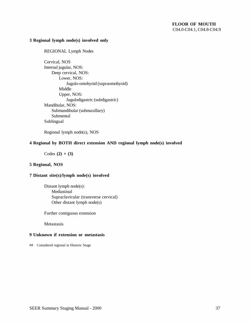
FLOOR OF MOUTHC04.0-C04.1, C04.8-C04.9
3 Regional lymph node(s) involved only
REGIONAL Lymph Nodes
Cervical, NOSInternal jugular, NOS:
Deep cervical, NOS:Lower, NOS:
Jugulo-omohyoid (supraomohyoid)MiddleUpper, NOS:
Jugulodigastric (subdigastric)Mandibular, NOS:
Submandibular (submaxillary)Submental
Sublingual
Regional lymph node(s), NOS
4 Regional by BOTH direct extension AND regional lymph node(s) involved
Codes (2) + (3)
5 Regional, NOS
7 Distant site(s)/lymph node(s) involved
Distant lymph node(s):MediastinalSupraclavicular (transverse cervical)Other distant lymph node(s)
Further contiguous extension
Metastasis
9 Unknown if extension or metastasis
## Considered regional in Historic Stage
38 SEER Summary Staging Manual - 2000
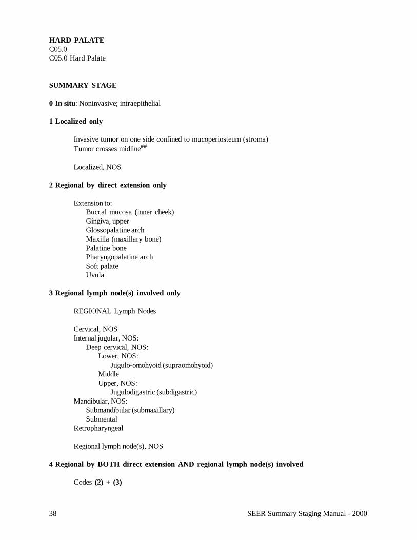
HARD PALATEC05.0C05.0 Hard Palate
SUMMARY STAGE
0 In situ: Noninvasive; intraepithelial
1 Localized only
Invasive tumor on one side confined to mucoperiosteum (stroma)Tumor crosses midline##
Localized, NOS
2 Regional by direct extension only
Extension to:Buccal mucosa (inner cheek)Gingiva, upperGlossopalatine archMaxilla (maxillary bone)Palatine bonePharyngopalatine archSoft palateUvula
3 Regional lymph node(s) involved only
REGIONAL Lymph Nodes
Cervical, NOSInternal jugular, NOS:
Deep cervical, NOS:Lower, NOS:
Jugulo-omohyoid (supraomohyoid)MiddleUpper, NOS:
Jugulodigastric (subdigastric)Mandibular, NOS:
Submandibular (submaxillary)Submental
Retropharyngeal
Regional lymph node(s), NOS
4 Regional by BOTH direct extension AND regional lymph node(s) involved
Codes (2) + (3)
39SEER Summary Staging Manual - 2000
HARD PALATEC05.0
5 Regional, NOS
7 Distant site(s)/lymph node(s) involved
Distant lymph node(s):MediastinalSupraclavicular (transverse cervical)Other distant lymph node(s)
Extension to:Floor of noseMaxillary antrum (sinus)##
Nasal cavity##
NasopharynxPterygoid plateSphenoid bone
Further contiguous extension
Metastasis
9 Unknown if extension or metastasis
## Considered regional in Historic Stage
40 SEER Summary Staging Manual - 2000
SOFT PALATE, UVULAC05.1-C05.2C05.1 Soft palate, NOSC05.2 Uvula
Note 1: AJCC includes inferior surface of the soft palate (C05.1) and uvula (C05.2) with oropharynx (C09._, C10._).Note 2: Soft palate excludes nasopharyngeal (superior) surface of soft palate (C11.3).
SUMMARY STAGE
0 In situ: Noninvasive; intraepithelial
1 Localized only
Invasive tumor on one side confined to:Lamina propriaMusculatureSubmucosa
Tumor crosses midline##
Localized, NOS
2 Regional by direct extension only
Extension to:Buccal mucosa (inner cheek)Gum (gingiva), upperHard palateLateral pharyngeal wallTonsillar pillars and fossaeTonsils
3 Regional lymph node(s) involved only
REGIONAL Lymph Nodes
Cervical, NOSInternal jugular, NOS:
Deep cervical, NOS:Lower, NOS:
Jugulo-omohyoid (supraomohyoid)MiddleUpper, NOS:
Jugulodigastric (subdigastric)Mandibular, NOS:
Submandibular (submaxillary)Submental###
Retropharyngeal###
Regional lymph node(s), NOS
41SEER Summary Staging Manual - 2000
SOFT PALATE, UVULAC05.1-C05.2
4 Regional by BOTH direct extension AND regional lymph node(s) involved
Codes (2) + (3)
5 Regional, NOS
7 Distant site(s)/lymph node(s) involved
Distant lymph node(s):MediastinalSupraclavicular (transverse cervical)Other distant lymph node(s)
Extension to:LarynxMandible##
Maxilla##
Maxillary antrum (sinus)Nasal cavity##**
Nasopharynx##
Palatine bone (bone of hard palate)##
Pterygoid muscleTongue##
Further contiguous extension
Metastasis
9 Unknown if extension or metastasis
## Considered regional in Historic Stage### Considered distant in Historic Stage** Considered regional in 1977 Summary Staging Guide
42 SEER Summary Staging Manual - 2000
CHEEK (Buccal) MUCOSA, VESTIBULEC06.0-C06.1C06.0 Cheek mucosaC06.1 Vestibule of mouth
Note: In ICD-O-3, C06.0 for buccal mucosa includes the membrane lining of the cheeks but not of the lips.(UICC includes labial mucosa with buccal mucosa.)
SUMMARY STAGE
0 In situ: Noninvasive; intraepithelial
1 Localized only
Invasive tumor confined to:Lamina propriaMusculature (buccinator)##**
Submucosa
Localized, NOS
2 Regional by direct extension only
Extension to:
GingivaLateral pharyngeal wallLip(s) including commissureSubcutaneous soft tissue of cheekTonsillar pillars and fossaeTonsils
3 Regional lymph node(s) involved only
REGIONAL Lymph Nodes
Cervical, NOSFacial: Buccinator (buccal)
NasolabialInternal jugular, NOS:
Deep cervical, NOS:Lower, NOS:
Jugulo-omohyoid (supraomohyoid)MiddleUpper, NOS:
Jugulodigastric (subdigastric)Mandibular, NOS:
Submandibular (submaxillary)Submental
Parotid, NOS:Infra-auricularPreauricular
Regional lymph node(s), NOS
43SEER Summary Staging Manual - 2000
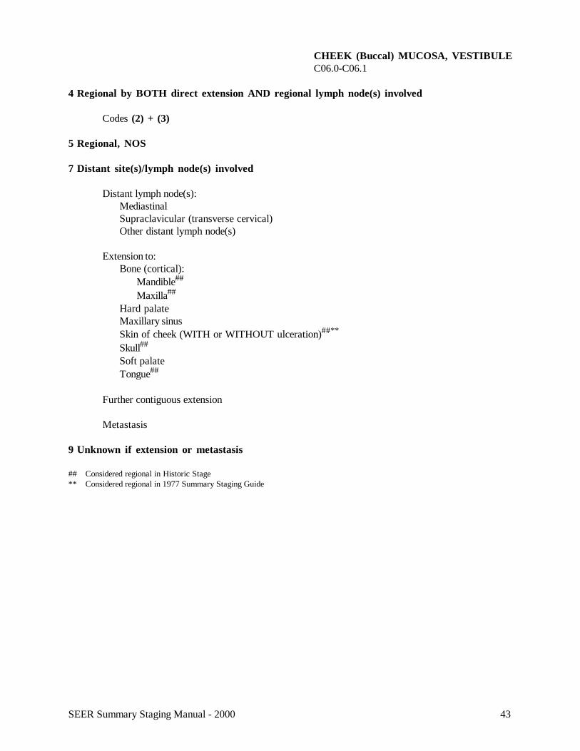
CHEEK (Buccal) MUCOSA, VESTIBULEC06.0-C06.1
4 Regional by BOTH direct extension AND regional lymph node(s) involved
Codes (2) + (3)
5 Regional, NOS
7 Distant site(s)/lymph node(s) involved
Distant lymph node(s):MediastinalSupraclavicular (transverse cervical)Other distant lymph node(s)
Extension to:Bone (cortical):
Mandible##
Maxilla##
Hard palateMaxillary sinusSkin of cheek (WITH or WITHOUT ulceration)##**
Skull##
Soft palateTongue##
Further contiguous extension
Metastasis
9 Unknown if extension or metastasis
## Considered regional in Historic Stage** Considered regional in 1977 Summary Staging Guide
44 SEER Summary Staging Manual - 2000
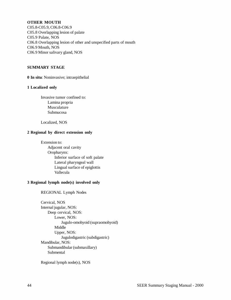
OTHER MOUTHC05.8-C05.9, C06.8-C06.9C05.8 Overlapping lesion of palateC05.9 Palate, NOSC06.8 Overlapping lesion of other and unspecified parts of mouthC06.9 Mouth, NOSC06.9 Minor salivary gland, NOS
SUMMARY STAGE
0 In situ: Noninvasive; intraepithelial
1 Localized only
Invasive tumor confined to:Lamina propriaMusculatureSubmucosa
Localized, NOS
2 Regional by direct extension only
Extension to:Adjacent oral cavityOropharynx:
Inferior surface of soft palateLateral pharyngeal wallLingual surface of epiglottisVallecula
3 Regional lymph node(s) involved only
REGIONAL Lymph Nodes
Cervical, NOSInternal jugular, NOS:
Deep cervical, NOS:Lower, NOS:
Jugulo-omohyoid (supraomohyoid)MiddleUpper, NOS:
Jugulodigastric (subdigastric)Mandibular, NOS:
Submandibular (submaxillary)Submental
Regional lymph node(s), NOS
45SEER Summary Staging Manual - 2000
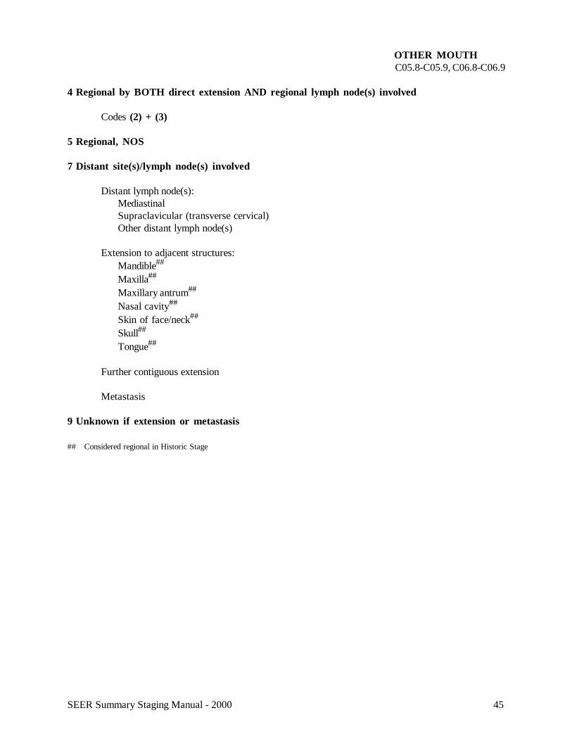
OTHER MOUTHC05.8-C05.9, C06.8-C06.9
4 Regional by BOTH direct extension AND regional lymph node(s) involved
Codes (2) + (3)
5 Regional, NOS
7 Distant site(s)/lymph node(s) involved
Distant lymph node(s):MediastinalSupraclavicular (transverse cervical)Other distant lymph node(s)
Extension to adjacent structures:Mandible##
Maxilla##
Maxillary antrum##
Nasal cavity##
Skin of face/neck##
Skull##
Tongue##
Further contiguous extension
Metastasis
9 Unknown if extension or metastasis
## Considered regional in Historic Stage
46 SEER Summary Staging Manual - 2000
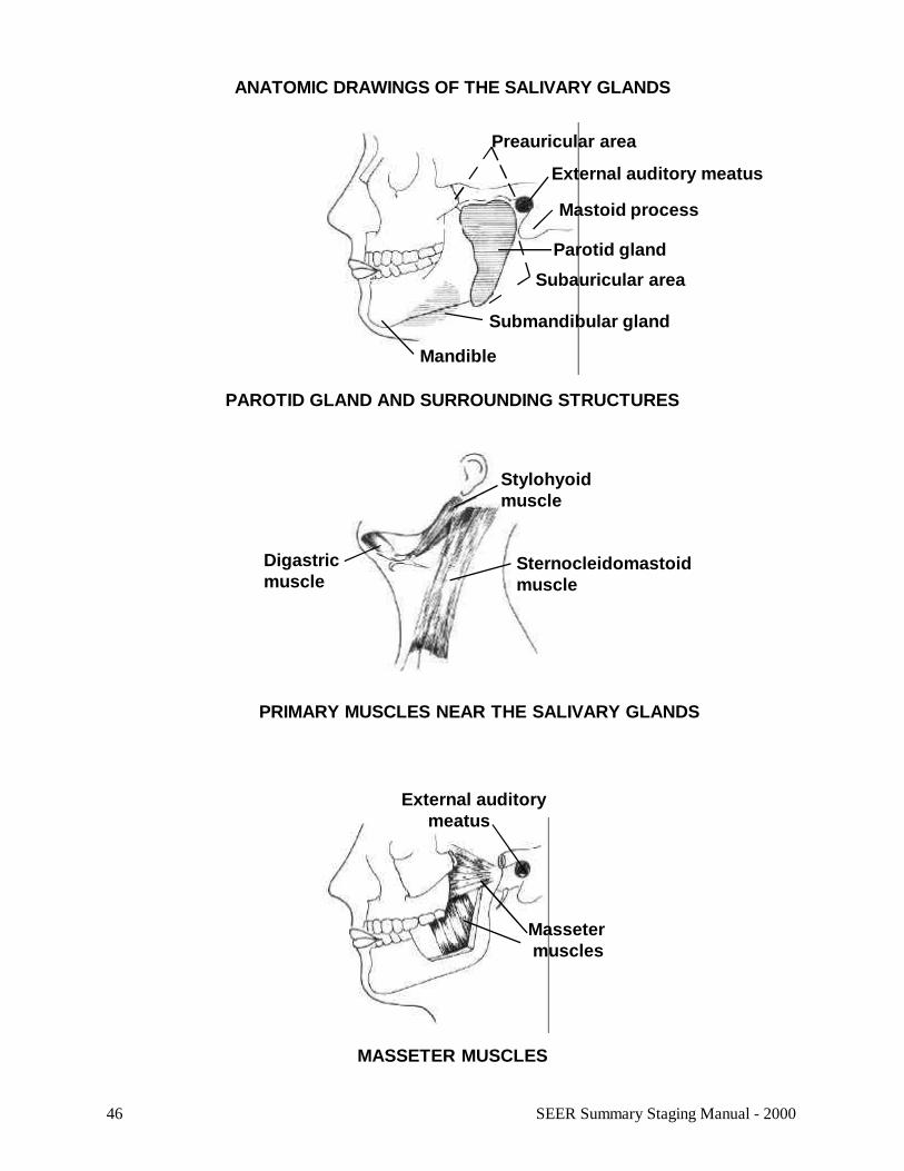
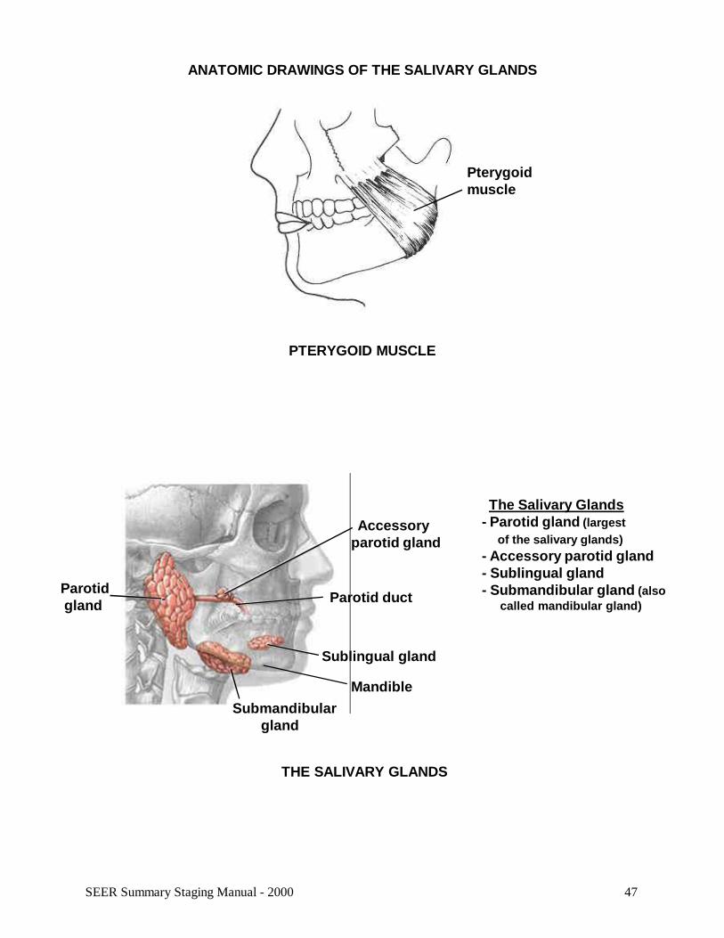
ANATOMIC DRAWINGS OF THE SALIVARY GLANDS
MASSETER MUSCLES
Stylohyoidmuscle
Sternocleidomastoidmuscle
Digastricmuscle
PRIMARY MUSCLES NEAR THE SALIVARY GLANDS
Masseter muscles
External auditory meatus
PAROTID GLAND AND SURROUNDING STRUCTURES
Mandible
Preauricular area
Mastoid process
Parotid gland
Subauricular area
Submandibular gland
External auditory meatus
47SEER Summary Staging Manual - 2000
ANATOMIC DRAWINGS OF THE SALIVARY GLANDS
THE SALIVARY GLANDS
Accessoryparotid gland
Mandible
Parotid gland Parotid duct
Sublingual gland
Submandibular gland
The Salivary Glands- Parotid gland (largest of the salivary glands)- Accessory parotid gland- Sublingual gland- Submandibular gland (also called mandibular gland)
Pterygoidmuscle
PTERYGOID MUSCLE
48 SEER Summary Staging Manual - 2000
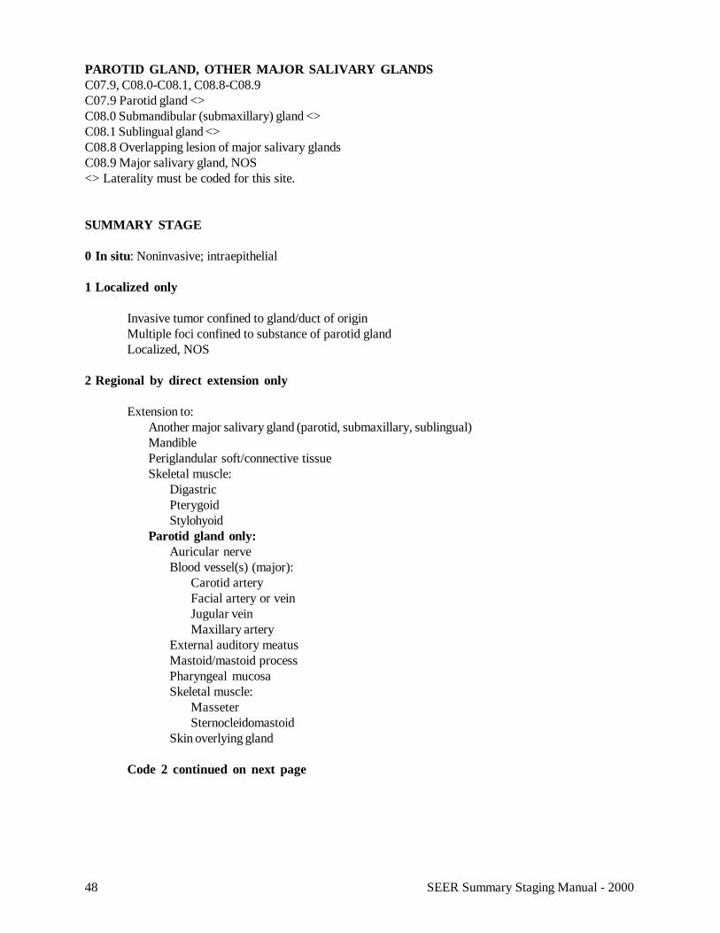
PAROTID GLAND, OTHER MAJOR SALIVARY GLANDSC07.9, C08.0-C08.1, C08.8-C08.9C07.9 Parotid gland <>C08.0 Submandibular (submaxillary) gland <>C08.1 Sublingual gland <>C08.8 Overlapping lesion of major salivary glandsC08.9 Major salivary gland, NOS<> Laterality must be coded for this site.
SUMMARY STAGE
0 In situ: Noninvasive; intraepithelial
1 Localized only
Invasive tumor confined to gland/duct of originMultiple foci confined to substance of parotid glandLocalized, NOS
2 Regional by direct extension only
Extension to:Another major salivary gland (parotid, submaxillary, sublingual)MandiblePeriglandular soft/connective tissueSkeletal muscle:
DigastricPterygoidStylohyoid
Parotid gland only:Auricular nerveBlood vessel(s) (major):
Carotid arteryFacial artery or veinJugular veinMaxillary artery
External auditory meatusMastoid/mastoid processPharyngeal mucosaSkeletal muscle:
MasseterSternocleidomastoid
Skin overlying gland
Code 2 continued on next page
49SEER Summary Staging Manual - 2000
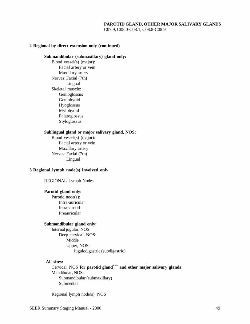
PAROTID GLAND, OTHER MAJOR SALIVARY GLANDSC07.9, C08.0-C08.1, C08.8-C08.9
2 Regional by direct extension only (continued)
Submandibular (submaxillary) gland only:Blood vessel(s) (major):
Facial artery or veinMaxillary artery
Nerves: Facial (7th)Lingual
Skeletal muscle:GenioglossusGeniohyoidHyoglossusMylohyoidPalatoglossusStyloglossus
Sublingual gland or major salivary gland, NOS:Blood vessel(s) (major):
Facial artery or veinMaxillary artery
Nerves: Facial (7th)Lingual
3 Regional lymph node(s) involved only
REGIONAL Lymph Nodes
Parotid gland only:Parotid node(s):
Infra-auricularIntraparotidPreauricular
Submandibular gland only:Internal jugular, NOS:
Deep cervical, NOS:MiddleUpper, NOS:
Jugulodigastric (subdigastric)
All sites:Cervical, NOS for parotid gland*** and other major salivary glandsMandibular, NOS:
Submandibular (submaxillary)Submental
Regional lymph node(s), NOS
50 SEER Summary Staging Manual - 2000
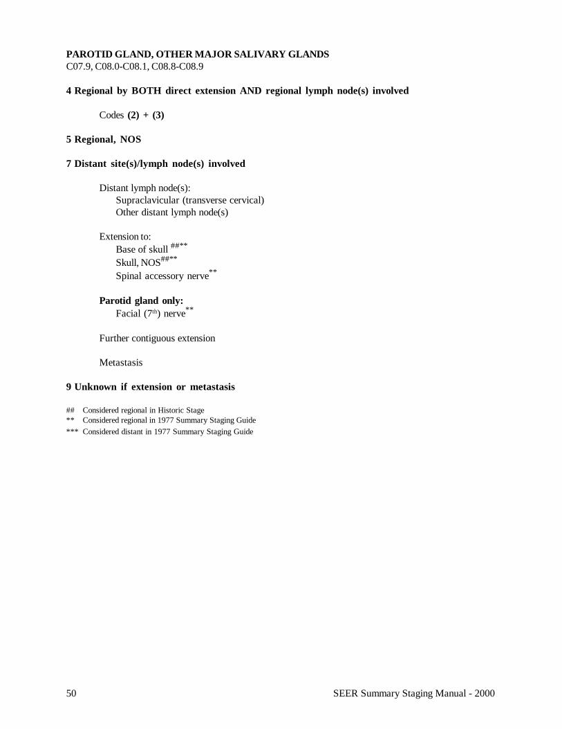
PAROTID GLAND, OTHER MAJOR SALIVARY GLANDSC07.9, C08.0-C08.1, C08.8-C08.9
4 Regional by BOTH direct extension AND regional lymph node(s) involved
Codes (2) + (3)
5 Regional, NOS
7 Distant site(s)/lymph node(s) involved
Distant lymph node(s):Supraclavicular (transverse cervical)Other distant lymph node(s)
Extension to:Base of skull ##**
Skull, NOS##**
Spinal accessory nerve**
Parotid gland only:Facial (7th) nerve**
Further contiguous extension
Metastasis
9 Unknown if extension or metastasis
## Considered regional in Historic Stage** Considered regional in 1977 Summary Staging Guide*** Considered distant in 1977 Summary Staging Guide

51SEER Summary Staging Manual - 2000
52 SEER Summary Staging Manual - 2000
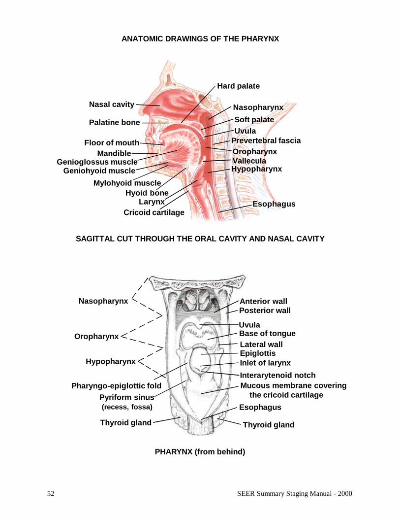
ANATOMIC DRAWINGS OF THE PHARYNX
SAGITTAL CUT THROUGH THE ORAL CAVITY AND NASAL CAVITY
Nasal cavity
Palatine bone
Floor of mouthMandible
Nasopharynx
Hard palate
Soft palate
Prevertebral fasciaUvula
LarynxCricoid cartilage
Esophagus
OropharynxGenioglossus muscle
Geniohyoid muscleMylohyoid muscle
Hyoid bone
HypopharynxVallecula
PHARYNX (from behind)
Anterior wall
Uvula
Posterior wall
Base of tongueLateral wall
Nasopharynx
Oropharynx
Hypopharynx
Pyriform sinus (recess, fossa)
Inlet of larynx
Esophagus
Thyroid glandThyroid gland
Epiglottis
Pharyngo-epiglottic fold Mucous membrane covering the cricoid cartilage
Interarytenoid notch
53SEER Summary Staging Manual - 2000
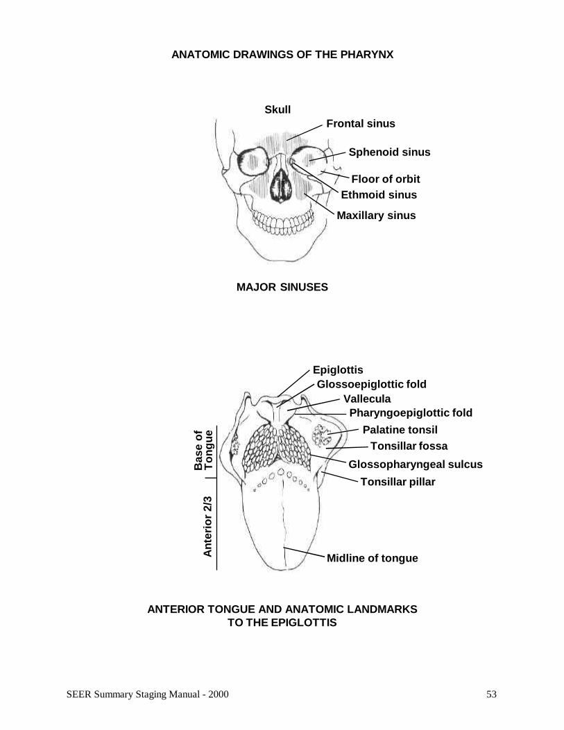
ANATOMIC DRAWINGS OF THE PHARYNX
MAJOR SINUSES
Frontal sinus
Floor of orbit
Sphenoid sinus
Ethmoid sinus
Maxillary sinus
Skull
EpiglottisGlossoepiglottic fold
ValleculaPharyngoepiglottic fold
Palatine tonsilTonsillar fossa
Glossopharyngeal sulcusTonsillar pillar
Midline of tongueAnt
erio
r 2/
3B
ase
ofT
ongu
e
ANTERIOR TONGUE AND ANATOMIC LANDMARKSTO THE EPIGLOTTIS
54 SEER Summary Staging Manual - 2000
TONSIL, OROPHARYNXC09.0-C09.1, C09.8-C09.9, C10.0-C10.4,C10.8-C10.9C09.0 Tonsillar fossa <> C10.0 ValleculaC09.1 Tonsillar pillar <> C10.1 Anterior surface of epiglottisC09.8 Overlapping lesion of tonsil <> C10.2 Lateral wall of oropharynxC09.9 Tonsil, NOS <> C10.3 Posterior wall of oropharynx<> Laterality must be coded for this site C10.4 Branchial cleft
C10.8 Overlapping lesion of oropharynxC10.9 Oropharynx, NOS
Note: AJCC includes base of tongue (C01.9) and lingual tonsil (C02.4) with oropharynx (C09._, C10._).
Note: See the introductory material for this section (page 18) forNote: AJCC includes lingual (anterior) surface detailed descriptions of the anatomic limits of the structures in theof epiglottis (C10.1) with larynx (C32._). oropharynx.
SUMMARY STAGE
0 In situ: Noninvasive; intraepithelial
1 Localized only
Invasive tumor confined to one of the following subsites:Anterior wall (including vallecula and lingual (anterior) surface of epiglottis)One lateral wallPosterior wall
Involvement of two or more subsites:##
Anterior, lateral or posterior wall(s)
Localized, NOS
2 Regional by direct extension only
Extension to:Base of tongueBuccal mucosa (inner cheek)###
Floor of mouth###
Gum (gingiva)###
Hypopharynx, NOSLarynx, NOSNasopharynx, NOS###
Posterior surface of epiglottisPrevertebral fascia or musclePterygoid musclePyriform sinus (pyriform fossa)Soft palate:
Inferior surfaceSuperior (nasopharyngeal) surfaceUvula
Soft tissue of neck
Fixation to adjacent tissues
55SEER Summary Staging Manual - 2000
TONSIL, OROPHARYNXC09.0-C09.1, C09.8-C09.9, C10.0-C10.4, C10.8-C10.9
3 Regional lymph node(s) involved only
REGIONAL Lymph Nodes
Cervical, NOSInternal jugular, NOS:
Deep cervical, NOS:MiddleUpper, NOS:
Jugulodigastric (subdigastric)Mandibular, NOS:
Submandibular (submaxillary)###***
Submental###***
Retropharyngeal###
Regional lymph node(s), NOS
4 Regional by BOTH direct extension AND regional lymph node(s) involved
Codes (2) + (3)
5 Regional, NOS
7 Distant site(s)/lymph node(s) involved
Distant lymph node(s):MediastinalSupraclavicular (transverse cervical)Other distant lymph node(s)
Extension to:Anterior 2/3 of tongueBoneExtrinsic muscles of tongue:
GenioglossusGeniohyoidHyoglossusMylohyoidPalatoglossusStyloglossus
Hard PalateMandibleParotid gland
Further contiguous extension
Metastasis
9 Unknown if extension or metastasis
## Considered regional in Historic Stage### Considered distant in Historic Stage*** Considered distant in 1977 Summary Staging Guide
56 SEER Summary Staging Manual - 2000
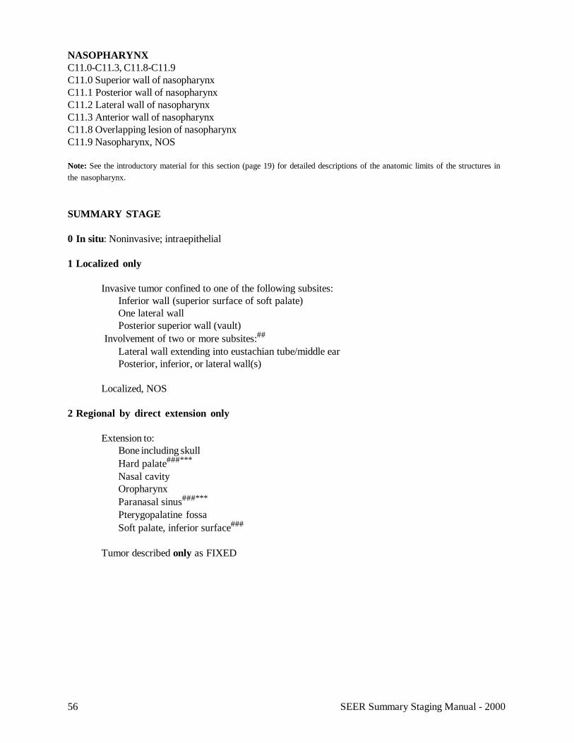
NASOPHARYNXC11.0-C11.3, C11.8-C11.9C11.0 Superior wall of nasopharynxC11.1 Posterior wall of nasopharynxC11.2 Lateral wall of nasopharynxC11.3 Anterior wall of nasopharynxC11.8 Overlapping lesion of nasopharynxC11.9 Nasopharynx, NOS
Note: See the introductory material for this section (page 19) for detailed descriptions of the anatomic limits of the structures inthe nasopharynx.
SUMMARY STAGE
0 In situ: Noninvasive; intraepithelial
1 Localized only
Invasive tumor confined to one of the following subsites:Inferior wall (superior surface of soft palate)One lateral wallPosterior superior wall (vault)
Involvement of two or more subsites:##
Lateral wall extending into eustachian tube/middle earPosterior, inferior, or lateral wall(s)
Localized, NOS
2 Regional by direct extension only
Extension to:Bone including skullHard palate###***
Nasal cavityOropharynxParanasal sinus###***
Pterygopalatine fossaSoft palate, inferior surface###
Tumor described only as FIXED
57SEER Summary Staging Manual - 2000
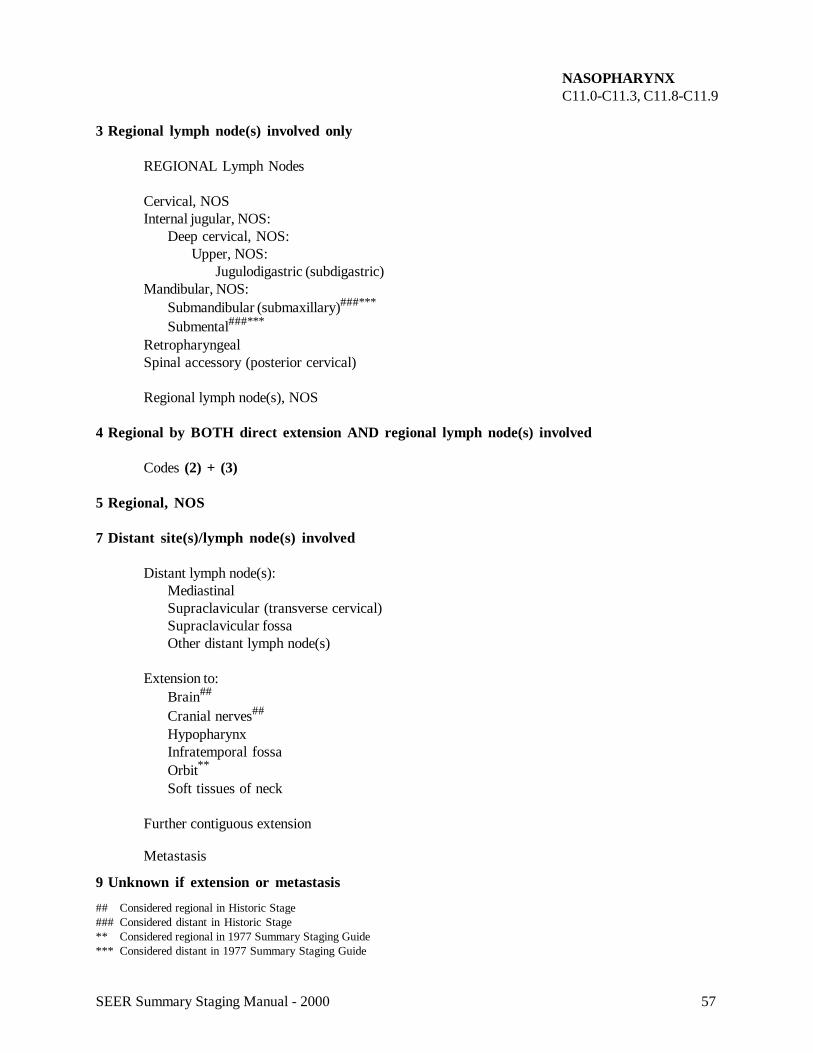
NASOPHARYNXC11.0-C11.3, C11.8-C11.9
3 Regional lymph node(s) involved only
REGIONAL Lymph Nodes
Cervical, NOSInternal jugular, NOS:
Deep cervical, NOS:Upper, NOS:
Jugulodigastric (subdigastric)Mandibular, NOS:
Submandibular (submaxillary)###***
Submental###***
RetropharyngealSpinal accessory (posterior cervical)
Regional lymph node(s), NOS
4 Regional by BOTH direct extension AND regional lymph node(s) involved
Codes (2) + (3)
5 Regional, NOS
7 Distant site(s)/lymph node(s) involved
Distant lymph node(s):MediastinalSupraclavicular (transverse cervical)Supraclavicular fossaOther distant lymph node(s)
Extension to:Brain##
Cranial nerves##
HypopharynxInfratemporal fossaOrbit**
Soft tissues of neck
Further contiguous extension
Metastasis
9 Unknown if extension or metastasis
## Considered regional in Historic Stage### Considered distant in Historic Stage** Considered regional in 1977 Summary Staging Guide*** Considered distant in 1977 Summary Staging Guide
58 SEER Summary Staging Manual - 2000
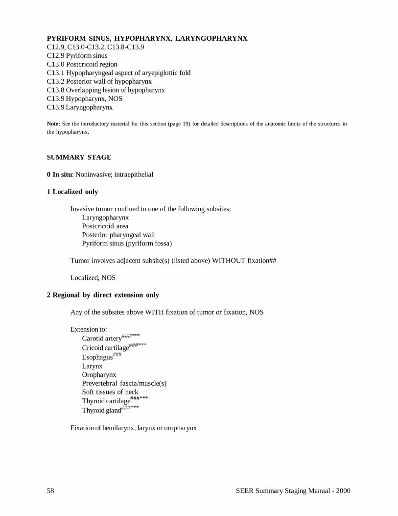
PYRIFORM SINUS, HYPOPHARYNX, LARYNGOPHARYNXC12.9, C13.0-C13.2, C13.8-C13.9C12.9 Pyriform sinusC13.0 Postcricoid regionC13.1 Hypopharyngeal aspect of aryepiglottic foldC13.2 Posterior wall of hypopharynxC13.8 Overlapping lesion of hypopharynxC13.9 Hypopharynx, NOSC13.9 Laryngopharynx
Note: See the introductory material for this section (page 19) for detailed descriptions of the anatomic limits of the structures inthe hypopharynx.
SUMMARY STAGE
0 In situ: Noninvasive; intraepithelial
1 Localized only
Invasive tumor confined to one of the following subsites:LaryngopharynxPostcricoid areaPosterior pharyngeal wallPyriform sinus (pyriform fossa)
Tumor involves adjacent subsite(s) (listed above) WITHOUT fixation##
Localized, NOS
2 Regional by direct extension only
Any of the subsites above WITH fixation of tumor or fixation, NOS
Extension to:Carotid artery###***
Cricoid cartilage###***
Esophagus###
LarynxOropharynxPrevertebral fascia/muscle(s)Soft tissues of neckThyroid cartilage###***
Thyroid gland###***
Fixation of hemilarynx, larynx or oropharynx
59SEER Summary Staging Manual - 2000
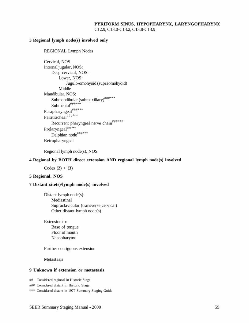
PYRIFORM SINUS, HYPOPHARYNX, LARYNGOPHARYNXC12.9, C13.0-C13.2, C13.8-C13.9
3 Regional lymph node(s) involved only
REGIONAL Lymph Nodes
Cervical, NOSInternal jugular, NOS:
Deep cervical, NOS:Lower, NOS:
Jugulo-omohyoid (supraomohyoid)Middle
Mandibular, NOS:Submandibular (submaxillary)###***
Submental###***
Parapharyngeal###***
Paratracheal###***
Recurrent pharyngeal nerve chain###***
Prelaryngeal###***
Delphian node###***
Retropharyngeal
Regional lymph node(s), NOS
4 Regional by BOTH direct extension AND regional lymph node(s) involved
Codes (2) + (3)
5 Regional, NOS
7 Distant site(s)/lymph node(s) involved
Distant lymph node(s):MediastinalSupraclavicular (transverse cervical)Other distant lymph node(s)
Extension to:Base of tongueFloor of mouthNasopharynx
Further contiguous extension
Metastasis
9 Unknown if extension or metastasis
## Considered regional in Historic Stage### Considered distant in Historic Stage*** Considered distant in 1977 Summary Staging Guide
60 SEER Summary Staging Manual - 2000
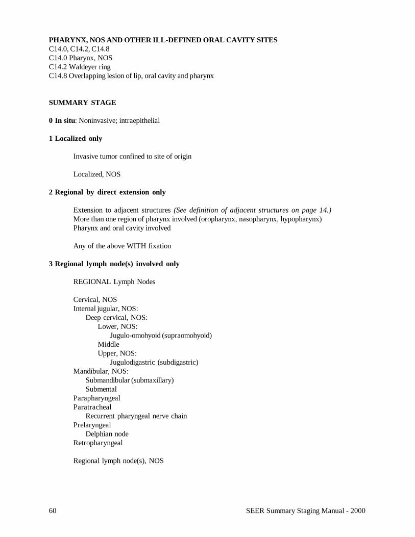
PHARYNX, NOS AND OTHER ILL-DEFINED ORAL CAVITY SITESC14.0, C14.2, C14.8C14.0 Pharynx, NOSC14.2 Waldeyer ringC14.8 Overlapping lesion of lip, oral cavity and pharynx
SUMMARY STAGE
0 In situ: Noninvasive; intraepithelial
1 Localized only
Invasive tumor confined to site of origin
Localized, NOS
2 Regional by direct extension only
Extension to adjacent structures (See definition of adjacent structures on page 14.)More than one region of pharynx involved (oropharynx, nasopharynx, hypopharynx)Pharynx and oral cavity involved
Any of the above WITH fixation
3 Regional lymph node(s) involved only
REGIONAL Lymph Nodes
Cervical, NOSInternal jugular, NOS:
Deep cervical, NOS:Lower, NOS:
Jugulo-omohyoid (supraomohyoid)MiddleUpper, NOS:
Jugulodigastric (subdigastric)Mandibular, NOS:
Submandibular (submaxillary)Submental
ParapharyngealParatracheal
Recurrent pharyngeal nerve chainPrelaryngeal
Delphian nodeRetropharyngeal
Regional lymph node(s), NOS
61SEER Summary Staging Manual - 2000
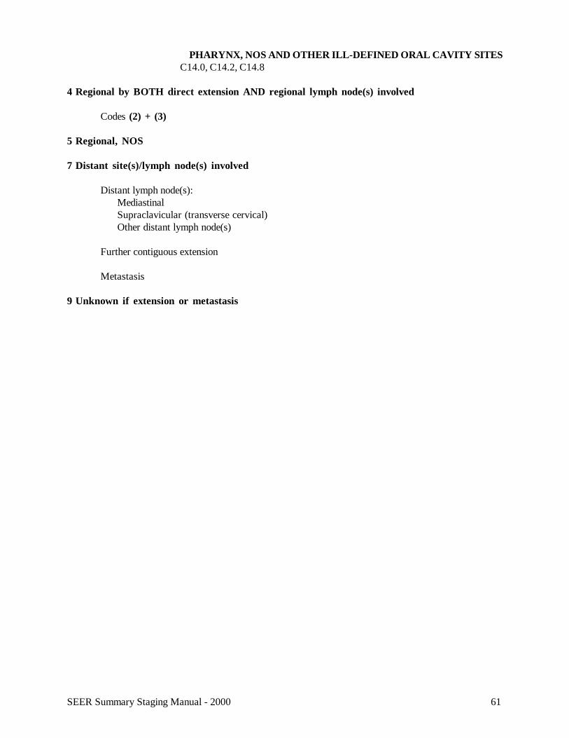
PHARYNX, NOS AND OTHER ILL-DEFINED ORAL CAVITY SITESC14.0, C14.2, C14.8
4 Regional by BOTH direct extension AND regional lymph node(s) involved
Codes (2) + (3)
5 Regional, NOS
7 Distant site(s)/lymph node(s) involved
Distant lymph node(s):MediastinalSupraclavicular (transverse cervical)Other distant lymph node(s)
Further contiguous extension
Metastasis
9 Unknown if extension or metastasis