Embed Size (px)
Citation preview
Journal of Periodontology; Copyright 2012 DOI: 10.1902/cap.2012.120015
1
Oral Rehabilitation by Dental Implants for Teeth Involved in a Maxillary Fibrous Dysplasia
Alberto Monje, D.D.S.*, Florencio Monje, MD, PhD†, Fernando Suarez, D.D.S.*, Raúl González-García, MD, PhD†, Laura Villanueva, MD† and Hom-Lay Wang, D.D.S.,
M.S., PhD*
* Graduate Periodontics, Department of Periodontics and Oral Medicine, University of Michigan School of Dentistry, Ann Arbor, MI, USA.
† Oral and Maxillofacial Surgeon, CICOM, Center of Implantology, Oral and Maxillofacial Surgery, Badajoz, Spain.
Introduction: Fibrous dysplasia (FD) is a rare but important benign lesion affecting the maxillofacial region, which presents bone pain, deformities and fracture. Although FD presents radiographic patterns, it has to be analyzed histologically for the final diagnosis. Currently, there is no guide for FD treatment universally accepted. We report a success case of excision of the entire lesion in the upper left maxilla and the placement of two dental implants for oral rehabilitation followed by guided bone regeneration.
Case presentation: The patient suffered a growth on the left side of the maxilla 4 years ago. Analyzed the lesion helped by cone-beam computer tomography machine, clinical features and confirmed by histologic analysis as a FD. We purposed her to extract the teeth involved by the lesion and the excision of the lesion. That was followed by two implants placement for oral rehabilitation grafting the cavity with an osteoconductor material. Both implants were successfully integrated and it was supported by the clinical examination as well as the resonance frequency test.
Conclusions: Dental implant and related guided bone regeneration can be successfully obtained in the patient who suffered from FD lesion.
Key words fibrous dysplasia, dental implant, maxillofacial abnormalities, bone graft(s), oral pathology.
BACKGROUND Fibro-osseous lesions are a group of processes characterized by replacement of normal bone by fibrous tissue containing a newly formed mineralized product. Fibrous dysplasia (FD) and ossifying fibroma are the most common fibro-osseous lesions, which may be associated with functional and cosmetic disturbance 1. These lesions usually present a diagnosis dilemma, which must be based upon the clinical evidence, histologic analysis and radiographic features. FD however, creates radiographic patterns that are virtually indistinguishable from other lesions affecting the bones 2.
FD is a rare but important benign lesion affecting the maxillofacial region that presents bone pain, bone deformities and fracture. FD is a genetically-based disease with mutations in the gene (GNSA I) encoding the α-subunit of stimulatory G-protein that results in the increased production of cyclic adenosine monophosphate, affecting the proliferation and differentiation of preosteoblast. The clinical severity of the lesion depends of the time of appearance of the mutation 3. The lesions of FD are divided in terms of “monostotic” and “polyostotic” forms depending on the number of bones involved. Monostotic form accounting for 70% to 85% of cases and does not progress into a polyostotic form of the disease 4. There is a slight female predilection usually presenting in the first 3 decades of life 5. The right unilateral nature of FD is noted in

Journal of Periodontology; Copyright 2012 DOI: 10.1902/cap.2012.120015
2
almost all the cases reported, being maxilla and frontal bones the most commonly involved 2, 5.
Radiological features of FD are a poorly defined fusiform enlargement especially in the mandible or generalized enlargement of the upper jaw. The margins of lesion, “ground glass” appearance and displacement of maxillary sinus are some other features related to the FD lesion in the maxilla 2. Although these features can be localized in a panoramic radiograph, computer tomography is often required for a more complete evaluation for the diagnosis and to evaluate treatment alternatives.
The increase rate of bone resorption in FD classically has been treated by bisphosphonates. However, the bone refilling of the place where the lesion was has been observed in only about 50% of the defects 6. Hence, the most predictable treatment, although more aggressive, is the excision of the entire affected area and graft it with bone grafts. While autologous bone has been considered the “gold standard” because of its osteogenic, osteoconductive and osteoinductive abilities, morbidity of the donor area, resorption and cost have leaded us to the use of bone substitutes. The refilling of the cavity will facilitate future placement of endosseous implants if patient wants to get back with their original function of the dentition. The use of endosseous implants as been well-established with a good long-term success 7. However, the outcome of dental implants has not been described in patients with fibro-osseous lesions that were repaired with osseous substitute yet. This paper reported a patient with monostotic FD in the upper maxilla was successfully rehabilitated dental implants.
CLINICAL PRESENTATION A 49-year-old Caucasian female presented to CICOM (Center of Implantology, Oral and Maxillofacial Surgery, Badajoz, Spain) on December 2009 with pain in the left second upper molar and an asymmetrical growth on the left side of the maxilla that started to grow up 4 years ago (Figure 1). She was a nonsmoker and otherwise healthy patient. Intraoral examination revealed good oral health and high level of hygiene. After exploring clinically, it was thought the extra mass on the left side of the maxilla due to the long-term asymptomatically history of the lesion probably was an osseous exostosis. However, at the same appointment, we decided to explore the lesion radiographically by a cone-beam CTφ. On it, we discovered a 19 mm of length and 20 mm of width well-defined “ground glass” lesion. Patient was then informed the necessity of biopsy to determine the final diagnosis of this lesion. Four weeks later an incisional biopsy was done under IV sedation and local anesthesia at the mass level in the left maxilla. A core was obtained for the histological evaluation (Figure 2). The lesion was the confirmed to be an FD lesion. In the microscopy was observed trabeculae of woven bone in a background of fibrous tissue. Radiographically, it was observed that the lesion was in close contact with the roots of left first and second upper premolars and left first upper molar (Figures 3 and 4). For these reasons, the teeth involved were extracted and the excision of the whole lesion by curettage was performed two months later (Figure 5).
CASE MANAGEMENT After a healing period of 10 months, oral rehabilitation was needed for the patient in the left maxillae area. Under IV sedation and local anesthesia two 4 x 15 mm γ were placed (Figure 6). This was followed by filling of ∆ into the remaining cavity due to its osteocontuctive abilities. A membrane was placed to promote the Ψ osseous regeneration. Implant stability quotient (ISQ) was measured by δ just before wound
Journal of Periodontology; Copyright 2012 DOI: 10.1902/cap.2012.120015
3
closure. The ISQ for both implants were measured right after the implant placement and 6 months later. These were 81 and 66; and 81 and 70 respectively. A ceramic-metal screw-retained fixed prostheses was delivered following the conventional loading 6 months after implant placement.
CLINICAL OUTCOMES One year after, no sign of inflammation, peri-implant bone loss or mobility were reported and, although no microscopic evidence of osseointegration, stability of implants was achieved for oral reconstruction/rehabilitation (Figure 7 and 8).
DISCUSSION Osseointegration is defined as the close contact between bone and implant material in histological observations and, in clinical terms, as the ankylosis of the implant in bone 8. However, in some clinical situations, bone grafting procedures are required prior to placement of dental implants. Bone augmentation permits implant treatment that could otherwise not be an option for some patients. High survival rates have been obtained in implants placed after bone augmentation procedures9. In the present case, a patient with a monostotic FD in the left side of the maxilla, was successfully managed by the excision of the whole lesion, grafting the defect with bone substituted and then placed two dental implants with implant stability without clinical detectable mobility.
FD is a benign fibro-osseous lesion that accounts for nearly 7% of all benign tumors of bone, although most experts believe that it should not be classified as a true bone neoplasm 10. This lesion is an hamartomatous condition related to mutations in gene (GNAS I) encoding the α-subunit of stimulatory G-protein (Gs-α) that result in the increased production of cyclic adenosine monophosphate (cAMP), affecting the proliferation and differentiation of preosteoblast 3. FD initially was described by Recklinghausen in 1891 and named in 1938 by Lichtenstein 10. In this disease, normal bone is replaced with a disorganized fibrous connective tissue and woven bone. Generally, is a painless process and often found by a routine clinical or radiologic examination 10. Our finding was due to patient´s concerns about the growth in the maxilla area and the pain related to the teeth closest to the lesion.
FD must be diagnosed by a complete clinical, radiographic and histologic study. This, frequently requires a differentiation with ossifying fibroma. Ossifying fibroma is a lesion of bone as well, but is a monofocal process that tends to grow centrifugally and be demarcated 10.
Clinically, the disease is classified as monostotic, polyostotic, or craniofacial. Monostotic form, as our finding, is isolated to a single bone, while polyostotic form involves more than one noncontiguous bone. The monostotic form is not a precursor of the polyostotic form and accounts for 70-85% of cases of FD. When the bones of the face are involved, the lesion often includes contiguous bones such as maxilla, sphenoid, zygoma and ethmoid bones. In the jaws, FD is more frequent to be found in the maxilla than in the mandible, mainly in the posterior region 4.
Currently, cone-beam CT (CBCT) provides comprehensive information for the diagnosis of a lesion in the maxillofacial area. CBCT can be used to determine the extent, specific dimension and radiodensity of FD 5. Besides, it allows to planning implant therapy virtually with special 3-dimension software programs. Furthermore, the emission of radiation of the CBCT is 25% less than a panoramic radiography and 40-60 times lower than a classical CT-scan 11.
Journal of Periodontology; Copyright 2012 DOI: 10.1902/cap.2012.120015
4
The 3 most common radiologic appearances of FD in the craniofacial skeleton are the pagetoid, sclerotic, and cystic patterns. These are appreciated in the CBCT as bone expansion and scattered islands of bone formation in a low-attenuation field, an homogeneous appearance with a “ground-glass” appearance and as a well-defined low-attenuation lesion with a sclerotic margin referred to as an “eggshell lesion”, respectively 2. The use of CBCT in the present case was required to plan our approach and the final treatment for oral rehabilitation with dental implants. Radiographic exploration must be realized in further appointments to be ensure the absence of reactivation of the FD.
Currently, there is no guideline for FD treatment that was universally accepted. Those cases in which the lesion is asymptomatic and does not cause deformities or functional impairment should simply be monitored 4. Surgical approach is required only when anatomical structures are in danger of being compressed or the function or cosmetic of the patient is compromised. However, although a complete excision was done, lifelong monitoring is often advocated because of the potential for late growth, dysfunction and a small risk of sarcomatous change 10. Recurrence of the disease is not common in adults but common during growth. Actually, in a 6-year follow-up study has shown almost all the surgically treated patients presented satisfactory outcomes (Only 1 out of 13 presented recurrence of FD) 12. In the present case, implant placement was delayed due to two main reasons. One: deficient amount of bone to accommodate proper implant placement. Two: the necessity of following up the lesion to ensure there is no recurrence of it. It is very important to point out that a short-term follow up of this specific case cannot draw any definitive conclusion with regards to the long-term predictability. Longer follow-up and larger sample size are needed to further validate the findings noted in here. When the surgery is not indicated, relief of bone pain and reduction of osteoclastic activity with partial filling of osteolytic lesions can be achieved with bisphosphonates therapy 6. In this specific case, we approached the lesion with completely excised of the lesion, filled with substitute to promote osseous regeneration, proper placement of two dental implants and then restored this area, as an implant-supported prostheses.
Cheung et al. reported a successful bone-to-titanium screw contact in patients with FD. Although the contact was higher in normal bone than in dysplasic bone, but was not statistically significant. Thus, they suggested that longer screws should be used to compensate for the reduced bone contact percentage 13. Bajwa et al. reported a case of oral rehabilitation using endosseous implants in both maxilla and mandible in a patient with craniofacial FD. After 5 years of follow-up, although osseointegration at microscopic was unable to prove, a direct functional connection between dysplasic bone and the surface of a load bearing implant was evidenced by the lack of symptoms, soft tissues inflammation and peri-implant bone loss 14. Uckan et al. nonetheless, preferred to treat their patient with a total maxillectomy and then place implants into the normal zygomatic bone to facilitate future oral rehabilitation 15. This case report proposed is one of the first attempts of using combination approaches (excise the lesion, augment lesion defect, place dental implants and restore with implant-supported prostheses) to restore patient back to its original function and esthetics.
SUMMARY Why is this case new information?
Journal of Periodontology; Copyright 2012 DOI: 10.1902/cap.2012.120015
5
• This is the first case that documented the success of dental implant osseointegration in a patient who suffered from Fibrous Dysplasia.
What are the keys to successful management of this case?
• Proper diagnosis (e.g., Computer Tomogram and histologic confirmation) and complete debridement of Fibrous Dysplasia lesion.
• Good oral hygiene is required.
• Carefully executed guided bone regeneration and wound closure. Abided by “PASS” principle for guided bone regeneration.
• Obtain implant primary stability at the time of implant placement and avoid any premature loading during healing.
What are the primary limitations to success in this case?
• Wrong diagnosis.
• Poorly executed lesion debridement, implant surgery and bone augmentation.
• Poor oral hygiene and premature implant loading.
ACKNOWLEDGMENTS The authors want to thanks FEDICOM (Foundation for the study of Implantology, Oral and Maxillofacial Surgery), Badajoz, Spain, for financial support. At the same time we want to thank Dr. Ramón Gómez de Tejada affiliated to the Pathology Service of Infanta Cristina Hospital, Badajoz, Spain, for the histologic analysis and also to to Dr. Miguel Padial-Molina of the Department of Periodontics and Oral Medicine of the University of Michigan due to his valuable helping to describe and understand the histologic aspect of the lesion.
Conflict of interests: None
Disclaimer: The authors do not have any financial interests, either directly or indirectly, in the products or information listed in the paper.
REFERENCES 1. Toyosawa S, Yuki M, Kishino M, et al. Ossifying fibroma vs fibrous dysplasia of the jaw: molecular
and immunological characterization. Mod Pathol 2007;20:389-396.
2. Sontakke SA, Karjodkar FR, Umarji HR. Computed tomographic features of fibrous dysplasia of maxillofacial region. Imaging Sci Dent 2011;41:23-28.
3. Cohen MM, Jr., Howell RE. Etiology of fibrous dysplasia and McCune-Albright syndrome. Int J Oral Maxillofac Surg 1999;28:366-371.
4. Lustig LR, Holliday MJ, McCarthy EF, Nager GT. Fibrous dysplasia involving the skull base and temporal bone. Arch Otolaryngol Head Neck Surg 2001;127:1239-1247.
5. MacDonald-Jankowski D. Fibrous dysplasia in the jaws of a Hong-Kong population: radiographic presentation and systematic review. Dentomaxillofac Radiol 1999;28:195-202.
6. Chapurlat RD, Hugueny P, Delmas PD, Meunier PJ. Treatment of fibrous dysplasia of bone with intravenous pamidronate: long-term effectiveness and evaluation of predictors of response to treatment. Bone 2004;35:235-242.
7. Buser D, Mericske-Stern R, Bernard JP, et al. Long-term evaluation of non-submerged ITI implants. Part 1: 8-year life table analysis of a prospective multi-center study with 2359 implants. Clin Oral Implants Res 1997;8:161-172.
8. Albrektsson T, Zarb GA. Current interpretations of the osseointegrated response: clinical significance. Int J Prosthodont 1993;6:95-105.
Journal of Periodontology; Copyright 2012 DOI: 10.1902/cap.2012.120015
6
9. Hammerle CH, Jung RE, Feloutzis A. A systematic review of the survival of implants in bone sites augmented with barrier membranes (guided bone regeneration) in partially edentulous patients. J Clin Periodontol 2002;29 Suppl 3:226-231; discussion 232-223.
10. Kim DD, Ghali GE, Wright JM, Edwards SP. Surgical treatment of giant fibrous dysplasia of the mandible with concomitant craniofacial involvement. J Oral Maxillofac Surg 2012;70:102-118.
11. Sukovic P. Cone beam computed tomography in craniofacial imaging. Orthod Craniofac Res 2003;6 Suppl 1:31-36; discussion 179-182.
12. Keijser LC, Van Tienen TG, Schreuder HW, Lemmens JA, Pruszczynski M, Veth RP. Fibrous dysplasia of bone: management and outcome of 20 cases. J Surg Oncol 2001;76:157-166; discussion 167-158.
13. Cheung LK, Samman N, Pang M, Tideman H. Titanium miniplate fixation for osteotomies in facial fibrous dysplasia--a histologic study of the screw/bone interface. Int J Oral Maxillofac Surg 1995;24:401-405.
14. Bajwa MS, Ethunandan M, Flood TR. Oral rehabilitation with endosseous implants in a patient with fibrous dysplasia (McCune-Albright syndrome): a case report. J Oral Maxillofac Surg 2008;66:2605-2608.
15. Uckan S, Oguz Y, Uyar Y, Ozyesil A. Reconstruction of a total maxillectomy defect with a zygomatic implant-retained obturator. J Craniofac Surg 2005;16:485-489.
Corresponding author:
Florencio Monje Gil
Calle Juan Miró s/n, local 16-17
06010 Badajoz (Spain)
Tel: 0034 924 205 235
Fax: 0034 924 260 773
E-mail address: [email protected]
Submitted February 16, 2012; accepted for publication March 24, 2012.
Figure 1.
Frontal view of the initial growth. (to be included on the first page)
Figure 2.
Histologic aspect of the lesion.
A. Microscopic detail of the relation between trabecular bone (*) and stroma (+) x 400.
B. Area of stroma showing more fibrous density (arrow) x 200.
C. Immature trabecular bone (*) divided by fibrous tissue (+) x 100.
D. Detail of the loose fibrillar stroma with fibroblastic cells x 400.
Figure 3.
Panoramic radiograph showing the extent of the involvement.
Figure 4.
Sagittal view of the FD in close contact to a tooth of the left upper maxilla.
Figure 5.
Journal of Periodontology; Copyright 2012 DOI: 10.1902/cap.2012.120015
7
Teeth after extraction with rest of the lesion on their roots.
Figure 6.
Implant placement stage 10 months after the excision of the lesion. (to be included on the first page)
Figure 7.
Frontal view of both implants placed after 12 months of healing.
Figure 8.
Sagittal view of one of the implants placed at 12 months follow-up. φ i-CAT (Imaging Sciences International, Hatfield, PA, USA) γ Speedy™ Groovy Nobel Biocare Implants (Nobel Biocare AB, Göteborg, Sweden) ∆ Bio-Oss® (Geistlich Pharma AG, Wolhusen, Switzerland) Ψ Bio-Gide® (Geistlich Pharma AG, Wolhusen, Switzerland) δ Osstell™ (Osstell AB, Göteborg, Sweden)

Figure 1. Frontal view of the initial growth.
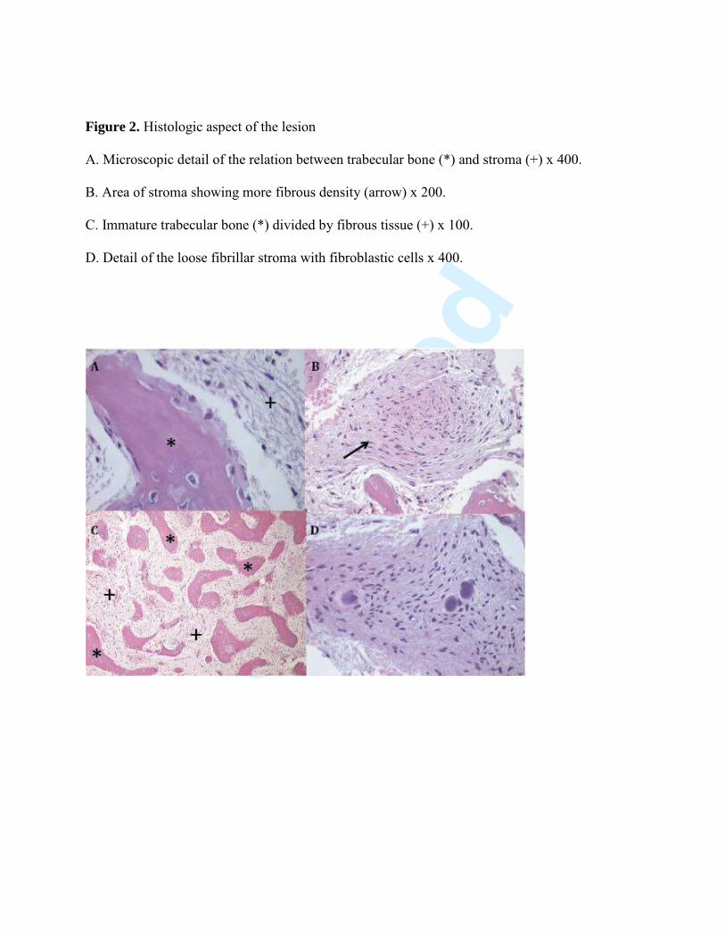
Figure 2. Histologic aspect of the lesion
A. Microscopic detail of the relation between trabecular bone (*) and stroma (+) x 400.
B. Area of stroma showing more fibrous density (arrow) x 200.
C. Immature trabecular bone (*) divided by fibrous tissue (+) x 100.
D. Detail of the loose fibrillar stroma with fibroblastic cells x 400.
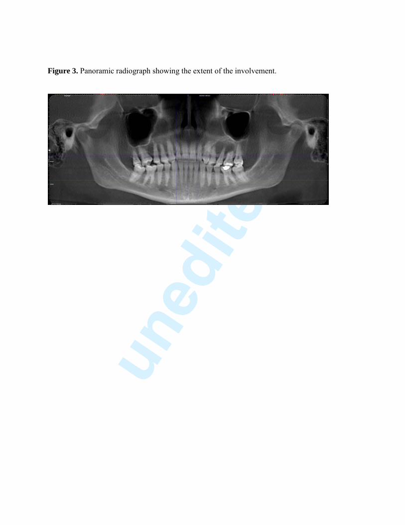
Figure 3. Panoramic radiograph showing the extent of the involvement.
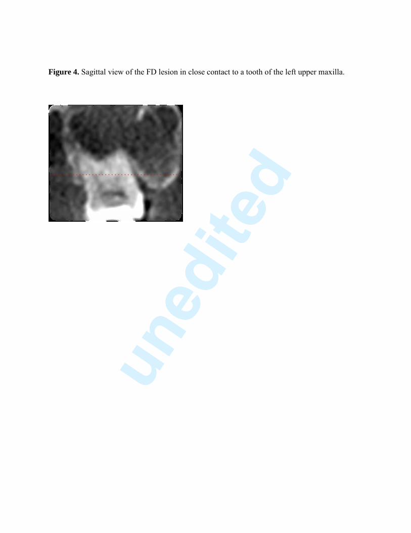
Figure 4. Sagittal view of the FD lesion in close contact to a tooth of the left upper maxilla.
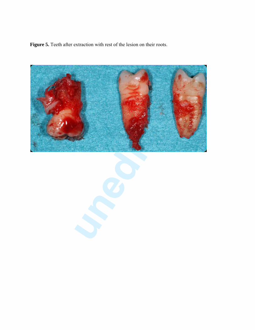
Figure 5. Teeth after extraction with rest of the lesion on their roots.
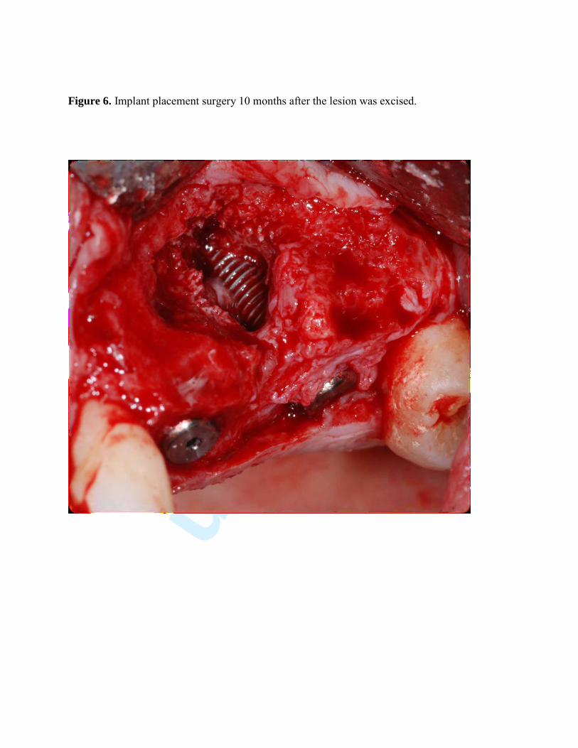
Figure 6. Implant placement surgery 10 months after the lesion was excised.

Figure 7. Frontal view of both implants placed after 12 months of healing.
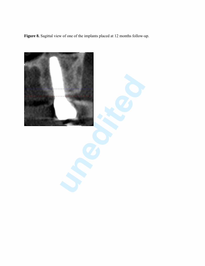
Figure 8. Sagittal view of one of the implants placed at 12 months follow-up.

First page image – Left side
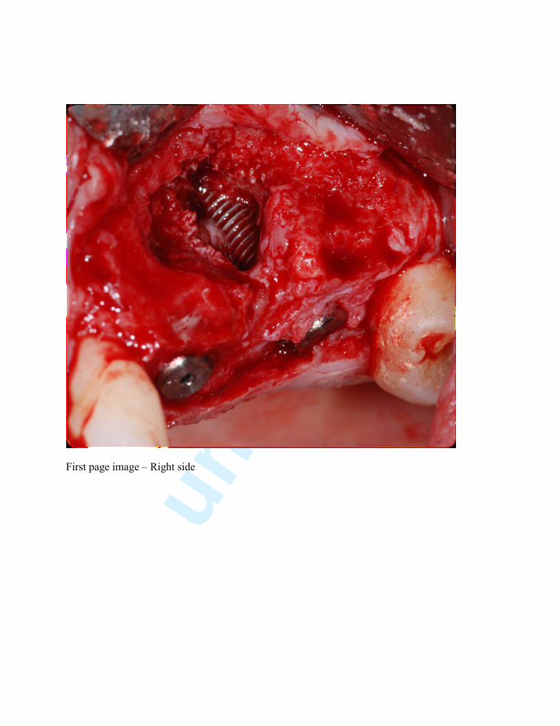
First page image – Right side