Embed Size (px)
Citation preview

Instructions for use
Title Dynamic photo-control of kinesin on a photoisomerizable monolayer ‒ hydrolysis rate of ATP and motility ofmicrotubules depending on the terminal group
Author(s) Rahim, M. K. Abdul; Kamei, Takashi; Tamaoki, Nobuyuki
Citation Organic & Biomolecular Chemistry, 10(16): 3321-3331
Issue Date 2012-04-28
Doc URL http://hdl.handle.net/2115/63001
Type article (author version)
File Information revised MS RSC Article Template2.pdf
Hokkaido University Collection of Scholarly and Academic Papers : HUSCAP
Journal Name
Cite this: DOI: 10.1039/c0xx00000x
www.rsc.org/xxxxxx
Dynamic Article Links ►
ARTICLE TYPE
This journal is © The Royal Society of Chemistry [year] [journal], [year], [vol], 00–00 | 1
Dynamic photo-control of kinesin on a photoisomerizable monolayer-Hydrolysis rate of ATP and motility of microtubule depending on the terminal group M. K. Abdul Rahim,a Takashi Kameia and Nobuyuki Tamaoki*a
Received (in XXX, XXX) Xth XXXXXXXXX 20XX, Accepted Xth XXXXXXXXX 20XX 5
DOI: 10.1039/b000000x
The reversibly and repeatedly altered gliding motility of microtubules driven by kinesin on the photoresponsive monolayer surface is studied. It was confirmed that an azobenzene monolayer surface needs to have free amino terminal groups for the successful dynamic control of the motility of microtubule. The surface of azobenzene monolayer with terminal amino groups can dynamically control 10
the ATP hydrolysis activity of kinesin which resulted in the change in motility of microtubule.
Introduction The regulation of nano assembly devices and molecular machines using biomolecular motors as promising components is one of the hot topic in nanotechnology and bioengineering.1-9 Kinesins are 15
prominent microtubule associated motor proteins, which play a central role in both structural and biological functions of the cells.10-12 The fundamental role of kinesin is to actively distribute the intracellular materials like organelles, vesicles, chromosomes along the microtubules13,14 and to drive the cell division process 20
by converting the chemical energy of adenosine triphosphate (ATP) into mechanical energy with the positional movement towards the fast polymerizing plus ends of microtubules.15-17 Each kinesin molecule has two “heads”, a 50 nm long semi-flexible coiled-coil region which binds to microtubules and 25
hydrolyses ATP and a “tail”, which is thought to bind to the cargo.18 Among the molecular motors, the kinesin-microtubule system is used widely for developing nanoscale biodevices, because the motility can be regenerated on a surface comparatively easily. In the common in vitro construction of 30
the kinesin-microtubule system, the kinesin are adsorbed onto a surface such as glass and microtubules are able to glide across the kinesin coated glass surface.14 The kinesin-microtubule system has other advantages such as, its small size, and the linear movement of single kinesin along the microtubule compared to 35
other molecular motor proteins.19,20 The ability to reversibly control the speed of microtubule gliding and the direction of movement on the surface of kinesin by external stimuli would greatly improve the sophistication of nano devices. Several methodologies have been used for the 40
artificial control of microtubule function which includes microlithographic tracks,21,22 electric and magnetic fields,23 antibody 24 and so on. When considering the biodevices in the
future, photochemical energy inputs offer advantages compared to chemical energy inputs because (i) light does not generate 45
waste products; (ii) it can be switched on/off easily and rapidly; (iii) photons, besides supplying the energy to the system, can also be useful to “read” the state of the system and thus to control and monitor the operation of the machine etc.. Higuchi et al.25 used the caged ATP as photo-controlled switch, because their inactive 50
caged states (OFF state of motility) can be converted to active uncaged states (ON state of motility) by UV light irradiation. Another group successfully used the photolysis of caged ATP to develop molecular shuttles on engineered kinesin tracks.26 On the other hand Tatsu et al. demonstrated the switching of kinesin’s 55
activity from the ON state to the OFF state by the photolysis of caged peptide derived from the kinesin C-terminus domain working as an inhibitor.27 In that study, they successfully demonstrated an 80% reduction of the initial gliding velocity of microtubules on the kinesin surface after the photochemical 60
deprotection of the o-nitrobenzyl protecting group on the caged peptide. All these methods can either trigger the initiation of the movement or cessation of the microtubule motility. To design the more useful nanoscale biodevices, it is important that the system should provide ON and OFF switching of gliding motility at any 65
desired time and at any desired position in space. To the best of our knowledge the light controlled reversible and repeated switching of microtubule motility was demonstrated only by our group.28 We reported the reversible and repeated regulation of the 70
motility of microtubule by the photoisomerization of the underlying monolayer using two different wavelengths of light.28
For the fabrication of such photoresponsive monolayer, we employed a derivative of azobenzene; one of the most studied photochromic compounds29-33 due to its strong photo-switching75
Journal Name
Cite this: DOI: 10.1039/c0xx00000x
www.rsc.org/xxxxxx
Dynamic Article Links ►
ARTICLE TYPE
This journal is © The Royal Society of Chemistry [year] [journal], [year], [vol], 00–00 | 2
HN
NH2O
a) b) c)N N
NH
HN
O O N N
NH2
H2N
N N
NH2
HN
N N
NH
NH
Si
HN
OOOO
d)
4 5
7, 8, 9 1b', 1c', 1d'
3
R
7, 1b': R =HN
O
HN HN
Ntert-Boc
tert-Boc
tert-Boc
O
HN tert-Boc
O
O tert-Bu
O O
O tert-Bu
8, 1c': R =
9, 1d': R =
R
Scheme 1 a) NaBO3, H3BO3, HOAc, HCl, MeOH, 60 °C. b) NaOH. c) For 7, 1b′ = Boc-Arg-(Boc)2-OH, for 8, 1c′ = Boc-Glu-(OtBu)-OH and for 9, 1d′ =
Mono-tert-butyl malonate, DCC, HOBt, DMF. d) OCN-(CH2)3-Si-(OEt)3, THF.
effect, reversibility and simplicity of incorporation,34 with a triethoxy silane group which react with the glass surface to be 5
anchored and a lysine group which can interact with motor proteins. Using this approach, we described the reversible and repeated control of the gliding velocity of microtubules driven by kinesin on the azobenzene monolayer (with a maximum of 15% difference in velocity) upon irradiation with UV and visible 10
light.35 In this paper we are reporting the generality of the system in the repeated regulation of fast and slow modes of microtubule gliding motility by devising a series of surface of azobenzene monolayers with various terminal groups. We also propose a mechanism that accounts for the changes in the velocity of 15
microtubule when the photoresponsive monolayer surface is isomerized between E and Z states upon irradiation of different wavelengths of light. We believe that the method developed here should be applicable in forthcoming nanoscale biodevices based on the kinesin-microtubule system. 20
Experimental Materials
Unless otherwise noted, all reagents including solvents were obtained from major commercial suppliers such as TCI, Sigma-Aldrich and Wako used directly without further purification. 25
DMF was routinely dried and/or distilled prior to use and stored over molecular sieves (4A). Column chromatography was performed with silica gel (63-210μm).
General methods, instrumentation and measurements 1H NMR spectra were recorded at 400 MHz using TMS as an 30
internal standard. Matrix-Assisted Laser Desorption Ionization Time-Of-Flight Mass Spectrometry (MALDI-TOF MS) was performed on an applied Biosystems Voyager- DE pro instrument with positive-ion mode. Absorption spectra were recorded on an Agilent 8453 spectrophotometer and also a Hitachi U-3100 35
absorption spectrophotometer. Photo-isomerization studies were
Journal Name
Cite this: DOI: 10.1039/c0xx00000x
www.rsc.org/xxxxxx
Dynamic Article Links ►
ARTICLE TYPE
This journal is © The Royal Society of Chemistry [year] [journal], [year], [vol], 00–00 | 3
N N
O
HN
ONH
NHBoc
Boc
OO
N N
O
H2N
OO
N N
OH
HN
O
11N N
OH
H2N
N N
O
HN
ONH
NHBoc
Boc
OHO
NH2
HN
Oa)
N N
O
HN
ONH
NHBoc
Boc
HN
OSi O
O O
b) c)
d) e)
f)
10 12
13 14
15 1e' Scheme 2 a) HCl, H2O, NaNO2, NaOH, PhOH, 0~2 °C. b) 10% NaOH, Reflux 2h. c) Br-(CH2)5-COOEt, K2CO3, (CH3)2CO, 50-60 °C. d) Boc-Lys-(Boc)-
OH.DCHA, DCC, HOBt, DMF. e) Dioxane, 1M aq. KOH. f) OCN-(CH2)3-Si-(OEt)3, THF.
conducted using a mercury-xenon lamp (Hamamatsu photonics K.K 200W) after passage through appropriate filters (366 or 440 5
or >500 nm). Microtubules motility assay were carried out using fluorescence microscope (Olympus BX50 or Nikon TEi) equipped with a high NA objective lens (100x, 1.30 NA for Olympus BX50 and 100x, 1.45 NA for Nikon TEi) and the 10
fluorescence image was recorded with appropriate filters (Chroma) to remove the excitation light, back-illuminate electron-cooled CCD camera (EM9100-12, HAMAMATSU), and the image processing system AQUACOSMOS (HAMAMATSU). Both were performed at room temperature (22 °C). 15
Synthesis
The synthetic route to compounds 1b′-e′ and 2a are illustrated in Scheme 1-3 and compound 2b was commercially purchased. Synthetic details of 1a′ were reported in our previous paper. Detailed synthetic procedures for 1b′-e′ and 2a are described 20
below. Compounds 4 and 5 were prepared according to the literature.36
Compound 7. Hydroxybenzotriazole (115 mg, 0.848 mmol) and N,N'-Dicyclohexylcarbodiimide (175 mg, 0.848 mmol) were added under an argon atmosphere to a solution of Boc-Arg-25
(Boc)2-OH (268 mg, 0.565 mmol) in dry DMF (5 mL) at room temperature. After 1 h, 4,4´-diaminoazobenzene (5) (150 mg, 0.707 mmol) was added and the mixture was stirred at room temperature overnight. The mixture was then diluted with brine and was extracted with ethyl acetate (EtOAc); the combined 30
extracts were dried (MgSO4) and the solvent was removed under reduced pressure. The residue was purified by column chromatography (eluent: dichloromethane (DCM) / EtOAc). Yield: 41%. 1H NMR (400 MHz, CDCl3): δ = 1.40 (s, 9H), 1.51 (d, J = 4 Hz, 18H), 1.65–1.71 (m, 2H), 1.82–1.95 (m, 4H), 3.81 35
(m, 1H), 4.07 (br, 2H), 4.55 (m, 1H), 5.95 (q, J = 4 Hz, 1H), 6.72 (d, J = 8 Hz, 2H), 7.61 (d, J = 8 Hz, 2H), 7.77 (d, J = 8 Hz, 2H), 7.82 (d, J = 8 Hz, 2H), 9.19 (br, 1H), 9.29 (br, 1H). MS (MALDI): Calculated for C33H48N8O7 [M + H]+ m/z 669.36; found m/z 669.66. 40
Compound 1b′. Triethoxysilylpropyl isocyanate (62 mg, 0.251 mmol) was added to a solution of 7 (252 mg, 0.377 mmol) in anhydrous tetrahydrofuran (THF) (3 mL). The mixture was refluxed under N2 atmosphere until the completion of the reaction. The residue was washed with copious amounts of hexane and was 45
then dried under vacuum. Yield: 71%. 1H NMR (400 MHz, CDCl3): δ = 0.61 (t, J = 8 Hz, 2H), 1.17 (t, J = 8 Hz, 9H), 1.43 (d, J = 4 Hz, 18H), 1.52 (s, 9H), 1.63 (m, 2H), 1.77 (m, 2H), 1.89 (m, 2H), 2.89 (q, J= 4 Hz, 2H), 3.23 (q, J = 4 Hz, 2H), 3.79 (q, J= 8 Hz, 6H), 3.96 (m, 1H), 4.34 (br, 1H), 5.97 (q, J = 4 Hz, 1H), 6.33 50
(m, 2H), 7.67 (d, J = 8 Hz, 2H), 7.81 (d, J = 4 Hz, 2H), 7.83 (d, J = 4 Hz, 2H), 7.85 (d, J = 4 Hz, 2H), 8.27 (s; urea H atom close to aryl, 1H), 9.26 (br, 1H). 13C-NMR (400 MHz, CDCl3): δ = 171.10, 170.83, 161.17, 155.75, 155.15, 154.80, 149.29, 148.18, 141.84, 141.70, 124.07, 123.55, 120.14, 119.34, 82.96, 82.33, 55
80.00, 58.52, 55.63, 48.47, 42.70, 28.43, 28.09, 24.99, 23.47, 22.99, 18.31, 7.66. MS (MALDI): Calculated for C43H69N9O11Si
4 | Journal Name, [year], [vol], 00–00 This journal is © The Royal Society of Chemistry [year]
[M + H]+ m/z 917.48; found m/z 917.43. Compound 8. Hydroxybenzotriazole (115 mg, 0.848 mmol) and N,N'-Dicyclohexylcarbodiimide (175 mg, 0.848 mmol) were added under an argon atmosphere to a solution of Boc-Glu-(OtBu)-OH (172 mg, 0.565 mmol) in dry DMF (5 mL) at room 5
temperature. 4,4´-diaminoazobenzene (5) (150 mg, 0.707 mmol) was added after 1 h and the mixture was stirred at room temperature overnight. The mixture was then diluted with brine and was extracted with ethyl acetate (EtOAc); the combined extracts were dried (MgSO4) and the solvent was removed under 10
reduced pressure. The residue was purified by column chromatography (eluent: DCM / EtOAc). Yield: 43%. 1H NMR (400 MHz, CDCl3): δ = 1.45 (d, J = 8 Hz, 18H), 1.59–1.63 (m, 2H), 2.45–2.49 (t, J = 8 Hz, 2H), 4.09 (br, 2H), 4.24 (br, 1H), 5.4 (q, J = 4 Hz, 1H), 6.71 (d, J = 8 Hz, 2H), 7.74 (d, J = 8 Hz, 2H), 15
7.77 (d, J = 8 Hz, 2H), 7.83 (d, J = 8 Hz, 2H), 9.12 (Br, 1H). MS (MALDI): Calculated for C26H35N5O5 [M + H]+ m/z 498.26; found m/z 498.36. Compound 1c′. Triethoxysilylpropyl isocyanate (76.6 mg, 0.31 mmol) was added to a solution of 8 (235 mg, 0.465 mmol) in 20
anhydrous tetrahydrofuran (THF) (3 mL). The mixture was refluxed under N2 atmosphere till the reaction was completed. The residue was washed with copious amounts of hexane and was then dried under vacuum. Yield: 79%. 1H NMR (400 MHz, CDCl3): δ = 0.65 (t, J = 8 Hz, 2H), 1.20 (t, J = 8 Hz, 9H), 1.46 (d, 25
J = 8 Hz, 18H), 1.64 (m, 2H), 2.23–2.33 (m, 2H), 2.47–2.51 (t, J = 8 Hz, 2H), 3.26 (q, J = 4 Hz, 2H), 3.79 (q, J = 6 Hz, 6H), 4.24 (br, 1H), 5.39 (br, 1H), 5.42 (q, J = 4 Hz, 1H), 7.06 (br, 1H), 7.44 (d, J = 8 Hz, 2H), 7.73 (d, J = 8 Hz, 2H), 7.79 (d, J = 8 Hz, 2H), 7.84 (d, J = 8 Hz, 2H), 9.24 (s; urea H atom close to aryl, 1H). 30 13C-NMR (400 MHz, CDCl3): δ = 171.21, 155.31, 149.63, 149.02, 142.22, 141.68, 124.00, 123.70, 119.94, 119.42, 82.94, 80.71, 58.53, 53.20, 42.68, 34.25, 31.00, 28.35, 27.96, 25.80, 23.48, 22.65, 18.33, 7.62. MS (MALDI): Calculated for C36H56N6O9Si [M+H]+ m/z 745.39; found m/z 745.43. 35
Compound 9. Hydroxybenzotriazole (191 mg, 1.4 mmol) and N,N'-Dicyclohexylcarbodiimide (292 mg, 1.4 mmol) were added under an argon atmosphere to a solution of mono-tert-butyl malonate (151 mg, 0.94 mmol) in dry DMF (3 mL) at room temperature. After 1 h, 4,4´-diaminoazobenzene (5) (250 mg, 40
1.18 mmol) was added and the mixture was stirred at room temperature overnight. The mixture was then diluted with brine and was extracted with ethyl acetate (EtOAc); the combined extracts were dried (MgSO4) and the solvent was removed under reduced pressure. The residue was purified through column 45
chromatography (eluent: DCM / EtOAc). Yield: 46%. 1H NMR (400 MHz, CDCl3): δ = 1.45 (s, 9H), 3.34 (s, 2 H), 3.97 (br, 2H), 6.64 (d, J = 8 Hz, 2H), 7.62 (d, J = 8 Hz, 2H), 7.70 (d, J = 8 Hz, 2H), 7.76 (d, J = 8 Hz, 2H), 9.50 (br, 1H). MS (MALDI): Calculated for C19H22N4O3 [M + H]+ m/z 355.17; found m/z 50
355.08. Compound 1d′. Triethoxysilylpropyl isocyanate (104 mg, 0.42 mmol) was added to a solution of 9 (150 mg, 0.42 mmol) in anhydrous tetrahydrofuran (THF) (3 mL). The mixture was refluxed under N2 atmosphere until the completion of the reaction. 55
The residue was washed with copious amounts of hexane and was then dried under vacuum. Yield: 83%. 1H NMR (400 MHz, CDCl3): δ = 0.64 (t, J = 8 Hz, 2H), 1.19 (t, J = 8 Hz, 9H), 1.52 (s,
9H), 1.60-1.69 (m, 2H), 3.23 (q, J = 4 Hz, 2H), 3.44 (s, 2 H), 3.78 (q, J = 6 Hz, 6H), 5.72 (m, 2H), 7.40 (d, J = 8 Hz, 2H), 7.55 (br, 60
1H), 7.63 (d, J = 4 Hz, 2H), 7.76 (d, J = 4 Hz, 2H), 7.80 (d, J = 4 Hz, 2H), 9.64 (s; urea H atom close to aryl, 1H). 13C-NMR (400 MHz, CDCl3): δ = 169.08, 164.07, 155.60, 149.44, 147.87, 142.16, 139.24, 124.02, 123.65, 120.55, 119.13, 83.36, 58.48, 53.45, 42.68, 28.04, 18.31, 14.20, 7.66. MS (MALDI): Calculated 65
for C29H43N5O7Si [M + H]+ m/z 602.29; found m/z 602.39. Compounds 11 and 12 were prepared according to the literature.37
Compound 13. 4-(4´-Hydroxyphenylazo) aniline (12) (2.10 g, 10 mmol), 6-Bromo-hexanoic acid ethyl ester (3.4 g, 15.2 mmol) 70
and potassium carbonate (2.8 g, 38 mmol) was dissolved in acetone (60 mL) and the mixture was refluxed for 12 hours under argon atmosphere. The mixture was then diluted with brine and was extracted with ethyl acetate (EtOAc); the combined extracts were dried (MgSO4) and the solvent was removed under reduced 75
pressure. The resulting mixture was submitted for silica column chromatography using a mixed solvent (DCM / EtOAc, 9:1) and 13 was obtained in 91% yield. 1H NMR (400 MHz, CDCl3): δ = 1.24 (t, J = 6 Hz, 3H), 1.50 (m, 2H), 1.68 (m, 2H), 1.80 (m, 2H), 2.32 (t, J = 8 Hz, 2H), 3.98 (t, J = 8 Hz, 2H), 4.02 (br, 2H), 4.10 80
(q, J = 8 Hz, 2H), 6.72 (d, J = 8 Hz, 2H), 6.95 (d, J = 8 Hz, 2H), 7.75 (d, J = 8 Hz, 2H), 7.81 (d, J = 8 Hz, 2H). MS (MALDI): Calculated for C20H25N3O3 [M+H]+ m/z 356.20; found m/z 356.23. Compound 14. To a DMF (10 mL) solution of Boc-Lys-85
(Boc).DCHA (520 mg, 1.4 mmol) at 0 °C, HOBt (200 mg, 1.48 mmol), EDC (284 mg, 1.48 mmol) and Et3N (0.196 µL, 1.4 mmol) were added under argon atmosphere. After keeping for 15 minutes at 0 °C and 30 minutes at room temperature, the compound 13 (510 mg, 1.44 mmol) was added and the mixture 90
was stirred overnight at room temperature. The mixture was diluted with brine and was extracted with ethyl acetate, dried over MgSO4 and the solvent was removed under reduced pressure. The residue was purified using column chromatography (DCM / AcOEt mixture as eluents (8:2)) and 14 were obtained as yellow 95
powder in 48% yield. 1H NMR (400 MHz, CDCl3): δ = 1.19 (m, 2H), 1.24 (t, J = 6 Hz, 3H), 1.31 (m, 2H), 1.43 (d, J = 8 Hz, 18H), 1.52 (m, 2H), 1.70 (m, 2H), 1.80 (m, 2H), 1.88 (m, 2H), 2.32 (t, J = 6 Hz, 2H), 3.15 (m, 2H), 3.79 (q, J = 8 Hz, 2H), 4.02 (t, J = 6 Hz, 2H), 4.17 (br, 1H), 4.61 (m, 1H), 5.21 (br, 1H), 6.96 (d, J = 100
12 Hz, 2H), 7.68 (d, J = 8 Hz, 2H), 7.85 (d, J = 4 Hz, 2H), 7.87 (d, J = 4 Hz, 2H), 8.64 (br, 1H). MS (MALDI): Calculated for C36H53N5O8 [M+H]+ m/z 684.40; found m/z 684.47. Compound 15. To a solution of 14 (200 mg, 0.29 mmol) in dioxane (6.0 mL), 1M KOH aqu.solution (3 mL) was added. 105
After stirring for 2 h at room temperature the mixture was neutralized with 2N NH4
+Cl-, extracted with AcOEt and was dried over MgSO4. The residue was purified by column chromatography (AcOEt) and 15 was obtained in 47% yield. 1H NMR (400 MHz, CDCl3): δ = 1.24 (m, 2H), 1.45 (d, J = 4 Hz, 110
18H), 1.50 (m, 2H), 1.68 (m, 2H), 1.81 (m, 2H), 1.86 (m, 2H), 2.03 (m, 2H), 2.33 (t, J = 8 Hz, 2H), 3.09 (m, 2H), 4.02 (t, J = 6 Hz, 2H), 4.05 (br, 1H), 4.65 (m, 1H), 5.21 (br, 1H), 6.96 (d, J = 12 Hz, 2H), 7.68 (d, J = 8 Hz, 2H), 7.85 (d, J = 4 Hz, 2H), 7.87 (d, J = 4 Hz, 2H), 8.65 (br, 1H). MS (MALDI): Calculated for 115
C34H49N5O8 [M+H] + m/z 656.34; found m/z 656.36.
Journal Name
Cite this: DOI: 10.1039/c0xx00000x
www.rsc.org/xxxxxx
Dynamic Article Links ►
ARTICLE TYPE
This journal is © The Royal Society of Chemistry [year] [journal], [year], [vol], 00–00 | 5
NH2 SiOO
OOCNTHF SiO
OON
HNH
O
16 17 2a Scheme 3 Synthetic scheme of compound 2a.
Compound 1e′. The mixture solution of 100 mg (0.15 mmol) of 15, 67 mg (0.31 mmol) of (3-aminopropyl) triethoxy silane and 67 mg (0.31 mmol) of dicyclohexylcarbodiimide in DCM (12 5
mL) was stirred for 2 h under ice cooling. The precipitated dicyclohexylurea was removed by filtration. The filtrate was evaporated under reduced pressure, washed with copious amounts of hexane and was then dried under vacuum affording the product as orange solid. The crude residue 1e′ was used without any 10
further purification. 1H NMR (400 MHz, CDCl3): δ = 0.86 (t, J = 4 Hz, 2H), 1.24 (t, J = 6 Hz, 9H), 1.45 (d, J = 4 Hz, 18H), 1.51-1.61 (m, 10H), 1.68-17.76 (m, 2H), 1.80-1.88 (m, 2H), 1.95 (t, J = 8 Hz, 2H), 2.33 (t, J = 6 Hz, 2H), 3.09 (m, 2H), 3.79 (t, J = 4 Hz, 1H), 4.02 (t, J = 6 Hz, 2H), 4.11 (q, J = 4 Hz, 6H), 4.19 (br, 15
1H), 4.64 (m, 1H), 5.21 (br, 1H), 6.96 (d, J = 4 Hz, 2H), 7.67 (d, J = 12 Hz, 2H), 7.85 (d, J = 4 Hz, 2H), 7.87 (d, J = 4 Hz, 2H), 8.65 (br, 1H). Yield was 86%. 13C-NMR (400 MHz, CDCl3): δ = 172.35, 169.57, 160.27, 155.22, 152.92, 148.04, 145.87, 138.77, 123.49, 122.47, 118.71, 113.58, 79.47, 78.25, 66.88, 58.81, 52.46, 20
48.60, 47.96, 34.52, 32.83, 31.64, 29.88, 27.93, 27.37, 27.25, 25.25, 21.52, 18.35, 17.21, 8.59. MS (MALDI): Calculated for C34H49N5O8 [M+H]+ m/z 859.49; found m/z 859.62. Compound 2a. To a solution of hexylamine (16) (1.53 g, 15.18 mmol) in THF (6 mL) was added to triethoxysilylpropyl 25
isocyanate (17) (2.5 g, 10.1 mmol) and was refluxed for 36 hour at argon atmosphere. After the completion of reaction, the residue was washed with copious amount of hexane and was dried under vacuum. Yield: 78%. 1H NMR (400 MHz, CDCl3): δ = 0.52 (t, J = 12 Hz, 2H), 0.79 (t, J = 4 Hz, 3H), 1.11 (t, J = 8 Hz, 9H), 1.20 30
(m, 4H), 1.37 (m, 2H), 1.50 (m, 2H), 1.78 (m, 2H), 3.05 (m, 4H), 3.71 (q, J = 6 Hz, 6H), 5.23 (m, 2H). MS (MALDI): Calculated for C16H36N2O4Si [M + H]+ m/z 349.24; found m/z 349.36.
Preparation of monolayer
Preparation of monolayer was done by a modification of the 35
methods described elsewhere38 through the formation of siloxane linkages between 1a-d or 2a and the substrate. Glass or quartz plates were purified ultrasonically in acetone, in concentrated nitric acid and aqueous solution of saturated sodium bicarbonate in sequence. Each ultra-sonication was carried out for 10 min and 40
followed by washing with Milli-Q water. After drying at 120 °C for 30 minutes, the plates were dipped in a mixed THF solution containing compound 1a′-e′ along with compound 2a or 2b (total concentration: 2 mM) at room temperature for 30 minutes. The modified plates were dried at 120 °C for 30 minutes and washed 45
ultrasonically in THF for 10 minutes and dried again at 120 °C
for 30 minutes. The tert-Boc protecting groups of 1a′-e′ were removed by dipping the monolayer in a 30 % trifluoroacetic acid (TFA) in DCM solution39 which yielded the target compounds 1a-e. The quantitative removal of the tert-Boc protecting group 50
was verified by the measurements of contact angle before and after deprotection.
Optical measurements and photoisomerization
For photo-switching the azobenzene 1a′-d′, samples (in acetonitrile, 22 °C) were irradiated at 366 nm (trans to cis) and 55
>500 nm (cis to trans), and 1e′ sample (in DCM, 22 °C) were irradiated with 366 nm (trans to cis) and 440 nm (cis to trans) respectively, using a mercury-xenon lamp (Hamamatsu photonics K.K 200W) and band-pass filters (366, >500 and 440 nm). Thermal relaxation of the cis to the trans isomer was monitored 60
with the same spectrophotometer under dark condition. The absorption spectra of mixed monolayer of 1a-e incorporated on a quartz plate were recorded with same method and time as in solution, before and after photoirradiation using appropriate band-pass filters (366, >500 and 440 nm) on a Hitachi U-3100 65
absorption spectrophotometer.
Contact angle measurements
The sample consisting of the monolayer was placed on a flat surface and water which is used as fluid was dropped on the sample using syringe. The image of the sample was obtained by 70
adjusting the light and focus of the camera. To confirm the deprotection of tert-Boc group, images were taken before and after deprotection and to know the reversible changes in surface property upon photoirradiation; images were taken before and after 366 nm wavelength light irradiation. 75
Proteins preparations Tubulins were purified from porcine brains through two cycles of polymerization-depolymeraization processes in the presence of a high-molarity PIPES buffer.40 MTs were polymerized using the purified tubulins and labelled with tetramethylrhodamine 80
succinimidyl ester. Kinesin utilized in this research was a recombinant kinesin consist of 573 amino acid residues from N-terminus of a conventional human kinesin. This recombinant kinesin fused with His-tag in the N-terminus (plasmid; pET30b) was expressed in Escherichia coli Rosetta (DE3) pLysS and 85
purified by the general method utilizing Ni-NTA-agarose.
Flow cells
Journal Name
Cite this: DOI: 10.1039/c0xx00000x
www.rsc.org/xxxxxx
Dynamic Article Links ►
ARTICLE TYPE
This journal is © The Royal Society of Chemistry [year] [journal], [year], [vol], 00–00 | 6
N N
NH
NH
Si
HNR
OOO
O
NH2
NH21a : R =
1b : R =
1c : R =O
HN
O
NH2NH2
NH
O
NH2
O
OH
2a =
N N
O
HN
OH2N
NH2
HN
OSi O
O O
1e =
2b =
HN Si O
O OHN
O
Si O
O O
1d : R =
O O
OH
Chart 1 Chemical structure of photoresponsive azobenzene silane compound (1a-e) along with alkyl silane chain (2a and 2b).
The flow cells were prepared by placing two strips of double-stick tape on a glass slide ca. 6–9 mm apart and covered with an 18x18 or 22x22 mm cover slip consisting of monolayer. The 5
inner volume of the obtained flow cell was ca. 10-12 µL. Solutions were pipetted on one side and sucked out on the other side by capillary action using Whatman filter paper or a Kim wipe as shown somewhere.41, 42
Motility assays 10
For these experiments, a simple protocol by using the standard capillary flow technique reported elsewhere for kinesin motility assay with polarity marked taxol MTs11 was employed. For all experiment the starting buffer was BRB80 (80 mM PIPES, 2 mM
MgCl2, 1 mM EGTA, pH 6.95 with KOH). The azobenzene 15
embedded flow cells were sequentially filled with a standard kinesin solution (0.3069 mg/mL, which is just sufficient for the movement of MTs) and kept in moist condition for five minutes. Unbound kinesins were washed out with a solution containing 20 μl of BRB80 assay buffer + 1 mM DTT + 1 mM MgATP + 10 20
µM taxol. And then a motility buffer consisting of BRB80 and microtubules labelled by tetramethylrhodamine succinimidyl ester were stabilized with taxol (5–10 μm in length). After keeping this for five minutes, the unbound MTs were washed out by the assay buffer containing taxol with an added oxygen 25
scavenger system (2-mercaptoethanol: 0.14 M, glucose 20 mM, catalase: 20 µg/ml, glucose oxidase: 100 µg/ml). The flow cells
Journal Name
Cite this: DOI: 10.1039/c0xx00000x
www.rsc.org/xxxxxx
Dynamic Article Links ►
ARTICLE TYPE
This journal is © The Royal Society of Chemistry [year] [journal], [year], [vol], 00–00 | 7
300 400 500 6000.0000
0.0005
0.0010
0.0015
0.0020
0.0025
0.0030
0 1 2 3 4 50.00140.00160.00180.00200.00220.0024
Abs
orba
nce a
t 37
6 nm
Number of cycles
c
b
a
A
bsor
banc
e
Wavelength / nm
300 400 500 6000.0
0.2
0.4
0.6
0.8
300 400 500 6000.0
0.2
0.4
0.6
0.8 a 24 h
1 hb
Abs
orba
nce
Wavelength/ nm
c
b
a
Abs
orba
nce
Wavelength / nm
300 400 500 6000.0000
0.0005
0.0010
0.0015
0.0020
0.0025
0 1 2 3 4 50.0012
0.0014
0.0016
0.0018
0.0020
Abs
orba
nce a
t 37
6 nm
Number of cycles
b
ca
Abs
orba
nce
Wavelength / nm
300 400 500 6000.0
0.2
0.4
0.6
0.8
300 400 500 6000.00.20.40.60.81.0
24 h
1 h
a
b
Abs
orba
nce
Wavelength/ nm
b
ca
Abs
orba
nce
Wavelength / nm
300 400 500 6000.0000
0.0005
0.0010
0.0015
0.0020
0.0025
0.0030
0 1 2 3 4 50.00160.00180.00200.00220.00240.0026
Abso
rban
ce a
t 37
6 nm
Number of cycles
b
ca
Abs
orba
nce
Wavelength/ nm
300 400 500 6000.0
0.2
0.4
0.6
0.8
300 400 500 6000.0
0.2
0.4
0.6
0.8
1 h
24 ha
b
Abs
orba
nce
Wavelength/ nm
c
b
a
Abs
orba
nce
Wavelength/ nm
300 400 500 6000.0000
0.0005
0.0010
0.0015
0.0020
0.0025
0 1 2 3 4 50.00100.00120.00140.00160.00180.0020
Abs
orba
nce a
t
376
nm
Number of cycles
b
ca
Abs
orba
nce
Wavelength/ nm
300 400 500 6000.0
0.2
0.4
0.6
300 400 500 6000.0
0.2
0.4
0.6 24 h
1 hb
a
Abs
orba
nce
Wavelength/ nm
c
b
a
Abs
orba
nce
Wavelength/ nm
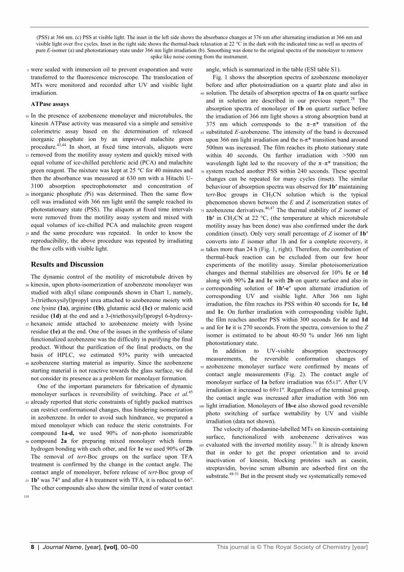
Fig. 1 Absorption spectral changes of 10% of 1b-e and 90% of 2a or 2b in quartz surface after deprotection of tert-Boc group [left (1), (3), (5), (7)] and 5
that of 1b′-d′ in acetonitrile and 1e′ in DCM before deprotection of tert-Boc group [right (2), (4), (6), (8)]. (a) Before irradiation. (b) photostationary state
(1)
(3)
(5)
(7)
(6)
(4)
(2)
(8)
8 | Journal Name, [year], [vol], 00–00 This journal is © The Royal Society of Chemistry [year]
(PSS) at 366 nm. (c) PSS at visible light. The inset in the left side shows the absorbance changes at 376 nm after alternating irradiation at 366 nm and visible light over five cycles. Inset in the right side shows the thermal-back relaxation at 22 °C in the dark with the indicated time as well as spectra of pure E-isomer (a) and photostationary state under 366 nm light irradiation (b). Smoothing was done to the original spectra of the monolayer to remove
spike like noise coming from the instrument.
were sealed with immersion oil to prevent evaporation and were 5
transferred to the fluorescence microscope. The translocation of MTs were monitored and recorded after UV and visible light irradiation.
ATPase assays
In the presence of azobenzene monolayer and microtubules, the 10
kinesin ATPase activity was measured via a simple and sensitive colorimetric assay based on the determination of released inorganic phosphate ion by an improved malachite green procedure.43,44 In short, at fixed time intervals, aliquots were removed from the motility assay system and quickly mixed with 15
equal volume of ice-chilled perchloric acid (PCA) and malachite green reagent. The mixture was kept at 25 °C for 40 minutes and then the absorbance was measured at 630 nm with a Hitachi U-3100 absorption spectrophotometer and concentration of inorganic phosphate (Pi) was determined. Then the same flow 20
cell was irradiated with 366 nm light until the sample reached its photostationary state (PSS). The aliquots at fixed time intervals were removed from the motility assay system and mixed with equal volumes of ice-chilled PCA and malachite green reagent and the same procedure was repeated. In order to know the 25
reproducibility, the above procedure was repeated by irradiating the flow cells with visible light.
Results and Discussion The dynamic control of the motility of microtubule driven by kinesin, upon photo-isomerization of azobenzene monolayer was 30
studied with alkyl silane compounds shown in Chart 1, namely, 3-(triethoxysilyl)propyl urea attached to azobenzene moiety with one lysine (1a), arginine (1b), glutamic acid (1c) or malonic acid residue (1d) at the end and a 3-(triethoxysilyl)propyl 6-hydroxy-hexanoic amide attached to azobenzene moiety with lysine 35
residue (1e) at the end. One of the issues in the synthesis of silane functionalized azobenzene was the difficulty in purifying the final product. Without the purification of the final products, on the basis of HPLC, we estimated 93% purity with unreacted azobenzene starting material as impurity. Since the azobenzene 40
starting material is not reactive towards the glass surface, we did not consider its presence as a problem for monolayer formation. One of the important parameters for fabrication of dynamic monolayer surfaces is reversibility of switching. Pace et al.45 already reported that steric constraints of tightly packed matrixes 45
can restrict conformational changes, thus hindering isomerization in azobenzene. In order to avoid such hindrance, we prepared a mixed monolayer which can reduce the steric constraints. For compound 1a-d, we used 90% of non-photo isomerizable compound 2a for preparing mixed monolayer which forms 50
hydrogen bonding with each other, and for 1e we used 90% of 2b. The removal of tert-Boc groups on the surface upon TFA treatment is confirmed by the change in the contact angle. The contact angle of monolayer, before release of tert-Boc group of 1b′ was 74° and after 4 h treatment with TFA, it is reduced to 66°. 55
The other compounds also show the similar trend of water contact
angle, which is summarized in the table (ESI table S1). Fig. 1 shows the absorption spectra of azobenzene monolayer before and after photoirradiation on a quartz plate and also in solution. The details of absorption spectra of 1a on quartz surface 60
and in solution are described in our previous report.28 The absorption spectra of monolayer of 1b on quartz surface before the irradiation of 366 nm light shows a strong absorption band at 375 nm which corresponds to the π–π* transition of the substituted E-azobenzene. The intensity of the band is decreased 65
upon 366 nm light irradiation and the n-π* transition band around 500nm was increased. The film reaches its photo stationary state within 40 seconds. On further irradiation with >500 nm wavelength light led to the recovery of the π–π* transition; the system reached another PSS within 240 seconds. These spectral 70
changes can be repeated for many cycles (inset). The similar behaviour of absorption spectra was observed for 1b′ maintaining tert-Boc groups in CH3CN solution which is the typical phenomenon shown between the E and Z isomerization states of azobenzene derivatives.46,47 The thermal stability of Z isomer of 75
1b′ in CH3CN at 22 °C, (the temperature at which microtubule motility assay has been done) was also confirmed under the dark condition (inset). Only very small percentage of Z isomer of 1b′ converts into E isomer after 1h and for a complete recovery, it takes more than 24 h (Fig. 1, right). Therefore, the contribution of 80
thermal-back reaction can be excluded from our few hour experiments of the motility assay. Similar photoisomerization changes and thermal stabilities are observed for 10% 1c or 1d along with 90% 2a and 1e with 2b on quartz surface and also in corresponding solution of 1b′-e′ upon alternate irradiation of 85
corresponding UV and visible light. After 366 nm light irradiation, the film reaches its PSS within 40 seconds for 1c, 1d and 1e. On further irradiation with corresponding visible light, the film reaches another PSS within 300 seconds for 1c and 1d and for 1e it is 270 seconds. From the spectra, conversion to the Z 90
isomer is estimated to be about 40-50 % under 366 nm light photostationary state. In addition to UV-visible absorption spectroscopy measurements, the reversible conformation changes of azobenzene monolayer surface were confirmed by means of 95
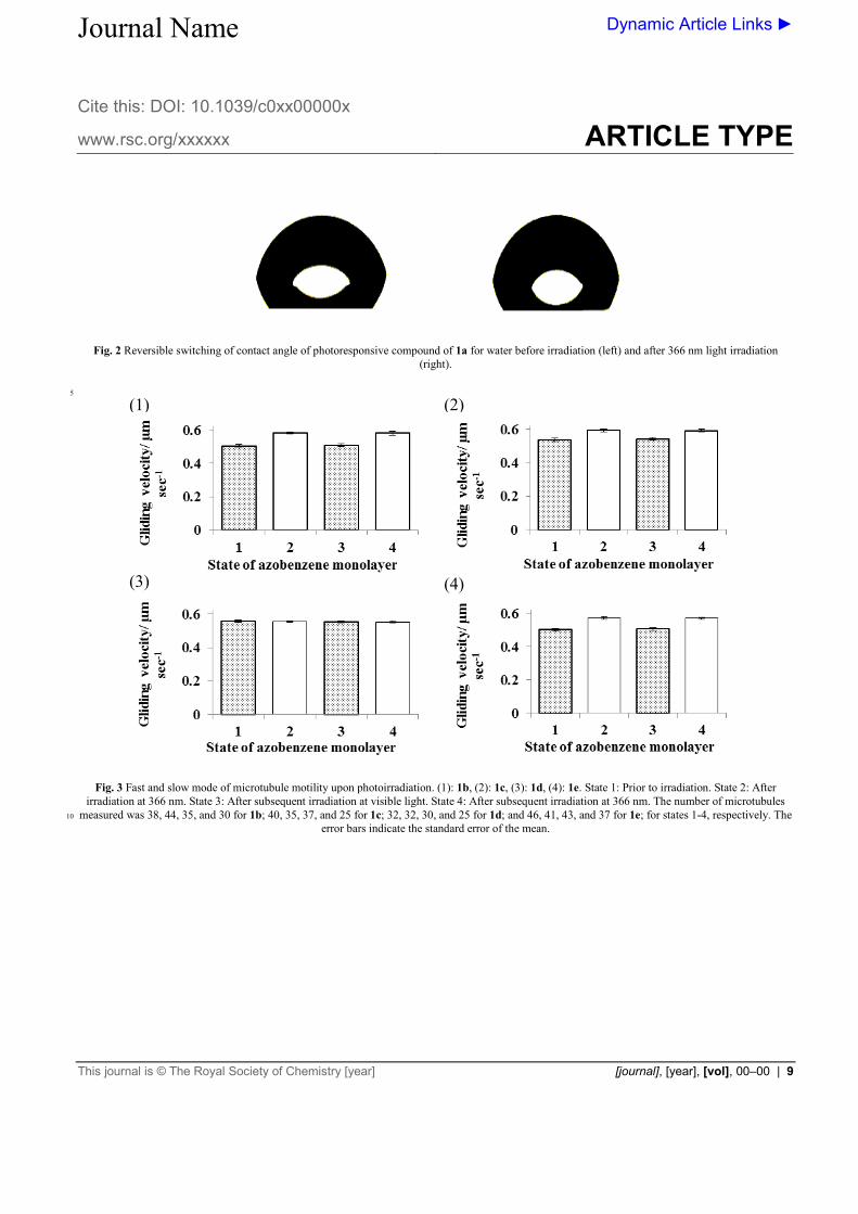
contact angle measurements (Fig. 2). The contact angle of monolayer surface of 1a before irradiation was 65±1º. After UV irradiation it increased to 69±1º. Regardless of the terminal group, the contact angle was increased after irradiation with 366 nm light irradiation. Monolayers of 1b-e also showed good reversible 100
photo switching of surface wettability by UV and visible irradiation (data not shown). The velocity of rhodamine-labelled MTs on kinesin-containing surface, functionalized with azobenzene derivatives was evaluated with the inverted motility assay.11 It is already known 105
that in order to get the proper orientation and to avoid inactivation of kinesin, blocking proteins such as casein, streptavidin, bovine serum albumin are adsorbed first on the substrate.48-51 But in the present study we systematically removed
110
Journal Name
Cite this: DOI: 10.1039/c0xx00000x
www.rsc.org/xxxxxx
Dynamic Article Links ►
ARTICLE TYPE
This journal is © The Royal Society of Chemistry [year] [journal], [year], [vol], 00–00 | 9
Fig. 2 Reversible switching of contact angle of photoresponsive compound of 1a for water before irradiation (left) and after 366 nm light irradiation
(right).
5
Fig. 3 Fast and slow mode of microtubule motility upon photoirradiation. (1): 1b, (2): 1c, (3): 1d, (4): 1e. State 1: Prior to irradiation. State 2: After
irradiation at 366 nm. State 3: After subsequent irradiation at visible light. State 4: After subsequent irradiation at 366 nm. The number of microtubules measured was 38, 44, 35, and 30 for 1b; 40, 35, 37, and 25 for 1c; 32, 32, 30, and 25 for 1d; and 46, 41, 43, and 37 for 1e; for states 1-4, respectively. The 10
error bars indicate the standard error of the mean.
(1) (2)
(3) (4)
Journal Name
Cite this: DOI: 10.1039/c0xx00000x
www.rsc.org/xxxxxx
Dynamic Article Links ►
ARTICLE TYPE
This journal is © The Royal Society of Chemistry [year] [journal], [year], [vol], 00–00 | 10
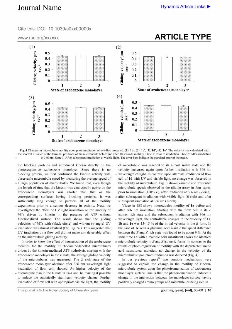
Fig. 4 Changes in microtubule motility upon photoirradiation of tert-Boc protected. (1): 1b′, (2): 1c′, (3): 1d′, (4): 1e′. The velocity was calculated with
the shortest distance of the terminal positions of the microtubule before and after 10 seconds motility. State 1: Prior to irradiation. State 2: After irradiation at 366 nm. State 3: After subsequent irradiation at visible light. The error bars indicate the standard error of the mean. 5
the blocking proteins and introduced kinesin directly on the photoresponsive azobenzene monolayer. Since there is no blocking protein, we first confirmed the kinesin activity with observable microtubule speed by measuring the average speed of a large population of microtubules. We found that, even though 10
the length of time that the kinesin was catalytically active on the azobenzene monolayers was shorter than that on the corresponding surfaces having blocking proteins, it was sufficiently long enough to perform all of the motility experiments prior to a serious decrease in activity. Next, we 15
investigated the effect of UV light irradiation on the motility of MTs driven by kinesin in the presence of ATP without functionalized surface. The result shows that the gliding velocities of MTs with (black circle) and without (triangle) UV irradiation was almost identical (ESI Fig. S2). This suggested that, 20
UV irradiation on a flow cell did not make any detectable effect on the microtubule gliding motility. In order to know the effect of isomerization of the azobenzene moieties for the motility of rhodamine-labelled microtubules driven by the kinesin-mediated ATP hydrolysis, starting with the 25
azobenzene monolayer in the E state, the average gliding velocity of the microtubules was measured. The Z rich state of the azobenzene monolayer obtained after 366 nm wavelength light irradiation of flow cell, showed the higher velocity of the microtubule than in the E state in 1a-c and 1e, making it possible 30
to induce the statistically significant velocity change. Further irradiation of flow cell with appropriate visible light, the motility
of microtubule was reached to its almost initial state and the velocity increased again upon further irradiation with 366 nm wavelength of light. In contrast, upon alternate irradiation of flow 35
cell of 1d with UV and visible light, no change was observed in the motility of microtubule. Fig. 3 shows variable and reversible microtubule speeds observed in the gliding assay in four states: prior to irradiation (100% E), after irradiation at 366 nm (Z-rich), after subsequent irradiation with visible light (E-rich) and after 40
subsequent irradiation at 366 nm (Z-rich). Video in ESI shows microtubules motility of 1a before and after 366 nm irradiation. Starting with the flow cell in its E isomer rich state and the subsequent irradiation with 366 nm wavelength light, the controllable changes in the velocity of 1a, 45
1b and 1e was 13~15 % of the initial velocity in the E form. In the case of 1c with a glutamic acid residue the speed difference between the E and Z rich state was found to be about 9 %. At the same time 1d with a malonic acid substituent shows the identical microtubule velocity in E and Z isomeric forms. In contrast to the 50
results of photo-regulation of motility with the deprotected amino acid substituted moieties; no change in the velocity of the microtubules upon photoirradiation was detected (Fig. 4). In our previous report28 two possible mechanisms were suggested to explain the change in the motility of kinesin-55
microtubule system upon the photoisomerization of azobenzene monolayer surface. One is that the photoisomerization induced a change in the interaction between the monolayer surface having positively charged amino groups and microtubules being rich in
(1) (2)
(4) (3)
Journal Name
Cite this: DOI: 10.1039/c0xx00000x
www.rsc.org/xxxxxx
Dynamic Article Links ►
ARTICLE TYPE
This journal is © The Royal Society of Chemistry [year] [journal], [year], [vol], 00–00 | 11
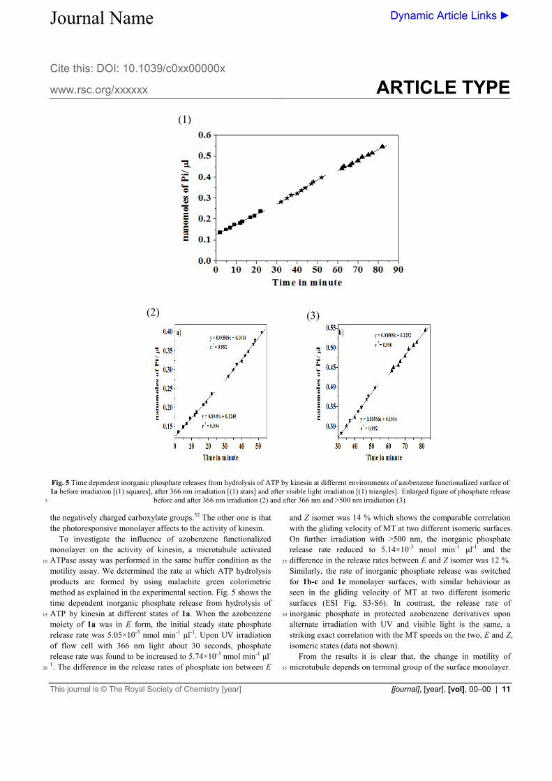
Fig. 5 Time dependent inorganic phosphate releases from hydrolysis of ATP by kinesin at different environments of azobenzene functionalized surface of 1a before irradiation [(1) squares], after 366 nm irradiation [(1) stars] and after visible light irradiation [(1) triangles]. Enlarged figure of phosphate release
before and after 366 nm irradiation (2) and after 366 nm and >500 nm irradiation (3). 5
the negatively charged carboxylate groups.52 The other one is that the photoresponsive monolayer affects to the activity of kinesin. To investigate the influence of azobenzene functionalized monolayer on the activity of kinesin, a microtubule activated ATPase assay was performed in the same buffer condition as the 10
motility assay. We determined the rate at which ATP hydrolysis products are formed by using malachite green colorimetric method as explained in the experimental section. Fig. 5 shows the time dependent inorganic phosphate release from hydrolysis of ATP by kinesin at different states of 1a. When the azobenzene 15
moiety of 1a was in E form, the initial steady state phosphate release rate was 5.05×10⁻³ nmol min-1 μl-1. Upon UV irradiation of flow cell with 366 nm light about 30 seconds, phosphate release rate was found to be increased to 5.74×10⁻³ nmol min-1 μl-
1. The difference in the release rates of phosphate ion between E 20
and Z isomer was 14 % which shows the comparable correlation with the gliding velocity of MT at two different isomeric surfaces. On further irradiation with >500 nm, the inorganic phosphate release rate reduced to 5.14×10⁻³ nmol min-1 μl-1 and the difference in the release rates between E and Z isomer was 12 %. 25
Similarly, the rate of inorganic phosphate release was switched for 1b-c and 1e monolayer surfaces, with similar behaviour as seen in the gliding velocity of MT at two different isomeric surfaces (ESI Fig. S3-S6). In contrast, the release rate of inorganic phosphate in protected azobenzene derivatives upon 30
alternate irradiation with UV and visible light is the same, a striking exact correlation with the MT speeds on the two, E and Z, isomeric states (data not shown). From the results it is clear that, the change in motility of microtubule depends on terminal group of the surface monolayer. 35
(1)
(2) (3)
12 | Journal Name, [year], [vol], 00–00 This journal is © The Royal Society of Chemistry [year]
When our photoresponsive azobenzene monolayer 1a, 1b or 1e presenting two or more free amino groups having a net positive charge in the buffer solution was used for the inverted motility assay experiment, the gliding velocity of microtubule in the Z state was 13~15 % higher than the gliding velocity in the E state. 5
The azobenzene monolayer surface of 1c offering one negatively charged carboxyl group and one positively charged amino group gave an increase in microtubule velocity of 9 % in the Z state than the gliding velocity in the E state. In contrast negatively charged surface of 1d and non-polar surface of protected 10
monolayer didn’t show any change in the gliding velocity between two isomeric forms. The striking exact correlation between the difference in the gliding velocity of microtubule driven by kinesin and the difference in the rate at which the ATP hydrolysis products 15
formed at two isomeric surfaces, E and Z, allows us to conclude that, the change in the microtubule gliding motility is mainly due to the change in activity of kinesin. It is already reported that the activity or the reaction kinetics of kinesin is changed depending on the nature of the surface of the substrate and in some extreme 20
cases, the kinesin is completely inactivated in the absence of a blocking protein on some substrates.50 Recently Martin et al. reported that when the polymer of hydroxymethylated derivative of EDOT, (2,3-dihydrothieno [3,4-b][1,4]dioxin-methanol, ‘CH2OH-EDOT’) on surface was electrochemically transformed 25
from a cationic (doped) state to a neutral (dedoped) state and vice versa, the ATPase activity of the adsorbed kinesin can be controlled reversibly, resulted in the reversible control of the gliding speed of the associated microtubule between fast (in dedoped state) and slow (in doped state) mode.53 It is reasonably 30
expected that the terminal amino groups with positive charge dive into the forest of alkyl chain of 2a or 2b during E-to-Z photoisomerization upon 366 nm wavelength light irradiation, which is proved by the increase in contact angle of the monolayer. So when the positively charged amino groups are exposed to the 35
surface at E state, the slowdown of microtubule motility is observed and when the alkyl chain is exposed to the surface at Z state, the motility of microtubule increased.
The relationship between the kinesin activity and the presence of positive charges on the surface is not clear. The possible 40
mechanism which can explain the change in the kinesin activity and the gliding motility of the microtubule is that the positive charge might have affected the secondary structure of the kinesin, resulting in a change in its activity of ATP hydrolysis.
Conclusions 45
We observed that an azobenzene monolayer surface needs to have free amino terminal groups for the dynamic control of the motility of microtubule. The surface of azobenzene monolayer with terminal amino groups can dynamically control the ATP hydrolysis activity of kinesin which further induces the change in 50
motility of microtubule. We believe that the reported method could facilitate the rational design of new type of photo controllable molecular devices if we could control the motility of microtubule between complete ON/ OFF switching. Currently we are trying to find out the more suitable terminal group which can 55
control completely the ON/ OFF switching of the function of kinesin.
Acknowledgments This work was supported by a grant-in-aid for science research in a priority area “New Frontiers in Photochromism (no. 471)”from 60
the Ministry of Education, Culture, Sports, Science, and Technology (MEXT), Japan. M.K.A.R. acknowledges The Ushio Foundation for a scholarship. We thanks to Dr. Y. Hiratsuka, Dr. Y. Hachikubo and Dr. T. Q. P. Uyeda for kindly giving recombinant kinesin construct and Dr. A. Kakugo, Y. Tamura 65
and Prof. J. P. Gong for help in purification of protein specimen.
Notes and references a Research Institute for Electronic Science, Hokkaido University, N20, W10, Kita-ku, 001-0020,Sapporo, Hokkaido, Japan. Fax: +81 11 706 9357. Tel: +81 11 706 9356. E-mail: [email protected]. 70
† Electronic Supplementary Information (ESI) available: [details of any supplementary information available should be included here]. See DOI: 10.1039/b000000x/ ‡ Footnotes should appear here. These might include comments relevant to but not central to the matter under discussion, limited experimental and 75
spectral data, and crystallographic data. 1 A. Agarwal and H. Hess, Prog. Polym. Sci., 2010, 35, 252–277. 2 A. Goel and V. Vogel, Nat. Nanotechnol., 2008, 3, 465–475. 3 M. G. L van den Heuvel and C. Dekker, Science, 2007, 317, 333–336. 4 T. Fischer and H. Hess, J. Mater. Chem., 2007, 17, 943–951. 80
5 J. R. Dennis, J. Howard and V. Vogel, Nanotechnology, 1999, 10, 232-236.
6 M. G. L. van den Heuvel, M. P. De Graaff and C. Dekker, Science, 2006, 312, 910–914.
7 L. Ionov, M. Stamm and S. Diez, Nano Lett., 2005, 5, 1910–1914. 85
8 K. J. Bohm, J. Beeg, G. M. zu Horste, R. Stracke and E. Unger, IEEE Trans. Adv. Packaging, 2005, 28, 571–576.
9 R. K. Doot, H. Hess and V. Vogel, Soft Matter, 2007, 3, 349–356. 10 R. D Vale, T. S. Reese and M. P. Sheetz, Cell, 1985, 42, 39-50. 11 J. Howard, A. J. Hudspeth and R. D. Vale, Nature, 1989, 342, 154-158. 90
12 B. J. Schnapp, T. S. Reese and R. Bechtold, J. Cell Biol., 1992, 119, 389-399.
13 S. T. Brady, Nature, 1985, 317, 73-75. 14 R. D. Vale, T. S. Reese and M. P. Sheetz, Cell, 1985, 42, 39-50. 15 N. Hirokawa, Y. Noda and Y. Okada, Curr. Opin. Cell Biol., 1998, 10, 95
60-73. 16 N. Hirokawa and R. Takemura, Nat. Rev. Neurosci., 2005, 6, 201-214. 17 R. D. Vale, Cell, 2003, 112, 467-480. 18 D. L. Coy, M. Wagenbach and J. Howard, J. J. Biol. Chem., 1999, 274,
3667-3671. 100
19 S. M. Block, L. S. Goldstein and B. J. Schnapp, Nature, 1990, 348, 348-352.
20 W. O. Hancock and J. Howard, J. Cell biol., 1998, 140, 1395-1405. 21 Y. Hiratsuka, T. Tada, K. Oiwa, T. Kanayama and T. Q. P. Uyeda,
Biophys. J., 2001, 81, 1555-1561. 105
22 M. G. L. van denHeuvel, C. T. Butcher, R. M. M. Smeets, S. Diez and C. Dekker, Nano Lett., 2005, 5, 1117-1122.
23 M. Platt, G. Muthukrishnan, W. O. Hancock and M. E. Williams, J. Am. Chem. Soc., 2005, 127, 15686-15687.
24 L. Limberis, J. J. Magda and R. J. Stewart, Nano Lett., 2001, 1, 277-110
280. 25 H. Higuchi, E. Muto, Y. Inoue and T. Yanagida, Proc. Natl. Acad. Sci.
U.S.A., 1997, 94, 4395-4400. 26 H. Hess, J. Clemmens, D. Qin, J. Howard and V. Vogel, Nano Lett.,
2001, 1, 235-239. 115
27 A. Nomura, T. Q. P. Uyeda, N. Yumoto and Y. Tatsu, Chem. Commun., 2006, 3588-3590.
28 M. K. A. Rahim, T. Fukaminato, T. Kamei and N. Tamaoki, Langmuir, 2011, 27, 10347–10350.
29 F. Ercole, T. P. Davis and R. A. Evans, Polym. Chem., 2010, 1, 37-54. 120
30 N. Tamaoki, and T. Kamei, J. Photochem. Photobiol. C., 2010, 11, 47-61.
31 T. Seki and S. Nagano, Chem. Lett., 2008, 37, 484-489.
This journal is © The Royal Society of Chemistry [year] Journal Name, [year], [vol], 00–00 | 13
32 S. Yagai and A. Kitamura, Chem. Soc. Rev., 2008, 37, 1520-1529. 33 K. G. Yager and C. J. Barrett, J. Photochem. Photobiol. A., 2006, 182,
250-261. 34 C. J. Barrett, J. Mamiya, K. G. Yager and T. Ikeda. Soft Matter, 2007,
3, 1249–1261. 5
35 The following article describes the photocontrol of the ATPase activity of kinesin but not the motility using an azobenzene derivative: M. D. Yamada, Y. Nakajima, H. Maeda and S. Maruta, J. Biochem., 2007, 142, 691-698.
36 P. Santurri, F. Robbins and R. Stubbings, Organic Syntheses, 1973, 10
Coll. Vol. 5, 341; 1960, 40, 18. 37 F. Cisnetti, R. Ballardini, A. Credi, M. T. Gandolfi, S. Masiero, F.
Negri, S. Pieraccini and G. P. Spada, Chem. Eur. J., 2004, 10, 2011-2021.
38 K. Aoki, T. Seki, Y. Suzuki, T. Tamaki, A. Hosoki and K. Ichimura, 15
Langmuir, 1992, 8, 1007-1013. 39 M. Dasog, A. Kavianpour, M. F. Paige, H. B. Kraatz and R. W. J.
Scott, Can. J. Chem., 2008, 86, 368-375. 40 M. Castoldi and A. V. Popov, Protein Expression and Purification,
2003, 32, 83-88. 20
41 H. Suzuki, K. Oiwa, A. Yamada, H. Sakakibara, H. Nakayama and S. Mashiko, Jpn. J. Appl. Phys., 1995, 34, 3937-3941.
42 J. Howard, A. J. Hunt and S. Baek, Methods in Cell Biology, 1993, 39,137-147.
43T. Kodama, K. Fukui and K. Kometani, J. Biochem., 1986, 99, 1465-25
1472. 44 H. H. Hess and J. E. Derr, Anal. Biochem., 1975,63, 607-613. 45 G. Pace, V. Ferri, C. Grave, M. Elbing, C. von Hanisch, M. Zharnikov,
M. Mayor, M. A. Rampi and P. Samori, Proc. Natl. Acad. Sci. U.S.A., 2007, 104, 9937–9942. 30
46 K. G. Yager and C. J. Barrett, J. Photochem. Photobiol. A., 2006, 182, 250-261.
47 K. Ichimura, Y. Suzuki, T. Seki, A. Hosoki and K. Aoki, Langmuir, 1988, 4, 1214-1216.
48 T. Ozeki, V. Verma, M. Uppalapati, M.; Y. Suzuki, M. Nakamura, J. 35
M. Catchmark and W. O. Hancock, Biophys. J., 2009, 96, 3305-3318. 49 D. J. G. Bakewell and D. V. Nicolau, Aust. J. Chem., 2007, 60, 314-
332. 50 C. Brunner, K. H. Ernst, H. Hess and V. Vogel, Nanotechnology, 2004,
15, S540-S548. 40
51 E. Berliner, H. K. Mahtani, S. Karki, L. F. Chu, J. E. Cronan and J. Gelles, J. Biol. Chem., 1994, 269, 8610–8615.
52 D. C. Turner, C. Chang, K. Fang, S. L. Brandow and D. B. Murphy, Biophys. J., 1995, 69, 2782-2789.
53 B. D. Martin, L. M. Velea, C. M. Soto, C. M. Whitaker, B. P .Gaber 45
and B. Ratna, Nanotechnology, 2007, 18, 055103.