Embed Size (px)
Citation preview

1
Organic Synthesis and Fungicidal
Activity of Oxylipin-Based Compounds
Yi Zhou
Doctor of Philosophy
ASTON UNIVERSITY
January 2011
This copy of the thesis has been supplied on condition that anyone who
consults it is understood to recognise that its copyright rests with its author
and that no quotation from the thesis and no information derived from it
may be published without proper acknowledgement.

2
Thesis summary
Organic Synthesis and Fungicidal Activity of Oxylipin-Based
Compounds
Yi Zhou
Doctor of Philosophy
ASTON UNIVERSITY
January 2011
Previous research has shown that the naturally occurring reactive electrophilic species
(RES), 12-oxophytodienoic acid (OPDA), not only serves as a precursor for jasmonic
acid but is also a potent antifungal compound. However, both the low amount present in
plants and the multistep synthesis required to produce this compound on a scale viable
for agrochemical use currently limits its practical value. The aim of this research was to
generate a range of molecular mimics of OPDA with a minimum number of synthetic
steps and screen for antifungal activity. Synthetic 4-octyl-cyclopentenone containing the
cyclopentenone ring and an eight carbon alkyl chain was found to show the highest in
vitro antifungal activity against C. herbarum and B. cinerea with minimum inhibition
concentration (MIC) of 100-200μM. This indicates that structurally simplified
4-octyl-cyclopentenone can be successfully synthesised to mimic the antifungal activity
of OPDA against specific fungal strains. Application of 4-octyl-cyclopentenone could
act as surfactant by disrupting and disorganising the lipid membrane non-specifically,
resulting in the leakage of potassium ions, which was the proposed mode of action of
this compound. However, the sensitivity of fungi to this compound is not correlated to
the lipid composition of fungal spores. (E)-2-alkenals were also studied for their
antimicrobial activity and (E)-2-undecenal was found to have the highest antimicrobial
activity against a range of pathogens. The hydrophilic moiety (the α,β-unsaturated
carbonyl group), common to both (E)-2-undecenal and 4-octyl-cyclentenone is essential
to their bioactivity, and the hydrophobic moiety plays an important role in their
antimicrobial activities. 4-Octyl-cyclopentenone showed no visible toxicity to the test
plant, Arabidopsis thaliana, suggesting that its high antifungal activity against Botrytis
and Cladosporium could be exploited for commercialisation as a new generation of
agrochemical.
Key words: fungicides, oxylipins, OPDA, RES.

3
Acknowledgements
I would like to express my best regards, deepest thanks and sincere appreciation to Dr.
Gareth Griffiths for his continuous support, expert guidance and patience. This work
would not have been possible without his constant supervision and enthusiastic
encouragement. Dr. Griffiths provided me with all of the laboratory facilities and
instruments to undertake this project. I am grateful to him for giving me such an
opportunity to work in his group.
I am extremely grateful to Dr. Andy Sutherland for his professional guidance about
organic synthesis and his organic chemistry laboratory facilities and instruments to
carry on this project.
I would like to thank Professor Peter Lambert for his professional guidance about
leakage test, agar diffusion assay and MIC tests. His valuable advice and patience are
greatly appreciated.
I would like to thank Professor Eric Holub and Dr. Joana G. Vicente from Warwick
HRI for providing seeds of Arabidopsis thaliana (Col-5) and Brassica oleracea.
I would like to thank Dr. Katherine J. Denby (Warwick HRI) for her professional
guidance about in vivo antifungal test on leaves of Arabidopsis thaliana.

4
I would like to thank Dr. Joelle Fournier for her advice and help on fungal culture and
antifungal tests.
I would like to thank Dr. Mike Perry and Karen Farrow for their help on NMR and
APCI MS.
I would like to thank Dr. Richard Armstrong for his advice on statistical analysis.
My thanks are also extended to Dr. John Behrendt, Mr. Simon Smith, Mr. Anuj Chander,
Mr. Tulpesh Patel and Mr. Steve Farmer for their cooperation and help in the
laboratory.
Finally, I would like to express many thanks to my wife and parents for their endless
support in my life.

5
Contents
Pages
Abbreviations 11
Tables, Figures and Schemes 16
Chapter 1 Introduction
1.1 Introduction to fungicides 20
1.2 Natural fungicides 22
1.3 Plant oxylipins 25
1.3.1 Plant lipids 25
1.3.2 Lipid oxidation 31
1.3.3 Enzymatic oxidation pathway 34
1.3.3.1 Lipoxygenases 34
1.3.3.2 Allene oxide synthesis pathway 36
1.3.3.3 Hydroperoxide lyase pathway 42
1.3.4 Non-enzymatic oxidation pathways 45
1.3.4.1 Biosynthesis of phytoprostanes 46
1.3.4.2 Bioactivity of phytoprostanes 51
1.4 Fatty acid derived reactive electrophilic species (RES) 52
1.4.1 Oxylipin reactive electrophilic species 53
1.4.2 Electrophilicity and lipophilicity of RES 54

6
1.4.3 Biological activities of oxylipin RES in plants 57
1.4.4 Phytotoxicity of RES 60
1.5 Molecular design based on 12-oxo-PDA 62
1.6 Aims 66
Chapter 2 Organic Synthesis and Characterisation of Target Compounds
2.1 Introduction 68
2.1.1 Grignard reagent 69
2.1.2 Installation of alkyl group 70
2.1.3 Hydroxyl group oxidation 72
2.2 Materials 73
2.3 Synthetic procedure for target compounds 74
2.3.1 Synthesis of compounds 3-5 75
2.3.2 Synthesis of 8-(4-oxocyclopentenyl)octanoic acid (8) 78
2.4 Discussion 82
2.5 Conclusion 84
Chapter 3 Antifungal Activity Assays
3.1 Introduction 86
3.1.1 Fungal pathogens 86

7
3.1.2 Lab facilities and safety 91
3.2 In vitro antifungal activity assays 92
3.2.1 Material and equipment 92
3.2.1.1 Fungal strains 92
3.2.1.2 Antifungal activity of synthesised compounds 93
3.2.1.3 Chemicals 93
3.2.1.4 Apparatus 94
3.2.2 Methods 94
3.2.2.1 Fungal culture 94
3.2.2.2 Harvest of fungal spores 95
3.2.2.3 Antifungal tests 96
3.2.2.4 Spore germination assay 98
3.2.3 Results 99
3.2.3.1 Antifungal tests 99
3.2.3.2 Antifungal activities of 4-octyl-cyclopentenone 104
3.2.3.3 Effect of compounds on fungal spore germination 109
3.2.4 Discussion 111
3.2.5 Conclusion 116
3.3 In vivo Antifungal assay of 4-octyl-cyclopentenone 116
3.3.1 Materials 117
3.3.2 Methods 117
3.3.2.1 Growing of the test plants 117

8
3.3.2.2 Phytotoxicity test of 4-octyl-cyclopentenone on Brassica oleracea 118
3.3.2.3 In vivo antifungal test on leaves of Arabidopsis thaliana 119
3.3.2.4 In vivo antifungal test on apricot fruits 120
3.3.3 Results 120
3.3.3.1 Phytotoxicity test of 4-octyl-cyclopentenone 120
3.3.3.2 In vivo antifungal test on leaves of Arabidopsis thaliana 121
3.3.3.3 Antifungal test on apricot fruits 123
3.3.4 Discussion 124
3.3.5 Conclusion 126
3.4 Summary 126
Chapter 4 Study on the mode of action of 4-octyl-cyclopentenone
4.1 Introduction 128
4.1.1 Mode of action of fungicides 128
4.1.2 Plasma membrane structure of cells 131
4.1.3 Electrolyte leakage 132
4.1.4 Lipid extraction and analysis 133
4.2 Materials 134
4.3 Methods 134
4.3.1 Adsorption of compounds to spore 135
4.3.2 Leakage test of potassium ions from spores 135

9
4.3.3 Lipid extraction and analysis 136
4.4 Results 138
4.4.1 Adsorption of compounds to spores 138
4.4.2 Leakage of potassium ions from spores 140
4.4.3 Lipid composition of fungal spores 143
4.5 Discussion 150
4.6 Conclusion 155
Chapter 5 Antimicrobial Activities of (E)-2-alkenals
5.1 Introduction 156
5.2 Materials 157
5.3 Methods 158
5.3.1 Antifungal test against plant fungal strains 158
5.3.2 Agar diffusion tests 159
5.3.3 Antimicrobial MIC assays 159
5.4 Results 160
5.4.1 Antifungal activity against plant pathogens 160
5.4.2 Antimicrobial activity against human pathogens 164
5.5 Discussion 168
5.6 Conclusion 173

10
Chapter 6 Summary and Future Work
6.1 Summary 174
6.2 Future work 176
References 182
Appendices 194
11
Abbreviations
ACP Acyl carrier protein
AOC Allene oxide cyclase
AOS Allene oxide synthase
APCI Atmospheric pressure chemical ionisation
ATCC American type culture collection
Avr Avirulence
CDCl3 Deuterated chloroform
COI1 Coronatine Insensitive 1
CrO3 Chromium trioxide
CuCN Copper(I) cyanide
CuI Copper(I) iodide
DAG Diacylglycerol
DCM Dichloromethane
DES Divinyl ether synthase
DGDG Digalactosyl diacylglycerol
DMF Dimethylformamide
DMSO Dimethylsulfoxide
α-DOX α-Dioxygenase
EAS
ESI
Epoxy alcohol synthase
Electrospray ionization

12
ER Endoplasmic reticulum
Et2O Diethyl ether
EtOH Ethanol
EtOAc Ethyl acetate
FAME Fatty acid methyl esters
FID Flame-ionisation detector
GC Gas Chromatography
GLV Green leaf volatile
GPL Glycerophospholipid
GSH Glutathione
GST Glutathione-S-transferase
HEPA High efficiency particulate air
HPL Hydroperoxide lyase
HPLC High performance liquid chromatography
HR Hypersensitive reaction
IsoP Isoprostane
JA Jasmonic acid
KAT L-3-ketoacyl-CoA thiolase
KODE Ketodiene
KOTE Ketotriene
α-LeA α-Linolenic acid
LA Linoleic acid

13
LOX Lipoxygenase
LRMS
LUMO
Low resolution mass spectroscopy
Lowest unoccupied molecular orbital
MAG Monoacylglycerol
MeJA Methyl jasmonic acid
MDA Malondialdehyde
MFC Minimum fungicidal concentration
MFP Multifunctional protein
MGDG Monogalactosyl diacylglycerol
MIC Minimum inhibition concentration
MP Melting point
MS Mass spectra
MVK Methyl vinyl ketone
NA
NAD
NADP
Nutrient agar
Nicotinamide adenine dinucleotide
Nicotinamide adenine dinucleotide phosphate
NB Nutrient broth
NCTC National collection of type cultures
NEFA Nonesterified fatty acid
NMR Nuclear magnetic resonance
OPDA (12-oxo-PDA) 12-oxo-10,15c-phytodienoic acids
dnOPDA Dinor 12-oxo-phytodienoic acid

14
OPR 12-oxo-phytodienoate reductase
PCD Programmed cell death
PDA Potato dextrose agar
PDB Potato dextrose broth
PE Phosphatidylethanolamine
PG Phosphatidylglycerol
RES Reactive electrophilic species
PP Phytoprostane
PR Pathogenesis-related
PUFA Polyunsaturated fatty acid
PXG Peroxygenase
QSAR Quantitative structure-activity relationship
ROS Reactive oxidative species
RT Room temperature
SA Salicylic acid
SDA Sabouraud dextrose agar
SDB Sabouraud dextrose broth
SE Sterol esters
SRA System acquired resistance
TAG Triacylglycerol
TBAF Tetrabutylammonium fluoride
TBDPS tert-Butylchlorodiphenylsilane
15
TEM Transmission electron microscopy
TFA Trienoic fatty acid
THF
TMS
Tetrahydrofuran
Tetramethylsilane
TLC Thin layer chromatography

16
Figures, Tables and Schemes
Table 1.1 Some naturally occurring fatty acids: structure and nomenclature 27
Figure 1.1 Example of triacylglycerol 28
Figure 1.2 Structure-based classifications of membrane lipids 30
Figure 1.3 Amphipathic lipid aggregates that form in water 31
Figure 1.4 Overview of oxylipin biosynthesis 33
Figure 1.5 Oxidation catalysed by LOX 35
Figure 1.6 JA biosynthesis and OPDA metabolism in Arabidopsis thaliana 38
Figure 1.7 Antifungal activities of oxylipins 41
Figure 1.8 Hydroperoxide lyase pathway from 9- and 13- hydroperoxides of
linolenic acid
45
Table 1.2 Two different oxidation pathways generate hydroperoxides 47
Figure 1.9 Mechanism of linolenic acid autoixdation 48
Figure 1.10 Non-enzymatic phytoprostane biosynthesis from α-linolenic acid 49
Figure 1.11 The phytoprostane pathway in plants 50
Table 1.3 RES oxylipins in plants and their bio-synthetic pathway 54
Figure 1.12 Product formations after reaction with thiol and primary amine
nucleophiles
56
Figure 1.13 Designed target compounds 66
Scheme 2.1 Pathway to synthesise compound simplified from OPDA 69
Scheme 2.2 Installation of alkyl group 71

17
Table 2.1 Synthetic conditions of the reactions with high yield of
1,4-isomer
72
Scheme 2.3 Hydroxyl group oxidation 73
Table 2.2 Chemicals used in organic synthesis 74
Scheme 2.4 Synthesis of compounds 3-5 75
Scheme 2.5 Synthesis of 8-(4-oxocyclopentenyl)octanoic acid (8) 78
Figure 2.1 Synthetic compounds mimic of OPDA 85
Figure 3.1 Flowchart summarising classification of fungus-like organisms 87
Figure 3.2 Fusarium oxysporum 89
Figure 3.3 Alternaria brassicicola 89
Figure 3.4 Cladosporium herbarum 90
Figure 3.5 Botrytis cinerea 91
Table 3.1 Fungal strains tested 93
Figure 3.6 Hemocytometer (Bright-Line, Sigma-Aldrich) 96
Figure 3.7 Results of in vitro antifungal assay 102
Figure 3.8 In vitro antifungal assay of 4-octyl-cyclopentenone (3) 107
Figure 3.9 Effect of compounds on spore germination 110
Table 3.2 Lipophilicity (log P) of compounds tested for in vitro antifungal
activity
112
Figure 3.10 Phytotoxicity assay of 4-octyl-cyclopentenone on Brassica
oleracea
121
Figure 3.11 In vivo antifungal test on leaves of Arabidopsis thaliana 122

18
Figure 3.12 In vivo antifungal test on apricots 123
Table 4.1 The various modes of action of different fungicides 129
Figure 4.1 Fluid mosaic model for general cell membranes and their
composition
132
Figure 4.2 The adsorption test 139
Figure 4.3 Leakage test of potassium ions 141
Table 4.2 Comparison of the yield of lipid extractions using different
methods
143
Table 4.3 Comparison of the composition of NEFA from extractions using
different methods for fungal strains
143
Figure 4.4 Lipid classes of three fungal strains 145
Table 4.4 The major fatty acid composition of four fungal strains 146
Table 4.5 Lipid class distribution of four fungal strains 146
Figure 4.5 Fatty acid composition of lipid classes 147
Table 4.6 The values of UNSAT/SAT and degree of unsaturation (Δ/mol)
of four fungal strains: A. brassicicola, F. oxysporum, C. hebarum
and B. cinerea(GLUK-1)
149
Table 5.1 Classification and culture method of human pathogens 158
Figure 5.1 Antifungal activity of (E)-2-alkenals against plant pathogens 162
Table 5.2 Antifungal (MIC) activity (micrograms per milliliter) of
(E)-2-alkenals against C. albicans, S. aureus and E. coli
166

19
Figure 5.2 Effect of (E)-2-alkenals on C. albicans (a), E. coli (b right) and
S. aureus (b left) on SDA
167
Table 5.3 Major phospholipid components of bacteria 171
Table 6.1 Potential applications in the future 181
.

20
Chapter1
Introduction
The development and application of agrochemicals to crops has a history over
thousands of years. A growing population is predicted to reach 6 billion by the middle
of this century, so there is a huge pressure to increase food production to sustain the
global population. Considering the constraints, one of the efficient ways to increase
production is to improve the yield of unit area using agrochemicals.
Up to now, most of the chemically synthesised traditional agrochemicals are
non-biodegradable, and their residues left in soil and water will have, potentially, severe
detrimental effects on environment and biosphere. In addition, some of them are toxic to
humans and animals [1]. To address these problems, recent research and development of
novel agrochemicals, which are environmental friendly and which have low toxicity
towards mammals is being undertaken.
1.1 Introduction to fungicides
Agrochemicals are classified according to their target organisms, e.g. insecticides,
herbicides, and fungicides, clearly depict their targets. For example, fungicides are used

21
extensively in agricultural production to control fungi. In 2004, the market value of
fungicides reached over 7 billion dollars, accounting for nearly 25% of the whole
agrichemical market share[1]. Since the 1960s, the world fungicide market has been
dominated by man-made organic chemicals and most of them pose an environmental
risk [2]. In the future, a primary concern for the development of new fungicides will be
their effect on the environment and safety application issues. Indeed the concept of an
agrochemical could change from killing (-cide) to controlling [1]. Generally speaking, it
is extremely expensive and difficult to discover a new compound with antifungal
activity. Five approaches of developing fungicides are generally recognised [2]:
Random screening: This is a traditional method for fungicide development based on
wide-scale screening of compounds. The approach works well since it does not
depend on knowledge of the modes of action of the compound. However, it
requires a high throughout method to efficiently screen the large amount of
candidate compounds.
Combinatorial chemistry: Used widely in pharmaceutical design, in which cheap
and low molecular weight compounds are systematically synthesised and tested.
Analogue synthesis: Based on existing fungicides, some chemical modification can
lead to a new fungicide showing enhanced properties.
Biorational design: This is becoming more increasingly important in novel
fungicide discovery, candidates could be synthesised for special mode of action
from prior knowledge of related modes of action.
Speculative approach: Using a simple chemical starting point and an array of

22
potential substitutions that lie within predetermined physicochemical limits, a
family of structural variants can be produced.
To date, most commercial fungicides are chemically, rather than biologically,
synthesised. Meanwhile, more and more natural compounds are being discovered to
have antifungal activity, which could play an important role in the market of fungicides
in future. Using the above approaches, the chemical modification of natural antifungal
molecules with enhanced properties can be envisaged to develop a new generation of
fungicides. Modifications of some naturally occurring fungicides might mimic natural
compound activity, or even improve their activity and increase the efficiency. In short,
the approaches mentioned above could be used for the study and exploitation of a new
generation of antifungal compounds based on naturally occurring products with
fungicidal properties to identify such lead molecules. A range of target compounds with
simplified structure from naturally occurring molecules need to be developed to satisfy
commercial demand.
1.2 Natural fungicides
Aside from synthesised agrochemicals, fundamental research has revealed that many
natural products contribute to plant protection against pathogens. These compounds,
including fungicides, insecticides and herbicides, could be derived from bacteria, fungi,

23
plants and animals. Recently, some of them have been used commercially [3]. They are
able to be used directly for industrial manufacture or as a starting point in developing
simpler and more efficiently synthesisable compounds, such as pyrrolnitrin, produced
by Pseudomonas pyrrocinia, and strobilurin A, produced by Strobilurus tenacellus [4].
In this research, plant-derived antifungal agents were investigated due to their important
participation in plant defence against fungal pathogens. Significant progress in our
understanding of plant-pathogen interactions has been made in recent years [5].
Although our knowledge about this area is still fragmented, some antifungal agents
derived from plants have been discovered, which can be broadly classified into two
groups:
1. Direct antifungal substances; they are secreted constitutively by plants or in
response to external stresses and have toxicity against fungal strains, inhibiting their
growth. These substances include antimicrobial proteins/ peptides [6], oils [7, 8],
fatty acids [9, 10], oxylipins [11] and phytoalexins [12]. The expression of some
antimicrobial peptides and proteins, like pathogenesis related (PR) proteins, is
triggered once plants are attacked by pathogens. Phytoalexins are secondary
metabolites of plants and have been well known for their antimicrobial activities for
many years. Several chemical types of phytoalexins are also well known, such as
isoprenoids and flavonoids [13]. Many oils and fatty acids extracted from different
species exhibit the ability of inhibiting fungal growth due to antifungal components

24
in the complex mixture. A major systemic study of 43 oxylipins revealed their
antimicrobial activities towards a range of bacterial and fungal pathogens [11].
2. Indirect antifungal agents; when pathogen invasion is recognised by disease
resistance (R) genes binding to specific pathogen-derived avirulence (Avr) proteins
and pathogen derived elicitors, a complex signalling network is induced by stimuli
[5]. Such stimuli, which normally have no antifungal activity per se, initiate plant
defence mechanisms by triggering the expression of genes which encode, for
example, pathogen related proteins, or enzymes involved in the biosynthesis of
phytoalexins, or the enzymes of oxidative stress protection, or regulation of
hypersensitive reaction (HR) that lead to rapid host cell death around infected sites.
Such events often arrest further microbial invasion. Some of the stimuli contributes
to both local inducible host resistance and „systemic acquired resistance‟ (SAR) to
pathogens. In plants, salicylic acid (SA) and reactive oxidative species (ROS) were
well studied as such stimuli of SAR in the 1990s [14, 15]. In the last decade,
oxylipins, especially the jasmonate family, such as jasmonic acid (JA) and its
precursor 12-oxo-phytodienoic acid (12-oxo-PDA/OPDA), were found to be closely
related to plant defence signalling and gene expression during wounding and
pathogen attack [16-19]. Non-enzymatically formed oxylipins, phytoprostanes,
which are generated from oxidised lipids by oxygen or reactive oxygen species
(ROS), were also reported to play a similar role in plants in response to biotic and
abiotic stress [20].

25
Interestingly, oxylipins can play both direct and indirect antifungal roles in plants. In
this research, oxylipins are the starting point for developing a new generation of
antifungal compounds.
1.3 Plant oxylipins
1.3.1 Plant lipids
Lipids, ubiquitous in all organisms, are fatty acids and their derivatives, and substances
related (biosynthetically or functionally) to these compounds [21]. Generally speaking,
fatty acids are monocarboxylic acids with a long unbranched chain containing an even
number of carbon atoms. The carbon chain length and degree of unsaturation is
generally given in a standard nomenclature style, e.g. oleic acid (18:1Δ9), where „18‟
represents chain length and the number after the colon is the number of double bond (i.e.
„1‟). The delta (Δ9) indicates the carbon number at which the double bond occurs
counting from the carboxylic end of the fatty acid. In plants the de novo synthesis of
fatty acids occurs in the plastids, in which long chain fatty acids, such as palmitic acid
(16:0), stearic acid (18:0) and oleic acid (18:1), are synthesised from the preformed
acetyl-CoA through several cycles of two carbon addition. The reactions involve a
series of condensation, reduction, dehydration and further enoyl reduction of acyl
thioester on acyl carrier protein (ACP) [22]. The end product of fatty acid synthesis is

26
palmitoyl-ACP (16:0-acyl-ACP) and this can be elongated by specific enzymes to
stearoyl-ACP (18:0-acyl-ACP). Most of the newly formed 18:0-acyl-ACP (typically
80-85%) are converted to oleoyl-ACP (18:1-acyl-ACP) by Δ9 desaturase [23, 24]. These
fatty acids are transported to the endoplasmic reticulum (ER) membrane for further
elongation and desaturation of chains to form other fatty acids, such as linoleic acid,
18:2 (Δ9,12
), α-linolenic acid, 18:3 (Δ9,12,15
) and longer chain fatty acids (C20-C24).
Elongation proceeds in a similar way to fatty acid synthesis. For desaturation, after the
incorporation of fatty acid acyl groups into glycerophospholipids, such as
phosphatidylcholine (PC), desaturases bound to the membrane of ER convert oleate
(18:1) to linoleate (18:2) and then to linolenate (18:3) [13, 25]. These fatty acids are
mainly esterified onto the glycerol back bone to form glycerolipids in plastids and ER;
small amount of fatty acids are consumed to generate minor lipids, such as
sphingolipids and sterol esters. Over 1,000 fatty acids are known with different chain
lengths, positions, configurations and degrees of unsaturation. However, only around 20
fatty acids occur widely in nature; of these, palmitic, oleic, and linoleic acids make up
~80% of commodity oils and fats [23]. The most commonly occurring fatty acids
having an even number of carbon atoms in an unbranched chain are listed in table 1.1.

27
Table 1.1 Some naturally occurring fatty acid: structure and nomenclature [24].
Carbon
skeleton
Systematic name Common name Melting
point
(°C)
12:0 n-Dodecanoic acid Lauric acid 44.2
14:0 n-Tetradecanoic acid Myristic acid 53.9
16:0 n-Hexadecanoic acid Palmitic acid 63.1
16:1(Δ9) cis-9-Hexadecenoic acid Palmitoleic acid 0.5-1
18:0 n-Octadecanoic acid Stearic acid 69.6
18:1(Δ9) cis-9-Octadecenoic acid Oleic acid 13.4
18:2(Δ9,12
) cis-,cis-9,12-Octadecadienoic acid Linoleic acid 1-5
18:3(Δ9,12,15
) cis-,cis-,cis-9,12,15-Octadecatrienoic acid α-Linolenic acid -11
18:3(Δ6,9,12
) cis-,cis-,cis-6,9,12-Octadecatrienoic acid γ-Linolenic acid -11
20:0 n-Eicosanoic acid Arachidic acid 76.5
20:4(Δ5,8,11,14
) cis-,cis-,cis-,cis-5,8,11,14-lcosatetraenoic
acid
Arachidonic acid -49.5
24:0 n-Tetracosanoic acid Lignoceric acid 86.0
There are three main biological functions of lipids:
1. Non-polar lipids, mainly triacylglycerols (Figure 1.1) assembled in the ER
membrane (which consist of a glycerol molecule that is esterified with three fatty
acids), are used for energy storage. In plant seeds, triacylglycerols function as a

28
carbon store to supply the carbon required for biosynthetic process during seed
germination [13].
Figure 1.1 Example of triacylglycerol [24]. A variety of fatty acids could be esterified to
one of three carbons of glycerol which are named sn-1, sn-2, and sn-3, with "sn"
standing for "sterospecifical numbering". For example, in this figure at sn-1 is stearic
acid; linoleic acid and palmitic acid are linked to sn-2 and sn-3 respectively.
2. Amphiphilic polar lipids can be categorised as glycerophospholipids (GPLs),
sphingolipids and sterols (it should be noted that GPLs have been erroneously
referred to as „phospholipids‟ – this is incorrect as not all phospholipids contain
glycerol, Figure 1.2), which are composed of a polar head group (hydrophilic part)
and non-polar carbon chains (hydrophobic part). GPLs differ from each other with
respect to polar alcoholic head groups linked to phosphate group whose OH group is
esterified to the sn-3 carbon of glycerol. The polar alcoholic head groups are choline,
ethanolamine, serine, glycerol and inositol. In GPLs, the fatty acid at sn-1 often has

29
a saturated chain with 16 or 18 carbon atoms. The longer fatty acids (at least 18
carbon atoms) at sn-2 are nearly always unsaturated, with one or more cis double
bonds (Figure 1.2 a). The backbone structure of sphingolipids contains a so-called
sphingo base, and a saturated fatty acid with a long chain (up to 24 carbon atoms) is
linked to the amino group of sphingo base with an amide linkage. The acylated
sphingosine is referred to as a ceramide. When a sugar or an oligosaccharide is
bonded to the hydroxyl group of ceramide, a glycosphingolipid (GSL) forms (Figure
1.2 b). The polar head group of sterol is a single OH group, whereas the sterane
skeleton with the side chain serves as the hydrophobic moiety [26]. The hydroxyl
group of sterols can be acylated to form sterol ester molecules (Figure 1.2 c). A large
variety of sterols (such as cholesterol, β-sitosterol, stigmasterol and ergosterol)
constitutes membranes of different species. For example, cholesterols are important
membrane constituents of animals; plants mainly have β-sitosterol and stigmasterol
in membrane; and ergosterol is a sterol present in the cell membrane of fungi.
Amphiphilic polar lipids are the major membrane forming components which form
a bilayer structure (Figure 1.3).
3. It is evident that membranes are key sites of signal perception and transduction. For
example, phosphoinositides are the inositol-containing glycerophospholipids of
plasma membrane of plant cells and are known to be involved both in transmission
of signal across the plasma membrane and in intracellular signalling [27]. Some
other lipids derivatives, especially oxylipins which arise from the oxidation of

30
unsaturated fatty acids (e.g. jasmonates), regulate gene expression and signal
transduction in plants [17].
Figure 1.2 Structure-based classifications of membrane lipids [26]. Plasma membrane
lipids consist of glycerophospholipids (GPLs), sphingolipids and sterols.

31
Figure 1.3 Amphipathic lipid aggregates that form in water [24]. (a) In micelles, there is
virtually no water in the hydrophobic interior. (b) Open bilayer. Hydrophobic chains are
protected by the hydrophilic heads from interaction with water. (c) Liposome is a closed
bilayer, a three-dimensional hollow vesicle enclosing an aqueous cavity.
1.3.2 Lipid oxidation
Lipid oxidation is a complex array of important reactions that occur constantly during
the life cycle of all aerobic organisms. This process can either be catalysed
enzymatically or non-enzymatically, where the later is referred to as autoxidation. On
the one hand, lipids can be metabolised by oxidative reactions to maintain health, for
example, the fatty acids of triacylglycerol can undergo β-oxidation to supply about 40%
calories for animals/plants and sufficient carbon source for the germination of plant
seeds. Conversely, excessive oxidation of lipids, especially polar-lipids and
polyunsaturated fatty acids in membrane, are potentially harmful to cells due to damage
to plasma membrane and toxic derivatives, such as (2E)-4-hydroxyl hexenal and
(2E)-4-hydroxyl nonenal formed during oxidation. Interestingly, when plants are
stressed by ROS, wounded, or attacked by pathogens, some compounds derived from
lipids oxidation, which have a negative effect on plant cells at high levels, were found to

32
be able to trigger some defence gene expression to protect plants at sub lethal level.
During last decade, this family of lipid metabolites, known as oxylipins, has been
shown to have an important role in plant defence systems [28, 29].
Oxylipins are a group of compounds derived from the oxidation of unsaturated fatty
acids, either through enzymatic or non-enzymatic pathways. In animals, oxylipins
including enzymatically formed prostaglandins and non-enzymatically formed
neuroprostanes and isoprostanes, have been well studied and their specific roles in the
regulation of physiological processes are known to some extent [30, 31].
Plant oxylipins are a diverse class of lipid metabolites that are derived from the
oxidation of polyunsaturated fatty acids, linoleic acid (LA, 18:2 n-6), α-linolenic acid
(α-LeA, 18:3 n-3) or roughanic acid (16:3 n-3). In general, the first step of fatty acid
oxidation forms hydroperoxides, either through enzymatic oxidation or non-enzymatic
routes, e.g. chemical reactions (Figure 1.4).
So far, research on oxylipins has mainly focused on their important roles in plant
defence in response to biotic/abiotic stresses [32], such as wounding, pathogen attack,
ROS stresses; and their roles in the regulation of development. In order to elucidate the
roles of oxylipins in a given biological context, comprehensive analytical assays are
available for the determination of the oxylipin profiles of plant tissue. Methods for
oxylipin analysis have been recently summarised in reviews [33, 34].

33
Figure 1.4 Overview of oxylipin biosynthesis [35]. The formation of oxylipins starts
with the conversion of polyunsaturated fatty acids (PUFAs) containing a (1Z,
4Z)-pentadiene system. LOX, lipoxygenase, α-DOX, α-dioxygenase, AOS, allene oxide
synthase; AOC, allene oxide cyclase; DES, divinyl ether synthase; EAS, epoxy alcohol
synthase; HPL, hydroperoxide lyase; PXG, peroxygenase. PUFAs can also be
non-enzymatically converted into fatty acid hydroperoxides and hydroxy fatty acids.
Non-enzymatic reactions are shaded in dark grey (right), enzymatic reactions in light
grey (left).

34
1.3.3 Enzymatic oxidation pathway
The routes of enzymatic oxidation pathways are more known in comparison to
non-enzymatic routes, because of their important functions in plants. Several enzymatic
synthesis pathways including those initiated by allene oxide synthase (AOS), divinyl
ether synthase (DES), hydroperoxide lyase (HPL), peroxygenase (PXG), or epoxy
alcohol synthase (EAS) (Figure 1.4 ), have been established [28].
1.3.3.1 Lipoxygenases
In enzymatic oxidation, the first key step in oxylipin metabolism is the formation of
regio- and stereo-specific hydroperoxides from free or esterified polyunsaturated fatty
acids (PUFAs), which is activated by lipoxygenases (LOXs) or α-dioxygenase (α-DOX)
[36, 37]. LOXs are non-heme iron containing monomeric proteins
(dioxygenases/oxidoreductases) which are very active in catalysing the oxidation
reactions of PUFA with a (1Z, 4Z)-pentadiene motif [28]. The functions of LOX have
been reviewed elsewhere [38]. When plants suffer external stresses, LOXs gene
expression can be up-regulated by several effectors such as wounding, pathogen and
even oxylipins, such as jasmonic acid (JA). Plant LOXs can be classified with respect to
their positional specificity of fatty acid oxygenation against linolenic acid. With
α-linolenic acid as a substrate molecule in plants, only the hydrogen at C11 could be
stereoselectively removed then the oxygen could be added to either the 9 or 13 carbon,
35
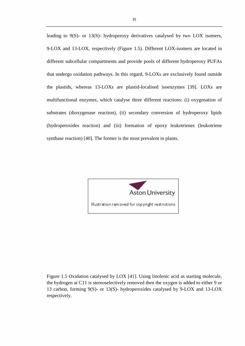
leading to 9(S)- or 13(S)- hydroperoxy derivatives catalysed by two LOX isomers,
9-LOX and 13-LOX, respectively (Figure 1.5). Different LOX-isomers are located in
different subcellular compartments and provide pools of different hydroperoxy PUFAs
that undergo oxidation pathways. In this regard, 9-LOXs are exclusively found outside
the plastids, whereas 13-LOXs are plastid-localised isoenzymes [39]. LOXs are
multifunctional enzymes, which catalyse three different reactions: (i) oxygenation of
substrates (dioxygenase reaction), (ii) secondary conversion of hydroperoxy lipids
(hydroperoxides reaction) and (iii) formation of epoxy leukotrienes (leukotriene
synthase reaction) [40]. The former is the most prevalent in plants.
Figure 1.5 Oxidation catalysed by LOX [41]. Using linolenic acid as starting molecule,
the hydrogen at C11 is stereoselectively removed then the oxygen is added to either 9 or
13 carbon, forming 9(S)- or 13(S)- hydroperoxides catalysed by 9-LOX and 13-LOX
respectively.

36
1.3.3.2 Allene oxide synthesis pathway
The 13(S)-hydroperoxy derivatives from linoleic and linolenic acid will be metabolised
largely through the pathway catalysed by allene oxide synthase (AOS), an enzyme of
the cytochrome hemoproteins of P450 family. This enzyme is responsible for the
biosynthesis of jasmonic acid (JA). In Arabidopsis thaliana, the level of this enzyme
depends on a complex control through wounding, pathogen attack, ethylene (generated
from ethephon), SA and feedback regulation through other octadecanoids (like JA and
12-oxo-10,15c-phytodienoic acids (12-oxo-PDA, OPDA)) [42].
(a) Jasmonate family and biosynthesis
JA is synthesised through the allene oxide synthesis pathway in higher plants, with
OPDA being the key intermediate. When α-linolenic acid is released from membranes,
LOX activity converts it into either 9(S)- or 13(S)-hydroperoxides. 13(S)-hydroperoxide
(13(S)-hydroperoxy-9c,11t,15c-octadecatrienoic acid) is then converted to
12,13(S)-epoxy-9c,11t,15c-octadecatrienoic acid by an AOS (Figure 1.6) [42]. This
unstable compound readily undergoes further reaction to form α-ketol through
nucleophilic substitution and γ- ketol through electrophilic substitution by ions in the
chloroplast, but more importantly is cyclised to OPDA by allene oxide cyclase (AOC)
[28]. Four possible enantiomers of OPDA can be synthesised from the two chiral carbon
centres (9, 13 C). However only 12-oxo-9(S),13(S)-phytodienoic acid (the

37
cis-(+)-enantiomer) exists in most plants and is useful as a precursor of (+)-7-iso-JA
which has JA biochemical and physiological properties [43]. These reactions occur in
the stroma phase of chloroplasts. The OPDA is transported from the plastids to the
peroxisomes as an uncharged free acid and OPDA-CoA esterified from or assisted by
the peroxisomal ATP-binding cassette (ABC) transporter COMATOSE (CTS) [44]. In
peroxisomes, the cis-(+)-OPDA-specific OPDA- reductase (isozyme 3: OPR3) reduces
OPDA to 3-oxo-2-(2-pentenyl)- cyclopentane-1-octanoic acid (OPC-8:0) followed by
three cycles of β-oxidation catalysed by acyl-CoA oxidase (ACX), multifunctional
proteins (MFP) and 3-ketoacyl-CoA thiolase (KAT) to form (+)-7-iso-JA [45]. Three
cycles of β-oxidation are responsible for shortening the carboxylic acid side chain by
removing two carbons in each cycle. During the whole pathway, the export of OPDA
from chloroplasts to peroxisomes is the rate limiting step [14], resulting in an
accumulation of OPDA in the membrane of the chloroplast. Interestingly, some early
research reported that OPDA was saturated at the 10, 11-double bond to yield OPC-8:0
in cytoplasm, which was followed by transportation into peroxisomes and β-oxidation
[46, 47]. In Arabidopsis, most OPDA and dinor 12-oxo-phytodienoic acid (dnOPDA)
(derived from roughanic acid (16:3 n-3)) is mainly esterified in mono- and digalactosyl
diacylglycerol (MGDG and DGDG). These galactolipids probably serve as a storage
compartment for the rapid synthesis of jasmonates following both biotic and abiotic
stress [48-50]. Lipids with esterified OPDA and dnOPDA have only been found in
Arabidopsis to date, and are termed arabidopsides.

38
Figure 1.6 JA biosynthesis and OPDA metabolism in Arabidopsis thaliana [51]. After
cis-(+)-OPDA is synthesised from α-LA in chloroplast, they can be transported form
chloroplast to peroxisome for further oxidation to form JA, added to glutathione (GSH)
to form OPDA-GSH, or directly trigger expression of genes in nucleus. 13-LOX,
13-lipoxygenase; ACX, acyl-CoA oxidase; AOC, allene oxid cyclase; AOS, allene
oxide synthase; CTS ⁄ PXA1 ⁄ PED3, ABC transporter for OPDA or OPDA–CoA import;
COI1, F-box protein in JA signal transduction; GST, glutathione-S-transferase; KAT,
L-3-ketoacyl-CoA thiolase; MFP, multifunctional protein; OPR, 12-oxo-phytodienoate
reductase; PLAI, plastidic acyl hydrolase.

39
(b) Biological activity of jasmonates
In plants, jasmonates are by far the most studied fatty acid derived signalling molecules.
They are important plant signal molecules in response to stresses including wounding,
insect attack, interaction with pathogens, ROS, and plant development [52]. Jasmonic
acid, a member of jasmonate family, is well documented. Its roles include information
(signal) transduction to trigger expression of genes related to plant defence [53, 54] and
development of plants, such as reproduction in Arabidopsis [55]. Its volatile methylated
form, methyl jasmonic acid (MeJA), can act in interplant communication [17, 56]. It
was also reported that exogenous treatment of MeJA on plants could activate
broad-spectrum disease resistance against several fungal pathogens [57, 58]. For the last
decade, another member of the jasmonate class of plant hormones, OPDA, has been
revealed to be able to regulate gene expression independently or in concert with JA to
fine-tune the expression of defence genes [59]. OPDA was proposed to regulate two
distinct signal pathways [60, 61], one through COI1 (Coronatine Insensitive 1) similar
to JA, and the other COI1-independent pathway via the electrophile effect of its
cyclopentenone moiety (such as biological activities mediated through TGA
transcription factors by covalently binding to free thiol groups of transcription factors
[62]). OPDA and JA have individual signalling activities, however, they share
overlapping biological activities [17, 63]. Both of them contribute to plant defence
against some species of fungi, like Botrytis cinerea, and OPDA has been shown more
recently to also play an important role against necrotrophic pathogens [64]. However, in

40
some cases, the conversion of OPDA to JA is necessary for the resistance of plants [55].
For example, it is JA, rather than OPDA, that plays an essential role as a signal for
wounding-induced defence response in tomatoes [65]. Interestingly, the exogenous
treatment of JA could induce the endogenous accumulation of OPDA in some species
[66]. Evidently, OPDA and JA appear to operate in concert to contribute to the gene
expressions.
Besides its ability to regulate gene expression, OPDA exhibits direct antifungal activity.
OPDA (100 µM) has the highest in vitro antifungal activity and chemical stability
among 43 oxylipins tested against several plant pathogens, such as C. herbarum, B.
cinerea and P. parasitica (Figure 1.7) [11]. In contrast, the JA does not show any direct
in vitro antifungal activity. The accumulation of OPDA-containing arabidopsides was
found in wounded (arabidopside A, B, E, G) and HR (arabidopside E and G) plant
tissue, which were induced by two stimuli; wounding [48] and recognition of
HR-caused phytopathogenic avirulence peptides [49]. The term arabidopsides has been
coined to describe galactolipids (MGDG and DGDG) which contain esterified OPDA
and dnOPDA (at sn-2 position). In one respect, the arabidopside E and G was reported
to have high antifungal activity against B. cinerea [49]. However, in the same report,
OPDA at concentration of 100 µM was showed to have very limited antifungal activity
against B. cinerea in comparison with arabidopsides (100 µM).

41
Figure 1.7 Antifungal activities of oxylipins [11]. Each test fungal or oomycete strain
was exposed to each compound (100 μM) in the appropriate liquid medium for 24 h.
Inhibition of growth over 24 h was expressed as a percentage relative to the ethanol
control. In the diagram, colour intensity is inversely proportional to inhibitory activity
of the oxylipin toward the organism. The names of tested organisms are abbreviated as
follows: P. infestans, Phytophthora infestans; R. spp., Rhizopus species; A. brassicicola,
Alternaria brassicicola; F. oxysporum, Fusarium oxysporum; P. parasitica,
Phytophthora parasitica nicotianae; B. cinerea, Botrytis cinerea; and C. herbarum,
Cladosporium herbarum.

42
1.3.3.3 Hydroperoxide lyase pathway
Hydroperoxide lyase (HPL), like AOS, belongs to the cytochrome P450 family, and can
be classified roughly into two groups, according to their substrate specificity. 13-HPL
can cleave the 13-hydroperoxy derivatives of linoleic or linolenic acids to form a group
of C6 volatiles (such as (Z)-3-hexenal, (E)-2-hexenal, hexanal, and hexanol [67]) and
C12-oxo acids (including traumain and traumatic acid), at the carbon-carbon bond
adjacent to the 13-hydroperoxide functionality (Figure 1.8) [28]. In parallel to 13-HPL,
9-HPL will catalyse the cleavage of 9-hydroperoxy isomers of linoleic and linolenic
acids, yielding C9 compounds, such as (2E),(6Z)-nonadienal, (Z)-3-nonenal and
(E)-2-nonenal. Both C6 and C9 volatiles are referred to as green leaf volatiles (GLVs).
As a group of bio-active compounds, GLVs are formed rapidly when tissues are
disrupted and contribute to the plant defence system, especially when they are enhanced
by the presence of α,β-unsaturated carbonyl groups [68]. For example, (E)-2-hexenal is
a very common GLV in plants and has been well studied for their multifunctional
properties correlated with plant defence [69], several features of which are listed below:
1. (E)-2-hexenal was found to be more effective than the corresponding saturated
aldehydes and hexanols at inhibiting the growth of fungal spores against some
species of fungi, like Colletorichum acutatum. This green volatile compound was
found to be able to alter the structures of the cell wall and plasma membrane,
causing disorganisation and lysis of organelles and, eventually, cell death [70]. Also

43
Hamilton-Kemp et al. [71] demonstrated that the α,β-unsaturated aldehyde,
(E)-2-hexenal, was considerably more active in the inhibition of hyphal growth of
Botrytis cinerea than hexenol and hexanal.
2. (E)-2-hexenal exhibits antibacterial activity against several bacterial species, such as
Pseudomonas syringae, Xanthomonas campestris and Erwinia carotovora [11].
3. (E)-2-hexenal can trigger the expression of defence-related genes and induce
antifungal proteins and phytoalexin accumulation in plants. Therefore, these
responses contribute to enhanced resistance against pathogens [72, 73].
4. (E)-2-hexenal accumulates in plants and occurs along with rapidly increased
activities of LOX and HPL after wounding [69]. Acetylated C6-aldehydes were
reported to be the predominant wound-inducible volatile signal that mediates the
indirect defence response by directing tritrophic (plant-herbivore-natural enemy)
interactions, whereas the jasmonates are responsible for the activation of the direct
plant-defence response [74].
Some recent reports indicate that (E)-2-hexenal antifungal activity was induced by its
electrophilic properties, α,β-unsaturated carbonyl groups, that interact with proteins
through the reaction with thiol and amine groups of proteins in the cell membrane [75,
76]. Besides (E)-2-hexenal, another 13-HPL derived plant hormone, traumatin
(12-oxo-(E)-10-dodecenoic acid), is formed through α-cleavage of fatty acid
hydroperoxides, which was considered to trigger cell division near infected or wound
sites [28]. Unsaturated C9-aldehydes, like (E)-2-nonenal, form rapidly after the

44
disruption of cucumber tissues from the cleavage of fatty acid by 9-HPL from
9-hydroperoxide [77]. Compared with (E)-2-hexenal, this compound is more active
towards hindering fungal growth [68, 77]. However, the high concentration required to
inhibit growth was also toxic to host cells, leading to cell death and preventing the
further spread of pathogens [77].
From the 9-LOX pathway, another important antifungal oxylipin is DES derived
divinylethers (colnelenic and colneleic acid), which are formed as fungicides in very
high amounts in potato after infection with the oomycete Phytophtera parasitica [78].
Therefore, the 9-LOX pathway was reported to perform essential roles in plant defence
against microbial pathogens [29, 79], probably resulting from the activity of these
antipathogenic substances (nonenal, colnelenic and colneleic acid).
In short, apart from jasmonates, derivatives from other oxylipin bio-synthetic pathways,
like HPL and DES pathways, participate and contribute to the plant defence system.

45
Figure 1.8 Hydroperoxide lyase pathway from 9- and 13- hydroperoxides of linolenic
acid [28]. LOX: lipoxygenase; HPL: Hydroperoxide lyase; IF: isomerisation factor. In a
similar reaction sequence, linoleic acid yields hexanal as well as (Z)-3- and
(E)-2-nonenal.
1.3.4 Non-enzymatic oxidation pathways
ROS, including free radical and oxygen, catalysed non-enzymatic oxidation of PUFAs
occur constitutively in membranes of all aerobic organisms, due to the existence of two
or three non-conjugated double bonds. Enzymatically formed oxylipins can be classified
according to different pathways, such as AOS, DES, HPL, PXG and EAS (Figure 1.4).
In contrast, free radicals and oxygen can attack all reactive sites, so it is difficult to give
46
a classification to non-enzymatically formed oxylipins. Some examples of them are
shown in figure 1.4, like cyclopentanoids, (2E)-4-hydroxy alkenals, hydroperoxides,
and some ketols, of which the group of cyclopentanoids are of interest because they are
structurally related to the established animal and plant defence mediators of the
prostaglandin and jasmonate type. In animals, non-enzymatically-formed
cyclopentanoids from arachidonic acid (C20:4ω6) are racemic isomers of
enzymatically-formed prostaglandins, so they are termed isoprostanes [80]. In plant
membrane, α-linolenic acid (C18:3ω3) is the predominate source for dinor isoprostanes
that are termed phytoprostanes (PPs) [81, 82]. Currently, it has been proposed that
omega-3 trienoic fatty acids (TFAs) in plants, in particular α-linolenic acid, serve in the
protection of cells by absorbing ROS such that they are oxidised in a
free-radical-dependent (non-enzymatic) manner [83]. However, this is debatable,
because some products from the oxidation of TFAs could be another source of ROS and
harmful to plants.
1.3.4.1 Biosynthesis of phytoprostanes
As in the case of the enzymatic oxidation pathway, the first step of non-enzymatic
oxidation is the formation of hydroperoxides. Enzymatic and non-enzymatic pathways
differ from each other with respect to the generation of different hydroperoxides (Table
1.2) [35].

47
Table 1.2 Two different oxidation pathways generate hydroperoxides.
Enzymatic Oxidation Non-enzymatic Oxidation
LOXs catalyse forms either S or R
hydroperoxides
Free radical reaction forms a racemic
mixture of hydroperoxides.
LOXs insert molecular oxygen only at
C-9 and C-13 of LA or α-LnA
In case of α-LnA, different positional
isomers at C-9, C-10, C-12, C-13, C-15,
C-16 could be formed, of which C-10 and
C-15 are oxidised by singlet oxygen that
is inserted at either end carbon of a
double bond [84].
The mechanism of autoxidation of α-LnA has been reviewed [84]. Briefly, hydrogen
abstraction at C-11 and C-14 between two 1,4-diene systems will generate two
pentadienyl radicals which are not stable and can undergo oxidation at the end-carbon
of each pentadienyl radical leading to four hydroperoxides isomers, conjugated diene 9-,
12-, 13- and 16-hydroperoxides (Figure 1.9). Although 12- and 13- isomers are of lower
concentration in comparison to 9- and 16- isomers, the presence of a homoallylic cis
double bond in 12- and 13- isomers favours their tendency to undergo 1,3 cyclisation by
intramolecular radical addition to the double bond. The newly formed radicals from 12-
and 13- peroxyl radicals can rapidly react with oxygen to generate the type I
phytoprostane G ring (PPG) and the type II PPG, respectively [80]. There are 16
phytoprostane G ring (PP) isomers formed from each starting peroxyl radical and 32
PPGs in total from α-LnA (Figure 1.10). PPGs are very unstable and their
non-enzymatic degradation pathway leads to D-, E-, and F- ring compounds, of which

48
D- and E- phytoprostanes may dehydrate and isomerise to J-, deoxy-J, A- and B- ring
phytoprostanes (Figure 1.11). Plant E1- and F1- phytoprostane in several species were
first identified in the late 1990s [81, 82]. As plant analogues of the animal isoprostanes,
the importance and perspective of the discovery of phytoprostanes for future research
has been reviewed [85]. Up to now, some sensitive and specific analysis methods have
been developed to detect low levels of phytoprostanes in plants, such as GC-MS, HPLC,
HPLC-ESI-MS/MS [20, 86]. Among PPs mentioned above, PPD1 and dPPJ1 were found
to be the most abundant PPs, not only in tomato leaves [20], but also in some other plant
species, such as Arabidopsis [87].
Figure 1.9 Mechanism of linolenic acid autoxidation [84].

49
Figure 1.10 Non-enzymatic phytoprostane biosynthesis from α-linolenic acid [86].

50
Figure 1.11 The phytoprostane pathway in plants [20]. G1-phytoprostanes rapidly
decompose in aqueous environment. Non-enzymatic or enzymatic reduction of both
peroxy groups yields chemically stable F1-phytoprostanes. The rearrangement of the
endoperoxide group and reduction of the side chain hydroperoxy group of
G1-phytoprostanes yields D1- and E1-phytoprostanes which may dehydrate/isomerise to
J1- and deoxy-J1-phytoprostanes or A1- and B1-phytoprostanes, respectively.

51
1.3.4.2 Bioactivity of phytoprostanes
It was found that the levels of PP in fresh plant tissues are much higher than isoprostane
(IsoP) in mammalian tissues, and the levels increase dramatically when plants are dried
and stored after harvest [88]. Recent research shows that singlet oxygen generated
during photosynthesis in chloroplast may accelerate radical driven formation of lipid
hydroperoxides which are the precursors of PPs [89]. It is also known that the
production of PPs in plants could be induced in several ways, such as reactive oxygen
species (ROS) in vivo [82, 90], wounding [82] and pathogen infection [90].
Two distinct groups of PPs are classified by chemical structure difference. The first
group comprises reactive electrophiles with a cyclopentenone ring moiety, which have
thiol and amine reactivity. More research was focused on the biological activities of
these PPs including A-, B-, and J- ring PPs (due to their important roles in inducing
several plant-protection mechanisms), such as gene triggering for plant defence and
detoxification [91], induction of phytoalexins [90] and recently they were found to have
anti-inflammatory activities similar to prostaglandin A1 (PGA1) and
15-Deoxy-Δ12,14
-prostaglandin J2 (dPGJ2) in human cells [92]. These cyclopentenone
PPs belong to a large family of compounds with α,β-unsaturated carbonyl structure,
which are termed reactive electrophilic species (RES). The second group of PPs
includes D-, E- and F- ring phytoprostanes. These compounds have been shown to have
lower bioactivity than PPs with cyclopentenone rings. Like malondialdehyde (MDA),

52
the F-ring of PPs can also be used as an oxidation marker, not only because of their
stability but their direct generation from the G-ring of PPs [82]. Some reports also
showed that E1-phytoprostane could possibly induce secondary metabolism (scopoletin
levels in tobacco cell) [90]. However, the conversion of PPE to the active PPA (RES)
could not be ruled out.
Most studies have used a mixture of various PP isomers because of the difficulty to
purifying individual isomers. Only a few reports indicated the effect of different
regioisomers with interesting results [90, 93]. Some organic synthetic approaches to
prepare PPs have been published [86, 94], which provide good standards and biological
controls for further phytoprostane research on the structure activity relationship.
Although the interest and discovery of PPs were enlightened by analogy to the
mammalian pathway, our knowledge about phytoprostanes and their functional
importance is limited compare to the analogous (iso- and neuro- prostanes in humans).
More effort is needed to elucidate their full spectrum of biological activities.
1.4 Fatty acid derived reactive electrophilic species (RES)
As discussed above, oxylipins are composed of various fatty acid derivatives, of which
those compounds with an α,β-unsaturated carbonyl group, like (E)-2-hexenal, OPDA
and cyclopentenone PPs, have attracted the most interest due to their bioactivities. In

53
general, RES are classified as compounds having chemical reactivity with nucleophilic
groups, such as thiol- (SH-) and amine- (H2N-) groups. In plants, compounds containing
α,β-unsaturated carbonyl groups or other types of RES derived from fatty acids are
termed „oxylipin RES‟ [95, 96].
1.4.1 Oxylipin reactive electrophilic species
Fatty acid derived α,β-unsaturated epoxides, α,β-unsaturated aldehydes, cyclopentenone
products and other electrophilic hydroperoxides or ketones are classified as oxylipin
RES [96]. These representative oxylipin RES could be generated, enzymatically or
non-enzymatically, from lipid oxidation of PUFAs (Table 1.3 and Figure 1.4) [97].
Both enzymatically and non-enzymatically derived oxylipin RES are constantly
generated in vivo even in healthy plant tissues, however, both pathways could be
activated by a variety of biotic and abiotic stress conditions leading to accumulation of
RES oxylipins [52]. These oxylipin RES are derived from different synthetic origins
and play different roles in plants. However, they have two common properties:
electrophilicity and lipophilicity, which endow them with important biological
activities.

54
Table 1.3 RES oxylipins in plants and their bio-synthetic pathway [97].
Synthetic Pathway RES Example
Enzymatic
Pathway
Jasmonate pathway Jasmonates OPDA, dinor-OPDA,
arabidopside
Reduction of
enzymatically formed
LOOH
Hydroxyl and
oxo-fatty acids
KOTE,
KODE,
Traumatin
Fragmentation of
hydroperoxides through
enzymatic HPL pathway
Aldehydes and
other small
fragments
Hexenals, Nonenals
Non-enzymatic
Pathway
PPs pathway Cyclopentenone
phytoprostanes
J1-, A1- and B1- PPs
Reduction of
nonenzymatically
formed LOOH
Hydroxyl and
oxo-fatty acids
KOTE,
KODE
Fragmentation of
formed LOOH under
free radical catalysis
Aldehydes and
other small
fragments
Hexenals, Nonenals,
MDA,
4-hydroxyl-(2E)-nonenal
1.4.2 Electrophilicity and lipophilicity of RES
Oxylipin RES normally consist of an electrophilic part (e.g. an α,β-unsaturated carbonyl
moiety) and saturated or unsaturated carbon chains together with other functional
groups, like carboxyl or hydroxyl moieties.

55
As for electrophilic α,β-unsaturated carbonyl groups, it should be noticed that hard
electrophiles having small atomic radii, high electronegativity (high ELUMO) and low
polarisability, tend to react with hard nuleophiles (e.g. amino groups of lysine and DNA)
(Figure 1.12). Soft electrophiles, on the other hand, having larger radii, high
polarisability, containing unshared pairs of electrons and low electronegativity (low
ELUMO) prefer soft nuleophiles (such as, thiol group of cysteine or glutathione) [97, 98].
Theoretically, the α,β-unsaturated carbonyl group in oxylipins maybe (or are) closed as
soft electrophilic species which tend to react with soft nucleophiles, like cysteine
residues in GSH [99, 100]. Similar to RES in animals, it is hypothesised that bioactive
plant RES could act as Michael acceptors, reacting not only with GSH, but also directly
with other enzymes and transcription factors [51]. Different RES have different
reactivities with nucleophiles as a consequence of their different electrophilicities.
Notably, the smallest RES molecule, acrolein, has much higher reactivity with thiol and
amine groups in comparison with other α,β-unsaturated aldehydes, explaining its high
cytotoxicity [100] and bioactivity [61]. In addition, conjugates of the RES with protein
could be used as markers of oxidative stress in tissues.

56
Figure 1.12 Product formations after reaction with thiol and primary amine nucleophiles
[100]. (a) Michael addition, product in the reaction between glutathione or thiol and an
α,β-unsaturated aldehyde; (b) Michael addition, product in the reaction between primary
amine (e.g. lysine) and α,β-unsaturated aldehyde; (c) Schiff base, product in the reaction
between primary amine and α,β-unsaturated aldehyde.
Besides electrophilicity, lipophilicity(or hydrophobicity) is another important parameter
of oxylipin RES. Lipophilicity refers to the ability of a chemical compound to dissolve
in lipophilic substances, such as oils, fats, lipids and non-polar solvents, and obey the
axiom that like dissolves like. The partition coefficient, log P, is often used as a
measurement of lipophilicity, which is defined to be the logarithm of the ratio of the
concentrations of an unionised compound in the two phases of a mixture of two
immiscible solvents (normally a water and octanol solvent system) at equilibrium [101].

57
Log P is one of the major determinants of estimating the behaviour of drugs in
organisms; therefore it is likely to be an important factor for the design of drugs and
agrochemicals. In plant biochemistry, log P has a major importance in all biological
activities as it represents the ability of molecules to reach the active sites [102]. With
respect to RES, log P could affect the biological activity and cytotoxicity in two ways.
The first is the ability of the compound to permeate through a membrane since plasma
membranes are composed typically of a bilayer structure, in the middle of which are
non-polar tails of fatty acids. Secondly, lipophilicity is an important physicochemical
parameter referring to the affinity for the hydrophobic binding sites of proteins. The
different affinities with the proteins will also influence the subsequent reaction rates
with the substrates.
1.4.3 Biological activities of oxylipin RES in plant defence
In mammals, lipid derived RES, such as prostaglandins and isoprostanes, and their
biological activities have been well studied [30]. By comparison, research on plant RES
is somewhat lagging behind. Recently, a growing number of biological roles of oxylipin
RES in plants have been unveiled and some of them are related to the plant defence
system.

58
Low levels of RES oxylipins are ubiquitously in all higher organisms and are naturally
present, even in unstressed cells [91]. Studies showed that in general, the amount of
RES increases rapidly following both biotic and abiotic stresses leading to a transient
over accumulation [35]. In order to keep RES to low levels in healthy organisms, the
expressions of genes related to their detoxification are increased. Some genes encoding
detoxification proteins (such as glutathione-S-transferases (GST)) and other plant
defence related gene expressions have been reported to be inducible by various kinds of
RES with α,β-unsaturated carbonyls, such as PPs [62, 90], OPDA [18, 52, 63, 103],
MDA [104], hexenal [72, 105], KODE and KOTE [106]. Interestingly, RES B1-PP, that
can not react with thiol groups, was also reported to trigger plant defence and
detoxification [93]. RES oxylipins with similar structures could induce a common set of
defence genes. For example, two cyclopentenone rings containing compounds, PPA1
and OPDA share similar biological activities (reviewed elsewhere [107]), although
phytoprostanes may also induce a unique set of responses. In addition, exogenous RES
can influence endogenous RES pools. For example, in Arabidopsis the level of OPDA
can be increased by the treatment with acrolein, which may be attributed to the action of
lipase on esterified OPDA [61]. Enzymatic and non-enzymatic lipid peroxidation is
implicated in the hypersensitive response (HR), during which, some of the oxylipins
serve as signals necessary for defence gene activation whereas others contribute to
pathogen killing or participate in the execution of programmed cell death (PCD)
associated with this resistance [17, 79, 96]. A range of RES oxylipins, such as
derivatives of 9, 13-LOX pathways, jasmonates and arabidopsides were found in

59
Arabidopsis during HR [17, 78, 96, 108, 109]. These enzymatically-formed oxylipins
were found at an early stage, whereas non-enzymatically oxidised lipids were at low
levels [109]. ROS mediated non-enzymatic peroxidation appeared as a late stage [79,
110] and both of them contribute to HR. This reveals the defence-related functions of
enzymatically and non-enzymatically-formed oxylipins RES during biotic and abiotic
stress.
Phytoalexins are disease-inducible low molecular mass secondary metabolites, which
are commonly toxic to fungi, bacteria, plants and animal cells. Therefore, most of
phytoalexins are absent from unchallenged, healthy tissue. When invaded by pathogens,
plants will endeavour to protect themselves by a rapid accumulation of phytoalexin at
the site of invasion. The production of phytoalexins often accompanies the
hypersensitive response and is elicited by a variety of stimuli [111], of which RES
compounds are important. Phytoalexins accumulate in plant cells in response to
treatment with some oxylipin RES. For example, camalexin accumulates in cells after
pre-treatment with cyclopentenone phytoprostanes [90, 93] and (E)-2-hexenal [73]. The
accumulation of scopoletin was found in tobacco cells in response to OPDA and
cyclopentenone phytoprostanes [94]. In short, RES induced accumulation of
phytoalexins contributes to plant defence against some pathogens through their general
toxicity.

60
Some oxylipins also show direct antimicrobial activities. 43 oxylipins were screened
against several fungal and bacterial strains [11]. Oxylipin RES, like OPDA, KOTE,
hexenals, have high antimicrobial activities. OPDA was considered as the best
antifungal candidate and was also more stable in comparison to other effective
compounds. (E)-2-hexenal had the best antibacterial activity. Interestingly, JA does not
show any ability to inhibit fungal growth and (E)-2-nonenal that is reported to have
antifungal activities [77] is not as active as (E)-2-hexenal against bacterial strains. The
simple RES, α,β-unsaturated aldehydes, were revealed to have antifungal activities
against Saccharomyces cerevisiae and the length of the carbon chain plays an important
role in balancing the hydrophilic α,β-unsaturated carbonyl and hydrophobic carbon
chain [68]. During HR, the accumulation of arabidopside E along with keto and
hydroxyl fatty acids and jasmonates, display direct antipathogenic effects against
secondary infectious agents. Considering that PPs, particularly cyclopentenone
containing PPs, appear in the late stage of HR and some of them have similar chemical
structure with OPDA, it is possible that these compounds also have antimicrobial
activity.
1.4.4 Phytotoxicity of RES
Oxylipin RES play important roles in plant defence. However, this group of compounds
can also cause severe damage to host cells due to their reactive chemical properties.
They can affect plant cells in at least two ways. Firstly, the α,β-unsaturated carbonyl can

61
be reduced by enzymes such as NADPH-oxidoreductases and oxo-phytodienoic acid
reducase, and cause indirect damage by the depletion of pools of reductants (NADPH
and NADH). Secondly, RES can damage cells directly by modifying cell substances
(proteins) due to their chemical reactivity with nucleophiles [95]. The photosystem II
fluorescence of Arabidopsis was measured to quantify the damaging effects of lipid
derivatives, especially oxylipin RES and was in the order: acrolein > methyl vinyl
ketone (MVK) > 2E,6Z-nonadienal > (E)-2-nonenal > (E)-2-hexenal >
OPDA=13-KOTE > 13-KODE > traumatin [61]. This investigation shows a correlation
of activities with molecular weight and the increased degree of unsaturation. The
general cytotocixity of α,β-unsaturated RES largely depends on electrophilic reactivity
and lipophilicity. For example, the hepatocyte toxicity of α,β-unsaturated aldehydes
generally increases with increasing electrophilic reactivity with thiol and amine groups
and often increases with lipophilicity value higher than 1.8 [100]. Although the
phytotoxicity of RES was believed to have a similar relationship with their chemical
reactivity, more work is necessary to clarify the mechanism. Summarising, oxylipin
RES have powerful biological activity at relatively low levels, but are highly phytotoxic.
Therefore, it is important to control the accumulation of oxylipin RES to a sub-lethal in
plant cells.
In short, oxylipin RES play an important role in plants, and to some extent, are more
active than their non-electrophilic counterparts, such as (E)-2-hexenal vs hexanal,
OPDA vs JA and A-, B-, J- ring PPs vs E-, D-, F- ring PPs. Enzymatically or

62
non-enzymatically formed oxylipins RES accumulate in response to biotic and abiotic
stresses, however, the detailed mechanism of RES recognition and signalling remains to
be identified.
1.5 Molecular design based on 12-oxo-PDA
So far, oxylipin RES contribute to the plant defence system through several mechanisms
in response to environmental stresses. They are very promising candidates for the
development of a new generation of agrochemicals, of which OPDA with a
cyclopentenone ring is one of the most interesting compounds. This is not only because
of its biological activity [63, 103], but also it has the most effective in vitro antifungal
activity and stability among 43 oxylipins tested against fungal strains [11]. The
cyclopentenone moiety is considered to confer the bioactivity of OPDA due to its
electrophilicity as discussed above.
In this project, 12-oxo-PDA (OPDA) was considered as a natural lead molecule for the
development of new agrochemicals. OPDA can be found in almost all higher plants.
The biosynthetic pathway takes place in chloroplasts where most OPDA exists as free
and esterified lipids, but only at low levels. OPDA is only present in plant tissue at
nanogram levels [112] and the degradation could easily happen during extraction.
Hence, it is difficult to extract and purify a large amount of this compound directly from

63
plant tissues. OPDA can be successfully produced in the laboratory through organic
synthesis [113], however, the published process is complicated and consists of many
steps. Therefore, at the start of this research, neither natural nor organic synthesis was
reviewed as viable for large-scale commercial production of the compound for
antifungal applications. Consequently, a new approach needed to be developed to
enable the production of a new generation of agrochemicals based on the modification
of OPDA.
The basic idea and fundamental principle underlying our approaches is to modify the
chemical structure of OPDA, keeping the functional cyclopentenone ring and
simplifying the substituents whilst preserving the biological and antifungal activities of
the molecule. Undoubtedly, the electrophilic cyclopentenone ring is the key structural
motif that needed to be retained in the design and development of new compounds. The
substituent on C9 of OPDA is a hydrophobic carbon chain terminated with a carboxyl
group, which is known to be related to the membrane permeability of OPDA [44]. The
hydrophobic carbon chain provides the lipophicility which is balanced by the
hydrophilic moiety of OPDA. The terminal carboxyl group forms an ester with glycerol
and plays an important role in β-oxidation during metabolism. In addition, the formation
of OPDA-CoA esters makes it easier to traverse the membrane of peroxisomes [44].
Similar to OPDA, another 16-carbon cyclopentenoic acid present at low levels in plants,
dinor 12-oxo-phytodienoic acid (dnOPDA), is derived from plastid 16:3 fatty acids
rather than by β-oxidation of the 18-carbon OPDA [114]. With respect to different chain

64
length, dnOPDA is 2C shorter than OPDA. However, both free and esterified dnOPDA
were reported to have similar behaviour to OPDA [59] in plants but their antifungal
activities have not been evaluated. The other substitute on C13 of OPDA is the pentenyl
side-chain and very little is known about its role. With a similar structure to OPDA, the
15,16-dihydro-12-oxo-phytodienoic acid (DH-OPDA), which was proposed to be
generated from linoleic acid (18:2 n-6), has a saturated pentyl substituent on C13 and is
involved in a defence signalling pathway parallel to OPDA but somewhat lower in
terms of overall activity [115]. Another function of the pentenyl side-chain on C13
could be that the presence of the double bond (C15=C16) favours the cyclisation similar
to the function of the homoallylic cis double bond involved in both enzymatic and
non-enzymatic 1,3-cyclisation during synthesis [84, 116]. The delocalisation of this
double bond assists in epoxide opening and undergoes subsequent cyclisation catalysed
by the enzyme AOC [117]. So far, the side chain on C9 is known to be important in the
activities of OPDA and closely associated with its bio-activity. The pentenyl substituent
on C13 seems to enable the formation of OPDA. However, limited information is
known on how it contributes to the biological properties of OPDA.
According to two common properties of RES, electrophilicity and lipophilicity, the
preferred modification is to synthesise a compound lacking a substituent on the C13 of
OPDA, i.e. compounds composed of just the cyclopentenone ring along with
hydrophobic carbon chains of varying lengths. Altering the length of the hydrophobic
chain will mainly affect the lipophilic properties of the molecule. Appropriate

65
lipophilicity is required for antifungal performance of the compound and in this project
it is determined by the length of the hydrophobic chain. Experimentally, the log P value
of most of commercial agrochemicals is lower than 5 and was suggested to be
controlled between 3 and 4.5 to achieve high permeability to cells [1]. The calculated
log P value of the lead molecule, OPDA is around 4.6. A low lipophilicity is helpful for
solubility of the compound in tissue and transportation of the compound to reach
infected sites in plants. Another reason for maintaining low lipophilicity is the increase
in toxicity with increasing lipophilicity [100, 118]. Furthermore, the shorter the chain,
the shorter the half-life of the compound, this could translate to the compound having
less adverse effects on the environment. Bearing in mind the need to control two
common properties of the designed compound (electrophilicity and lipophilicity), a
targeted organic synthesis will be more efficient and economical than random screening.
The synthetic design centred therefore on producing compounds with a cyclopentenone
ring along with a hydrophobic carbon chain substituent on the C4 of the cyclopentenone
ring with varying lengths to control the log P value to less than 5. Some designed target
compounds to be synthesised for evaluation of antifungal activity are given in Figure
1.13 and their log P values were calculated by Molinspiration log P calculator.

66
Figure 1.13 Designed target compounds. The value of log P was calculated by
Molinspiration log P calculator.
1.6 Aims
The aim of this project was to synthesise compounds with a cyclopentenone ring and
different length alkyl chains on the C4 of the cyclopentenone ring. These compounds
have a similar chemical structure to OPDA and may thus mimic its natural antifungal
properties. These designed compounds needed to be highly effective on specific
targeted fungal pathogens rather than possessing a general toxicity towards many
organisms, which would thus minimise their side effects on users. Like many naturally
occurring compounds, the ready biodegradation of these synthetic compounds will
make them environmentally friendly by reducing persistent application residue. In
67
addition, the synthetic pathway for the production of these compounds should have as
few steps as possible, which favours large scale manufacture and commercial
application. The main work focuses on the following research:
Synthesis of OPDA analogues
Screening of synthesised OPDA analogue for antifungal activity
Investigation into the mode of action of active compounds
Antimicrobial activity of (E)-2-alkenals

68
Chapter 2
Organic Synthesis and Characterisation
of Target Compounds
2.1 Introduction
The Nazarov and Pauson-Khand reactions provide two common methods to synthesise
cyclopentenone rings [94, 119, 120], however, neither of them have been used to
synthesise OPDA. Instead, other methods have been developed to synthesise OPDA and
three synthetic pathways were published by Grieco [121], Ernst [122] and Kobayashi
[113, 123]. In this project, the synthetic pathway published by Kobayashi was
simplified and used for the preparation of the target compounds.
The most recent method of synthesising OPDA [113] starts with
(1R,3S)-(+)-cis-4-cyclopentene-1,3-diol-1-acetate and requires several further steps to
complete. As mentioned in the introduction, the function of the pentenyl side group on
the cyclopentenone ring is not clear, thus the hydrophobic long carbon chain on the
cyclopentenone ring can be installed first of all. The synthesis can then be generally
simplified to less than five steps to produce the target compounds in Figure 1.13.
Copper-catalysed installation of an alkyl chain onto the cyclopentene ring published by
Kobayashi [124], provides a simple method for the addition of varying lengths of

69
carbon chains to the cyclopentenone ring which, in turn, should change the balance
between the hydrophilic and hydrophobic portions of the molecule. Target compounds
were synthesised as given below (Scheme 2.1).
Scheme 2.1 Pathway to synthesise compound simplified from OPDA. Reagents and
conditions: (a) imidazole (1.5 equiv), TBDPS-Cl DMF, RT; (b) Mg, 1,2-dichloroethane;
(c) CuI, THF, -18°C; (d) TBAF, 4Å molecular sieves, 55°C; (e) CrO3/H2SO4, acetone.
2.1.1 Grignard reagent
The important step in the copper-catalysed addition of the alkyl chain to the
cyclopentenone ring is the preparation of the corresponding Grignard reagent. The
Grignard reaction is widely used to form carbon- carbon bonds and some carbon-
heteroatom bonds. Preparation of a Grignard reagent is achieved via reaction between
an alkyl or aryl halide and magnesium. This is a relatively simple reaction, however
reaction conditions are critical. Moisture inhibits the reaction by quenching the reagent

70
to the corresponding hydrocarbon, so the synthesis should be conducted using dry
solvents and under an inert atmosphere. Other factors can also influence the preparation
of Grignard reagent [125]:
1. Quantity of the magnesium. Usually, a 5-10% stoichiometric excess, relative to the
alkyl halide of fresh magnesium turnings are used.
2. Activators and inhibitors. In order to increase the activity of the magnesium, iodine
and ethyl bromide are often used as activators. The concentration of the activator
and the reaction temperature will affect the efficiency of the activation process.
3. Effect of halide purity and of the rate of its addition. High purity of the halide and
slow addition are required for carrying out a high yielding reaction. In addition,
vigorous stirring is necessary to eliminate the local overheating that can occur
during this exothermic reaction.
4. Effect of the nature of the halogen and of the alkyl group. Given the same organic
moiety, iodides react more readily with magnesium than bromides do, which in turn
are more active than chlorides. This order of reactivity is entirely consistent with
any nucleophilic substitution reaction employing these leaving groups.
2.1.2 Installation of alkyl group
The synthetic pathway for the larger compounds starts with (1R,
3S)-(+)-cis-4-cyclopentene-1,3-diol-1-acetate, with the key step being the
copper-catalysed addition of the alkyl chain onto the unsaturated five-member ring

71
(scheme 2.2).
Scheme 2.2 Installation of alkyl group [124]. RMgX (X=Cl, Br), CuX (X=CN, I), R:
alkyl chain
This reaction generates trans 1,4- or 1,2- isomers (Scheme 2.2). The ratio of these two
regioisomeric products and their yields of formation depends on the composition of
RMgX (X= Cl, Br)/CuX (X= CN, I) and the choice of solvent (Et2O or THF) [124].
Typically, the 1,4-isomer predominates and is the major product. This is fortunate as
this regioisomer is the precursor of the target compounds. The composition of
RMgX/CuX and the choice of solvent were found to be the most important factors for
increasing the yield of the 1,4-isomer. According to the previous report [124], several
combinations of reagent/solvent were tested, and the two of which gave the optimum
yield and highest ratio of 1,4-isomer: 1,2-isomer are shown in Table 2.1.
Since there was very little difference between the CuCN and CuI catalysed reaction, CuI
was used to catalyse the reaction since it is far less toxic than CuCN. Compared with
Et2O, THF is more polar and tends to determine the regioselectivity, preferentially
forming the 1,4-isomer in this reaction. In addition, it was found that a bromide
Grignard reagent will influence the regioselectivity of the installation and increase the

72
Table 2.1 Synthetic conditions of the reactions with high yield of 1,4-siomer [124].
yield of 1,2-isomer [124], thus chloride Grignard reagent is needed to achieve high yield
of 1,4-isomer. This is an efficient method to add an alkyl chain onto the cyclo-ring. In
this project, a RMgCl/CuI (10 mol %) in THF reaction system was used to add an alkyl
substituent to the cyclo-ring and achieve high yield of 1,4-isomer followed by the
oxidation of hydroxyl group to produce the cyclopentenone ring.
2.1.3 Hydroxyl group oxidation
Jones reagent (CrO3/H2SO4) is an ideal oxidation reagent for the oxidation of primary
and secondary hydroxyl groups to carboxylic acids and ketones, respectively, without
influencing the double bond in cyclopentenone ring (Scheme 2.3). Jones reagent is
prepared by dissolving CrO3 into concentrated H2SO4 and diluting this mixture with
distilled water.

73
Scheme 2.3 Hydroxyl group oxidation.
2.2 Materials
All reactions involving moisture-sensitive and/or air-sensitive reagents were conducted
under a nitrogen atmosphere, in oven dried (120oC) glassware. Tetrahydrofuran (THF)
was dried by refluxing over sodium/benzophenone and was distilled immediately prior
to use. Distilled water was used throughout. All other reagents were used as received
from chemical suppliers Sigma-Aldrich Chemical Co. Acros, Fluka and Fisher. Thin
layer chromatography (TLC) and column chromatography were carried out using Merck
Kieselgel 60 TLC plates and Merck Kieselgel 60 silica gel, respectively.
Infra red (IR) spectra were recorded on a PerkinElmer FT-10 FT-IR spectrometer.
Nuclear Magnetic Resonance (NMR) spectra, 1H NMR (300 MHz) and
13C NMR
(75.58 MHz), were recorded on a Brucker AC300 spectrometer. Deuterated chloroform
(CDCl3) was used as the solvent (7.24 for 1H and 77.0 for
13C respectively) and
chemical shift values are reported relative to TMS. 13
C NMR spectra were recorded
using the PENDANT program [126]. Mass spectra (MS) were recorded by Karen
Farrow (LHS, Aston University, UK). A Sartorius ME36S microbalance was used to

74
measure the weight of synthesis products. The chemicals used in organic synthesis and
their supplier are listed in Table 2.2.
Table 2.2 Chemicals used in the organic synthesis.
Chemicals Manufacture
(1R,3S)-(+)-cis-4-cyclopentene-1,3-diol 1-acetate ≥99% Sigma-Aldrich
8-Chloro-1-octanol 98% Sigma-Aldrich
Butylmagnesium chloride 2.0M in THF Sigma-Aldrich
Hexylmagnesium chloride 2.0M in THF Sigma-Aldrich
Octylmagnesium chloride 2.0M in THF Sigma-Aldrich
tert-Butyl(chloro)diphenylsilane 98% Sigma-Aldrich
Ammonium chloride 99.5% Sigma-Aldrich
Ammonium Hydroxide solution 28% Sigma-Aldrich
Tetrabutylammonium fluoride solution 1.0 M in tetrahydrofuran Sigma-Aldrich
Silica Gel 200-400mesh 60Å for column chromatography Sigma-Aldrich
Celite 521 Acros
Chromium (VI) trioxide 99+% Acros
Copper (I) iodide 98% Acros
1, 2-Dichloroethane puriss over MS (H2O≤0.005%) Fluka
2.3 Synthetic procedure of target compounds
A simplified version of Kobayashi‟s synthesis of OPDA [113] was employed to produce
the target compounds 3, 4, 5, and 8 (Scheme 2.4 and 2.5). FT-IR, 1H NMR,
13C NMR
spectra and APCI-MS of target compounds can be found in the Appendices.

75
2.3.1 Synthesis of compounds 3-5
Scheme 2.4 Synthesis of compounds 3-5. Reaction conditions: (a) CuI, THF, -18°C, (b)
CrO3/H2SO4, acetone.
The synthetic method was modified from Kobayashi‟s method [124]. A two necked
round bottomed flask was charged with CuI (14.0 mg, 0.071 mmol), suspended in dry
THF (1.8 mL), the resultant slurry stirred magnetically and cooled to -18 °C (NaCl/ice
bath: 1:3). A solution of RMgCl (octylmagnesium chloride in THF 1.5 mL, 1.39 M) was
then added dropwise whilst maintaining this temperature. Stirring was
then continued for a further 20 minutes at -18 °C, after which a solution of
(1R,3S)-(+)-cis-4-cyclopentene-1,3-diol 1-acetate 1 (100.3 mg) in THF (0.2 mL) was
added dropwise. The reaction was then stirred at -18oC for 5 hours before being
quenched by the addition of a saturated aqueous solution of NH4Cl (5 mL) and an
aqueous solution of NH4OH (28%, 5 mL) with vigorous stirring. The crude product was
extracted with Et2O (3 x 25 mL). And the combined organic extracts were then dried by
standing over anhydrous MgSO4, collected by filtration and concentrated under reduced
pressure to furnish the crude product as a colourless oil. The crude product was purified
by column chromatography (Hexane/EtOAc: 75/25) to give the pure and major 1,

76
4-isomer 2 (132.1 mg, 0.674 mmol) (TLC Hexane/EtOAc: 75/25, Rf= 0.40). This
product was used in subsequent oxidation reactions directly without further analysis.
The pure 1, 4-isomer 2 (132.1 mg 0.674 mmol) was dissolved in freshly distilled
acetone (1.7 mL) and the resultant solution cooled to 0 °C. Freshly prepared Jones
reagent was then added dropwise until the colour of the reagent persisted and the
resultant orange solution were stirred for a further 30 minutes at 0 °C. After this time,
the reaction was quenched by the addition of 2-propanol, and the resultant mixture
filtered through a pad of silica gel using Et2O as the eluent. The filtrate was washed
with brine (3 x 25 mL) before being dried by standing over anhydrous MgSO4, collected
by filtration and concentrated under reduced pressure to furnish the crude products 3-5
as yellow oils. The crude products were purified by column chromatography
(Hexane/EtOAc: 50/50) to give the pure compounds 3-5.
4-octyl-cyclopentenone (3) was obtained as a yellow oil (47.3 mg, 0.24 mmol, yield
35.6%). (TLC Hexane/EtOAc: 50/50 Rf= 0.75). IR (neat): νmax /cm-1
3052, 1716, 1669,
1178. 1H NMR (300 MHz, CDCl3): δ 0.97 (t, 3,
3J =6.9 Hz, CH3), 1.28-1.80 (m, 14,
CH2), 2.07 (dd, 1, 2J5a,5b=18.9 Hz,
3J5a,4 =2.1 Hz, 5-Ha), 2.61 (dd, 1,
2J5b,5a=18.9 Hz,
3J5b,4 =6.3 Hz, 5-Hb), 2.95-3.08 (m, 1, 4-H), 6.22 (dd, 1,
3J3,2 =5.7 Hz, J = 2.1 Hz, 3-H),
7.71 (dd, 1, 3J2,3 =5.7 Hz, J =2.4 Hz, 2-H).
13C NMR (75.58 MHz, CDCl3, PENDANT):
δ 14.15 (CH3), 22.70 (CH2), 27.67 (CH2), 29.28 (CH2), 29.50 (CH2), 29.64(CH2), 31.89
(CH2), 34.80 (CH2), 41.11 (C-5), 41.56 (C-4), 133.59 (C-3), 168.83 (C-2), 210.20 (C-1).

77
LRMS (APCI+): m/z 195 ([M+H]+); calculated for C13H22O: 194.32.
4-hexyl-cyclopentenone (4) was obtained as a yellow oil (11.0 mg, 0.066 mmol, yield
25.8%). (TLC Hexane/EtOAc: 50/50 Rf= 0.75). IR (neat): νmax /cm-1
3046, 1716, 1669,
1181. 1H NMR (300 MHz, CDCl3): δ 0.95 (t, 3,
3J =6.9 Hz, CH3), 1.25-1.72 (m, 10,
CH2), 2.05 (dd, 1, 2J5a,5b=18.8 Hz,
3J5a,4 =2.0 Hz, 5-Ha), 2.59 (dd, 1,
2J5b,5a=19.0 Hz,
3J5b,4 =6.4 Hz, 5-Hb), 2.93-3.06 (m, 1, 4-H), 6.22 (dd, 1,
3J3,2 =5.6 Hz, J = 2.0 Hz, 3-H),
7.71 (dd, 1, 3J2,3 =5.7 Hz, J =2.4 Hz, 2-H).
13C NMR (75.58 MHz, CDCl3, PENDANT):
δ 14.10 (CH3), 22.63 (CH2), 27.63 (CH2), 29.30 (CH2), 31.73 (CH2), 34.79 (CH2), 41.11
(C-5), 41.55 (C-4), 133.60 (C-3), 168.79 (C-2), 210.13 (C-1). LRMS (APCI+): m/z 167
([M+H]+); calculated for C11H18O: 166.26.
4-butyl-cyclopentenone (5) was obtained as a yellow oil (9.2 mg, 0.067 mmol, yield
25.7%). (TLC Hexane/EtOAc: 50/50 Rf= 0.75). IR (neat): νmax /cm-1
3049, 1716, 1669,
1183. 1H NMR (300 MHz, CDCl3): δ 0.99 (t, 3,
3J =6.9 Hz, CH3), 1.26-1.74 (m, 6, CH2),
2.14 (dd, 1, 2J5a,5b=18.8 Hz,
3J5a,4 =2.1 Hz, 5-Ha), 2.61 (dd, 1,
2J5b,5a=18.9 Hz,
3J5b,4 =6.3
Hz, 5-Hb), 2.90-3.08 (m, 1, 4-H), 6.22 (dd, 1, 3J3,2 =5.6 Hz, J = 2.0 Hz, 3-H), 7.72 (dd, 1,
3J2,3 =5.6 Hz, J =2.4 Hz, 2-H).
13C NMR (75.58 MHz, CDCl3, PENDANT): δ 14.01
(CH3), 22.72 (CH2), 29.84 (CH2), 34.48 (CH2), 41.11 (C-5), 41.53 (C-4), 133.61 (C-3),
168.85 (C-2), 210.42 (C-1). LRMS (APCI+): m/z 139 ([M+H]+); calculated for C9H14O:
138.21.

78
2.3.2 Synthesis of 8-(4-oxocyclopentenyl)octanoic acid (8)
Scheme 2.5 Synthesis of 8-(4-oxocyclopentenyl)octanoic acid (8). Reaction conditions:
(a) CuI, ClMg(CH2)8OTBDPS, THF, -18°C; (b) TBAF, 4Å molecular sieves, 55°C; (c)
CrO3/H2SO4, acetone.
Grignard preparation of ClMg(CH2)8OTBDPS (Scheme 2.1)
A one-necked flask (25 ml) was charged with 8-chloro-1-octanol (1.0 g, 6.0 mmol),
TBDPSCl (2.0 g, 7.2 mmol) and DMF (10 ml) in ice bath with stirring. Imidazole (1.0 g,
15.0 mmol) was added slowly and stirred until complete dissolution in DMF was
achieved. The reaction was carried out at room temperature (RT) overnight. The
reaction was quenched by the addition of water and extracted with DCM (25 ml x 3)
then the combined extracts were washed with HCl solution (1 M) (25 ml x 3). After
being dried with MgSO4, the mixture was concentrated to a transparent oil. The crude
oil was purified by column chromatography (Hexane/EtOAc: 90/10) to give the pure oil
(Cl(CH2)8OTBDPS, 2.27 g, 5.64 mmol, yield: 94%) (TLC Hexane/EtOAc: 90/10, Rf=
0.86). IR (neat): νmax /cm-1
3071.3, 2931.7, 2857.5, 1428.0, 1111.8, 701.8. 1HNMR (300
MHz, CDCl3): δ 1.13 (s, 9, CH3), 1.31-1.54 (m, 8, CH2), 1.63 (m, 2, CH2), 1.83 (m, 2,
CH2), 3.61 (t, 2, 3JHCCH =6.7 Hz, CH2), 3.73 (t, 2,
3JHCCH =6.7 Hz, CH2), 7.41-7.54 (m, 6,

79
Ph-H), 7.70-7.79 (m, 4, Ph-H). 13
CNMR (75.58 MHz, CDCl3, PENDANT): δ 22.34
(Si-C), 25.71 (CH2), 26.60 (CH2), 26.92 (CH3), 28.88 (CH2), 29.22 (CH2), 32.55 (CH2),
32.67 (CH2), 45.23 (CH2), 63.97 (CH2), 127.61 (Ph-C), 129.54 (Ph-C), 133.97 (Ph-C),
135.63 (Ph-C). HRMS (TOF MS ES+): m/z 403.221 ([M+H]+); calculated for
C24H35OClSi: 402.214.
A two necked round bottom flask (25 ml) was charged with fresh magnesium turnings
(54.1 mg) and dry THF (0.5 ml) which just covered the turnings. Several drops of
Cl(CH2)2Cl (0.06 ml) were added, then the mixture was heated up to 50 °C for a few
minutes without stirring. When the heat removed, effervnescence was observed from the
surface of the turnings and solution became grey and turbidity. TBDPSO(CH2)8Cl (0.45
g) in THF (1.5 ml) was added dropwise slowly with vigorous stirring. Then heat the
reaction up to refluxing temperature and leave it overnight. Grignard reagent
(ClMg(CH2)8OTBDPS) prepared was used in next step without any purification and
characterisation.
4-(8-hydroxyoctyl)cyclopentenol (7)
The synthetic pathway was simplified from Kobayashi‟s method [123]. Grignard
reagent, ClMg(CH2)8OTBDPS, (2.0 mL, 0.53 M in THF, 1.06 mmol) was added slowly
to a slurry of CuI (10.5 mg, 0.054 mmol) in THF (1.0 mL) at -18 °C. The reaction
mixture was then stirred for a further 20 minutes at -18 °C, after which time a solution
of (1R, 3S)-(+)-cis-4-cyclopentene-1,3-diol-1-acetate 1 (50 mg, 0.35 mmol) in THF
80
(0.2 mL) was added dropwise. The resultant reaction mixture was then stirred at -18 °C
for 5 hours before being quenched by the addition, with vigorous stirring, of a saturated
aqueous solution of NH4Cl (5 mL) and an aqueous solution of NH4OH (28%, 5 mL).
The crude product was extracted with EtOAc (3 x 10 mL). The combined organic
extracts were dried by standing over anhydrous MgSO4, collected by filtration and
concentrated under reduced pressure to furnish the crude product as an oil. After that,
the crude product was purified by column chromatography (Hexane/EtOAc: 75/25) to
afford the major 1, 4-isomer 6 (142.5 mg, 0.316 mmol) (TLC Hexane/EtOAc: 75/25,
Rf= 0.35). Finally, the purified product was used directly in a subsequent deprotection
step without being subjected to any analysis.
A mixture of 6 (107.9 mg, 0.24 mmol), tetrabutylammonium fluoride (TBAF) (1.20 mL,
1.0 M in THF, 1.20 mmol), 4Å molecular sieves (89 mg) and THF (2.50 mL) was
stirred and heated to 55 °C for one hour before being cooled to 0 °C and diluted by the
addition of EtOAc (2.5 mL) and an aqueous solution of saturated NH4Cl (2.5 mL). The
resultant mixture was filtered through a pad of silica gel using EtOAc as the eluent.
After filtration, the phases were separated and the aqueous phase was extracted with
EtOAc (3 x 25 mL). The combined organic extracts were dried by standing over
anhydrous MgSO4, collected by filtration and concentrated under reduced pressure to
furnish the crude product as a yellow oil. The crude product was purified by column
chromatography (DCM/Acetone: 75/25) to give 4-(8-hydroxyoctyl)cyclopentenol 7
(34.7 mg, 0.158 mmol, yield: 68.3%). (TLC DCM/Acetone: 75/25, Rf= 0.40). IR (neat):

81
νmax /cm-1
3418, 3034, 1465, 1022. 1HNMR (300 MHz, CDCl3): δ 1.20-1.51 (m, 14,
CH2), 1.59-1.61 (m, 2, OH), 1.80 (ddd, 1, 2J5a,5b=14.0 Hz,
3J5a,1 =7.1 Hz,
3J5a,4 =5.2 Hz,
5-Ha), 1.92 (ddd, 1, 2J5b,5a=14.1 Hz,
3J5b,4 =7.5 Hz,
3J5b,1 =2.9 Hz, 5-Hb), 2.82-2.90 (m, 1,
4-H), 3.71 (dd, 2, 2J=12.0 Hz,
3J=6.6 Hz, CH2CH2OH), 4.83-4.91 (m, 1, 1-H), 5.83 (dt,
1, 3J3,2 =5.4 Hz, J =2.2 Hz, 3-H), 5.96 (ddd, 1,
3J2,3 =5.6 Hz, J =2.0 Hz, J =0.8 Hz, 2-H).
13CNMR (75.58 MHz, CDCl3, PENDANT): δ 25.76 (CH2), 27.95 (CH2), 29.43 (CH2),
29.59 (CH2), 29.72 (CH2), 32.83 (CH2), 35.92 (CH2), 40.73 (CH2), 44.09 (C-4), 63.13
(CH2-OH), 77.20 (C-1), 132.30 (C-3), 140.40 (C-2).
8-(4-oxocyclopentenyl)octanoic acid (8) 4-(8-hydroxyoctyl)cyclopentenol 7 (34.7
mg 0.158 mmol) was dissolved in freshly distilled acetone (1.0 mL) and the resultant
solution cooled 0 °C. Freshly prepared Jones reagent was then added dropwise until the
colour of the reagent persisted and the resultant orange solution was stirred for a further
30 minutes at 0 °C. After this time, the reaction was quenched by the addition of
2-propanol (5 mL), and the resultant mixture filtered through a pad of silica gel using
Et2O as the eluent. The filtrate was washed with brine (3 x 25 mL) before being dried by
standing over anhydrous MgSO4, collected by filtration and concentrated under reduced
pressure to furnish the crude product as a white solid. The crude product was
purified by column chromatography (DCM/Acetone: 75/25) to give pure
8-(4-oxocyclopentenyl)octanoic acid 8 as a white crystalline solid (8.7 mg, 0.039 mmol,
yield: 24.3%) (TLC DCM/Acetone: 75/25, Rf= 0.70). Melting Point (MP): 83-85 °C. IR
(KBr disc): νmax /cm-1
3430, 3050, 1734, 1698, 1670, 1172. 1H NMR (300 MHz, CDCl3):

82
δ 1.33-1.68 (m, 12, CH2), 2.07 (dd, 1, 2J5a,5b= 18.9 Hz,
3J5a,4 =2.1 Hz, 5-Ha), 2.43 (t, 2, J
=7.5 Hz, CH2CH2COOH), 2.61 (dd, 1, 2J5b,5a=18.6 Hz,
3J5b,4 =6.3 Hz, 5-Hb), 2.95-3.05
(m, 1, 4-H), 6.22 (dd, 1, 3J3,2=5.7 Hz, J =1.8 Hz, 3-H), 7.75 (dd, 1,
3J2,3 =5.4Hz, J =2.4Hz,
2-H), 9.00-11.0 (br, 1, COOH). 13
C NMR (75.58 MHz, CDCl3, PENDANT): δ 24.64
(CH2), 27.56 (CH2), 28.97 (CH2), 29.11 (CH2), 29.39 (CH2), 34.02 (CH2), 34.70 (CH2),
41.07 (C-5), 41.53 (C-4), 133.62 (C-3), 168.89 (C-2), 179.68 (COOH), 210.41 (C-1).
LRMS (APCI NEG): m/z 223 ([M-H+]
-); calculated for C13H20O3: 224.30.
2.4 Discussion
In the synthesis of 8-(4-oxocyclopentenyl)octanoic acid, tert-butylchlorodiphenylsilane
(TBDPS) was used to protect the hydroxyl group because of its stability both under
acidic and basic conditions [127]. The preparation of the Grignard reagent,
ClMg(CH2)8OTBDPS, was proved to be the most difficult step to accomplish in the
whole pathway (Scheme 2.1), since the less active catalyst, Cl(CH2)2Cl, recommended
by Kobayashi was used. The purpose of using this catalyst is to control the
regioselectivity during the installation step to achieve a high yield of the desired
1,4-isomer [113]. Given the same organic moiety, chlorides have lower reactivity with
magnesium than bromides and iodides. In addition, the bulky end hydroxyl protecting
group, TBDPS, made the reaction hard to start, so it was time consuming to establish
the optimum concentration of catalyst and temperature to initiate the Grignard reaction.

83
The key point in the initiation of the Grignard formation reaction is to achieve high
concentration of Cl(CH2)2Cl by adding this catalyst into the mixture of magnesium
turnings in only a very small volume of dry THF. The freshly prepared Grignard
reagent was used immediately for the chain installation.
Side chain installation is the key step of the whole pathway and the reaction conditions
should be strictly controlled. CuI is used as the catalyst rather than CuCN which is
much more toxic. Mechanistically the ζ-allylcopper intermediates was produced by
coordination of the copper reagent to the olefin moiety present within the starting
molecule, (1R,3S)-(+)-cis-4-cyclopentene-1,3-diol 1-acetate, followed by the oxidative,
nucleophilic substitution of the Grignard reagent at C1 position of the ring [124].
Jones reagent (CrO3/H2SO4) was used for oxidation in this project. The yield of this step
is lower than expected; only 20-30%. This finding might be caused by the moisture of
improperly prepared acetone. Distilled acetone should be dried using molecular sieves
before use as the solvent for the Jones reagent. In future work, distilled acetone needs to
be dried to overcome this problem.
The 8-(4-oxocyclopentenyl)octanoic acid synthesised in this project is a new molecule
that has not been published before, however, 4-octyl-cyclopentenone has been
synthesised as an intermediate which is subsequently converted to other target
compounds through different synthetic pathways [128, 129]. The biological properties

84
of this cyclopentanone intermediate (4-octyl-cyclopentenone) are not disclosed.
Compared with the two synthetic routes published, the synthetic pathway in this project
is much shorter and simpler. Interestingly, the same final target compound and an
intermediate (4-octyl-cyclopentenone) were synthesised using different synthetic routes
in these two published works [128, 129]. The final target compound is alkylidene
cyclopentanone prostaglandin, TEI 9826, which is known to have anti-tumour activity.
With respect to a similar synthetic method, it is possible to add different chains onto the
ring of 4-octyl-cyclopentenone to generate prostaglandins and phytoprostanes-like
compounds, which may be of interest in other applications. Therefore,
4-octyl-cyclopentenone could also be used as a multifunctional intermediate for other
interesting biological compounds and potential antimicrobial agents.
2.5 Conclusion
The successful synthesis of target compounds listed below (Figure 2.1) was achieved by
successfully modifying of Kobayashi‟s method [124]. All of the target compounds were
characterised by IR, NMR and MS (see Appendix). The structural similarity of these
molecules to OPDA is evident and they were tested for antifungal activity in order to
assess their efficacy in comparison with the lead compound, OPDA.

85
Figure 2.1 Synthetic compounds mimic of OPDA.

86
Chapter 3
Antifungal Activity Assays
3.1 Introduction
3.1.1 Fungal pathogens
Fungi are now recognised as one of five kingdoms of life and they differ from plants
and animals with respect to unique biological features. So far, more than 70,000 species
of fungi have been described and about 700 new species are discovered each year [111].
Some of them we are familiar with in daily life, such as mushrooms, Penicillium, yeasts
and so on. The fungi studied by mycologists include three groups of fungus-like
organisms, the straminipila, the slime moulds and the true fungi. Formal classification
and nomenclature of fungi are set out in Figure 3.1.
Humans have cultivated and harvested fungi for millennia as food source and for the use
in fermentation processes. For example, mushrooms are an important food; yeasts are
required for fermentation of wine and bread making; fungal derived antibiotics are
essential against some diseases. However, many fungi are harmful to human society,
especially serious fungal pathogens of cultivated plants. For example, a million people
died of starvation in Ireland when the late blight fungus, Phytophthora infestans,

87
Figure 3.1 Flowchart summarising classification of fungus-like organisms [130].
infected potatoes in the 1840s [131]. Some fungi are necrotrophic pathogens, like
Botrytis spp [132], which first kill plant cells by producing toxins, then feed upon the
host plants by producing enzymes to degrade the constituents. In comparison with
necrotrophs, in biotrophic associations, the plant tissues are invaded but remain alive.
Necrotrophic parasites survive and are easily cultured in the absence of their hosts,
whereas most biotrophic pathogens can only live together with their hosts.
In order to control fungal growth and minimise damage to crops, a variety of fungicides
have been developed and applied for thousands of years. Most research on the
interaction of fungi and plants has been motivated by crop loss caused by parasitic or
pathogenic fungi through two distinct infection sites, aerial surfaces and root surfaces.

88
The first step in the development of fungicides is targeting the pathogens, since for each
crop, there are more than one pathogen causing disease. Fungicide pathogens which
become targets for development are driven by their economic impact, such as disease
severity, value of economic loss and commercial risk. The grapevine fungicide market,
for example, with an estimated a million dollar market in Europe, is largely targeted at
Botrytis cinerea infections.
In this research, four major plant fungal pathogens of phylum Ascomycota were chosen
to test the antifungal activity of the synthesised compounds, they were: F. oxysporum,
Fusarium oxysporum; A. brassicicola, Alternaria brassicicola; C. herbarum,
Cladosporium herbarum and B. cinerea, Botrytis cinerea.
Fusarium oxysporum
Fusarium oxysporum is a fungus that causes Fusarium wilt disease in more than a
hundred species of plants. F. oxysporum has been characterised as causing the following
symptoms: vascular wilt, yellows, corm rot, root rot, and damping-off. The most
important of these is vascular wilt. Losses often exceed 30,000 tons of canning tomatoes,
or 10% to 35% of the crop in the USA [133]. In solid media culture, such as potato
dextrose agar (PDA), the aerial mycelium of F. oxysporum first appears white and then
may change to a variety of colours - ranging from violet to dark purple - according to
the strain (Figure 3.2).
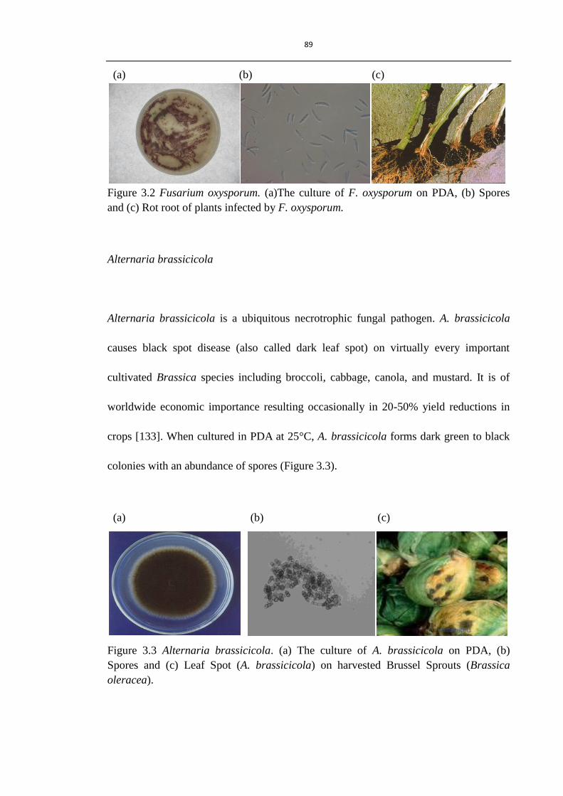
89
(a) (b) (c)
Figure 3.2 Fusarium oxysporum. (a)The culture of F. oxysporum on PDA, (b) Spores
and (c) Rot root of plants infected by F. oxysporum.
Alternaria brassicicola
Alternaria brassicicola is a ubiquitous necrotrophic fungal pathogen. A. brassicicola
causes black spot disease (also called dark leaf spot) on virtually every important
cultivated Brassica species including broccoli, cabbage, canola, and mustard. It is of
worldwide economic importance resulting occasionally in 20-50% yield reductions in
crops [133]. When cultured in PDA at 25°C, A. brassicicola forms dark green to black
colonies with an abundance of spores (Figure 3.3).
(a) (b) (c)
Figure 3.3 Alternaria brassicicola. (a) The culture of A. brassicicola on PDA, (b)
Spores and (c) Leaf Spot (A. brassicicola) on harvested Brussel Sprouts (Brassica
oleracea).

90
Cladosporium herbarum
Cladosporium herbarum is the most frequently encountered mold in the air. It causes
leaf mold and is one of the most common colonisers of dying and dead plants and also
occurs in various soil types [133]. Also, this mold is frequently found in uncleaned
refrigerators, foodstuffs, on moist window frames, in houses with poor ventilation, with
straw roofs, situated in low, damp areas. Cladosporium herbarum is cultured on PDA
and the colour of the colony ranges from dark green to black (Figure 3.4).
(a) (b) (c)
Figure 3.4 Cladosporium herbarum. (a) The culture of C. herbarum on PDA, (b) Spores
and (c) Sweet corn infected by C. herbarum.
Botrytis cinerea
Botrytis cinerea, the cause of grey mould disease, is an important commercial target for
fungicide, as it is the major necrotrophic fungal pathogen for vine, vegetables and top
fruits world-wide [134]. Juicy fruits favour the growth of this pathogen in which it
spread much quicker than on plant leaves, hence B. cinerea causes huge damage to fruit
crops and is responsible for much post harvest spoilage. Apart from traditional azoles

91
and dicarboximides fungicides, many new fungicides have been developed to eliminate
the crop loss caused by this pathogen, such as pyrimethanil and mepanipyrim [132]. The
west European fungicide market of B. cinerea was estimated to be worth about 50
million dollars in 2004 [2]. In culture on PDA, as on host plant, the colonies are grey in
colour (Figure 3.5).
(a) (b) (c)
Figure 3.5 Botrytis cinerea. (a) The culture of B. cinerea on PDA, (b) Spores and (c)
bunch rot B. cinerea of grapes.
3.1.2 Laboratory facilities and safety
Some micro-organisms are hazardous to humans, for example, spores of Botrytis,
Cladosprium, Aspergillus, and Rhizopus, give rise to allergic (hypersensitive) reactions
[135]. Although the fungi tested in this project have low pathogenicity to humans,
safety precautions should still been followed carefully. In the laboratory, cultural
procedures were carried out in a designated culture room which was maintained clean
and key facilities were sterilised with 70% EtOH. A class II bio-safety cabinet was used
to reduce contamination of both culture and operator. In such a cabinet, the air pressure
inside was higher than outside, and the circulating air was sterilised by a high efficiency

92
particulate air (HEPA) filter which removes over 99.7% of airborne particles of 0.3 μm
or greater in diameter. All apparatus and culture media were sterilised to eliminate any
transmissible agents (such as fungus, bacterial, virus and so on) by steam autoclave
(121 °C, 20 min). After experimentation, bio-hazard waste was sterilised by autoclave
before disposal.
3.2 In vitro antifungal activity assays
In vitro antifungal assay was established in a 96-well plate in order to assess the effect
of compounds on different fungal species in a controlled environment.
3.2.1 Material and equipment
3.2.1.1 Fungal strains
Four fungal strains tested were from the fungal stocks used by Prost I et al. [11]
including C. herbarum, Cladosporium herbarum (Pers.:Fr.); B. cinerea, Botrytis cinerea
(MUCL30158); F. oxysporum, Fusarium oxysporum (unspecified) and A. brassicicola,
Alternaria brassicicola (MUCL20297). Another isolate of Botrytis cinerea, B. cinerea
(GLUK-1), was supplied by Dr. Katherine J. Denby (Warwick HRI) (Table 3.1).

93
Table 3.1 Fungal strains tested.
Fungal strain Plant disease Growth condition
C. herbarum Leaf mould,
Indoor and outdoor mould
PDA, 25 °C, in darkness.
B. cinerea
(MUCL30158)
Major pathogen for vine,
Grey mould,
Root and fruit rotting,
PDA, 25 °C, 12 h light, 12 h
dark.
B. cinerea
(GLUK-1)
Major pathogen for vine,
Grey mould,
Root and fruit rotting,
Sterilised apricot half, 25 °C,
in darkness.
F. oxysporum Breakdown of xylem,
Leaf wilting,
Head blight (wheat),
PDA, 25 °C, in darkness.
A. brassicicola Brassica dark leaf spot on most
Brassica species,
PDA, 25°C, in darkness.
3.2.1.2 Antifungal activity of synthesised compounds
Four synthetic compounds (Figure 2.1) and two compounds, (E)-2-nonenal and a
cyclopentenone ring, purchased from Sigma-Aldrich were tested.
3.2.1.3 Chemicals
Distilled water was used to prepare aqueous media for all experiments. Other solvents
used were of high purity (High performance liquid chromatography (HPLC) grade) for
all routine experiments.

94
3.2.1.4 Apparatus
A Max Mono Monocular biological Microscope (Fisher) was used for spore counting;
Omega sterilising system (Prestige Medical Autoclave) for sterilisation; Versatile
Environmental test Chamber MRL-350H (SANYO) for fungal culture; Microplate
reader Model 680 (Bio-rad) for monitoring turbidity. All the solvents, glassware and
pipette tips used for fungal culture were sterilised by autoclaving.
3.2.2 Methods
3.2.2.1 Fungal culture
The method used for fungal culture was modified from that used for the evaluation of
oxylipins [11, 135]. Briefly, C. herbarum, F. oxysporum and A. brassicicola were
cultured on autoclaved potato dextrose agar (PDA 39g/L, Sigma-Aldrich). These
samples were sealed with parafilm (Fisher) and stored in an incubator at 25 °C. F.
oxysporum and A. brassicicola were sub-cultured every week. C. herbarum was
sub-cultured every two weeks.
B. cinerea (MUCL30158) was grown on PDA (24 g/L, Sigma-Aldrich) for 3 to 4 weeks
with a photoperiod of 12 hours at 25 °C in a growth chamber (SANYO, Versatile
Enviromental Test Chamber, Fluorescent light: 40W×3(FL40SSW/37)). B. cinerea

95
(GLUK-1) was grown on half apricots purchased from Tesco, and these samples were
sealed with micro-pore tape (Boots) and cultured for 2 to 3 weeks in darkness at 25 °C
in an incubator.
3.2.2.2 Harvest of fungal spores
Spore numbers were counted in suspension using a hemocytometer (Bright-Line,
Sigma-Aldrich) (Figure 3.6).
F. oxysporum and A. brassicicola
Petri dishes were flushed with sterilised potato dextrose broth (PDB, 48 g/L,
Sigma-Aldrich) solution and newly cultured fungal spores were removed by pipette.
The spore suspension was filter through a 50 μm autoclaved Cell Micro Sieves (VWR)
and adjusted to 100,000 to 120,000 spores/mL by dilution with PDB.
C. herbarum and B. cinerea (MUCL30158 & GLUK-1)
For C. herbarum spore production, petri dishes were flooded with distilled water.
Spores were scraped off into distilled water using a glass pipette which had been heat
treated to form a right angle configuration. The spores were filtered through an
autoclaved Cell Micro Sieves (VWR) with micropore size of 50 μm and the

96
concentration was adjusted to 40,000 spores/mL with distilled water. A similar method
was used to collect B. cinerea (MUCL30158 & GLUK-1) spore suspension. Once B.
cinerea mycelium was suspended in water, spores were gently scraped into distilled
water. Spore concentration was adjusted to 100,000 spores/mL after filtration through
Cell Micro Sieves (VWR) with micropore size of 50 μm.
Figure 3.6 Hemocytometer (Bright-Line, Sigma-Aldrich). The volume of each squares
marked with 1 to 5 = 1 mm (length) ×1 mm (width)×0.1 mm (depth) = 1×10⁻⁴
mL. The
concentration of cell (cells/mL) = (Total cells counted in five squares) ÷5 x 10⁴.
3.2.2.3 Antifungal tests
The method used for antifungal test was modified from the one used for the evaluation
of oxylipins [11]. F. oxysporum and A. brassicicola were grown in potato dextrose

97
broth (PDB, 48g/L, Sigma-Aldrich) in an incubator at a speed of 250 rpm at 25°C. C.
herbarum and B. cinerea were grown in clarified V8 juice broth (5% V8 juice (100%,
Campbell Foods, Belgium) in water, pH was adjusted to 5.0 (Hydrus 300 pH meter,
Fisher) by the addition of NaOH and centrifuged three times (10,000 g, 5 min,
Eppendorf Centrifuge 5804)) in an incubator at 25 °C. Cultures were started with 2,000
spores/well (C. herbarum) or 5,000 spores/well (B. cinerea, F. oxysporum and A.
brassicicola). Microorganisms were grown in sterile, flat-bottom, 96-well flat-bottom
microplates (NUNC Sigma-Aldrich), sealed with parafilm, in a final volume of 100 μL.
Test compounds were dissolved in ethanol and the ethanol concentration was kept to a
maximum of 1% final concentration in each well. Controls were treated with 1%
ethanol. Fungal growth was detected by the absorbance of the microcultures at 595 nm
with Bio-rad plate reader Model 680, after 24 to 72 h of incubation in the presence or
absence of compounds. For C. herbarum and B. cinerea, compounds were added after
16 h of growth and A595 measured at 16 h and 24 to 72 h period. The amount of light
transmitted through the culture suspension was compared with that through a blank,
measuring the turbidity of each well according to the Lambert-Beer Law:
lcI
IA
0
log
A: absorbance (also called optical density (OD))
I0: amount of incident light
I: amount of transmitted light
ε: extinction coefficient
l: path length across the suspension
c: concentration

98
For homogenous spore suspensions, turbidity equates with spore germination and
mycelium growth and largely follows the Lambert-Beer Law [135]. However, the
germination and growth of some fungal species (e.g. Rhizopus) form non-uniform
suspensions which clearly can lead to wide discrepancies in values. Fungal strains tested
here can form relatively homogenous growing suspensions and provide reliable results.
3.2.2.4 Spore germination assay
This method was modified from the one published by Prost I [11]. Spore germination
was assessed in the presence of the test compounds in a 96-well plate. Compounds were
added to each well to a final concentration of 100 μM (all compounds) together with the
spore suspension. For B. cinerea (GLUK-1) and C. herbarum, spores were suspended in
clarified 5% V8 juice broth, whereas for F. oxysporum and A. brassicicola, potato
dextrose broth was used instead. The plate was incubated for a few hours (16 h for C.
herbarum, 10 h for B. cinerea (GLUK-1), 8 h for F. oxysporum, A. brassicicola), before
the observation was carried out under the microscope (Max Mono Monocular biological
Microscope, magnification: 100×) and 50 spores were assessed for germination status.
Spores with visible germ tubes were counted as being germinated and results were
expressed as a percentage of germination.

99
3.2.3 Results
3.2.3.1 Antifungal tests
A. brassicicola, F. oxysporum, C. herbarum, B. cinerea (MUCL30158) and B. cinerea
(GLUK-1) were exposed to each of the test compounds and their growth was measured
over a 72 h period (Figure 3.7). None of the test compounds (100 μM) had significant
antifungal activity against F. oxysporum. Only 4-octyl-cyclopentenone showed limited
inhibition (60% inhibition, P<0.01) to fungal growth after 24 h. However, fungal
growth recovered and overcame inhibition after 72 h (Figure 3.7 a). Similar to F.
oxysporum, none of the test compounds at 100 μM showed good antifungal activities
against A. brassicicola and appeared to have significant antifungal activity in
comparison to the control after 72 h. 4-Octyl-cyclopentenone, 4-hexyl-cyclopentenone
and (E)-2-nonenal showed limited inhibition (50% inhibition, P<0.01) on fungal growth
within 24 h and these two target microorganisms overcame inhibition after 72 h (Figure
3.7 b). In contrast to A. brassicicola and F. oxysporum, C. herbarum were more
sensitive to test compounds, especially to 4-octyl-cyclopentenone. The absorbance
value of the treatment of 4-octyl-cyclopentenone (100 μM) after 24 h culture was nearly
zero and visually the suspension was clear without any fungal growth observable to the
naked eye, whereas fungal growth could be seen in the other treatments at the same
concentration after 24 h culture. Fungal growth with the treatment of
4-octyl-cyclopentenone (100 μM) recovered gradually but slower than the other
100
treatments. After 72 h culture, its inhibition decreased to nearly 50% (P<0.01).
Therefore, this compound (at 100 μM) was just able to control fungal growth rather than
kill it. None of the other test compounds showed as good antifungal activity against C.
herbarum as 4-octyl-cyclopentenone. (E)-2-nonenal showed very limited inhibition
against this fungal strain (Figure 3.7 c).
The inhibition of B. cinerea (MUCL30158) was similar to that of C. herbarum.
Mycelium growth following exposure to 4-octyl-cyclopentenone (100 μM) was
inhibited by nearly 100% (P<0.01) after 24 h. Fungal growth recovered by about 50%
(P<0.01) after 48 h culture. No other test compounds showed significant antifungal
activity in comparison to the control after 48 h (Figure 3.7 d). As to the other fungal
isolate of Botrytis, B. cinerea (GLUK-1), the antifungal activities of treatments at higher
concentration of 200 μM were assessed. The antifungal activity of
4-octyl-cyclopentenone at 200 μM against B. cinerea (GLUK-1) is similar to that of B.
cinerea (MUCL30158) treated with 4-octyl-cyclopentenone at 100 μM. None of the
other compounds tested at the concentration of 200 μM inhibited fungal growth (Figure
3.7 e). The interesting finding is that fungal growth recovery occurred after 48 h in
wells of these two B. cinerea isolates, and it was found that among eight wells treated
with 4-octyl-cyclopentenone, fungal growth was only found in three to four wells and
meanwhile the other wells were completely clear. This accounted for the wide
varieties in the absorbance/optical density (OD) values of 4-octyl-cyclopentenone
treatments of these two B. cinerea isolates had large variations (Figure 3.7 d and e) after

101
72 h culture.
In short, previous work reported that (E)-2-nonenal (100 μM) has no effect on all tested
fungal strains [11], which was confirmed in this work. The cyclopentenone ring on its
own did not appear to have any antifungal activity. 4-Hexyl-cyclopentenone and
4-butyl-cyclopentenone having six and four carbon alkyl chains, respectively, are much
less effective than 4-octyl-cyclopentenone (containing an eight carbon alkyl chain) on
fungi, which shows the importance of the length of alkyl chain on antifungal activity of
compounds. Surprisingly, 8-(4-oxocyclopentenyl)octanoic acid is very similar to the
lead molecule, OPDA, yet little antifungal activity was observed. Overall,
4-octyl-cyclopentenone showed the best antifungal activities among these test
compounds. It has similar antifungal activities to its natural analogue, OPDA, which is
also more active against C. herbarum and B. cinerea (MUCL30158) than F. oxysporum
and A. brassicicola [11]. Further tests were therefore focused on 4-octyl-cyclopentenone
using different concentrations of the compounds.

102
(a) Effect of test compounds on the growth of F. oxysporum.
(b) Effect of test compounds on the growth of A. brassicicola.
Figure 3.7 Results of in vitro antifungal assay. Fungal strains were exposed to a series
of test compounds in appropriate liquid culture medium, starting from the time of spore
addition to wells (F. oxysporum and A. brassicicola) or 16 h later (C. herbarum and B.
cinerea). Results show means±SD (n=6). Asterisk indicates a significant difference at
P< 0.01 (Dunnett‟s test) against control. 4-octyl-cyclopentenone (3),
4-hexyl-cyclopentenone(4), 4-butyl-cyclopentenone(5), 8-(4-oxocyclopentenyl)octanoic
acid (8).
* *
*
*
0.000
0.100
0.200
0.300
0.400
0.500
0.600
0.700
0.800
Turb
idit
y (O
D a
t 5
95
nm
)
24h48h72h
* * *
* * * *
0.000
0.100
0.200
0.300
0.400
0.500
0.600
0.700
Turb
idit
y (O
D a
t 5
95
nm
)
24h48h72h

103
(c) Effect of test compounds on the growth of C. herbarum.
(d) Effect of test compounds on the growth of B. cinerea (MUCL30158).
Figure 3.7 Results of in vitro antifungal assay. Fungal strains were exposed to a series
of test compounds in appropriate liquid culture medium, starting from the time of spore
addition to wells (F. oxysporum and A. brassicicola) or 16 h later (C. herbarum and B.
cinerea). Results show means±SD (n=6). Asterisk indicates a significant difference at
P< 0.01 (Dunnett‟s test) against control. 4-octyl-cyclopentenone (3),
4-hexyl-cyclopentenone(4), 4-butyl-cyclopentenone(5), 8-(4-oxocyclopentenyl)octanoic
acid (8).
**
**
*
0.000
0.100
0.200
0.300
0.400
0.500
Turb
idit
y (O
D a
t59
5n
m)
24h48h72h
-** **
-0.200
-0.100
0.000
0.100
0.200
0.300
0.400
0.500
0.600
Turb
idit
y (O
D a
t59
5n
m)
24h48h72h

104
(e) Effect of test compounds on the growth of B. cinerea (GLUK-1).
Figure 3.7 Results of in vitro antifungal assay. Fungal strains were exposed to a series
of test compounds in appropriate liquid culture medium, starting from the time of spore
addition to wells (F. oxysporum and A. brassicicola) or 16 h later (C. herbarum and B.
cinerea). Results show means±SD (n=6). Asterisk indicates a significant difference at
P< 0.01 (Dunnett‟s test) against control. 4-octyl-cyclopentenone (3),
4-hexyl-cyclopentenone(4), 4-butyl-cyclopentenone(5), 8-(4-oxocyclopentenyl)octanoic
acid (8).
3.2.3.2 Antifungal activities of 4-octyl-cyclopentenone
4-Octyl-cyclopentenone at various concentrations (400 µM, 200 µM and 100 µM) was
tested against fungal strains (Figure 3.8). F. oxysporum was not very sensitive to
4-octyl-cyclopentenone at the lowest concentration, 100 μM. However, at higher
concentrations significant inhibition (P<0.01) occurred. At 200 µM, the inhibition of
mycelium growth decreased from 70% (P<0.01) after 24 h to nearly 40% after 72 h,
reaching 50% growth inhibition (P<0.01) after 48 h (Figure 3.8 a).
4-Octyl-cyclopentenone at 100 µM showed limited antifungal activity against A.
**
*
0.000
0.100
0.200
0.300
0.400
Turb
idit
y (O
D a
t 5
95
nm
)
24h
48h
72h

105
brassicicola, although this activity increased with increase in concentration.
4-Octyl-cyclopentenone only inhibits hyphal growth rather than kill it, and growth
recovery was found after 48 h. At concentrations of 200 µM and 400 µM,
4-octyl-cyclopentenone showed increased antifungal activity against A. bassicicola than
F. oxysporum, and the inhibitions of 200 µM and 400 µM were approximately 50%
(P<0.01) and 65% (P<0.01) respectively after 72 h culture (Figure 3.8 b). C. herbarum,
4-octyl-cyclopentenone at 400 µM, 200 µM and 100 µM had nearly 100% inhibition
(P<0.01) of fungal growth after 24 h and no mycelium growth was evident within a
week in 400 µM and 200 µM treated samples (Figure 3.8 c). For concentrations lower
than 100 µM, very limited growth inhibition was found and growth recovered very
quickly in two day culture (data not shown). Similar to C. herbarum, the fungal growth
of B. cinerea (MUCL30158) was totally inhibited at 200 µM and 400 µM and no
mycelium growth was detected even after one week culture. For 100 µM application,
inhibition was nearly 100% (P<0.01) after 24 h and recovered a little after 48 h (Figure
3.8 d). The fungal growth of B. cinerea (GLUK-1) was inhibited by this compound
within 48 h, however the recovery of fungal growth was found in 72 h culture. As long
as the concentration increased to 400 μM, 4-octyl-cyclopentenone inhibited the growth
of this isolate within 72 h, and no recovery was observed (Figure 3.8 e).
In short, antifungal activity against all fungi increases with an increase in concentration
of 4-octyl-cyclopentenone. For the two less sensitive fungi, F. oxysporum and A.
brassicicola, their spores can survive and grow when exposed to

106
4-octyl-cyclopentenone at high concentration of 400 μM after 24 h, i.e. the minimum
inhibitory concentration (MIC) must be over 400 μM. For C. herbarum and B. cinerea
(MUCL30158), no visual fungal growth was found in the treatment of
4-octyl-cyclopentenone at 100 μM after 24 h, e.g. MIC is 100 μM, and the fungal
growth recovered slowly after 48 h. When the concentration of 4-octyl-cyclopentenone
increased to 200 μM, no recovery of fungal growth of these two fungi was detected
within 72 h and even after a week (data not shown), e.g. the minimum fungicidal
concentration (MFC) of this compound against C. herbarum and B. cinerea
(MUCL30158) is between 100 and 200 μM. The other isolate of B. cinerea (GLUK-1)
(from infected pepper in Scotland) is less susceptible to 4-octyl-cyclopentenone than B.
cinerea (MUCL30158) isolated in mainland Europe (source not given).
4-Octyl-cyclopentenone at 200 μM can only inhibit spore germination and partially
inhibit spore germination/mycelial growth within 24 h. The fungicidal effect against B.
cinerea (GLUK-1) can be achieved by doubling the concentration to 400 μM. Hence,
the MIC of 4-octyl-cyclopentenone against B. cinerea (GLUK-1) is 200 μM and
corresponding MFC is 400 μM. In summary, although the antifungal activity increases
with concentration, 4-octyl-cyclopentenone is effective against C. herbarum and B.
cinerea at concentrations lower than 400 μM.

107
(a) Effect of 4-octyl-cyclopentenone on the growth of F. oxysporum.
(b) Effect of 4-octyl-cyclopentenone on the growth of A. brassicicola.
Figure 3.8 In vitro antifungal assay of 4-octyl-cyclopentenone (3). Fungal strains were
exposed to 4-octyl-cyclopentenone (3) of different concentrations of 100, 200 and 400
μM, starting from the time of spore addition to wells (F. oxysporum and A. brassicicola)
or 16 h later (C. herbarum and B. cinerea). Results show means±SD (n=6). Asterisk
indicates a significant difference at P< 0.01 (Dunnett‟s test) against control.
* * *
**
*
**
*
0.000
0.100
0.200
0.300
0.400
0.500
0.600
0.700
0.800
Control(EtOH 1%) 3(100µM) 3(200µM) 3(400µM)
Turb
idit
y O
D(5
95
nm
)
24h48h72h
* * *
**
*
*
*
0.000
0.100
0.200
0.300
0.400
0.500
0.600
0.700
Control(EtOH 1%) 3(100µM) 3(200µM) 3(400µM)
Turb
idit
y (O
D a
t 5
95
nm
)
24h48h72h

108
(c) Effect of 4-octyl-cyclopentenone on the growth of C. herbarum.
(d) Effect of 4-octyl-cyclopentenone on the growth of B. cinerea (MUCL30158).
Figure 3.8 In vitro antifungal assay of 4-octyl-cyclopentenone (3). Fungal strains were
exposed to 4-octyl-cyclopentenone (3) of different concentrations of 100, 200 and 400
μM, starting from the time of spore addition to wells (F. oxysporum and A. brassicicola)
or 16 h later (C. herbarum and B. cinerea). Results show means±SD (n=6). Asterisk
indicates a significant difference at P< 0.01 (Dunnett‟s test) against control.
* * ** * ** *
-0.100
0.000
0.100
0.200
0.300
0.400
0.500
0.600
Control(EtOH 1%) 3(100 µM) 3(200 µM) 3(400 µM)
Turb
idit
y O
D a
t 5
95
nm
)
24h
48h
72h
* * ** * ** *
-0.100
0.000
0.100
0.200
0.300
0.400
0.500
Control(EtOH 1%) 3(100µM) 3(200µM) 3(400µM)
Turb
idit
y (O
D a
t 5
95
nm
)
24h
48h
72h

109
(e) Effect of 4-octyl-cyclopentenone on the growth of B. cinerea (GLUK-1)
Figure 3.8 In vitro antifungal assay of 4-octyl-cyclopentenone (3). Fungal strains were
exposed to 4-octyl-cyclopentenone (3) of different concentrations of 100, 200 and 400
μM, starting from the time of spore addition to wells (F. oxysporum and A. brassicicola)
or 16 h later (C. herbarum and B. cinerea). Results show means±SD (n=6). Asterisk
indicates a significant difference at P< 0.01 (Dunnett‟s test) against control.
3.2.3.3 Effect of compounds on fungal spore germination
Spore germination is an important developmental stage in the life cycle of fungi,
therefore it is worthy to show the performance of compounds on spore germination after
short time exposure. Spores were incubated for a few hours in the presence of selected
compounds. For each fungus, the incubation time for germination assessment was
optimised for high number of germinated spores in the controls for better visualisation
under the microscope. The percentage of germinated spores of each treatment was
calculated and compared with the percentage of germination in control treated with 1%
EtOH. The results are shown in figure 3.9. In the controls of C. herbarum and B.
** *
*
* *
**
*
-0.100
0.000
0.100
0.200
0.300
0.400
0.500
Control(EtOH 1%) 3(100µM) 3(200µM) 3(400µM)
Turb
idit
y (O
D a
t 5
95
nm
)
24h
48h
72h

110
cinerea, nearly all spores germinated after 16 hours and 10 hours cultured, however,
only 65% of spores of F. oxysporum germinated after 8 h culture.
4-Octyl-cyclopentenone is again the most active compound tested, which has about 80%
inhibition of the spore germination of test species at the concentration of 100 μM and
100% inhibition at 200 μM. Other test compounds showed very limited inhibition of
fungal spore germination. The effect of compounds on spore germination of A.
bassicicola was not able to be evaluated by this method, because the clustering of
spores made it difficult to count them under the microscope. However, it was still
evident that 4-octyl-cyclopentenone at test concentrations showed good inhibition of
spore germination of A. bassicicola after 7-8 h culture (data not shown).
Figure 3.9 Effect of compounds on spore germination. Freshly prepared spores of C.
herbarum (20,000 spores/mL), F. oxysporum (50,000 spores/mL) and B. cinerea (50,000
spores/mL) were exposed to each compounds and assessed for germination status after
16 h (C. herbarum) or 8 h (F. oxysporum and B. cinerea (GLUK-1)). Each bar
represents mean of three independent experiments ±SD. 4-octyl-cyclopentenone (3),
4-hexyl-cyclopentenone(4), 4-butyl-cyclopentenone(5), 8-(4-oxocyclopentenyl)octanoic
acid (8).
-20.0
0.0
20.0
40.0
60.0
80.0
100.0
120.0
% g
erm
inat
ion
sp
ore
s
C. herbarum
F. oxysporum
B.cinerea (GLUK-1)

111
3.2.4 Discussion
Apart from cyclopentenone and 8-(4-oxocyclopentenyl)octanoic acid, synthetic RES
compounds belong to non-ionised surfactants, with polar heads and non-polar tails,
which contribute to their different log P values. Surfactants are known as an important
group of materials having active properties, of which antimicrobial activity is an
important application. Lipophilicity is a key property and plays an important role in
modulating their antimicrobial activities.
4-Octyl-cyclopentenone, 4-hexyl-cyclopentenone and 4-butyl-cyclopentenone have the
same hydrophilic head, the cyclopentenone ring, and differ in the length of alkyl chain.
This compound is the most effective antifungal agent and significantly better than the
other two compounds against the tested fungal species. Their cyclopentenone ring will
provide similar electrophilicity and corresponding biological activities. The different
lengths of the hydrophobic chain that have influence on the lipophilicity (log P) of
compounds seem to affect the antifungal activity of compounds. In this project, log P
value was calculated by Molinspiration log P (milog P) calculator (Table 3.2).
The antifungal activities of some other non-ionised surfactants against several fungal
species have been well studied [9, 68, 136-143]. Their conclusion was that the
antifungal activity of non-ionised surfactants primarily depends on the properties of the
biological hydrophilic head of test compounds, however, the length of the alkyl group

112
Table 3.2 Lipophilicity (log P) of compounds tested for in vitro antifungal activity. The
value of log P was calculated by Molinspiration log P calculator.
Chemicals log P value
OPDA 4.6
4-octyl-cyclopentenone (3) 4.5
4-hexyl-cyclopentenone (4) 3.5
4-butyl-cyclopentenone (5) 2.5
8-(4-oxocyclopentenyl)octanoic acid (8) 2.8
Cyclopentenone 0.4
also plays an important role. This alkyl chain is considered to balance the hydrophobic
and hydrophilic moieties to achieve an optimal lipophilicity which favours compounds
to reach the active sites. Given the same hydrophilic part, the antifungal effectiveness of
test compounds, to some extent, depends on their lipophilicity, i.e. the balance between
hydrophobic and hydrophilic parts.
4-Octyl-cyclopentenone with an eight carbon aliphatic chain has the highest antifungal
activity of the compounds tested in this project. From Table 3.2, it can be seen that the
log P value of this compound is 4.5. 4-Hexyl-cyclopentenone and
4-butyl-cyclopentenone are much less effective on inhibiting fungal growth and their
log P values are 3.5 and 2.5, respectively. Interestingly, 4-octyl-cyclopentenone and
OPDA have very close values of log P, 4.5 and 4.6, respectively. In addition, OPDA
was elucidated to have very similar antifungal activities against several fungal strains to
4-octyl-cyclopentenone. Both have more effective antifungal activity against C.
herbarum and B. cinerea (MUCL30158) than F. oxysporum and A. brassicicola. As

113
RES, their similar eletrophilicity and lipophilicity might endow them comparable
performances, such as solubility in tissue matrices, affinity to amphiphilic substances
(lipids and proteins) and membrane interaction. However, OPDA and other oxylipins
tested were reported to be less effective than those commercial fungicides (such as
benomyl and metalaxyl) against the same target organisms [11]. The synthetic
analogous of OPDA, 4-octyl-cyclopentenone, behave similar antifungal activity to
OPDA, therefore further modification will be needed to enhance its performance which
would compete with those commercial products.
(E)-2-nonenal is an effective compound among plant volatile compounds derived from
HPL pathway displaying higher antifungal activity than other common GLVs, like
(E)-2-hexenal and (Z)-3-nonenal [144]. (E)-2-nonenal was reported to have antifungal
activity against A. brassicicola, F. oxysporum and B. cinerea [71, 77]. It showed
inhibition of spore germination of F. oxysporum and B. cinerea at concentration lower
than 100 μM. However, here we observed that (E)-2-nonenal (100 μM) showed little
effect on spore germination and fungal growth of test fungal species. The different
results may be caused by using different pathogen isolates and experimental systems in
our laboratory.
8-(4-Oxocyclopentenyl)octanoic acid which contains a carboxyl group on the end of the
alkyl chain like OPDA was not as effective as 4-octyl-cyclopentenone against these
fungal species. One possibility is that the loss of antifungal activity could be due to the

114
lack of a pentenyl chain on C13 of OPDA leading to a much lower log P value (2.8)
than that of OPDA (log P = 4.6) and 4-octyl-cyclopentenone (log P = 4.5). Such lower
lipophilicity will affect the membrane affinity and permeability of compounds to fungal
pathogens. Another reason for the inactivity of 8-(4-oxocyclopentenyl)octanoic acid
could be that the presence of two polar heads (cyclopentenone ring and carboxyl end
group) on 8-(4-oxocyclopentenyl)octanoic acid could interact with each other forming a
intramolecular ring when the eight carbon chain bends. Such a change will form a
two-ring structure of 8-(4-oxocyclopentenyl)octanoic acid so that it is hard to penetrate
the plasma membrane. In the case of natural occurring cis- (+) OPDA, the presence of
side pentenyl substitution will block the bending of the longer chain due to steric
hindrance and its antifungal activity was preserved or even enhanced. Pure OPDA and
4-octyl-cyclopentenone have the physical appearance of oil, whereas
8-(4-oxocyclopentenyl)octanoic acid with carboxyl end forms crystal when
concentrated. This observation reflects the influence of the pentenyl side chain of
OPDA on its physical property. Besides the two possibilities mentioned, the
biodegradation of 8-(4-oxocyclopentenyl)octanoic acid might cause the suppression of
its bio-activity. Although it is known that OPDA is very stable during in vitro antifungal
assays [11], the degradation of 8-(4-oxocyclopentenyl)octanoic acid by β-oxidation
cannot be excluded due to the presence of the carboxyl group. A carboxyl group is
needed for the esterification to CoA which is subsequently necessary for β-oxidation
[25, 65]. The β-oxidation would shorten the alkyl chain, and in this way, the antifungal
activity of 8-(4-oxocyclopentenyl)octanoic acid could be degraded. From another view,

115
the oxidation of compounds will accelerate the speed of biodegradation, which would
decrease the adverse effect of compounds on the environment. If
8-(4-oxocyclopentenyl)octanoic acid was sprayed onto plants to induce host defence
response, this compound could also be a potential indirect antifungal candidate. These
hypotheses need to be evaluated in the future and more research undertaken to clarify
the detailed biological activities of 8-(4-oxocyclopentenyl)octanoic acid.
For B. cinerea, 4-octyl-cyclopentenone shows a different performance against different
isolates. B. cinerea (MUCL30158) is more sensitive to this compound than B. cinerea
(GLUK-1). The large variation of results after 48 h culture was found for Botrytis
(MUCL30158) treated with 4-octyl-cyclopentenone at 100 μM and B. cinerea (GLUK-1)
treated with the same compound at 200 μM. As the antifungal action mode of
4-octyl-cyclopentenone is still unknown, it is difficult to explain this interesting finding.
One explanation could be that 4-octyl-cyclopentenone inhibited most of the fungal cells
and the recovery of fungal growth only started from the cells which survived from the
treatment. As discussed above, the antifungal activity of 4-octyl-cyclopentenone at
concentration of 100 μM against B. cinerea (MUCL30158) is comparable to that of
4-octyl-cyclopentenone at 200 μM against B. cinerea (GLUK-1). This observation
indicated that although different antifungal sensitivities were found for different isolates,
they might share similar antifungal modes of action. In this project, B. cinerea
(MUCL30158) was used for in vivo test and since B. cinerea (GLUK-1) provides a
larger yield of spore than B. cinerea (MUCL30158), this makes it more convenient for

116
the study of modes of action due to the requirement of large amount of spores used in
these types of assays.
3.2.5 Conclusion
In conclusion, 4-octyl-cyclopentenone is the most active compound against the fungal
species tested and has similar activities to OPDA. The simplified
4-octyl-cyclopentenone, therefore, can mimic some biological activities of OPDA. Our
data shows that as a RES, the series of synthesised compounds has a range of antifungal
activity even though they have the same electrophilic cyclopentenone ring. The different
lengths of alkyl chains therefore play an important role in balancing hydrophobic and
hydrophilic moieties (i.e. lipophilicity, measured as log P) which is likely to improve
the membrane permeability of the compound to reach the active sites of target
pathogens.
3.3 In vivo antifungal assay of 4-octyl-cyclopentenone
From the in vitro tests, 4-octyl-cyclopentenone was established as the strongest
antifungal compound against the test organisms. However, the controlled environment
of the in vitro test differs from the condition in whole tissues (in vivo test). It is
therefore necessary to determine whether in vitro results would translate into the desired

117
efficacy in vivo. To this end, a series of in vivo experiments was conducted to evaluate
4-octyl-cyclopentenone‟s antifungal activity.
3.3.1 Materials
Test fungal strains were mentioned in 3.2.1. Seeds of two plant species, Arabidopsis
thaliana (Col-5) and Brassica oleracea were supplied by Professor Eric Holub from
Warwick HRI. The in vivo test on leaves of Arabidopsis thaliana was carried out in the
laboratory of Dr. Katherine J. Denby (Warwick HRI), following their standard
procedure given below.
3.3.2 Methods
3.3.2.1 Growing of the test plants
Arabidopsis thaliana and Brassica oleracea are popular as model organisms in plant
biology and genetics. Their wild-type and mutants are widely used in many laboratories
to study plant defence and interface between pathogens and host plants [49, 63,
145-147].

118
(a) Growing of Arabidopsis thaliana (Col-5)
The basic method used for growing Arabidopsis thaliana in this project was modified
from the classic method published by the Arabidopsis Biological Resource Centre [148].
Seeds were placed on the surface of premixed soil (4 scoops of potting soil, 2 scoops of
sand and 2 scoops of vermiculite) in cell planting trays. Trays with seeds were placed at
3-4 °C for at least 2-4 days to break dormancy and improve germination rate. The trays
were then placed in a culture chamber (SANYO, Versatile Environmental Test Chamber)
under 100-150 μmol/m-2
s-1
photosynthetic photon flux density light with a 12 hour
photoperiod at 24 °C. Plants were regularly watered and the tray was covered with
plastic wrap which provided a high humidity level for successful seed germination.
(b) Growing of Brassica oleracea
Seeds were grown on the surface of premixed soil (6 scoops of potting soil, 1 scoop of
sand and 1 scoop of vermiculite) under 100-150 μmol/m-2
s-1
photosynthetic photon flux
density light with a 16 hour photoperiod in SANYO culture chamber at 24 °C. One
week old plants were used for phytotoxicity testing.
3.3.2.2 Phytotoxicity test of 4-octyl-cyclopentenone on Brassica oleracea
One week old Brassica oleracea plants were used and 20 μL of 4-octyl-cyclopentenone

119
at different concentrations (100 μM, 200 μM and 400 μM) were applied to the surface
of the leaves virtually covering the small leaves at this early growing stage. 20 μL of 1%
EtOH treatment was used as a control. The plants were placed back in SANYO culture
chamber under the same growth conditions (100-150 μmol/m-2
s-1
photosynthetic photon
flux density light with a 16 hour photoperiod at 24 °C). Visual evidence of disease and
difference between control and 4-octyl-cyclopentenone treatments were recorded every
day and photos were taken 2 weeks later.
3.3.2.3 In vivo antifungal test on leaves of Arabidopsis thaliana
Experiments to test how 4-octyl-cyclopentenone can inhibit the growth of B. cinerea
(MUCL30158) on leaves of plants followed the method published by Denby (Warwick
HRI) [146, 149]. Leaves were harvested from five-week old Arabidopsis thaliana and
placed into plastic humidity trays containing 1.5% phytoagar at a depth of 1 cm. Leaves
were inoculated with 10 μL of suspension of 16 hour pre-cultured germinated spores in
5% V8 juice (10,000 spores/mL) treated with 4-octyl-cyclopentenone to achieve a final
concentration of 200 μM. 10 μL of germinated spore suspension treated with 1% EtOH
as a control. Total leaf area and diameter of the developing lesion were measured after
48 and 72 h using IMAGEJ and high resolution digital pictures of the leaves. A 1 cm
scale was included in each image, which enabled calibration of the measurements.

120
3.3.2.4 In vivo antifungal test on apricot fruits
The in vivo antifungal activity of 4-octyl-cyclopentenone was assessed by culturing B.
cinerea (MUCL30158) on apricot halves. This test was evaluated to determine whether
the compound was effective in vivo using a susceptive host substrate. Apricots were
purchased from Tesco and cleaned by washing surface three times with autoclaved
distilled water, then placed in a petri-dish. The fruit surface was punctured to form 3-4
holes with a depth of 4 mm using a glass rod of 3 mm diameter. The 16 h pre-cultured
germinated spores (50,000 spores/mL) suspension was mixed with
4-octyl-cyclopentenone to achieve a final concentration of 200 μM and 10-20 μL of the
suspension was injected into the holes. The petri-dish was sealed with parafilm and
cultured in an incubator. After 48 h, the growth of the pathogen on the fruit was
recorded photographically and compared to 1% EtOH control treated sample.
3.3.3 Results
3.3.3.1 Phytotoxicity test of 4-octyl-cyclopentenone
Phytotoxic means harmful or lethal to plants and phytotoxic effects can range from
slight burning or browning of the leaves to death of the plants. The phytotoxic effect of
4-octyl-cyclopentenone on Brassica leaves was monitored visually and recorded
photographically. The results in Figure 3.10 showed that there were no damage lesions,

121
burning, browning or death of leaves. No abnormal growth was observed in the
following three weeks with regard to the number and radii of leaves. In short, there was
no visual difference between control and all the treatments, even at highest
concentration of 400 μM. Although this visualisation approach cannot measure the
overall phytotoxic effects, the results indicate that no obvious damage had occurred to
the plant leaves after compound application.
Figure 3.10 Phytotoxicity assay of 4-octyl-cyclopentenone on Brassica oleracea.
4-octyl-cyclopentenone (3) with concentration up to 400 μM has no visual effects on the
growth of Brassica oleracea even after two weeks culture.
3.3.3.2 In vivo antifungal test on leaves of Arabidopsis thaliana
Although B. cinerea is a main pathogen of fruits, it can be pathogenic on Arabidopsis
thaliana resulting in an interaction which is amenable to biological analysis [149].
4-Octyl-cyclopentenone at 200 μM showed significant antifungal activity against B.
cinerea (MUCL30158) on leaves of Arabidopsis thaliana in 72 h culture and the

122
damage lesion area of the treatment is approximately 70-75% less than the control
(Figure 3.11). By contrast, in vitro, complete growth inhibition of this fungal strain was
observed in the treatment of this compound at 200 μM. However, given that conditions
on a leaf surface are quite different from a controlled sterile environment, the inhibition
achieved in vivo was still quite significant.
(a) Control (1% EtOH)
(b) Treatment of 4-Octyl-cyclopentenone (3) (200 μM)
Figure 3.11 In vivo antifungal test on leaves of Arabidopsis thaliana.
4-Octyl-cyclopentenone (3) (200 μM) was added to suspension of B. cinerea
(MUCL30158) after 16 h. The significant difference of damage lesion between control
(a) and treatment (b) indicates the inhibition of fungal growth after 72 h. Asterisk
indicates a significant difference at P< 0.01 (Dunnett‟s test) against control. Error bars
indicate ±SD (n=13).
*
0.000
0.020
0.040
0.060
0.080
0.100
Control(EtOH 1%) Compound3(200μM)
Lesi
on
Are
a (c
m2 )

123
3.3.3.3 Antifungal test on apricot fruits
Apricot halves were used to test the antifungal activity of 4-octyl-cyclopentenone (200
μM) against B. cinerea (MUCL30158). After 48 h, fungal growth was found around all
three inoculation sites for control treatments, in comparison, only one out of three
inoculation sites was infected with B. cinerea growth for 4-octyl-cyclopentenone
treatment and the radii of the infected site was smaller than that of the control (Figure
3.12). However, unlike the in vitro tests, 4-octyl-cyclopentenone at 200 μM could not
inhibit growth of Botrytis completely and slow recovery of fungal growth was observed
on all inocula sites after 72 h culture. Summarising, complete inhibition of fungal
growth of B. cinerea (MUCL30158) was achieved by in vitro treatment of
4-octyl-cyclopentenone (200 μM), however, in vivo fungal growth recovered and
mycelium growth initiated after 48 h delay.
(a) (b)
Figure 3.12 In vivo antifungal test on apricots. B. cinerea (MUCL30158) was inoculated
to apricot halves in the absence (b) and presence (a) of 4-octyl-cyclopentenone and
incubated for 48 h. Fungal growth was found around all three inoculated sites without
4-octyl-cyclopentenone (a) and only a little growth was found in one inoculated site
incubated with 4-octyl-cyclopentenone (b).

124
3.3.4 Discussion
Most commercial agrochemicals are known to be toxic to host plants if not applied
according to their instructions; therefore any commercial agrochemical should be tested
for its phytotoxicity before being introduced to the market. RES oxylipins with
α,β-unsaturated carbonyl group have both positive and negative effects on the host plant
cells. On the one hand they have important biological activities during the life span of
plants, on the other hand, this group of compounds has phytotoxicity to host plants and
certain cytotoxicity to mammal cells at high levels [61, 100]. The application of
4-octyl-cyclopentenone up to 400 μM on one-week old leaves of Brassica shows no
visual adverse effect on plants. In comparison, OPDA was reported to cause harmful
effects to plant tissue at higher concentration than 200 μM [115]. When this compound
accumulates in cells, it can be detoxified by GSH reductants as shown for
phytoprostanes [93]. Although it is still unknown whether 4-octyl-cyclopentenone was
taken up by the plant or not, it is predicted that the similar electrophilicity and
lipophilicity between this compound and OPDA might endow them similar biological
and physical properties. In addition, the exogenous application of α,β-unsaturated RES
on plants could induce the accumulation of endogenous RES oxylipins [61] and
phytoalexin [90], which were reported to be related to HR response in tobacco leaves
[150]. So far, the exogenous application of RES, 4-octyl-cyclopentenone, was not found
to induce any HR response or other visual symptoms of disease on Brassica.
Nevertheless, more experiments need to be performed to evaluate the overall

125
phytotoxicity related to this compound.
As to the antifungal test of 4-octyl-cyclopentenone on the system of Arabidopsis and B.
cinerea, this compound at concentration of 200 μM does not show 100% inhibition of
fungal growth. The direct antifungal activity of 4-octyl-cyclopentenone has been
already established in vitro. Whilst the compound was effective in vitro, complete
inhibition of fungal growth was not achieved in vivo and some fungal growth recovery
was observed. The antifungal activity of 4-octyl-cyclopentenone against B. cinerea
during in vivo testing is attributed to at least two factors:
1. Complex environment, including light, air flow and contaminations that will affect
both on plants and pathogens.
2. The changes of chemical and physical properties of the applied compound,
including unwanted concentration reduction due to degradation (chemical
degradation and biological degradation by the host tissue).
Therefore, these common factors will contribute to the difference between in vitro and
in vivo results.
Apricot fruits are natural hosts for B. cinerea, whereas Arabidopsis and B. cinerea
system is mainly used as a model in the laboratory [149]. For the test on apricots, the
environment that the compound is exposed to is likely to consist of an array of
degradation enzymes that may inactivate it. In addition, the diffusion of the compound
into fruit tissue will reduce local concentration. 4-Octyl-cyclopentenone at 200 μM was

126
effective against B. cinerea cultured in apricots within 48 h, however, some fungal
growth was evident after 72 h incubation. It is therefore necessary to do more chemical
structural modifications of compound in order to improve the performance of these
compounds.
3.3.5 Conclusion
RES oxylipins are known to have an adverse effect on host plants and the accumulation
of hydroperoxides and OPDA was found to have a harmful effect on plants [61].
4-Octyl-cyclopentenone (with similar antifungal activity to OPDA) so far shows no
visual phytotoxicity to B. oleracea at 400 μM. Complex conditions in vivo reduce the
antifungal efficacy of 4-octyl-cyclopentenone observed in vitro by about 25%.
Improved antifungal activity may be achieved through further modification of its
chemical structure.
3.4 Summary
4-octyl-cyclopentenone was found to be the most effective antifungal compound among
six tested compounds in vitro. This compound showed the ability of inhibiting spore
germination of all tested fungal strains and growth of two fungal strains, C. herbarum
and B. cinerea, at low concentration. Fungal strain inhibition was also achieved against

127
B. cinerea on a leaf surface of Arabidopsis and on apricot fruits.

128
Chapter 4
Study on the mode of action of
4-octyl-cyclopentenone
4.1 Introduction
4-octyl-cyclopentenone has good in vitro antifungal activity against two fungi, C.
herbarum and B. cinerea, however, the knowledge about the mode of action is still
lacking, which is important for further modification to improve its fungicidal activity. In
order to better understand the observations on the antifungal activity of
4-octyl-cyclopentenone, we have analysed electrolyte leakage and lipid composition of
the spores of both sensitive and non-sensitive fungi to determine whether the difference
in spore lipids composition related to the sensitivity of the applied compound.
4.1.1 Mode of action of fungicides
Fungicides have been used for thousands of years, but research into their modes of
action is relatively recent (last 40 years). Some modes of actions are well known and
used to direct the research and development of fungicides [1, 2, 151, 152], which will be
more effective to pathogens, less harmful to the environment and lower chance of
resistance risk. Some modes of action for fungicides are listed in table 4.1:

129
Table 4.1 The various modes of action of different fungicides [2].
For the past decade, many research efforts have been focused on the concept of
antifungal agents as so called „plant activators‟, and their modes of action related to host
plant resistance, such as systemic acquired resistance (SAR). As mentioned in Chapter 1,
this group of compounds could have direct antifungal activity against several fungal
species or act indirectly on host plants by triggering host resistance. The benefit of the
application of these compounds is to minimise the adverse effect on the environment.

130
4-Octyl-cyclopentenone modified from the naturally occurring antifungal agent, OPDA,
was shown to have good antifungal activity (see Chapter 3). Two candidate modes of
action could contribute to its antifungal activity.
1. Membrane disruption and disorganisation: As a non-ionised surfactant with
optimum lipophilicity, it would cause the non-specific disruption of cell membrane
integrity. In this way, the fluidity of cell membranes would be disturbed, resulting in
increased permeability, modification of proteins, nutrient leakage and subsequent
cell death. In addition, the reactivity of this RES with nucleophiles of protein should
be taken account as well.
2. Induced host resistance (induced gene expression): As discussed in the introduction,
the biological activities of RES, such as OPDA, green leaf volatiles (GLVs) and
phytoprostanes, are reported to act through complex defence gene expression and
induction of phytoalexins in host plants. The synthetic RES, 4-octyl-cyclopentenone,
was predicted to be able to mimic these self-defence activities of plant RES due to
the bioactivity of the α,β-unsaturated carbonyl group of cyclopentenone ring, which
would reacts with nucleophiles, such as –SH containing proteins.
In this chapter, the research focuses on the effect of 4-octyl-cyclopentenone on
membrane disruption and disorganisation.

131
4.1.2 Plasma membrane structure of cells
In general, the main function of the plasma membrane is to separate the interior of a cell
from the external environment and maintain the transportation and exchange of
substances. The plasma membrane is mainly composed of several groups of molecules,
such as polar lipids, sterols and membrane proteins (intrinsic and extrinsic), which are
essential for the unique structure and biological functions of the membrane (Figure 4.1).
Glycerophospholipids are the major lipids of most biological membranes supplemented
with sphingolipids and sterols. The basic bilayer structure formed by amphiphilic
membrane lipids and their lateral diffusion contribute to membrane fluidity which plays
an important role in maintaining membrane structure and regulating basic biological
functions. The fluidity of the membrane largely depends on the nature of the
concentration of sterols and lipophilicity of lipids. It is commonly assumed that more
saturated fatty acids esterified to membrane lipids or adding more sterols makes
membrane less fluid. However, the influence of adding sterols on fluidity is still
debatable. For example the introduction of cholesterol into the phosphatidylcholine
model membrane has no effect on the speed of movement of lipids [25]. Membrane
proteins interact with lipids through amphiphilic parts or are anchored to the membrane
by covalent lipid modification and are essential for cell function, such as receptors,
transporters and signalling molecules.

132
Figure 4.1 Fluid mosaic model for general cell membranes and their composition [24].
Polar lipids bilayer of membrane forms a fluid structure in which membrane proteins
are held by hydrophobic interaction with non-polar chains and free to move laterally in
the plane of bilayer.
4.1.3 Electrolyte leakage
One of the important fungicidal modes of action is disruption of cell membrane caused
by dodine (guanidine) or polyenes [2]. The exact structural and functional modification
of the cell membrane caused by these compounds is not fully understood. However, the
cellular membrane disfunction due to the exposure of these compounds is well
expressed in increased fluidity and leakage of ions (e.g. potassium ions), which can be
readily measured by the efflux of electrolytes. Hence, the estimation of membrane
disruption by measuring cellular electrolyte leakage from affected spores into an
aqueous medium is finding a growing use as a measure of cell membrane stability.

133
Polyene antibiotics are a group of important antifungal antibiotics, including nystatin,
amphotericin B, and pimaricin. These drugs interact with sterols in cell membranes
(ergosterol in fungal cells; cholesterol in human cells) to form channels through the
membrane, causing the cells to become leaky. In this project, nystatin was used to alter
the fluidity and permeability of the membrane by causing leakage of potassium ions and
other essential substances from cells [153, 154]. In order to evaluate potential cell
membrane disruption by 4-octyl-cyclopentenone, we have compared the effect of this
compound with nystatin on the efflux of potassium ions from spores.
4.1.4 Lipid extraction and analysis
Several efficient methods exist for the total extraction of lipids from tissues. Bligh &
Dyer and Folch methods using methanol and chloroform-methanol solvent system are
very efficient for total lipid extraction [155, 156]. Other solvents and methods have also
been used to extract lipids, such as hexane, iso-propanol and solid phase. In order to
increase the efficiency of extraction, tissue disruption, such as mechanical homogeniser
[9], ultrasonic sound [157] and microwaves [158], have all been used.
Gas chromatography (GC) and high-performance liquid chromatography (HPLC) are
two common methods used for analysis of lipids, especially their fatty acid components.
In this project, the lipid composition was determined and quantified by GC. Briefly,
after converted to their corresponding methyl ester derivatives, volatile fatty acid methyl

134
esters (FAME) were transported through the column by the flow of an inert, gaseous
mobile phase. The column itself contains a liquid stationary phase which separate the
FAMEs according to their different affinities for the two phases. The outlet gas from
column is ionised and detected by either a flame ionisation detector (FID) or mass
spectrometry (MS).
4.2 Materials
All the solvents used in the experiment are HPLC grade unless stated otherwise. Four
fungal strains, C. herbarum, F. oxysporum, A. brassicicol and B. cinerea (GLUK-1)
were used for the study of the modes of action. A large volume of spore suspension at
high concentration needed to be prepared in each case.
4.3 Methods
The methods of fungal culture and spore harvest were described in 3.2. The freshly
prepared spore suspension was diluted with growth medium and used for the adsorption
and leakage tests. For lipid extraction, spores were collected by centrifuge (4,000 g, 5
min, Eppendorf Centrifuge 5424) and the resultant pellet was dried in a speed vacuum
system (SPD 1010 SpeedVac system, Thermosavant) for 10 min and stored at -80°C, at

135
which temperature they could be stored until required without significant loss of
activity.
4.3.1 Adsorption of compounds to spores
The method used for measuring the adsorption of 4-octyl-cyclopentenone and
8-(4-oxocyclopentenyl)octanoic acid to spores was modified from the published method
[159]. Briefly, spores of four fungal strains (F. oxysporum, A. brassicicola, C. herbarum
and B. cinerea (GLUK-1)) were harvested and washed three times with HPLC grade
water. After 10 min pre-incubation at room temperature, 4-octyl-cyclopentenone and
8-(4-oxocyclopentenyl)octanoic acid (100 μM and 200 μM) were mixed with and
without fungal spores (concentration of 107 spores/mL) at room temperature. The
suspension was vortexed for 5 s, and the absorbance of the supernatants (obtained by
centrifugation at 4,000 g for 5 min) was measured at 227 nm (JENWAY 6505 uv/vis
Spectrophotometer).
4.3.2 Leakage test of potassium ions from spores
Fungal spores were incubated with 4-octyl-cyclopentenone and the leakage of
potassium ions was monitored. Nystatin is a polyene antifungal drug which is well
studied for its antifungal activity caused by bonding with sterols and the leakage of ions
from cells [153, 160]. In this experiment, leakage of potassium ions was monitored by

136
measuring the potassium concentration in the medium with a potassium selective
electrode (Corning Electrode, Corning Ltd.) and the potential derived from the
potassium-sensitive electrode was recorded on a mV/pH meter (Corning 240 pH meter).
A range of increasing concentrations of KCl solutions was measured to give a standard
curve of K+ before each test. The precultured fungal spores were harvested and washed
three times with HPLC grade water and then resuspended in HPLC water to give 2×10⁶
spores/mL. After one min pre-incubation at room temperature, 4-octyl-cyclopentenone
and nystatin were added and the conductivity of suspension was measured every 5-10
min with continuous stirring using a magnetic stirring bar. Spore suspensions without
compounds were used as controls.
4.3.3 Lipid extraction and analysis
Two methods were evaluated for lipid extraction from whole cells:
1. Method one was modified from Bligh & Dyer method [161]. The fungal spores
in potassium phosphate buffer (1 mL, pH 7.4) were homogenised using an
ultrasonic probe (Ultrasonic Processor UP 200S) for 30 min. The suspension
was acidified with 0.15 M acetic acid (1mL) followed by the addition of
chloroform/methanol (1:2 v/v, 7.5 mL) and this extraction system was kept
stirring with a magnetic stirrer for 2 hours at 0 °C. After which, HPLC grade
water (2.25 mL) and chloroform (2.25 mL) were added to form phase separation
assisted by low-speed centrifugation and the lower chloroform phase containing

137
the lipids was removed with a glass pipette and dried under N2 on a heating
block (Dri-Block DB-3A & Sample Concentrator). Samples were resuspended
in a small volume of chloroform and stored at -80 °C prior to analysis.
2. Method two was modified by Kates [162] from the method of Bligh & Dyer and
used for extraction of lipids from B. cinerea by some groups [163]. Hot
iso-propanol (IPA) is used in order to inactivate lipid degradative enzymes.
Dried spores were transferred to test tubes and extracted with hot IPA (70 °C, 3
mL). After vortexing, the mixture was left to extract for 15 min in a heating
block at 70 °C before centrifuging at 1,000 g for 10 min. The supernatant was
decanted and the pellet was washed twice more with hot IPA (2×3 mL) and then
with IPA/chloroform (3 mL, 1 : 1, v/v). The supernatants were combined, dried
under N2 at 50 °C, resuspended in chloroform-methanol (1 : 1, v/v) and stored at
-80 °C.
Lipids were separated and purified by thin-layer chromatography (TLC) on precoated
silica gel plates (Merck, TLC silica gel 60) using a non-polar solvent system of
iso-hexane/diethyl ether/acetic acid (70:30:1 by vol.). Lipids were identified by lightly
staining with iodine vapour. Each stained lipid band on the TLC plate was scraped off
into a test tube and fatty acid methylation was performed by the addition of
concentrated sulfuric acid (2.5% v/v) in anhydrous methanol (2 mL) [161]. The fatty
acid methyl esters (FAME) were extracted by iso-hexane and dried under N2 and either
analysed by GC or stored at -80 °C prior to analysis.

138
Individual FAME were quantified by a GC (PERKIN ELMER Auto System XL Gas
Chromatograph) coupled to a flame-ionisation detector (FID), using a DB-23 capillary
column (Agilent 30 m, 0.25 mm, J&W Scientific) with helium as carrier gas. The oven
temperature program used was as follows: 100 °C, 20 °C/min to 200 °C, held for 5 min,
10 °C/min to 240 °C, and finally held for 5 min. FID temperatures were 230 °C.
FAMEs were identified by comparing retention times with standard FAMEs and
quantified using an internal standard of methyl-heptadecanoate (17:0).
4.4 Results
4.4.1 Adsorption of compounds to spores
Ultraviolet spectroscopy is a useful method of spectroscopic analysis to detect organic
molecules containing conjugated systems, such as dienes, α,β-unsaturated carbonyl
compounds and aromatics [164]. The solution of 4-octyl-cyclopentenone and
8-(4-oxocyclopentenyl)octanoic acid containing cyclopentenone rings were scanned
from 200 nm to 300 nm and the absorbance peak was found near 227 nm. According to
the Beer-Lambert law, the absorbance of compounds is proportional to the
concentration. Fungal spores were mixed with compounds and incubated for 1 min
based on the published method [159]. Centrifuge was then used to obtain the
supernatant from the cell suspension. The residual of 4-octyl-cyclopentenone after

139
treatments of B. cinerea, C. herbarum and A. brassicicola was approximately 45%; with
respect to F. oxysporum, it was about 70%. In case of 8-(4-oxocyclopentenyl)octanoic
acid only 2% or less was adsorbed to all species after treatments. This results shows that
4-octyl-cyclopentenone has much higher affinity to spores than
8-(4-oxocyclopentenyl)octanoic acid that shows very little adsorption to cells.
Figure 4.2 Adsorption of 4-octyl-cyclopentenone (3) and
8-(4-oxocyclopentenyl)octanoic acid (8) to fungal spores. Compounds at two
concentrations of 100 μM and 200 μM were mixed with different spore suspensions
(Blank, B. cinerea, C. herabrum, A. brassicicola and F. oxysporum with 107 spores/mL)
for 1 min. Absorbance at 227 nm was measured in the supernatant obtained. Values are
means ± SD (n=3).
0.000
0.100
0.200
0.300
0.400
0.500
0.600
0.700
0.800
0.900
1.000
3(0μM) 3(100μM) 3(200μM)
Ab
sorb
ance
(A
22
7)
Blank
B. cinerea(GLUK-1)
C. herbarum
A. brassicicola
F. oxysporum
0.000
0.200
0.400
0.600
0.800
1.000
1.200
1.400
1.600
8(0μM) 8(100μM) 8(200μM)
Ab
sorb
ance
(A
22
7)
Blank
B. cinerea(GLUK-1)
C. herbarum
A. brassicicola
F. oxysporum

140
4.4.2 Leakage of potassium ions from spores
The leakage of K+
caused by the addition of 4-octyl-cyclopentenone (200 μM, 39 μg/mL)
was measured and compared with nystatin (50 μg/mL). Spores were incubated in the
presence of compounds for 1 hour and the efflux of K+ was recorded every 5 min.
Nystatin induced efflux of K+ to the highest level for all the fungal spores within 20 min.
4-Octyl-cyclopentenone caused leakage of K+ in the incubation of two fungal strains, B.
cinerea and C. herbarum, however, the leakages were only about 35% and 25%,
respectively, of that caused by nystatin. For the other two fungal strains, A. brassicicola
and F. oxysporum, the leakage caused by 4-octyl-cyclopentenone was almost
undetectable (Figure 4.3). Although the leakage caused by 4-octyl-cyclopentenone was
lower than nystatin. This result is still correlated to the results of the in vitro antifungal
test, i.e. the more sensitive the fungal strains (B. cinerea and C. herbarum), the more
leakage of K+ caused by 4-octyl-cyclopentenone. Summarising, leakages of K
+ were
only detected for two fungal strains sensitive to 4-octyl-cyclopentenone, however the
level was lower in comparison to the commercial polyene drug, nystatin.

141
(a) Leakage test of potassium ions from spores of B. cinerea (GLUK-1)
(b) Leakage test of potassium ions from spores of C. herbarum.
Figure 4.3 Leakage test of potassium ions. 4-octyl-cyclopentenone (3) at 200 μM and
nystatin at 50 μg/mL were added to spore suspensions (a) B. cinerea (GLUK-1), (b) C.
herbarum, (c) F. oxysporum and (d) A. brassicicola with concentration of 2×10⁶
spores/mL). Spore suspension in the absence of compounds was used as a control. K+
efflux into the medium was measured with a K+
selective electrode.
0
0.5
1
1.5
2
2.5
3
3.5
0 5 10 15 20 25 30 40 50 60
K+
(mg/
L)
Incubation time (min)
3(200uM)
Nystatin (50ug/mL)
Control
0
0.5
1
1.5
2
2.5
0 5 10 15 20 25 30 40 50 60
K+
(mg/
L)
incubation time (min)
3 (200uM)
Nystatin(50ug/ML)
Control

142
(c) Leakage test of potassium ions from spores of F. oxysporum.
(d) Leakage test of potassium ions from spores of A. brassicicola.
Figure 4.3 Leakage test of potassium ions. 4-octyl-cyclopentenone (3) at 200 μM and
nystatin at 50 μg/mL were added to spore suspensions (a) B. cinerea (GLUK-1), (b) C.
herbarum, (c) F. oxysporum and (d) A. brassicicola with concentration of 2×10⁶
spores/mL). Spore suspension in the absence of compounds was used as a control. K+
efflux into the medium was measured with a K+
selective electrode.
0
0.1
0.2
0.3
0.4
0.5
0.6
0.7
0.8
0.9
0 5 10 15 20 25 30 40 50 60
K+
(mg/
L)
incubation time (min)
3 (200uM)
Nystatin(50ug/ML)
Control
0
0.5
1
1.5
2
2.5
0 5 10 15 20 25 30 40 50 60
K+
(mg/
L)
incubation time (min)
3 (200uM)
Nystatin (50ug/mL)
Control

143
4.4.3 Lipid composition of fungal spores
Two lipid extraction methods, Bligh & Dyer and hot IPA extraction, were used for lipid
extraction and analysis. Hot IPA is considered to be a better extraction solvent than
chloroform/methanol (Bligh & Dyer method) and used for lipid extraction, due to its
reasonable yield (Table 4.2) and inactivation of lipid degradative enzymes (Table 4.3).
Table 4.2 Comparison of the yield of lipid extractions using different methods.
Methods of
Extraction
Yield (Total lipid/Dry weight %)
C. herbarum F. oxysporum B. cinerea A. brassicicola
Bligh & Dyer 0.54 0.52 0.10 0.61
Hot IPA 0.37 8.15 0.25 0.27
Table 4.3 Comparison of the composition of NEFA from extractions using different
methods for fungal strains.
Methods of
Extraction
Composition of NEFA (wt%)
A. Herbarum F. oxysporum B. cinerea A. brassicicola
Bligh & Dyer 31.5 33.5 22.4 8.7
Hot IPA 4.0 2.9 7.83 4.7
The fatty acids extracted from spores are mainly composed of palmitate (16:0), stearate
(18:0), oleate (18:1 n-9), linoleate (18:2 n-6) and α-linolenate (18:3 n-3). The major
fatty acid composition of four fungi is shown in table 4.4. Linoleic acid (18:2 n-6) was
the major fatty acid in three species, A. brassicicola, C. herbarum and B. cinerea. Two
fungi, F. oxysporum and C. herbarum, are rich in oleic acid (18:1 n-9). The amount of

144
linolenic acid (18:3 n-3) was higher in A. brassicicola and B. cinerea (GLUK-1) than
the other two fungal strains.
Lipid extracts were separated by TLC using a non-polar solvent system of
iso-hexane/diethyl ether/acetic acid (70:30:1 by vol.) to achieve the separated lipid
classes including polar lipids, monoacylglycerols (MAGs), diacylglycerols (DAGs),
non-esterified (free) fatty acids (NEFAs), triacylglycerols (TAGs) and sterol esters (SE).
Lipid class distribution of the four fungi examined is shown in Table 4.5, Figure 4.4 and
Figure 4.5. Lipid composition of fungal strains was found to be different for each fungal
strain. C. herbarum and B. cinerea (GLUK-1) had higher levels of polar lipids than
non-polar lipids. In contrast, there were lower polar lipids in the other two fungal strains
in comparison with non-polar lipids (Table 4.5). Non-polar lipids of all species were
composed mainly of TAG (over 70% of non-polar lipids, except for B. cinerea in which
TAG constitutes 54%), primarily used as lipid storage for subsequent germination and
growth of the spores. The other non-polar lipid classes (MAG, DAG, NEFA and SE) are
relatively minor components. Low amounts of NEFA indicate that little hydrolysis of
endogenous lipids occurred during the extraction.
Linoleic acids (18:2, n-6) was the main polyunsaturated fatty acid of both polar and
non-polar lipids, while linolenic acid (18:3, n-3) was only detected in A. brassicicola
and B. cinerea and at much lower levels than linoleic acids (18:2, n-6). Oleic acid (18:1,
n-9) was detected in non-polar lipids with high levels of TAG in F. oxysporum, while a

145
low level of oleic acid in the lipids was detected in B. cinerea. Palmitic acid (16:0) is
the dominant saturated fatty acids accumulated in both polar and non-polar lipids of all
fungal strains, which was higher than the other saturated fatty acid, stearic acid (18:0).
Figure 4.4 Lipid classes of three fungal strains. Lipid classes of three fungal strains (A:
A. brassicicola; F: F. oxysporum; C: C. herbarum) were separated by non-polar TLC
plate and stained in iodine vapour. Class 1, Polar lipids; 2, MAG; 3, DAG (Sterols were
shown in the middle of the band of DAG); 4, NEFA; 5, Unknown band; 6, TAG (TAG
standard was used.); 7, Sterol esters.

146
Table 4.4 The major fatty acid composition of four fungal strains.
Fatty Acid Lipid Composition (wt. %)
A. brassicicola F. oxysporum C. herbarum B. cinerea
(GLUK-1)
Palmitate(16:0) 21.6±1.3 27.6±3.0 12.8±0.5 27.0±0.8
Stearate(18:0) 9.9±1.7 11.3±2.5 5.2±0.4 12.5±1.0
Oleate(18:1 n-9) 15.2±2.1 29.1±2.7 32.1±0.5 4.3±0.2
Linoleate(18:2 n-6) 48.3±0.6 29.6±3.2 49.5±0.5 48.1±1.0
α-linolenate(18:3 n-3) 5.0±0.4 2.6±0.5 0.3±0.1 8.1±1.2
Results show means±SD for three separate experiments.
Table 4.5 Lipid class distribution of four fungal strains.
Lipids (fatty acids wt. %)
A. brassicicola F. oxysporum C. herbarum B. cinerea
(GLUK-1)
Polar lipids 34.6±3.1 11.9±0.8 62.2±2.6 60.7±0.8
Monoacylglycerols 1.0±0.7 0.2±0.3 0.4±0.1 2.7±0.2
Diacylglycerols 4.7±0.6 1.1±0.3 5.5±0.1 5.7±0.3
Nonesterified fatty
acids
5.2±0.8 2.9±1.5 4.0±0.1 6.6±2.4
Triacylglycerols 51.9±4.6 82.9±1.2 25.8±2.3 21.4±1.2
Sterol esters 2.6±0.6 1.1±0.2 2.2±0.4 3.0±0.1
Non-polar lipids 65.4±3.1 88.1±0.8 37.8±2.6 39.3±0.8
Total lipid extracts were separated TLC by the non-polar solvent system and the
separated class were quantified by GC of their derived fatty acids. Results show
means±SD for three separate experiments.

147
(a) Fatty acid composition of lipid classes of A. brassicicola.
(b) Fatty acid composition of lipid classes of F. oxysporum.
Figure 4.5 Fatty acid composition of lipid classes. Fatty acid abbreviations: 16:0,
palmitate; 18:0, stearate; 18:1 (n-9), oleate; 18:2 (n-6), linoleate; 18:3 (n-3), α-linolenate.
Lipid abbreviations: DAG, diacylglycerol; MAG, monoacylglycerol; NEFA,
nonesterified fatty acids; TAG, triacylglycerol. Results as means±SD for three separate
experiments.
-5.00
0.00
5.00
10.00
15.00
20.00
25.00
30.00
Fatt
y ac
id c
om
po
siti
on
(μg
fatt
y ac
id p
er
10
0μ
g lip
id e
xtra
ct)
16:0
18:0
18:1(n-9)
18:2(n-6)
18:3(n-3)
-5.00
0.00
5.00
10.00
15.00
20.00
25.00
30.00
35.00
Fatt
y ac
id c
om
po
siti
on
(μg
fatt
y ac
id p
er
10
0μ
g lip
id e
xtra
ct)
16:0
18:0
18:1(n-9)
18:2(n-6)
18:3(n-3)

148
(c) Fatty acid composition of lipid classes of C. herbarum.
(d) Fatty acid composition of lipid classes of B. cinerea (GLUK-1).
Figure 4.5 Fatty acid composition of lipid classes. Fatty acid abbreviations: 16:0,
palmitate; 18:0, stearate; 18:1 (n-9), oleate; 18:2 (n-6), linoleate; 18:3 (n-3), α-linolenate.
Lipid abbreviations: DAG, diacylglycerol; MAG, monoacylglycerol; NEFA,
nonesterified fatty acids; TAG, triacylglycerol. Results as means±SD for three separate
experiments.
-5.00
0.00
5.00
10.00
15.00
20.00
25.00
30.00
35.00
40.00
45.00
fatt
y ac
id c
om
po
siti
on
(μg
fatt
y ac
id p
er
10
0μ
g lip
id e
xtra
ct)
16:0
18:0
18:1(n-9)
18:2(n-6)
18:3(n-3)
0.00
5.00
10.00
15.00
20.00
25.00
30.00
35.00
40.00
45.00
Fatt
y ac
id c
om
po
siti
on
(μg
fatt
y ac
id p
er
10
0μ
g lip
id e
xtra
ct)
16:0
18:0
18:1(n-9)
18:2(n-6)
18:3(n-3)
149
The plasma membrane of spores is rich in polar lipids and its fluidity largely depends on
the degree of unsaturation of hydrophobic membrane lipids and the concentration of
sterols [25]. The degree of unsaturation (Δ/mol) is described as:
Δ/mol= [1×(%18:1)+2×(%18:2)+3×(%18:3)]/100
The polar lipids of A. brassicicola and B. cinerea are rich in linoleic acids (18:2, n-6)
which results in a higher Δ/mol value than for the other two species (Table 4.6). For C.
herbarum, the degree of unsaturation is lowered by the high level of oleic acid (18:1 n-9)
in polar lipids. F. oxysporum had the lowest UNSAT/SAT ratio and degree of
unsaturation. Since B. cinerea and C. herbarum are the species which are more sensitive
to 4-octyl-cyclopentenone, the data obtained from the lipid analysis of the species
reveals no obvious correlation between lipid composition and antifungal activity.
Table 4.6 The values of UNSAT/SAT and degree of unsaturation (Δ/mol) of four fungal
strains: A. brassicicola, F. oxysporum, C. hebarum and B. cinerea (GLUK-1).
Fungal strains UNSAT/SAT (mol/mol) Δ/mol
A. brassicicola 4.8±0.4 1.7±0.3
F. oxysporum 2.4±0.6 1.4±0.1
C. herbarum 8.2±0.3 1.5±0.1
B. cinerea (GLUK-1) 3.2±0.1 1.6±0.1
Results show means±SD for three separate experiments.

150
4.5 Discussion
The process by which test compounds go through cell wall is usually neglected in this
research. The fungal cell wall (consisting largely of chitin and other polysaccharides) is
usually known as a semi-permeable layer which permits the passage of small molecule
and proteins. In addition, there is no correlation between the antifungal activity of
oxylipins and different cell wall structures of fungal species (true fungi have chitin and
oomycete have cellulose) [11]. For these reasons, we have focussed our discussion of
the mode of action of these synthetic compounds on their effects on cell membrane
components. The adsorption test of compounds to spores revealed that
4-octyl-cyclopentenone had good affinity to fungal membranes for all fungal species.
As a non-ionic surfactant, polar and non-polar moieties of 4-octyl-cyclopentenone are
balanced to achieve the optimal lipophilicity resulting in a favourable membrane
interaction. Antifungal activity of other non-ionised surfactants [138] suggested that
they interact with the plasma membrane and disrupt its functions. The adsorption of
4-octyl-cyclopentenone is a pre-requisite for further investigation into its mode of action.
This finding could explain, at least in part, the inhibition of spore germination of fungal
species that were exposed to 4-octyl-cyclopentenone. In contrast,
8-(4-oxocyclopentenyl)octanoic acid shows very poor affinity to cells and has low
antifungal activity (as discussed in Chapter 3). The results of this experiment indicate
that its inactivity could be caused by its low affinity to fungal spores.

151
Having established that 4-octyl-cyclopentenone can be adsorbed to the spores, the next
question to address is by what mode of action 4-octyl-cyclopentenone impedes fungal
membrane activity. As a non-ionic surfactant, 4-octyl-cyclopentenone is first likely to
interact with the membrane and alter its fluidity leading to leakage of essential internal
substances. In this way, the membrane will be disrupted and spore growth will be
inhibited by electrolyte leakage. This is one of the established mechanisms of cell
disruption, which are shared by many antifungal substances such as other non-ionic
surfactants [159] (including (E)-2-alkenals [70]), polyenes [153] and plant defensins
[160]. In order to prove this, the efflux of potassium ions from spores was measured in
presence of 4-octyl-cyclopentenone. Nystatin was used as a reference in this experiment
as it has previously been shown to have broad antifungal activity. The notable leakage
of potassium ions from cells exposed to 4-octyl-cyclopentenone was only found in
suspensions of C. herbarum and B. cinerea whose fungal growths were inhibited by
4-octyl-cyclopentenone during in vitro tests. However, the leakage caused by
4-octyl-cyclopentenone was lower than nystatin, which has greater antifungal activity.
A very low level leakage of K+ was detected for the other two fungal strains, which are
less sensitive to 4-octyl-cyclopentenone on fungal growth. The leakage of K+ caused by
the disruption of the fungal membrane correlated with the results of in vitro antifungal
test (see Chapter 3). It is still unknown whether 4-octyl-cyclopentenone interacts with
lipids or sterols or lipid proteins; also other potential modes of action could not be ruled
out, therefore, it will be important in the future study to identify the active sites.

152
Lipids were extracted from organisms and analysed to explore the relationship between
antifungal mechanisms and lipid composition. Apart from the differences between
fungal species, there are many important factors involved in determining the influence
of the environment on fungal lipids, such as temperature, pH, carbon source, inorganic
nutrients and aeration (reviewed in [23]). Therefore, the growth conditions of each
fungus were controlled to minimise the difference between batches. Two lipid
extraction methods, Bligh & Dyer and hot IPA extraction, were compared for their
efficiency of lipid extraction. The classic Bligh & Dyer method using
chloroform/methanol is the most widely used and ultrasonic homogeniser was used to
aid cellular disruption to increase the yield. The yields of these two methods are listed
in Table 4.2. Hot IPA was found to be more efficient for lipid extraction from F.
oxysporum and B. cinerea, whereas Bligh & Dyer extraction had higher yield for A.
brassicicola and C. herbarum. Therefore, hot IPA also shows good effectiveness for
lipid extraction. The reason for choosing hot IPA for lipid analysis here is not only
because of the yield of extraction, but also to achieve a low level of NEFA (Table 4.3).
Using Bligh & Dyer extraction assisted by ultrasonic sound, a significant high level of
NEFA was detected from extracts of fungal spores, which indicates that fatty acids were
readily hydrolysed from TAG or polar lipids by endogenous enzymes. In addition, the
high energy and force generated from the ultrasonic homogeniser could also increase
the level of NEFA through hydrolysis. In contrast, hot IPA was used as a solvent to
inactivate enzymes of degradation of lipids to obtain the lowest NEFA level. In future,
further modifications could be done to increase yield and eliminate lipid degradation

153
during Bligh & Dyer extraction, such as using mechanical homogenisers at low
temperature and inactivating degradating enzymes (for example, inhibiting
phospholipase by using mepacrine [165]).
From the results of lipids analysis, there is little correlation between sensitivities of
fungal strains to 4-octyl-cyclopentenone and lipid/ fatty acid composition of the spores.
For polar lipids, A. brassicicola and C. herbarum have higher values of UNSAT/SAT
ratios than F. oxysporum and B. cinerea. In contrast, A. brassicicola and B. cinerea
have higher degrees of unsaturation (Δ/mol) than F. oxysporum and C. herbarum. Major
findings are as follows:
1. Sensitive fungal strains, C. herbarum and B. cinerea, have a higher proportion of
polar lipids.
2. F. oxysporum has the lowest proportion of polar lipids and also contains the lowest
content of linoleic acid (18:2 n-6) compared with that of polar lipids of other species
resulting in the lowest UNSAT/SAT ratio and Δ/mol of all four species examined.
Additionally, the lowest adsorption of 4-octyl-cyclopentenone to F. oxysporum and
the lowest leakage of K+
caused by either nystatin or 4-octyl-cyclopentenone were
found as well.
3. No correlation was found between the sensitivity of A. brassicicola to test
compounds and their lipid composition.
Up to now, it is known that 4-octyl-cyclopentenone is able to disrupt cell membranes of

154
selective species and causes leakage, although the exact mechanism requires further
investigation to elucidate the process. The data presented here suggested similar ion
leakage and membrane disruption caused by the application of the compound. However,
no clear relationship was evidenced showing the lipid composition and the degree of
unsaturation in relation to the sensitivity toward 4-octyl-cyclopentenone.
Besides the degree of unsaturation, two remaining important factors are the
concentration of ergosterols and modification of proteins, which could be potential
active sites for test compounds. Ergosterols in the membrane, influence membrane
fluidity lead to the leakage of K+. Nystatin (and some other compounds) target and bind
to sterols to change fluidity were reported [166]. It is therefore necessary to analyse the
concentration of ergosterols in the membrane. On non-polar TLC plates of lipids, strong
sterol bands were observed near DAG in all samples (Figure 4.4). The details of how
compound interacts with membrane protein and which protein is targeted are still
unknown. More research needs to be done to unveil the influence of these two factors
on the interaction between test compounds and pathogens. The utilisation of other
techniques could be helpful to reveal further information of the mode of action of
4-octyl-cyclopentenone. Transmission electron microscopy (TEM) is capable of
imaging at a significantly higher resolution than light microscopes and is used by many
research groups to show the structural changes and cell disruption of fungi exposed to
antifungal chemicals, in order to identify active sites [167]. Another approach is to use
radio-labelled compounds to highlight potential candidate target sites [76].

155
4.6 Conclusion
4-octyl-cyclopentenone was readily adsorbed to the spores of all fungal species tested.
However, fungal spore sensitivity to the test compound showed that high electrolyte
leakage might account for their antifungal mode of action. In this project, no correlation
was found between the lipid composition of spores and their sensitivity to test
compounds. In short, the fungicidal mode of action could be the direct electrolyte
leakage and membrane disruption caused by permeation of compound and binding to
membrane active sites.

156
Chapter 5
Antimicrobial Activities of
(E)-2-Alkenals
5.1 Introduction
As a reactive electrophilic species (RES), 4-octyl-cyclopentenone was simplified to
mimic the biological activity of OPDA. Could it be simplified even further and still
have antifungal activity? As mentioned above, the α,β-unsaturated carbonyl group is the
important motif which endows the compounds with biological activities. How essential
is the cyclopentenone ring or could just an α,β-unsaturated carbonyl group with alkyl
chain exhibits the same properties? For comparative purposes, we have used aliphatic
(E)-2-alkenals, in order to assess, indirectly, the relative importance of the
cyclopentenone ring. (E)-2-alkenals are a group of RES containing electrophilic
α,β-unsaturated carbonyl groups and hydrophobic alkyl chains. They have been well
studied due to their common availability. This group of compounds is reported to have
antimicrobial activities and potential cytotoxicities [100, 168]. In this chapter, their
antifungal activities against plant pathogens (A. brassicicola, F. oxysporum, C.
herbarum, B. cinerea (MUCL30158 and GLUK-1)) were tested and compared with
4-octyl-cyclopentenone. In addition, another two bacteria (Escherichia coli,
Staphylococcus aureus) and one human fungal pathogen (Candida albicans) were tested

157
to evaluate the antifungal activity of (E)-2-alkenals. Based on previous work [68, 169],
the antimicrobial activity of medium-chain (E)-2-alkenals proved to be the most
efficient in a range from C9 to C13. Here we evaluate (C9-C13) (E)-2-alkenals and
compare them directly with the synthetic compound, 4-octyl-cyclopentenone.
5.2 Materials
Medium-chain (C9-C13) (E)-2-alkenals were purchased from Sigma-Aldrich. HPLC
grade solvents were used in experiment unless stated otherwise; five plant pathogenic
fungal strains (A. brassicicola, F. oxysporum, C. herbarum, B. cinerea (MUCL30158
and GLUK-1)) and equipment used for antifungal test were used as reported in Chapter
3.
Antimicrobial minimum inhibitory concentration (MIC) assays of three new
microorganisms (Escherichia coli, Staphylococcus aureus and Candida albicans) were
supervised by Professor Peter Lambert (Life & Health Science, Aston University). The
organisms listed in table 5.1 were supplied by Professor Lambert.

158
Table 5.1 Classification and culture method of human pathogens.
Name Classification Growth condition
Escherichia coli
(NCTC 10418)
Gram negative
bacteria
Nutrient agar (NA) at 37°C
Staphylococcus aureus
(NCTC 6571)
Gram positive
bacteria
Nutrient agar (NA) at 37°C
Candida albicans
(ATCC 76615)
Fungus Sabouraud dextrose agar (SDA) at 30°C
NCTC: National Collection of Type Cultures; ATCC: American Type Culture
Collection
5.3 Methods
5.3.1 Antifungal tests against plant fungal strains
The methods used for these assays were the same as described in Chapter 3. Briefly,
fungal spores (for A. brassicicola and F. oxysporum, spore suspensions with
concentration of 5,000 spores/well in PDB were prepared) or precultured germinated
spores (C. herbarum 2,000 spores/well and B. cinerea (MUCL30158 & GLUK-1),
5,000 spores/well in 5% V8 juice) for 16 h, were incubated in the presence of test
compounds in 96 well microplate, sealed with parafilm, in a final volume of 100 µL.
The fungal growth/turbidity was measured by a Bio-rad microplate reader at 595 nm
after 24, 48 and 72 hours.

159
5.3.2 Agar diffusion tests
A modified agar diffusion test [170] was also set up to reveal any inhibition of growth
by the test compounds, which were placed on the agar. When the compound inhibits the
growth of microbes, it is seen as a clear zone on the agar gel. An inoculation loop was
used to transfer microbes from agar (E. coli, S. aureus on nutrient agar (NA) and C.
albicans on sabouraud dextrose agar (SDA), Oxoid) plates and suspended into broth (E.
coli, S. aureus in nutrient broth (NB) and C. albicans in sabouraud dextrose broth
(SDB), Oxoid) forming uniform solutions. Cells in broth solutions were swabbed
uniformly across culture plates. Compounds were dissolved in dimethyl sulfoxide
(DMSO, Sigma-Aldrich) and the concentration was diluted to 10 mg/mL. DMSO has
little effect on pathogen growth. 20 μL of compounds solution was added to the
application sites where a round piece of agar was removed by using the end of a
sterilised glass pipette (Figure 5.2). Petri-dishes were sealed with parafilm and
incubated at 37 °C for bacteria and at 30 °C for fungi. The effects of several compounds
on the inhibition of growth are shown photographically after 24 h.
5.3.3 Antimicrobial MIC assays
The method used for antimicrobial assays is that modified from Kubo [68, 169]. All
antimicrobial susceptibility tests in this study were performed under a standard
condition using fresh inoculums from agar culture (E. coli, S. aureus on NA and C.

160
albicans on SDA), with final inoculum size of 105 cells/mL in the broth (E. coli, S.
aureus in NB and C. albicans in SDB, Oxoid). Test compounds were dissolved in
DMSO and a wide range of concentrations were prepared for the tests against the
pathogens in order to calculate the minimum inhibitory concentration (MIC). A serial
2-fold dilution of test compounds was prepared in DMSO [170], and 30 μL of the
compound solution was added to 3 mL of inoculated suspension with a final
concentration of 105 cells/mL. The assay tubes were incubated at 37 °C for bacteria and
at 30 °C for fungi without shaking for 24 h. MIC values were recorded as the lowest
concentration of compound that demonstrated no visible growth.
5.4 Results
5.4.1 Antifungal activity against plant pathogens
The effect of medium-chain (C9-C13) (E)-2-alkenals on the growth of four plant
pathogens is given in Figure 5.1. There was no significant difference between control
and treatments of (E)-2-alkenals (100 μM) against A. brasiccicola and C. herbarum.
4-Octyl-cyclopentenone showed much better antifungal effectiveness than
(E)-2-alkenals against these two fungi (Figure 5.1 a and c). For F. oxysporum,
(E)-2-dodecenal at 100 μM was the most effective one against this fungal strain, which
was slight better than 4-octyl-cyclopentenone. The inhibition was nearly 90% (P<0.01)

161
after 24 h and fungal growth recovered after 48 h (Figure 5.1 b). (E)-2-dodecenal and
(E)-2-undecenal at 100 μM were effective on fungal growth of B. cinerea (MUCL30158)
and the inhibition was over 90% (P<0.01) after 24 h. However, fungal growth in wells
treated with (E)-2-alkenals recovered much quicker than those treated with
4-octyl-cyclopentenone (Figure 5.1 d). For B. cinerea (GLUK-1), only (E)-2-dodecenal
had limited antifungal activity (60% inhibition after 24 h, P<0.01) at 200 μM (Figure
5.1 e).
In conclusion, (E)-2-alkenals showed limited growth inhibition against F. oxysporum
and B. cinerea and had little effect on the growth of A. brassicicola and C. herbarum.
(E)-2-undecenal and (E)-2-dodecenal at concentration of 100 μM inhibited growth of F.
oxysporum and B. cinerea (MUCL30158) for the first 24 h, however, growth recovered
very quickly after 48 h culture for all fungal pathogens. 4-Octyl-cyclopentenone
exhibited higher antifungal activity than (E)-2-alkenals tested against C. herbarum and
both isolates of B. cinerea, especially its effect on recovery of fungal growth after 48 h
culture. Only (E)-2-dodecenal showed comparable antifungal activity with
4-octyl-cyclopentenone against F. oxysporum. Its inhibition achieved about 25% against
F. oxysporum after 72 h culture which was slightly more efficacious than
4-octyl-cyclopentenone. In summary, 4-octyl-cyclopentenone was still the most
effective antifungal compound against plant pathogenic fungi and was better than
medium-chain (C9-C13) (E)-2-alkenals.

162
(a) Effect of (E)-2-alkenals on the growth of A. brasiccicola.
(b) Effect of (E)-2-alkenals on the growth of F. oxysporum.
Figure 5.1 Antifungal activity of (E)-2-alkenals against plant pathogens. Fungal strains
were exposed to a series of (E)-2-alkenals and 4-octyl-cyclopentenone (3) in appropriate
liquid culture medium, starting from the time of spore addition to wells (F. oxysporum
and A. brassicicola) or 16 h later (C. herbarum and B. cinerea). Results show
means±SD (n=6). Asterisk indicates a significant difference at P< 0.01 (Dunnett‟s test)
against control.
*
* *
*
0.000
0.100
0.200
0.300
0.400
0.500
0.600
Turb
idit
y (O
D a
t 5
95
nm
)
24h
48h
72h
* * * * * *
* * *
** *
* *
* *
0.000
0.100
0.200
0.300
0.400
0.500
0.600
0.700
0.800
Turb
idit
y (O
D a
t 5
90
nm
)
24h
48h
72h

163
(c) Effect of (E)-2-alkenals on the growth of C. herbarum.
(d) Effect of (E)-2-alkenals on the growth of B. cinerea (MUCL30158).
Figure 5.1. Antifungal activity of (E)-2-alkenals against plant pathogens. Fungal strains
were exposed to a series of (E)-2-alkenals and 4-octyl-cyclopentenone (3) in appropriate
liquid culture medium, starting from the time of spore addition to wells (F. oxysporum
and A. brassicicola) or 16 h later (C. herbarum and B. cinerea). Results show
means±SD (n=6). Asterisk indicates a significant difference at P< 0.01 (Dunnett‟s test)
against control.
**
*
*
-0.100
0.000
0.100
0.200
0.300
0.400
Turb
idit
y (O
D a
t 5
95
nm
)
24h
48h
72h
* * **
**
-0.100
0.000
0.100
0.200
0.300
0.400
0.500
0.600
Turb
idit
y (O
D a
t 5
95
nm
)
24h
48h
72h

164
(e) Effect of (E)-2-alkenals on the growth of B. cinerea (GLUK-1).
Figure 5.1 Antifungal activity of (E)-2-alkenals against plant pathogens. Fungal strains
were exposed to a series of (E)-2-alkenals and 4-octyl-cyclopentenone (3) in appropriate
liquid culture medium, starting from the time of spore addition to wells (F. oxysporum
and A. brassicicola) or 16 h later (C. herbarum and B. cinerea). Results show
means±SD (n=6). Asterisk indicates a significant difference at P< 0.01 (Dunnett‟s test)
against control.
5.4.2 Antimicrobial activity against human pathogens
In the agar diffusion test, the compound diffuses into the agar. The concentration of the
compound will be highest around the application sites, and will decrease as the distance
from the sites increases. If the compound is effective against organisms at a certain
concentration, no colonies will grow where the concentration in the agar is greater than
or equal to the effective concentration. This is the zone of inhibition. Thus, the size of
the zone of inhibition is a measure of the compound's effectiveness: the larger the clear
area around application sites, the more effective the compound. The results of agar
diffusion tests show that the three human pathogens respond differently to (2E)-alkenals
**
*
*
-0.100
0.000
0.100
0.200
0.300
Turb
idit
y (O
D a
t 5
95
nm
)
24h
48h

165
(Figure 5.2). (E)-2-nonenal, (E)-2-decenal, and (E)-2-undecenal were found to be
effective on C. albicans. (E)-2-undecenal and (E)-2-dodecenal showed antibacterial
activity against S. aureus, but not against E. coli. From the agar diffusion test, S. aureus
was more sensitive to (E)-2-alkenals than E. coli.
Although agar diffusion tests can be used to predict the antimicrobial activity of
compounds, the antimicrobial MIC assay is a more accurate mean to measure the
inhibition of fungal growth. The MIC values against C. albicans, S. aureus and E. coli
are listed in Table 5.2. As expected, their antimicrobial activity against these two
pathogens correlated with the hydrophobic alkyl chain length. Inhibition activity
determination on various chain lengths indicates that an optimum chain length is
reached after which there is a dramatic decrease of activity with the further increase of
chain length. This is known as the „cut-off‟ phenomenon. (E)-2-undecenal and
(E)-2-decenal were found to be very effective against C. albicans with MIC of 16
μg/mL, whereas (E)-2-tridecenal showed little activity up to 128 μg/mL. As for S.
aureus, (E)-2-dodecenal and (E)-2-undecenal had the strongest antibacterial activity
with MIC less than 4 μg/mL. The antifungal effect of (E)-2-tridecenal against this
organism is lower with a MIC of 16 μg/mL. In conclusion, (E)-2-undecenal was found
to have the best antimicrobial activity against tested pathogens among (E)-2-alkenals.
4-Octyl-cyclopentenone with a MIC of 19 μg/mL against C. albicans has similar
antifungal activity to (E)-2-undecenal and (E)-2-decenal, whereas this compound was
ineffective against S. aureus. As for E. coli, MIC values of all test compounds were

166
higher than 128 μg/mL, which shows very little sensitivity of this bacteria to
(E)-2-alkenals. Thus, these non-ionic surfactants have specific antimicrobial activity
against certain species.
Table 5.2 Antimicrobial (MIC) activity (micrograms per milliliter) of (E)-2-alkenals
against C. albicans, S. aureus and E. coli.
Chemicals log P value MIC*
C. albicans S. aureus E. coli
(E)-2-hexenal 2.3 128 128 >128
(E)-2-nonenal 3.8 64 64 >128
(E)-2-decenal 4.3 16 16 >128
(E)-2-undecenal 4.8 16 <4 >128
(E)-2-dodecenal 5.4 32 <4 >128
(E)-2-tridecenal 5.9 >128 16 >128
4-octyl-cyclopentenone 4.5 19 >76 >76
Log P values were calculated by Molinspiration log P calculator.
*MIC values of compounds were monitored after incubated for 24 h and no visual
fungal growth in treatments.

167
(a) Effect of (E)-2-alkenals on C. albicans.
(b) Effect of (E)-2-alkenals on E. coli (b right) and S. aureus (b left).
Figure 5.2 Effect of (E)-2-alkenals on C. albicans (a), E. coli (b right) and S. aureus (b
left) on SDA. A series of (E)-2-alkenals with concentration of 10 mg/mL: (E)-2-hexenal
(1), (E)-2-nonenal (2), (E)-2-decenal (3), (E)-2-undecenal (4), (E)-2-dodecenal (5) and
(E)-2-tridecenal (6) were added onto agar. Photos of plates were taken after 24 h.

168
5.5 Discussion
The tests performed on a series of (E)-2-alkenals revealed that the chain length of the
alkyl group was a primary determinant of antifungal activity (Table 5.2).
(E)-2-undecenal has the best antimicrobial activity against C. albicans and S. aureus.
Our findings agree with the results of previous work done by other groups [68, 169]. In
Kubo‟s studies on the structure antimicrobial activity-structure relationship with a
homologous series of (E)-2-alkenals, the activity of medium chain (C9-C13)
(E)-2-alkenals against, Saccharomyces cerevisiae [68], and Salmonella choleraesuis
[169], was evident. They revealed that (E)-2-undecenal was the best antifungal agent
against Saccharomyces cerevisiae and (E)-2-dodecenal was the most effective against
Salmonella choleraesuis. In addition, the activity “cut-off” phenomena occurred in both
cases when the chain length reached C13. Their observations regarding the properties of
(E)-2-alkenals concur with our results against different microbes (Table 5.2), therefore,
it is plausible that they share similar mechanisms and interactions with sensitive
pathogens. It is plausible that the hydrophilic head group (the α,β-unsaturated carbonyl
group) is essential. This head binds like a „hook‟ to active sites, such as nucleophilic
groups of membrane protein and the hydrophilic portions of cytoplasmic membrane
surface or sterols. The hydrophobic moiety, the alkyl chain, is able to enter the
membrane lipid bilayers through hydrophobic effect. In this way, these aliphatic
(E)-2-alkenals can disrupt and disorganise the membrane. The “cut-off” phenomena
could be explained by the inappropriate lipophilicity of compounds, or the

169
bond-breaking concept between α,β-unsaturated carbonyl group and specific membrane
protein domains [68]. Apart from Kubo‟s group, another study of antibacterial activity
of α,β-unsaturated aldehydes shows that (E)-2-alkenals cause the release of
carboxylfluorescein (which is trapped in liposomes of phosphatidylcholine (PC)), and
the effectiveness order correlates well with the chain length and the presence of the
α,β-unsaturated carbonyl group [171]. Therefore, apart from the hydrophilic head group,
the lipophilicity plays an important role in determining activity [172]. Notably, two
effective compounds against C. albicans, including (E)-2-undecenal and
4-octyl-cyclopentenone, have log P value around 4.7, however they have different
antimicrobial activities against S. aureus. In short, the activity of the hydrophilic moiety
is important to the antimicrobial activity of (E)-2-alkenals through balancing the
hydrophobic and the hydrophilic moieties.
In this project, it was also found that none of the (E)-2-alkenals tested show any activity
against E. coli in the agar diffusion test. These antimicrobial agents, therefore, seem to
have specific activity to different organisms due to the biological structure of these
organisms, such as membrane structure (the gram-negative bacterium, E. coli, has outer
and inner membrane, whereas, gram-positive bacteria, only have one layer of
membrane). Potentially, the outer membrane barrier could cause the different
susceptibility of bacteria to test compounds. However, the detailed mechanism and
clarification of membrane structures of different species need to be further investigated.
The different response towards the α,β-unsaturated alkenals could also reflects the

170
diversity of glycerophospholipids composition, which would be particularly important
in the cell membrane. Unlike animals, plants and fungi, bacteria generally do not
contain polyunsaturated fatty acids and characteristically contain odd-numbered and
branched fatty acids. Glycerophospholipids are usually the major constituents of
bacterial membranes. The glycerophospholipid composition of bacterial membranes is
reported [23, 173] and summarised in Table 5.3. Phosphatidylethanolamine (PE),
phosphatidylglycerol (PG) and diphosphatidylglycerol (DPG) are the major
glycerophospholipids present in bacteria although the lipid compositions in species
differ. For example, the major membrane lipid of gram-negative bacteria, E. coli, is PE,
whereas there is very little PE detected in gram-positive Staphylococcus spp. (where PG
is the main component). This difference between membrane lipid compositions could
result in the different susceptibility of species to a series of alkenals. Possibly, the
presence of large amount of PE could consume (E)-2-alkenals through the reaction of
the α,β-unsaturated carbonyl group with the amine group on the polar head group of PE.
In this case, most of the (E)-2-alkenals would be anchored to the surface of the
membrane rich in PE, therefore they could not permeate the membrane or interact with
other active sites. More studies should be undertaken on the reactivity of (E)-2-alkenals
with the positive charged amine group of PE. Other possible active sites for test
compounds could be proteins. The diversity of cell membrane structure and the
existence of specific proteins of different pathogen species make it difficult to explain
the specificity of the compounds tested.

171
Table 5.3 Major phospholipid components of bacteria [23].
Some natural (E)-2-alkenal products, such as (E)-2-hexenal and (E)-2-nonenal, were
reported to play important roles in plant defence through direct antimicrobial activity
[11] and indirect signalling to trigger the expression of plant defence genes [56]. In this
test, these two (E)-2-alkenals with low lipophilicity showed very limited antimicrobial
activity, which disagrees with the antibacterial activity of (E)-2-hexenal against bacteria
Pseudomonas syringae, Xanthomonas campestris and Erwinia carotovora [11]. More
surprisingly, in the same paper, (E)-2-nonenal was reported to be much less effective
than (E)-2-hexenal against bacteria. Our finding is that (E)-2-nonenal is more active
than (E)-2-hexenal against S. aureus and C. albicans. Again, it indicates that the

172
antimicrobial activity of (E)-2-alkenals is species dependent.
It has been reported that (E)-2-alkenals have better antimicrobial activity than their
corresponding alkanals [68] and alkanols [137], which indicates that different properties
of the head groups will influence the performance of the molecule. Both
4-octyl-cyclopentenone and (E)-2-alkenals have α,β-unsaturated carbonyl groups, and
the only difference between them is the five-membered carbon ring, which causes the
difference in antimicrobial activity between them. Cyclopentenone is a relatively weak
electrophile (higher ELUMO than that of (E)-2-alkenals) [168] and has a lower reactivity
with soft nucleophiles than acyclic α,β-unsaturated aldehydes. However, no correlation
could be made between the reactivity and the antimicrobial activity of the test
compounds. The weaker electrophile, 4-octyl-cyclopentenone, is more active against
four plant pathogenic fungi; some stronger electrophiles, (E)-2-alkenals, are more
effective to S. aureus; and both of them have no effect on E. coli. Therefore,
antimicrobial activity of this series of compounds is not only related to reactivity of
α,β-unsaturated carbonyl groups with nucleophiles in cells, but also to other processes.
Nevertheless, it is still important to identify the target sites for these compounds.
As mentioned above, aliphatic (E)-2-alkenals can inhibit growth of some
microorganisms by disrupting and disorganising the membrane. In addition, it is also
assumed that similar to (E)-2-hexenal and (E)-2-nonenal, other (E)-2-alkenals are able
to play a role in plant signalling because of the presence of the α,β-unsaturated carbonyl

173
group which is considered to contribute to their biological activities, as mentioned in
Chapter 1. The details are still not known, and more research is needed to treat plants
with these compounds and check the gene expression. Widescale gene expression
patterns using genomic techniques, such as DNA microarrays, are required to unveil the
effects of these agents on the regulation of gene expressions.
5.6 Conclusion
In studies on the structure-antifungal activity relationship with a homologous series of
acyclic (E)-2-alkenals, the antimicrobial activity of (E)-2-alkenals against pathogens is
largely due to their non-ionic surface-active properties. The hydrophilic α,β-unsaturated
carbonyl head group could bind to active sites and the maximum activity can be
obtained when the hydrophilic and hydrophobic moieties becomes the most
appropriately balanced. The antimicrobial effects of (E)-2-alkenals likely result from
several processes, such as:
1. Direct perturbation of the plasma membrane lipids, resulting in increased membrane
permeability and in leakage of intracellular substances.
2. Enhanced permeability of the membrane caused by the alternation of other
membrane functions, e.g. binding to specific proteins which act as ion
gates/receptors or signal transduction mediators.
3. Penetration into the interior of the cell and interact with other intracellular sites.

174
Chapter 6
Summary and Future Work
6.1 Summary
Fungi have been competing with man for crops since early cultivation and fungicides
have been utilised since then. The big challenge of traditional fungicides in the 21st
century is their environmental risk and high toxicity. In order to cope with the adverse
effects of traditional fungicides and improve their efficacy against target pathogens,
more studies have been devoted to developing a new generation of fungicides for the
future which is environmental friendly.
After millions of years of evolution, mature plant defence systems have been developed
to protect themselves from external stresses. Molecular signalling and antimicrobial
substances are generated by plants in response to the attack of pathogens. These include
oxylipins which are a large family of lipid derivatives formed from either enzymatic or
non-enzymatic oxidation pathways, which are of interest, not only because of their
important role in defence signalling, but also their direct antimicrobial activities against
many pathogenic species. The precursor of JA, OPDA, is one of the active RES
oxylipins containing an α,β-unsaturated carbonyl group. This bio-active compound was
reported not only to be able to regulate gene expression related to defence but also has

175
the best antifungal activity and stability among 43 oxylipins [11]. OPDA is therefore a
promising naturally occurring antifungal agent. In this project, a series of synthetic
OPDA analogues were designed and synthesised from the installation of alkyl chains
onto the cyclopentenone ring. In this way, all of the synthetic compounds are composed
of a hydrophilic cyclopentenone ring containing an α,β-unsaturated carbonyl group and
a hydrophobic alkyl chain with different carbon chain lengths.
The antifungal activities of the synthetic compounds were tested against several fungal
species which cause severe damage to agricultural production. 4-Octyl-cyclopentenone
containing a cyclopentenone ring and an eight carbon alkyl chain was found to show the
best in vitro antifungal activity against C. herbarum and B. cinerea with a MIC of
100-200μM. However, this compound, like OPDA, was less effective against A.
brassicicola and F. oxysporum. The results indicate that the structurally simplified
4-octyl-cyclopentenone was successfully synthesised and could mimic the antifungal
activity of OPDA against specific fungal strains due to their close electrophicility and
lipophicility. In vivo assays showed that this compound does not induce any visible
damage to Brassica. Studies in vivo using apricots were promising although recovery
was noted on some inoculation sites, so it appears from the preliminary studies that the
almost total inhibition of growth in vitro may be more challenging to achieve in vivo.
The exploitation of the mode of action of 4-octyl-cyclopentenone indicates that its
interaction with the fungal plasma membrane causes the leakage of internal substances,

176
such as potassium ions, leading to the inhibition of fungal growth. However, the lipids
composition has no relationship with 4-octyl-cyclopentenone uptake and leakage of ions.
Straight chain (E)-2-alkenals were also evaluated as antimicrobial compounds. These
molecules have the same α,β-unsaturated carbonyl group as 4-octyl-cyclopentenone.
Straight chain alkenals are able to interact with the active sites on the membrane surface
and disrupt the cell membrane. This mode of action also reflects the importance of two
common properties of RES oxylipins: electrophilicity and lipophilicity.
In short, the synthetic 4-octyl-cyclopentenone is found to have similar antifungal
activity to the naturally occurring RES, OPDA, but has a simpler chemical structure
which makes it easier to synthesise in a large scale for commercial exploitation.
6.2 Future work
This project has gone some way to generating a useful antifungal agent with promise as
a commercial product. Although 4-octyl-cyclopentenone could mimic the antifungal
activity of OPDA, there are still several remaining problems awaiting resolution. They
are outlined below:
1. Organic synthesis. In chapter 2, a two step synthetic pathway was used to produce
4-octyl-cyclopentenone. The challenge is to improve the yield and minimised the
cost of the starting material which is very expensive from the chemical supplier. In

177
addition, large scale manufacture of the Grignard reagent comes with safety risks
and would have to be manufactured in accordance with stringent health and safety
regulations. Two other methods have been used to synthesise
4-octyl-cyclopentenone [128, 129], however, both of them proved to be more
complicated than the current method.
2. Antifungal activity. Fungi are highly mutagenic and the possibility of adapting to
new agrochemicals is inevitable. Therefore, it is not enough that the test compounds
only can simulate the properties of naturally occurring molecules, but that they can
elicit even stronger responses. As for in vitro antifungal activity against C. albicans,
4-octyl-cyclopentenone is more effective than some synthetic surfactant antifungal
agents [174, 175], but still much lower than most commercial fungicides against a
broad spectrum of fungal pathogens. More efforts are therefore required to improve
the in vitro antifungal performance. In addition, more tests still need to be done to
indicate the antifungal activity of 4-octyl-cyclopentenone against a broad spectrum
of microbes.
3. Mode of action. Although, it is assumed that 4-octyl-cyclopentenone and
(E)-2-alkenals share similar antifungal mode of action and the leakage of potassium
ions was detected, the actual active site is still unknown. Understanding of the active
site is essential for improving antifungal activity and dealing with the fungal
resistance. It has been shown that the analysis of the lipid composition of fungal
species does not appear to have any correlation to their sensitivity to
4-octyl-cyclopentenone. The remaining two major components of the fungal
178
membrane are sterols and membrane proteins, both of which could be the possible
target sites interacting with (E)-2-alkenals.
4. Biological activity. Aside from the direct antifungal activity, the other important
role of OPDA is to contribute to the plant defence system through regulating
defence gene expression. Many RES oxylipins, such as (E)-2-hexenal, PPA and
PPB, were reported to be able to trigger gene expression in response to stresses. It
would not be surprising that RES 4-octyl-cyclopentenone has such ability; however,
this requires direct testing.
This project is just the beginning of work on this promising group of compounds
modified from natural antifungal RES oxylipins. In order to cope with these problems
mentioned above, several lines of work should be carried out to improve and develop
the product for commercial applications:
1. Modification of the compound. The idea is to combine the bioactive part, i.e. the
cyclopentenone ring and an improved hydrophobic part to enhance antifungal
activity. The easiest change to make is the installation of longer chains (C9, C10,
C11, C12, C13…) onto the ring and hopefully the “cut-off” phenomena could be
found as for (E)-2-alkenals. Notably, the longer chain could increase phytotoxicity
and therefore further tests would be required. Other groups, such as double bonds,
hydroxyl, amine and aromatic substituents could be added to the alkyl chain.
Substituents could also be added to the cyclopentenone ring, however, it was noted
that the derivatives with different substituents on the ring will affect the

179
electrophilicity of the α,β-unsaturated carbonyl group, leading to different reactivity
with nucleophilic proteins [176]. For 8-(4-oxocyclopentenyl)octanoic acid, oxygen
could be inserted to the alkyl chain to form a 3-oxo-carboxyl end group which is
resistant toward β-oxidation and has the potential for displaying enhanced biological
activity in situations where activity is limited by metabolic degradation [177].
4-Octyl-cyclopentenone could be used as a multifunctional intermediate that offers
fast access to the cyclopentenone prostaglandin analogues with powerful biological
activities. Therefore, the synthesis of these cyclopentenone compounds is also a
useful step for more possibilities of synthetic pathways to prostaglandins and
phytoprostane analogues. All of these derivatives could have enhanced antifungal
activity. Again, more attention should be given to the potential increase in toxicity
with the increasing number of double bonds and hydroxyl groups. Some techniques
commonly used in drug development could be used to design new fungicides, such
as quantitative structure- activity relationship (QSAR) models and computer
modelling. They could predict the potential antimicrobial activity and toxicity of
newly designed molecules [168, 178] prior to screening.
2. Regulation of gene expression. The genes related to pathogen attack and
detoxification could be assessed by DNA microarray technology. In addition, the
accumulation of other secondary metabolites such as phytoalexins, could also be
monitored after the treatment.
3. Determination of the mode of action. The plasma membrane of fungi is rich in
sterols, e.g. ergosterol, which adjusts the fluidity of the membrane. Several
180
fungicides were reported to affect this important membrane component by inhibiting
its synthesis or binding onto its surface, leading to the disruption and disorganisation
of the membrane. For 4-octyl-cyclopentenone, ergosterol could be an important
active site; therefore the analysis of the sterol composition of the target organism is
necessary. As to the influence on membrane protein, it is more complicated than the
analysis of the sterol. TEM could be used to identify the subcellular active site with
fluorescence or molecules targeted by other means, such as radio-labelled
compounds/proteins. Two-dimensional gel electrophoresis could be used to identify
potential target proteins. Recently, OPDA has become commercially available from
Cayman Chemical and can be used to explore the details of its mode of action.
4. Successful small scale in vitro examples need to be scaled up to field trials to show
that these compounds are efficient in a natural environment. Nevertheless, the
antifungal agent should be applied to real crops to evaluate their performance. Field
trials are the definitive approach to assess antifungal activity of promising
compounds.
5. Applications in the future. Recently, some new anti-cancer properties of jasmonates
were published [52]. 4-Octyl-cyclopentenone and other cyclopentenone
prostaglandin analogues could share bio-activities of prostaglandins and OPDA.
Therefore, the application of these compounds could be used in a variety of
commercial applications. Some possible aspects of application of this family of
compounds are listed in Table 6.1.

181
Table 6.1 Potential applications in the future.
Applications Examples
Agriculture/Horticulture industry Crop protection
Seed treatment
Protection of fruit postharvest
Ornamental plant protection
Forest protection
Cosmetic industry Antifungal activity in solid fats
Antifungal activity in liquid oils
Food industry Preservatives
Additivies
Environmental Antifungal Sector Additivities to deodorising sprays
Human healthcare
Human antifungal drugs
Human antifungal creams
Anti-cancer agents
Veterinary industry Veterinary antifungal agents
In short, 4-octyl-cyclopentenone and other synthetic compounds derived from the lead
molecule, OPDA, are a group of promising antimicrobial agents. In order to develop
efficient products suitable for commercial use, more efforts and investment are needed
to continue the related research. This project has revealed the potential application of
one such analogue of OPDA as an antifungal agent. More research into the wide
application of this interesting group of molecules is wanted to satisfy societal demands
for safer, less toxic but efficient chemicals that benefit people‟s lives.

182
References
1. Zhang, Y. (2006). Development of World Novel Agrochemicals, (Beijing:
Chinese Chemical Industry Press).
2. Hewitt, H.G. (1998). Fungicides in Crop Protection (Oxford: Oxford University
Press ).
3. Copping, L.G., and Duke, S.O. (2007). Natural products that have been used
commercially as crop protection agents. Pest Management Science 63, 524-554.
4. Gullino, M.L., Leroux, P., and Smith, C.M. (2000). Uses and challenges of novel
compounds for plant disease control. Crop Protection 19, 1-11.
5. Borrás-Hidalgo, O. (2004). Basic insight in plant pathogen interaction.
Biotecnología Aplicada 21, 1-4.
6. Selitrennikoff, C.P. (2001). Antifungal proteins. Applied and Environmental
Microbiology 67, 2883-2894.
7. Chang, H.T., Cheng, Y.H., Wu, C.L., Chang, S.T., Chang, T.T., and Su, Y.C.
(2008). Antifungal activity of essential oil and its constituents from Calocedrus
macrolepis var. formosana Florin leaf against plant pathogenic fungi.
Bioresource Technology 99, 6266-6270.
8. Prashar, A., Hili, P., Veness, R.G., and Evans, C.S. (2003). Antimicrobial action
of palmarosa oil (Cymbopogon martinii) on Saccharomyces cerevisiae.
Phytochemistry 63, 569-575.
9. Avis, T.J., and Belanger, R.R. (2001). Specificity and mode of action of the
antifungal fatty acid cis-9-heptadecenoic acid produced by Pseudozyma
flocculosa. Applied and Environmental Microbiology 67, 956-960.
10. Walters, D., Raynor, L., Mitchell, A., Walker, R., and Walker, K. (2004).
Antifungal activities of four fatty acids against plant pathogenic fungi.
Mycopathologia 157, 87-90.
11. Prost, I., Dhondt, S., Rothe, G., Vicente, J., Rodriguez, M.J., Kift, N., Carbonne,
F., Griffiths, G., Esquerre-Tugaye, M.T., Rosahl, S., et al. (2005). Evaluation of
the antimicrobial activities of plant oxylipins supports their involvement in
defense against pathogens. Plant Physiology 139, 1902-1913.
12. Harborne, J.B. (1999). The comparative biochemistry of phytoalexin induction
in plants. Biochemical Systematics and Ecology 27, 335-367.
13. Heldt, H.-W. (2005). Plant Biochemistry Third Edition Edition, (London, Oxford,
Boston, New York and San Diego: Elsevier Academic Press).
14. Thomma, B., Eggermont, K., Penninckx, I., Mauch-Mani, B., Vogelsang, R.,
Cammue, B.P.A., and Broekaert, W.F. (1998). Separate jasmonate-dependent
and salicylate-dependent defense-response pathways in Arabidopsis are essential
for resistance to distinct microbial pathogens. Proceedings of the National
Academy of Sciences of the United States of America 95, 15107-15111.
15. Ryals, J.A., Neuenschwander, U.H., Willits, M.G., Molina, A., Steiner, H.Y., and
Hunt, M.D. (1996). Systemic acquired resistance. Plant Cell 8, 1809-1819.
183
16. Smith, J.L., De Moraes, C.M., and Mescher, M.C. (2009). Jasmonate- and
salicylate-mediated plant defense responses to insect herbivores, pathogens and
parasitic plants. Pest Management Science 65, 497-503.
17. Weber, H. (2002). Fatty acid-derived signals in plants. Trends in Plant Science 7,
217-224.
18. Landgraf, P., Feussner, I., Hunger, A., Scheel, D., and Rosahl, S. (2002).
Systemic accumulation of 12-oxo-phytodienoic acid in SAR-induced potato
plants. European Journal of Plant Pathology 108, 279-283.
19. Reinbothe, C., Springer, A., Samol, I., and Reinbothe, S. (2009). Plant oxylipins:
role of jasmonic acid during programmed cell death, defence and leaf
senescence. FEBS Journal 276, 4666-4681.
20. Thoma, I., Krischke, M., Loeffler, C., and Mueller, M.J. (2004). The
isoprostanoid pathway in plants. Chemistry and Physics of Lipids 128, 135-148.
21. Christie, W.W. (2010). What is a lipid? (Dundee: Scottish Crop Research
Institute (and MRS Lipid Analysis Unit)).
22. Byers, D.M., and Gong, H.S. (2007). Acyl carrier protein: structure-function
relationships in a conserved multifunctional protein family. Biochemistry and
Cell Biology-Biochimie Et Biologie Cellulaire 85, 649-662.
23. Frank D. Gunstone, J.L.H., Albert J. Dijkstra (2007). The Lipid Handbook, 3rd
Edition, (Boca Raton: CRC Press).
24. Michael Cox, D.L.N. (2005). Lehninger Principles of Biochemistry, 4th Edition,
(New York: W. H. Freeman and Company).
25. Michael I. Gurr , J.L.H., Keith N. Frayn (2002). Lipid Biochemistry: An
Introduction, 5th edition Edition, (Oxford: Wiley-Blackwell).
26. Jacques Fantini, N.G., Radhia Mahfoud and Nouara Yahi (2002). Expert
Reviews in Molecular Medicine, (Cambridge: Cambridge University Press).
27. Tuteja, N., and Sopory, S.K. (2008). Chemical signaling under abiotic stress
environment in plants. Plant Signal Behav 3, 525-536.
28. Blee, E. (1998). Phytooxylipins and plant defense reactions. Progress in Lipid
Research 37, 33-72.
29. Blee, E. (2002). Impact of phyto-oxylipins in plant defense. Trends in Plant
Science 7, 315-321.
30. Straus, D.S., and Glass, C.K. (2001). Cyclopentenone prostaglandins: New
insights on biological activities and cellular targets. Medicinal Research
Reviews 21, 185-210.
31. Montuschi, P., Barnes, P., and Roberts, L.J. (2007). Insights into oxidative stress:
The isoprostanes. Current Medicinal Chemistry 14, 703-717.
32. Howe, G.A., and Schilmiller, A.L. (2002). Oxylipin metabolism in response to
stress. Current Opinion in Plant Biology 5, 230-236.
33. Mueller, M.J., Mene-Saffrane, L., Grun, C., Karg, K., and Farmer, E.E. (2006).
Oxylipin analysis methods. Plant Journal 45, 472-489.
34. Gobel, C., and Feussner, I. (2009). Methods for the analysis of oxylipins in
plants. Phytochemistry 70, 1485-1503.
35. Mosblech, A., Feussner, I., and Heilmann, I. (2009). Oxylipins: Structurally

184
diverse metabolites from fatty acid oxidation. Plant Physiology and
Biochemistry 47, 511-517.
36. Koeduka, T., Matsui, K., Hasegawa, M., Akakabe, Y., and Kajiwara, T. (2005).
Rice fatty acid alpha-dioxygenase is induced by pathogen attack and heavy
metal stress: activation through jasmonate signaling. Journal of Plant Physiology
162, 912-920.
37. Hamberg, M., Sanz, A., Rodriguez, M.J., Calvo, A.P., and Castresana, C. (2003).
Activation of the fatty acid alpha-dioxygenase pathway during bacterial
infection of tobacco leaves - Formation of oxylipins protecting against cell death.
Journal of Biological Chemistry 278, 51796-51805.
38. Porta, H., and Rocha-Sosa, M. (2002). Plant lipoxygenases. Physiological and
molecular features. Plant Physiology 130, 15-21.
39. Liavonchanka, A., and Feussner, N. (2006). Lipoxygenases: Occurrence,
functions and catalysis. Journal of Plant Physiology 163, 348-357.
40. Andreou, A., and Feussner, I. (2009). Lipoxygenases - Structure and reaction
mechanism. Phytochemistry 70, 1504-1510.
41. Christie, W.W. (2006). Plant oxylipins: Chemistry and biology. (Dundee:
Scottish Crop Research Institute (and MRS Lipid Analysis Unit)).
42. Laudert, D., and Weiler, E.W. (1998). Allene oxide synthase: a major control
point in Arabidopsis thaliana octadecanoid signalling. Plant Journal 15,
675-684.
43. Laudert, D., Hennig, P., Stelmach, B.A., Muller, A., Andert, L., and Weiler, E.W.
(1997). Analysis of 12-oxo-phytodienoic acid enantiomers in biological samples
by capillary gas chromatography mass spectrometry using cyclodextrin
stationary phases. Analytical Biochemistry 246, 211-217.
44. Theodoulou, F.L., Job, K., Slocombe, S.P., Footitt, S., Holdsworth, M., Baker, A.,
Larson, T.R., and Graham, I.A. (2005). Jasmonoic acid levels are reduced in
COMATOSE ATP-binding cassette transporter mutants. Implications for
transport of jasmonate precursors into peroxisomes. Plant Physiology 137,
835-840.
45. Wasternack, C., Stenzel, I., Hause, B., Hause, G., Kutter, C., Maucher, H.,
Neumerkel, J., Feussner, I., and Miersch, O. (2006). The wound response in
tomato - Role of jasmonic acid. Journal of Plant Physiology 163, 297-306.
46. Leon, J., and Sanchez-Serrano, J.J. (1999). Molecular biology of jasmonic acid
biosynthesis in plants. Plant Physiology and Biochemistry 37, 373-380.
47. Schaller, F., and Weiler, E.W. (1997). Enzymes of octadecanoid biosynthesis in
plants - 12-oxo-phytodienoate 10,11-reductase. European Journal of
Biochemistry 245, 294-299.
48. Buseman, C.M., Tamura, P., Sparks, A.A., Baughman, E.J., Maatta, S., Zhao, J.,
Roth, M.R., Esch, S.W., Shah, J., Williams, T.D., et al. (2006). Wounding
stimulates the accumulation of glycerolipids containing oxophytodienoic acid
and dinor-oxophytodienoic acid in Arabidopsis leaves. Plant Physiology 142,
28-39.
49. Kourtchenko, O., Andersson, M.X., Hamberg, M., Brunnstrom, A., Gobel, C.,

185
McPhail, K.L., Gerwick, W.H., Feussner, I., and Ellerstrom, M. (2007).
Oxo-phytodienoic acid-containing galactolipids in Arabidopsis: Jasmonate
signaling dependence. Plant Physiology 145, 1658-1669.
50. Stelmach, B.A., Muller, A., Hennig, P., Gebhardt, S., Schubert-Zsilavecz, M.,
and Weiler, E.W. (2001). A novel class of oxylipins,
sn1-O-(12-oxophytodienoyl)-sn2-O-(hexadecatrienoyl)-monogalactosyl
diglyceride, from Arabidopsis thaliana. Journal of Biological Chemistry 276,
12832-12838.
51. Boettcher, C., and Pollmann, S. (2009). Plant oxylipins: Plant responses to
12-oxo-phytodienoic acid are governed by its specific structural and functional
properties. FEBS Journal 276, 4696-4704.
52. Wasternack, C. (2007). Jasmonates: An update on biosynthesis, signal
transduction and action in plant stress response, growth and development.
Annals of Botany 100, 681-697.
53. Turner, J.G., Ellis, C., and Devoto, A. (2002). The jasmonate signal pathway.
Plant Cell 14, S153-S164.
54. Katsir, L., Schilmiller, A.L., Staswick, P.E., He, S.Y., and Howe, G.A. (2008).
COI1 is a critical component of a receptor for jasmonate and the bacterial
virulence factor coronatine. Proceedings of the National Academy of Sciences of
the United States of America 105, 7100-7105.
55. Schilmiller, A.L., Koo, A.J.K., and Howe, G.A. (2007). Functional
diversification of acyl-coenzyme A oxidases in jasmonic acid biosynthesis and
action. Plant Physiology 143, 812-824.
56. Farmer, E.E. (2001). Surface-to-air signals. Nature 411, 854-856.
57. Seo, H.S., Song, J.T., Cheong, J.J., Lee, Y.H., Lee, Y.W., Hwang, I., Lee, J.S.,
and Choi, Y.D. (2001). Jasmonic acid carboxyl methyltransferase: A key enzyme
for jasmonate-regulated plant responses. Proceedings of the National Academy
of Sciences of the United States of America 98, 4788-4793.
58. Thomma, B., Eggermont, K., Broekaert, W.F., and Cammue, B.P.A. (2000).
Disease development of several fungi on Arabidopsis can be reduced by
treatment with methyl jasmonate. Plant Physiology and Biochemistry 38,
421-427.
59. Stintzi, A., Weber, H., Reymond, P., Browse, J., and Farmer, E.E. (2001). Plant
defense in the absence of jasmonic acid: The role of cyclopentenones.
Proceedings of the National Academy of Sciences of the United States of
America 98, 12837-12842.
60. Farmer, E.E. (2007). Plant biology - Jasmonate perception machines. Nature 448,
659-660.
61. Almeras, E., Stolz, S., Vollenweider, S., Reymond, P., Mene-Saffrane, L., and
Farmer, E.E. (2003). Reactive electrophile species activate defense gene
expression in Arabidopsis. Plant Journal 34, 202-216.
62. Stefan Mueller, B.H., Katharina Dueckershoff, Thomas Roitsch, Markus
Krischke, Martin J. Mueller and Susanne Berger (2008). General detoxification
and stress responses are mediated by oxidized lipids through TGA transcription
186
factors in Arabidopsis. Plant Cell 20, 768-785.
63. Taki, N., Sasaki-Sekimoto, Y., Obayashi, T., Kikuta, A., Kobayashi, K., Ainai, T.,
Yagi, K., Sakurai, N., Suzuki, H., Masuda, T., et al. (2005). 12-oxo-phytodienoic
acid triggers expression of a distinct set of genes and plays a role in
wound-induced gene expression in Arabidopsis. Plant Physiology 139,
1268-1283.
64. Raacke, I.C., Mueller, M.J., and Berger, S. (2006). Defects in allene oxide
synthase and 12-oxa-phytodienoic acid reductase alter the resistance to
Pseudomonas syringae and Botrytis cinerea. Journal of Phytopathology 154,
740-744.
65. Li, C.Y., Schilmiller, A.L., Liu, G.H., Lee, G.I., Jayanty, S., Sageman, C.,
Vrebalov, J., Giovannoni, J.J., Yagi, K., Kobayashi, Y., et al. (2005). Role of
beta-oxidation in jasmonate biosynthesis and systemic wound signaling in
tomato. Plant Cell 17, 971-986.
66. Stelmach, B.A., Muller, A., and Weiler, E.W. (1999). 12-oxo-phytodienoic acid
and indole-3-acetic acid in jasmonic acid-treated tendrils of Bryonia dioica.
Phytochemistry 51, 187-192.
67. Matsui, K. (2006). Green leaf volatiles: hydroperoxide lyase pathway of
oxylipin metabolism. Current Opinion in Plant Biology 9, 274-280.
68. Kubo, I., Fujita, K.I., Kubo, A., Nihei, K.I., and Lunde, C.S. (2003). Modes of
antifungal action of (2E)-Alkenals against Saccharomyces cerevisiae. Journal of
Agricultural and Food Chemistry 51, 3951-3957.
69. Myung, K., Hamilton-Kemp, T.R., and Archbold, D.D. (2006). Biosynthesis of
trans-2-hexenal in response to wounding in strawberry fruit. Journal of
Agricultural and Food Chemistry 54, 1442-1448.
70. Arroyo, F.T., Moreno, J., Daza, P., Boianova, L., and Romero, F. (2007).
Antifungal activity of strawberry fruit volatile compounds against
Colletotrichum acutatum. Journal of Agricultural and Food Chemistry 55,
5701-5707.
71. Hamiltonkemp, T.R., McCracken, C.T., Loughrin, J.H., Andersen, R.A., and
Hildebrand, D.F. (1992). Effects of some natural volatile compounds on the
pathogenic fungal Ternaria alternata and Botrytis cinerea. Journal of Chemical
Ecology 18, 1083-1091.
72. Kishimoto, K., Matsui, K., Wawa, R., and Takabayashi, J. (2006). Components
of C6-aldehyde-induced resistance in Arabidopsis thaliana against a
necrotrophic fungal pathogen, Botrytis cinerea. Plant Science 170, 715-723.
73. Kishimoto, K., Matsui, K., Ozawa, R., and Takabayashi, J. (2005). Volatile
C6-aldehydes and allo-ocimene activate defense genes and induce resistance
against Botrytis cinerea in Arabidopsis thaliana. Plant and Cell Physiology 46,
1093-1102.
74. Chehab, E.W., Kaspi, R., Savchenko, T., Rowe, H., Negre-Zakharov, F.,
Kliebenstein, D., and Dehesh, K. (2008). Distinct roles of jasmonates and
aldehydes in plant-defense responses. PLoS One 3, e1904.
75. Neri, F., Mari, M., and Brigati, S. (2006). Control of Penicillium expansum by

187
plant volatile compounds. Plant Pathology 55, 100-105.
76. Myung, K., Hamilton-Kemp, T.R., and Archbold, D.D. (2007). Interaction with
and effects on the profile of proteins of Botrytis cinerea by C-6 aldehydes.
Journal of Agricultural and Food Chemistry 55, 2182-2188.
77. Matsui, K., Minami, A., Hornung, E., Shibata, H., Kishimoto, K., Ahnert, V.,
Kindl, H., Kajiwara, T., and Feussner, I. (2006). Biosynthesis of fatty acid
derived aldehydes is induced upon mechanical wounding and its products show
fungicidal activities in cucumber. Phytochemistry 67, 649-657.
78. Fammartino, A., Cardinale, F., Gobel, C., Mene-Saffrane, L., Fournier, J.,
Feussner, I., and Esquerre-Tugaye, M.T. (2007). Characterization of a divinyl
ether biosynthetic pathway specifically associated with pathogenesis in tobacco.
Plant Physiology 143, 378-388.
79. Cacas, J.L., Vailleau, F., Davoine, C., Ennar, N., Agnel, J.P., Tronchet, M.,
Ponchet, M., Blein, J.P., Roby, D., Triantaphylides, C., et al. (2005). The
combined action of 9 lipoxygenase and galactolipase is sufficient to bring about
programmed cell death during tobacco hypersensitive response. Plant Cell and
Environment 28, 1367-1378.
80. Taber, D.F., Morrow, J.D., and Roberts, L.J. (1997). A nomenclature system for
the isoprostanes. Prostaglandins 53, 63-67.
81. Parchmann, S., and Mueller, M.J. (1998). Evidence for the formation of dinor
isoprostanes E-1 from alpha-linolenic acid in plants. Journal of Biological
Chemistry 273, 32650-32655.
82. Imbusch, R., and Mueller, M.J. (2000). Analysis of oxidative stress and
wound-inducible dinor isoprostanes F-1 (phytoprostanes F-1) in plants. Plant
Physiology 124, 1293-1303.
83. Mene-Saffrane, L., Dubugnon, L., Chetelat, A., Stolz, S., Gouhier-Darimont, C.,
and Farmer, E.E. (2009). Nonenzymatic oxidation of trienoic fatty acids
contributes to reactive oxygen species management in Arabidopsis. Journal of
Biological Chemistry 284, 1702-1708.
84. Frankel, E.N. (2005). Lipid Oxidation, Second Edition Edition, (Bridgwater:
The Oil Press).
85. Mueller, M.J. (1998). Radically novel prostaglandins in animals and plants: the
isoprostanes. Chemistry & Biology 5, R323-R333.
86. Durand, T., Bultel-Ponce, V., Guy, A., Berger, S., Mueller, M.J., and Galano,
J.-M. (2009). New bioactive oxylipins formed by non-enzymatic
free-radical-catalyzed pathways: the phytoprostanes. Lipids 44, 875-888.
87. Krischke, M., Loeffler, C., and Mueller, M.J. (2003). Biosynthesis of
13,14-dehydro-12-oxo-phytodienoic acid and related cyclopentenones via the
phytoprostane D-1 pathway. Phytochemistry 62, 351-358.
88. Imbusch, R., and Mueller, M.J. (2000). Formation of isoprostane F-2-like
compounds (phytoprostanes F-1) from alpha-linolenic acid in plants. Free
Radical Biology and Medicine 28, 720-726.
89. Triantaphylides, C., Krischke, M., Hoeberichts, F.A., Ksas, B., Gresser, G.,
Havaux, M., Van Breusegem, F., and Mueller, M.J. (2008). Singlet oxygen is the
188
major reactive oxygen species involved in photooxidative damage to plants.
Plant Physiology 148, 960-968.
90. Mueller, M.J. (2003). Cyclopentenone isoprostanes induced by reactive oxygen
species trigger defense gene activation and phytoalexin accumulation in plants.
Free Radical Research 37, 15-15.
91. Mueller, M.J. (2004). Archetype signals in plants: the phytoprostanes. Current
Opinion in Plant Biology 7, 441-448.
92. Karg, K., Dirsch, V.M., Vollmar, A.M., Cracowski, J.L., Laporte, F., and Mueller,
M.J. (2007). Biologically active oxidized lipids (phytoprostanes) in the plant diet
and parenteral lipid nutrition. Free Radical Research 41, 25-37.
93. Loeffler, C., Berger, S., Guy, A., Durand, T., Bringmann, G., Dreyer, M., von
Rad, U., Durner, J., and Mueller, M.J. (2005). B-1-phytoprostanes trigger plant
defense and detoxification responses. Plant Physiology 137, 328-340.
94. Iqbal, M., Evans, P., Lledo, A., Verdaguer, X., Pericas, M.A., Riera, A., Loeffler,
C., Sinha, A.K., and Mueller, M.J. (2005). Total synthesis and biological activity
of 13,14-dehydro-12-oxo-phytodienoic acids (deoxy-J(1)-phytoprostanes).
Chembiochem 6, 276-280.
95. Farmer, E.E., and Davoine, C. (2007). Reactive electrophile species. Current
Opinion in Plant Biology 10, 380-386.
96. Davoine, C., Falletti, O., Douki, T., Iacazio, G., Ennar, N., Montillet, J.L., and
Triantaphylides, C. (2006). Adducts of oxylipin electrophiles to glutathione
reflect a 13 specificity of the downstream lipoxygenase pathway in the tobacco
hypersensitive response. Plant Physiology 140, 1484-1493.
97. Mueller, M.J., and Berger, S. (2009). Reactive electrophilic oxylipins: Pattern
recognition and signalling. Phytochemistry 70, 1511-1521.
98. Ayers, P.W., Parr, R.G., and Pearson, R.G. (2006). Elucidating the hard/soft
acid/base principle: A perspective based on half-reactions. Journal of Chemical
Physics 124, 194107.
99. Schultz, T.W., Carlson, R.E., Cronin, M.T.D., Hermens, J.L.M., Johnson, R.,
O'Brien, P.J., Roberts, D.W., Siraki, A., Wallace, K.B., and Veith, G.D. (2006). A
conceptual framework for predicting the toxicity of reactive chemicals:
modeling soft electrophilicity. Sar and Qsar in Environmental Research 17,
413-428.
100. Chan, K., Poon, R., and O'Brien, P.J. (2008). Application of structure-activity
relationships to investigate the molecular mechanisms of hepatocyte toxicity and
electrophilic reactivity of alpha,beta-unsaturated aldehydes. Journal of Applied
Toxicology 28, 1027-1039.
101. Leo, A., Hansch, C., and Elkins, D. (1971). Partition coefficients and their uses.
Chemical Reviews 71, 525-616.
102. Cronin, M.T.D., Dearden, J.C., Duffy, J.C., Edwards, R., Manga, N., Worth, A.P.,
and Worgan, A.D.P. (2002). The importance of hydrophobicity and
electrophilicity descriptors in mechanistically-based QSAEs for toxicological
endpoints. Sar and Qsar in Environmental Research 13, 167-176.
103. Farmer, E.E., Almeras, E., and Krishnamurthy, V. (2003). Jasmonates and related

189
oxylipins in plant responses to pathogenesis and herbivory. Current Opinion in
Plant Biology 6, 372-378.
104. Weber, H., Chetelat, A., Reymond, P., and Farmer, E.E. (2004). Selective and
powerful stress gene expression in Arabidopsis in response to malondialdehyde.
Plant Journal 37, 877-888.
105. Bate, N.J., and Rothstein, S.J. (1998). C-6-volatiles derived from the
lipoxygenase pathway induce a subset of defense-related genes. Plant Journal 16,
561-569.
106. Vollenweider, S., Weber, H., Stolz, S., Chetelat, A., and Farmer, E.E. (2000).
Fatty acid ketodienes and fatty acid ketotrienes: Michael addition acceptors that
accumulate in wounded and diseased Arabidopsis leaves. Plant Journal 24,
467-476.
107. Eckardt, N.A. (2008). Oxylipin signaling in plant stress responses. Plant Cell 20,
495-497.
108. Montillet, J.L., Cacas, J.L., Garnier, L., Montane, M.H., Douki, T., Bessoule, J.J.,
Polkowska-Kowalczyk, L., Maciejewska, U., Agnel, J.P., Vial, A., et al. (2004).
The upstream oxylipin profile of Arabidopsis thaliana: a tool to scan for
oxidative stresses. Plant Journal 40, 439-451.
109. Andersson, M.X., Hamberg, M., Kourtchenko, O., Brunnstrom, A., McPhail,
K.L., Gerwick, W.H., Gobel, C., Feussner, I., and Ellerstrom, M. (2006).
Oxylipin profiling of the hypersensitive response in Arabidopsis thaliana -
Formation of a novel oxo-phytodienoic acid-containing galactolipid,
arabidopside E. Journal of Biological Chemistry 281, 31528-31537.
110. Berger, S., Weichert, H., Porzel, A., Wasternack, C., Kuhn, H., and Feussner, I.
(2001). Enzymatic and non-enzymatic lipid peroxidation in leaf development.
Biochimica Et Biophysica Acta-Molecular and Cell Biology of Lipids 1533,
266-276.
111. Michael J. Carlile, G.W.G., Sarah C. Watkinson (2001). The Fungi, 2nd Edition,
(London, Oxford, Boston, New York and San Diego: Academic Press).
112. Stelmach, B.A., Muller, A., Hennig, P., Laudert, D., Andert, L., and Weiler, E.W.
(1998). Quantitation of the octadecanoid 12-oxo-phytodienoic acid, a signalling
compound in plant mechanotransduction. Phytochemistry 47, 539-546.
113. Ainai, T., Matsuumi, M., and Kobayashi, Y. (2003). Efficient total synthesis of
12-oxo-PDA and OPC-8 : 0. Journal of Organic Chemistry 68, 7825-7832.
114. Weber, H., Vick, B.A., and Farmer, E.E. (1997). Dinor-oxo-phytodienoic acid: A
new hexadecanoid signal in the jasmonate family. Proceedings of the National
Academy of Sciences of the United States of America 94, 10473-10478.
115. Gundlach, H., and Zenk, M.H. (1998). Biological activity and biosynthesis of
pentacyclic oxylipins: The linoleic acid pathway. Phytochemistry 47, 527-537.
116. Gardner, T.M.K.a.H.W. (2002). Lipid Biotechnology, (New York: Marcel
Dekker, Inc.).
117. Schaller, A., and Stintzi, A. (2009). Enzymes in jasmonate biosynthesis -
Structure, function, regulation. Phytochemistry 70, 1532-1538.
118. Vladimir Pliska, B.T., Han van de Waterbeemd, Raimund Mannhold, Hugo

190
Kubinyi, Hendrik Timmerman (1996). Lipophilicity in Drug Action and
Toxicology (Oxford: Wiley-VCH ).
119. Frontier, A.J., and Collison, C. (2005). The Nazarov cyclization in organic
synthesis. Recent advances. Tetrahedron 61, 7577-7606.
120. Brummond, K.M., and Kent, J.L. (2000). Recent advances in the Pauson-Khand
reaction and related [2+2+1] cycloadditions. Tetrahedron 56, 3263-3283.
121. Grieco, P.A., and Abood, N. (1989). Cycloalkenone synthesis via lewis
acid-catalyzed retro diels-alder reactions of norbornene derivatives synthesis of
12-oxophytodienoic acid. Journal of Organic Chemistry 54, 6008-6010.
122. Ernst, M., and Helmchen, G. (2002). A new synthesis route to enantiomerically
pure jasmonoids. Angewandte Chemie-International Edition 41, 4054-4056.
123. Kobayashi, Y., and Matsuumi, M. (2002). Controlled syntheses of 12-oxo-PDA
and its 13-epimer. Tetrahedron Letters 43, 4361-4364.
124. Ito, M., Matsuumi, M., Murugesh, M.G., and Kobayashi, Y. (2001). Scope and
limitation of organocuprates, and copper or nickel catalyst-modified Grignard
reagents for installation of an alkyl group onto cis-4-cyclopentene-1,3-diol
monoacetate. Journal of Organic Chemistry 66, 5881-5889.
125. Kharasch, M.S. (1954). Grignard reactions of nonmetallic substances., (New
York : Prentice-Hall ; London : Constable).
126. Homer, J., and Perry, M.C. (1994). New method for NMR signal enhancement
by polarization transfer, and attached nucleus testing. Journal of the Chemical
Society-Chemical Communications, 373-374.
127. Wuts, T.W.G.a.P.G.M. (1991). Protective groups in organic synthesis, 2nd
Edition, (New York, Chichester, Brisbane, Toronto and Singapore: John Wiley &
Sons. Inc).
128. Weaving, R., Roulland, E., Monneret, C., and Florent, J.C. (2003). A rapid
access to chiral alkylidene cyclopentenone prostaglandins involving ring-closing
metathesis reaction. Tetrahedron Letters 44, 2579-2581.
129. Schelwies, M., Dubon, P., and Helmchen, G. (2006). Enantioselective modular
synthesis of 2,4-disubstituted cyclopentenones by iridium-catalyzed allylic
alkylation. Angewandte Chemie-International Edition 45, 2466-2469.
130. Deacon, J. (2006). Fungal Biology, 4th Edition, (Oxford: Wiley, John & Sons,
Incorporated).
131. Andrivon, D. (1996). The origin of Phytophthora infestans populations present
in Europe in the 1840s: A critical review of historical and scientific evidence.
Plant Pathology 45, 1027-1035.
132. Rosslenbroich, H.J., and Stuebler, D. (2000). Botrytis cinerea - history of
chemical control and novel fungicides for its management. Crop Protection 19,
557-561.
133. Horst, C.W.R.K. (2001). Westcott's plant disease handbook, 6th Edition, (New
York: Springer).
134. Williamson, B., Tudzynsk, B., Tudzynski, P., and van Kan, J.A.L. (2007).
Botrytis cinerea: the cause of grey mould disease. Molecular Plant Pathology 8,
561-580.

191
135. Jennings, S.I.a.D. (1995). Microbial culture, (Oxford: Bios Scientific
Publishers).
136. Fujita, K., and Kubo, I. (2002). Antifungal activity of octyl gallate. International
Journal of Food Microbiology 79, 193-201.
137. Kubo, I., Muroi, H., and Kubo, A. (1995). Structural functions of antimicrobial
long-chain alcohols and phenols. Bioorganic & Medicinal Chemistry 3, 873-880.
138. Kubo, I., Xiao, P., Nihei, K., Fujita, K., Yamagiwa, Y., and Kamikawa, T. (2002).
Molecular design of antifungal agents. Journal of Agricultural and Food
Chemistry 50, 3992-3998.
139. Kubo, I., Fujita, T., Kubo, A., and Fujita, K. (2003). Modes of antifungal action
of alkanols against Saccharomyces cerevisiae. Bioorganic & Medicinal
Chemistry 11, 1117-1122.
140. Kubo, I., Fujita, K., Nihei, K., and Nihei, A. (2004). Antibacterial activity of
akyl gallates against Bacillus subtilis. Journal of Agricultural and Food
Chemistry 52, 1072-1076.
141. Fujita, K.I., Fujita, T., and Kubo, I. (2007). Anethole, a potential antimicrobial
synergist, converts a fungistatic dodecanol to a fungicidal agent. Phytotherapy
Research 21, 47-51.
142. Fujita, K., and Kubo, I. (2005). Multifunctional action of antifungal polygodial
against Saccharomyces cerevisiae: Involvement of pyrrole formation on cell
surface in antifungal action. Bioorganic & Medicinal Chemistry 13, 6742-6747.
143. Carballeira, N.M., Sanabria, D., and Parang, K. (2005). Total synthesis and
further scrutiny of the in vitro antifungal activity of 6-nonadecynoic acid. Archiv
Der Pharmazie 338, 441-443.
144. Santino, A., Poltronieri, P., and Mita, G. (2005). Advances on plant products
with potential to control toxigenic fungi: A review. Food Additives and
Contaminants 22, 389-395.
145. Glazebrook, J., Rogers, E.E., and Ausubel, F.M. (1997). Use of Arabidopsis for
genetic dissection of plant defense responses. Annual Review of Genetics 31,
547-569.
146. Kliebenstein, D.J., Rowe, H.C., and Denby, K.J. (2005). Secondary metabolites
influence Arabidopsis/Botrytis interactions: variation in host production and
pathogen sensitivity. Plant Journal 44, 25-36.
147. Bruinsma, M., Pang, B.P., Mumm, R., van Loon, J.J.A., and Dicke, M. (2009).
Comparing induction at an early and late step in signal transduction mediating
indirect defence in Brassica oleracea. Journal of Experimental Botany 60,
2589-2599.
148. Center, A.B.R. Handling Arabidopsis plants and seeds methods used by the
Arabidopsis biological resource center. (Arabidopsis Biological Resource Center,
Ohio State Univeristy).
149. Denby, K.J., Kumar, P., and Kliebenstein, D.J. (2004). Identification of Botrytis
cinerea susceptibility loci in Arabidopsis thaliana. Plant Journal 38, 473-486.
150. Montillet, J.L., Agnel, J.P., Ponchet, M., Vailleau, F., Roby, D., and
Triantaphylides, C. (2002). Lipoxygenase-mediated production of fatty acid

192
hydroperoxides is a specific signature of the hypersensitive reaction in plants.
Plant Physiology and Biochemistry 40, 633-639.
151. Russell, P.E. (2005). A century of fungicide evolution. Journal of Agricultural
Science 143, 11-25.
152. Hewitt, H.G. (2000). New modes of action of fungicides. Pesticide Outlook,
28-32.
153. Venables, P., and Russell, A.D. (1975). Nystatin-induced changes in
Saccharomyces cerevisiae. Resilient liners are frequently used to treat denture
stomatitis, a condition often associated with Candida 7, 121-127.
154. Vermerie, N., Malbrunot, C., Azar, M., and Arnaud, P. (1997). Stability of
nystatin in mouthrinses; effect of pH, temperature, concentration and colloidal
silver addition, studied using an in vitro antifungal activity. Pharmacy World &
Science 19, 197-201.
155. Bligh, E.G., and Dyer, W.J. (1959). A rapid method of total lipid extraction and
purification. Canadian Journal of Biochemistry and Physiology 37, 911-917.
156. Folch, J., Lees, M., and Stanley, G.H.S. (1957). A simple method for the
isolation and purification of total lipids from animal tissues. Journal of
Biological Chemistry 226, 497-509.
157. Li, H.Z., Pordesimo, L., and Weiss, J. (2004). High intensity ultrasound-assisted
extraction of oil from soybeans. Food Research International 37, 731-738.
158. Young, J.C. (1995). Microwave-assisted extraction of the fungal metabolite
ergosterol and total fatty acids. Journal of Agricultural and Food Chemistry 43,
2904-2910.
159. Fujita, K., and Kubo, I. (2002). Plasma membrane injury induced by nonyl
gallate in Saccharomyces cerevisiae. Journal of Applied Microbiology 92,
1035-1042.
160. Thevissen, K., Ghazi, A., DeSamblanx, G.W., Brownlee, C., Osborn, R.W., and
Broekaert, W.F. (1996). Fungal membrane responses induced by plant defensins
and thionins. Journal of Biological Chemistry 271, 15018-15025.
161. Jackson, F.M., Michaelson, L., Fraser, T.C.M., Stobart, A.K., and Griffiths, G.
(1998). Biosynthesis of triacylglycerol in the filamentous fungus Mucor
circinelloides. Microbiology-Sgm 144, 2639-2645.
162. Kates, M. (1986). Techniques in lipidology 2nd Edition, (Amsterdam: The
Netherlands: Elsevier).
163. Griffiths, R.G., Dancer, J., O'Nea, E., and Harwood, J.L. (2003). Lipid
composition of Botrytis cinerea and inhibition of its radiolabelling by the
fungicide iprodione. New Phytologist 160, 199-207.
164. Laurence M. Harwood, C.J.M., Jonathan M. Percy (1998). Experimental
Organic Chemistry: Preparative and Microscale 2nd Edition, (Oxford, London:
Wiley-Blackwell).
165. Sterinspeziale, N.B., Setton, C.P., Kahane, V.L., and Speziale, E.H. (1989).
Increase of phophatidylinositol arachidonic acid incorporation induced by
mepacrine. Biochemical Pharmacology 38, 725-728.
166. Horn, R. (1991). Diffusion of nystatin in plasma-membrane is inhibited by a
193
glass-membrane seal. Biophysical Journal 60, 329-333.
167. Feio, S.S., Franca, S., Silva, A.M., Gigante, B., Roseiro, J.C., and Curto, M.J.M.
(2002). Antimicrobial activity of methyl cis-7-oxo deisopropyldehydroabietate
on Botrytis cinerea and Lophodermium seditiosum: ultrastructural observations
by transmission electron microscopy. Journal of Applied Microbiology 93,
765-771.
168. Schultz, T.W., Netzeva, T.I., Roberts, D.W., and Cronin, M.T.D. (2005).
Structure-toxicity relationships for the effects to Tetrahymena pyriformis of
aliphatic, carbonyl-containing, alpha,beta-unsaturated chemicals. Chemical
Research in Toxicology 18, 330-341.
169. Kubo, I., Fujita, K., Nihei, K., and Kubo, A. (2004). Anti-Salmonella activity of
(2E)-alkenals. Journal of Applied Microbiology 96, 693-699.
170. Richard Schwalbe, L.S.-M.a.A.C.G. (2007). Antimicrobial Susceptibilty Testing
Protocols, 1st Edition, (Boca Raton: CRC Press).
171. Trombetta, D., Saija, A., Bisignano, G., Arena, S., Caruso, S., Mazzanti, G.,
Uccella, N., and Castelli, F. (2002). Study on the mechanisms of the antibacterial
action of some plant alpha,beta-unsaturated aldehydes. Letters in Applied
Microbiology 35, 285-290.
172. Nihei, K., Nihei, A., and Kubo, I. (2003). Rational design of antimicrobial
agents: Antifungal activity of alk(en)yl dihydroxybenzoates and
dihydroxyphenyl alkanoates. Bioorganic & Medicinal Chemistry Letters 13,
3993-3996.
173. Epand, R.F., Savage, P.B., and Epand, R.M. (2007). Bacterial lipid composition
and the antimicrobial efficacy of cationic steroid compounds (Ceragenins).
Biochimica Et Biophysica Acta-Biomembranes 1768, 2500-2509.
174. Carballeira, N.M., O'Neill, R., and Parang, K. (2007). Synthesis and antifungal
properties of at-methoxy and alpha-hydroxyl substituted 4-thiatetradecanoic
acids. Chemistry and Physics of Lipids 150, 82-88.
175. Carballeira, N.M., Miranda, C., and Parang, K. (2009). The first total synthesis
of (+/-)-4-methoxydecanoic acid: a novel antifungal fatty acid. Tetrahedron
Letters 50, 5699-5700.
176. Schultz, T.W., Yarbrough, J.W., and Johnson, E.L. (2005). Structure-activity
relationships for reactivity of carbonyl-containing compounds with glutathione.
Sar and Qsar in Environmental Research 16, 313-322.
177. Hamberg, M., Chechetkin, I.R., Grechkin, A.N., de Leon, I.P., Castresana, C.,
and Bannenberg, G. (2006). Synthesis of 3-oxalinolenic acid and
beta-oxidation-resistant 3-oxa-oxylipins. Lipids 41, 499-506.
178. Benigni, R., Passerini, L., and Rodomonte, A. (2003). Structure-activity
relationships for the mutagenicity and carcinogenicity of simple and alpha-beta
unsaturated aldehydes. Environmental and Molecular Mutagenesis 42, 136-143.

194
Appendices

195
Appendix 1
Spectra of 1H NMR,
13C NMR, FT-IR and APCI-MS of 4-octyl-cyclopentenone (3)

196

197
Appendix 2
Spectra of 1H NMR,
13C NMR, FT-IR and APCI-MS of 4-hexyl-cyclopentenone (4)

198
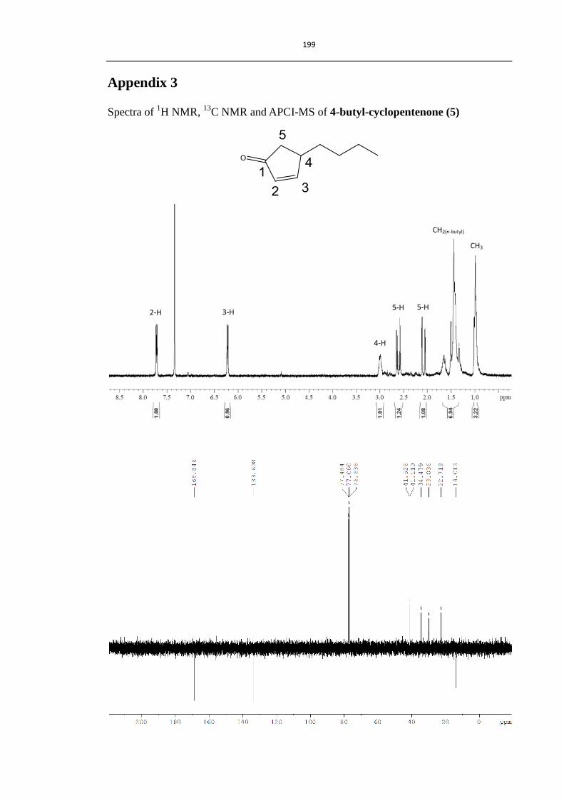
199
Appendix 3
Spectra of 1H NMR,
13C NMR and APCI-MS of 4-butyl-cyclopentenone (5)

200
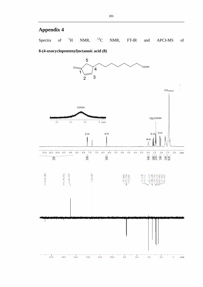
201
Appendix 4
Spectra of 1H NMR,
13C NMR, FT-IR and APCI-MS of
8-(4-oxocyclopentenyl)octanoic acid (8)

202