Embed Size (px)
Citation preview
Characterization of the OXPHOS system
in plant mitochondria
Von der Naturwissenschaftlichen Fakultät
der Gottfried Wilhelm Leibniz Universität Hannover
zur Erlangung des Grades
Doktorin der Naturwissenschaften
Dr. rer. nat.
genehmigte Dissertation
von
M.Sc. Katrin Peters
geboren am 16. März 1982 in Uelzen

Referent: Prof. Dr. Hans-Peter Braun
Korreferent: Prof. Dr. Christoph Peterhänsel
Tag der Promotion: 22. Dezember 2011
Abstract
This thesis aims to provide a deeper understanding of structure and function of the oxidative
phosphorylation (OXPHOS) system in plant mitochondria. Thanks to improvements in electron
microscopy (EM), a high number of structural details were obtained in the last years. Besides
the already known stable interactions of OXPHOS complexes, termed supercomplexes, even
higher-ordered oligomers are formed by those (section 2.2). Evidence is adduced for a row-like
organization of complexes I, III and IV, termed ‘respiratory string’. Furthermore, ATP-
synthase (complex V) forms long rows of dimers located at locally curved cristae membranes,
which are supposed to induce the bending of the inner mitochondrial membrane (IMM). New
details about the structure of complex I and the I+III2 supercomplex in Zea mays are outlined in
section 2.1. The typical L-shaped complex I structure resembles the one from Arabidopsis
thaliana and also in maize, a member of the group of C4 species, the plant specific carbonic
anhydrase (CA) domain could be detected. Comparing this domain with the X-ray structure of
homotrimeric γ-CA from the archaebacterium Methanosarcina thermophila, it is suggested that
the CA domain in maize most likely represents a trimer, too. Additionally, single-particle EM
of complex I from maize reveals structural heterogeneity in complex I, which is not known for
other plant species so far. Different physiological roles for these two complex I forms have to
be discussed. Moreover, the I+III2 supercomplex seems to be more stable in maize than in
Arabidopsis. In section 2.4 an immunoblot based quantification approach is presented,
revealing the ratio of OXPHOS complexes in different plant tissues. Drastic differences in
complex I to complex II ratio of individual plant tissues of Arabidopsis thaliana are displayed.
This leads to the suggestion of additional tissue dependent functions of these respiratory chain
complexes in plants besides electron transfer. Due to the results obtained by this study, light-
dependent side functions of complex I are discussed. Beyond these approaches on the
characterization of the OXPHOS system, a new application based on blue native
polyacrylamide gel electrophoresis (BN-PAGE), which is the method of choice for analyses of
membrane protein complexes, was established in this thesis (section 2.3). Different protein
samples are labelled with distinct fluorescent dyes, pooled and resolved in one single gel (blue
native difference gel electrophoresis, BN-DIGE). This technique allows systematic and
quantitative comparisons of protein complexes of related protein fractions, structural
investigations of protein complexes as well as assignments of protein complexes to subcellular
fractions like organelles.
Keywords: OXPHOS system, plant mitochondria, respiratory chain complexes
Zusammenfassung
Ziel dieser Arbeit ist es zur Aufklärung von Struktur und Funktion des oxidativen
Phosphorylierungs (OXPHOS) -Systems in pflanzlichen Mitochondrien beizutragen. Mit Hilfe
der Elektronenmikroskopie (EM) konnten neue strukturelle Erkenntnisse gewonnen werden.
Neben den bereits bekannten Interaktionen von OXPHOS Komplexen, sogenannten
Superkomplexen, lassen sich auch höher organisierte Strukturen bestehend aus diesen
nachweisen (Abschnitt 2.2). Bisher konnte eine Abfolge der Komplexe I, III und IV
(„respiratory string“) sowie eine Aneinanderreihung von ATP-Synthase (Komplex V) Dimeren
gezeigt werden. Letztere sind vermutlich für die Krümmung der inneren
Mitochondrienmembran (IMM), den sogenannten Cristae, verantwortlich. Neue strukturelle
Details des Komplex I sowie des I+III2 Superkomplex aus Zea mays werden in Abschnitt 2.1
vorgestellt. Die typische L-förmige Struktur von Komplex I ist ähnlich der aus Arabidopsis
thaliana und auch die pflanzenspezifische Carboanhydrase (CA) Domäne konnte in Mais, als
einem Vertreter der C4 Spezies, detektiert werden. Ein Vergleich mit der Röntgenstruktur der
homotrimeren γ-CA Domäne des Archaebakteriums Methanosarcina thermophila deutet darauf
hin, dass die CA Domäne in Mais ebenfalls als Trimer vorliegt. Zusätzlich konnte durch
„single-particle“ EM eine bisher unbekannte strukturelle Heterogenität des Komplex I gezeigt
werden, weshalb unterschiedliche physiologische Rollen dieser zwei Formen diskutiert werden.
Des Weiteren konnten in dieser Arbeit zum ersten Mal die OXPHOS Komplexe in
verschiedenen pflanzlichen Geweben mittels eines immunologischen Ansatzes quantifiziert
werden (Abschnitt 2.4). Es werden deutliche Unterschiede im Verhältnis von Komplex I zu
Komplex II aus einzelnen Geweben von Arabidopsis thaliana gezeigt. Die Ergebnisse lassen
vermuten, dass die Atmungskettenkomplexe in Pflanzen neben dem Elektronentransport noch
zusätzliche, gewebespezifische Funktionen besitzen. Darüber hinaus werden lichtabhängige
Nebenfunktionen des Komplex I diskutiert. Neben der Charakterisierung des pflanzlichen
OXPHOS Systems wurde im Rahmen dieser Arbeit eine neue Methode etabliert (Abschnitt
2.3), welche auf der blau nativen Polyacrylamid-Gelelektrophorese (BN-PAGE) basiert. Hierzu
werden Proteinproben mit unterschiedlichen Fluoreszenz-Farbstoffen markiert, vereint und
anschließend in einem einzigen Gel aufgetrennt. Diese Technik (blau native differentielle
Gelelektrophorese, BN-DIGE) ermöglicht einen systematischen und quantitativen Vergleich
von ähnlichen Proteinfraktionen, eine strukturelle Untersuchung von Proteinkomplexen sowie
eine Zuordnung von Proteinkomplexen zu subzellulären Fraktionen wie zum Beispiel
Organellen.
Schlagworte: OXPHOS System, pflanzliche Mitochondrien, Atmungsketten-Komplexe
Contents
Abbreviations 1
Chapter 1 General Introduction 3
1.1 Mitochondria: structure and evolution 1.2 The respiratory chain of mitochondria and its role in
oxidative phosphorylation 1.3 Supramolecular organization of the OXPHOS system 1.4 Characteristic features of the OXPHOS system in plants 1.5 Approaches used for the investigation of the OXPHOS system 1.6 Objective of the thesis
Chapter 2 Publications and Manuscripts 16
2.1 A structural investigation of complex I and I+III2 supercomplex from Zea mays at 11-13 Å resolution: assignment of the carbonic anhydrase domain and evidence for structural heterogeneity within complex I. Biochim. Biophys. Acta 1777: 84-93.
2.2 Structure and function of mitochondrial supercomplexes. Biochim. Biophys. Acta 1797: 664-670.
2.3 Comparative analyses of protein complexes by blue native DIGE.
Methods Mol. Biol. In press.
2.4 Complex I - complex II ratio strongly differs in various organs of Arabidopsis thaliana.
In preparation.
Chapter 3 Supplementary Discussion and Outlook 75
3.1 The highly branched electron transport chain of plant mitochondria 3.2 Outlook
References 83
Affix Curriculum vitae 91 Publications Danksagung Eidesstattliche Erklärung
Abbreviations
1D one-dimensional 2D two-dimensional 3D three-dimensional AOX alternative oxidase ADP adenosine diphosphate ATP adenosine triphosphate BN blue native CA carbonic anhydrase γCA gamma-type carbonic anhydrase γCAL gamma-type carbonic anhydrase like CAM carbonic anhydrase of Methanosarcina thermophila CCM CO2 concentrating mechanism CMS cytoplasmic male sterility Complex I NADH dehydrogenase Complex II succinate dehydrogenase Complex III cytochrome c reductase Complex IV cytochrome c oxidase Complex V ATP synthase COX cytochrome c oxidase DDM dodecyl maltoside DIGE difference gel electrophoresis DNA deoxyribonucleic acid e- electron EM electron microscopy ET electron tomography (m)ETC (mitochondrial) electron transfer chain ETFQ-OR electron transfer flavoprotein:quinone oxidoreductase F0 F0 part of complex V F1 F1 part of complex V FADH2 flavin adenine dinucleotide; reduced form FMN flavin mononucleotide G3P glycerol-3-phosphate GLDH L-galactono-1,4-lactone dehydrogenase H+ proton HSP70 heat shock protein 70 IEF isoelectric focusing IMM inner mitochondrial membrane IMS inter membrane space kDa kilo Dalton MDa mega Dalton MPP mitochondrial processing peptidase MS mass spectrometry NAD(P)+ nicotine amid dinucleotid (phosphate); oxidized form NAD(P)H nicotine amid dinucleotid (phosphate); reduced form OMM outer mitochondrial membrane OXPHOS oxidative phosphorylation PAGE polyacrylamide gel electrophoresis Pi phosphate
Abbreviations
1
PMF proton motive force RNA ribonucleic acid ROS reactive oxygen species SDS sodium dodecyl sulphate TIM translocases of the inner mitochondrial membrane TOM translocases of the outer mitochondrial membrane UQ ubiquinone UQH2 ubiquinol VDAC voltage-dependent anion channel WT wild type
Abbreviations
2
Chapter 1
1 General introduction
This chapter aims to provide an overview of the oxidative phosphorylation (OXPHOS) system
in general and the plant specific features of cellular respiration in particular. It will also present
the recent progress in this field of research.
1.1 Mitochondria: structure and evolution
The word mitochondrion comes from the Greek and is composed of the words ‘μίτος’ (or
‘mitos’) meaning thread and ‘χονδρίον’ (or ‘chondrion’) for granule. Mitochondria are double-
membrane bound organelles with a size of 1 to 3 µm and an abundance of 1 to 1000 per cell.
Their shape varies from tubular or reticulated (when mitochondria are attached to the
cytoskeleton) to ellipsoidal or spherical (isolated mitochondria in suspension) (Mannella 2006).
In most species, mitochondria are maternally inherited and propagate by division. They contain
their own small genome, often in a circular form, as well as a fully functional apparatus for
protein synthesis. Mitochondrial DNA encodes for some mitochondrial proteins, but the
majority of proteins are encoded by nuclear genes synthesized in the cytosol and imported into
the organelle. Two specific transport complexes in the outer and the inner mitochondrial
membrane, termed translocases of the outer/inner mitochondrial membrane (TOM and TIM),
as well as the matrix protein complex HSP70 (heat shock protein 70) are involved in the
protein import process (Braun and Schmitz 1999, Carrie et al. 2010).
According to the endosymbiontic hypothesis, the ancestor of mitochondria was an aerobe
prokaryote, taken up by a eukaryotic cell via endocystosis (Sagan 1967 and references therein).
It is assumed that this event happened only once in the evolution of the eukaryotic cell. Today,
the closest living relatives to this incorporated prokaryote are the α-proteobacteria (reviewed in
Gray et al. 2001). Mitochondria contain two membranes: inner and outer mitochondrial
membrane (IMM, OMM) (Fig. 1A). These two membranes enclose the inter membrane space
(IMS). The OMM separates the IMS from the cytosol and is permeable for proteins below 5000
Dalton due to voltage-dependent anion channels (VDAC) (Vander Heiden 2001) and due to
pores, formed by transmembrane proteins (porines), which allow the movement of solutes up to
1000 Dalton (Mannella and Tedeschi 1987). The IMM encloses the dense, protein-rich
mitochondrial matrix and is heavily folded, forming the so-called ‘cristae’, which results in an
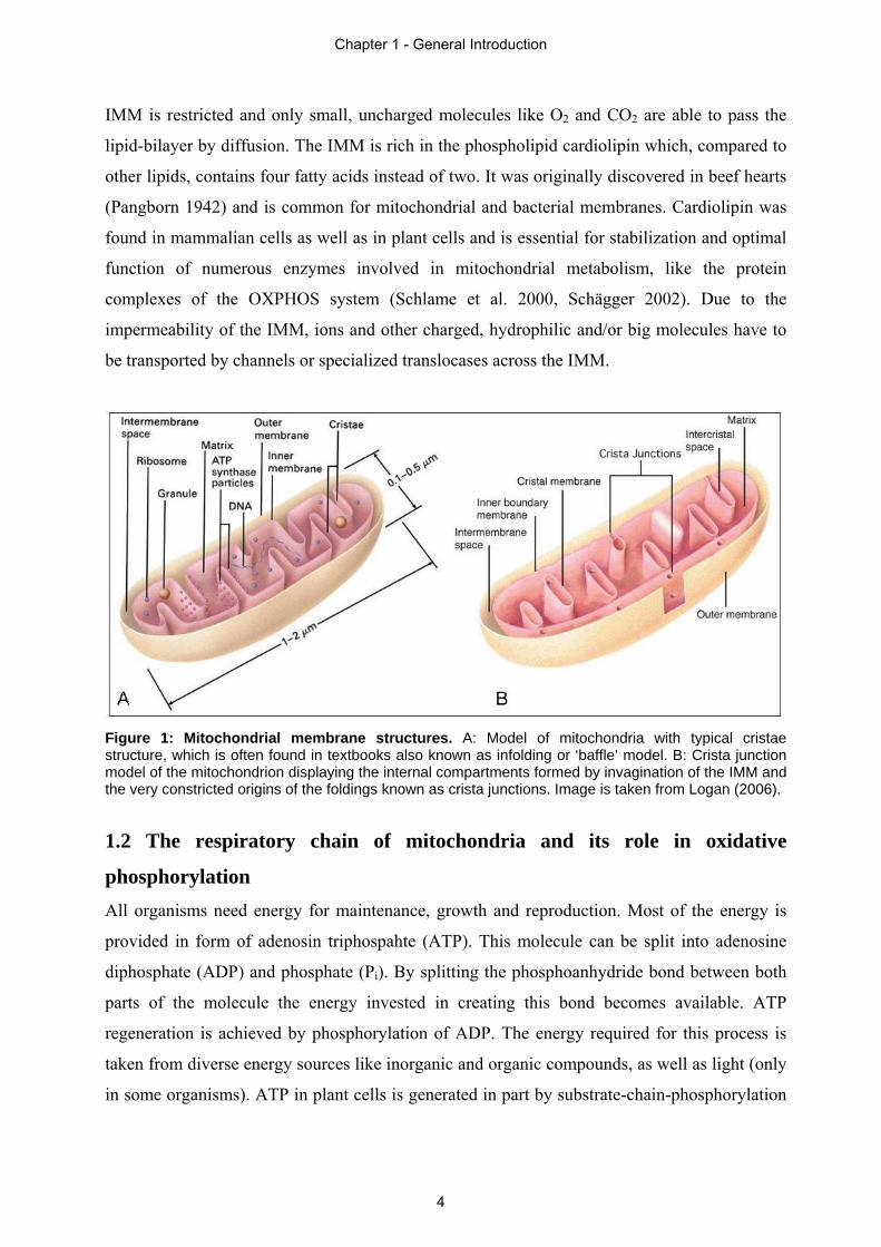
enormous enlargement of the membrane surface (Mannella 2006). The origins of the foldings,
called ‘crista junctions’, are constricted (Fig 1B) and therefore cristae can be regarded as
additional compartments (Mannella et al. 1997). The permeability for molecules across the
Chapter 1 - General Introduction
3
IMM is restricted and only small, uncharged molecules like O2 and CO2 are able to pass the
lipid-bilayer by diffusion. The IMM is rich in the phospholipid cardiolipin which, compared to
other lipids, contains four fatty acids instead of two. It was originally discovered in beef hearts
(Pangborn 1942) and is common for mitochondrial and bacterial membranes. Cardiolipin was
found in mammalian cells as well as in plant cells and is essential for stabilization and optimal
function of numerous enzymes involved in mitochondrial metabolism, like the protein
complexes of the OXPHOS system (Schlame et al. 2000, Schägger 2002). Due to the
impermeability of the IMM, ions and other charged, hydrophilic and/or big molecules have to
be transported by channels or specialized translocases across the IMM.
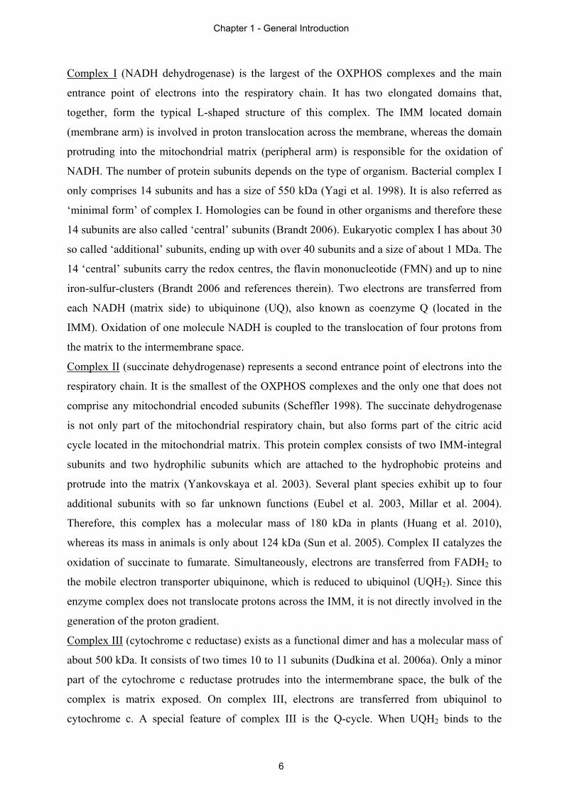
Figure 1: Mitochondrial membrane structures. A: Model of mitochondria with typical cristae structure, which is often found in textbooks also known as infolding or ‘baffle’ model. B: Crista junction model of the mitochondrion displaying the internal compartments formed by invagination of the IMM and the very constricted origins of the foldings known as crista junctions. Image is taken from Logan (2006).
1.2 The respiratory chain of mitochondria and its role in oxidative
phosphorylation
All organisms need energy for maintenance, growth and reproduction. Most of the energy is
provided in form of adenosin triphospahte (ATP). This molecule can be split into adenosine
diphosphate (ADP) and phosphate (Pi). By splitting the phosphoanhydride bond between both
parts of the molecule the energy invested in creating this bond becomes available. ATP
regeneration is achieved by phosphorylation of ADP. The energy required for this process is
taken from diverse energy sources like inorganic and organic compounds, as well as light (only
in some organisms). ATP in plant cells is generated in part by substrate-chain-phosphorylation
Chapter 1 - General Introduction
4
in the cytosol and in the mitochondrial matrix, but mainly by oxidative phosphorylation in
mitochondria or by photophosphorylation in chloroplasts.
During the photophosphorylation process in chloroplasts large amounts of ATP are produced,
but these are used directly in the plastids and therefore are not available to meet the energy
demands of the cell. The supply of ATP to the whole cell is mainly provided by oxidative
phosphorylation. This process takes place in the inner mitochondrial membrane and involves
the respiratory chain and the ATP-synthase complex. The respiratory chain, also termed
electron transport chain (ETC), transfers electrons from reduced nicotinamide adenine
dinucleotide (NADH) or flavin adenine dinucleotide (FADH2), generated by the action of the
citric acid cycle, via four different multi protein complexes and two mobile electron
transporters (ubiquinone and cytochrome c) onto molecular oxygen (O2) which is reduced to
water (H2O) (Fig. 2). Since this process is exergonic, the stepwise transfer of electrons allows
translocation of protons from the matrix to the intermembrane space resulting in a proton
gradient. Controlled reflux of these protons into the matrix drives the ATP-synthase complex in
the IMM which phosphorylates ADP to yield ATP (Mitchell 1961).
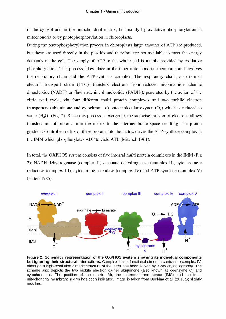
In total, the OXPHOS system consists of five integral multi protein complexes in the IMM (Fig
2): NADH dehydrogenase (complex I), succinate dehydrogenase (complex II), cytochrome c
reductase (complex III), cytochrome c oxidase (complex IV) and ATP-synthase (complex V)
(Hatefi 1985).
Figure 2: Schematic representation of the OXPHOS system showing its individual components but ignoring their structural interactions. Complex III is a functional dimer, in contrast to complex IV, although a high-resolution dimeric structure of the latter has been solved by X-ray crystallography. The scheme also depicts the two mobile electron carrier ubiquinone (also known as coenzyme Q) and cytochrome c. The position of the matrix (M), the intermembrane space (IMS) and the inner mitochondrial membrane (IMM) has been indicated. Image is taken from Dudkina et al. (2010a); slightly modified.
Chapter 1 - General Introduction
5
Complex I (NADH dehydrogenase) is the largest of the OXPHOS complexes and the main
entrance point of electrons into the respiratory chain. It has two elongated domains that,
together, form the typical L-shaped structure of this complex. The IMM located domain
(membrane arm) is involved in proton translocation across the membrane, whereas the domain
protruding into the mitochondrial matrix (peripheral arm) is responsible for the oxidation of
NADH. The number of protein subunits depends on the type of organism. Bacterial complex I
only comprises 14 subunits and has a size of 550 kDa (Yagi et al. 1998). It is also referred as
‘minimal form’ of complex I. Homologies can be found in other organisms and therefore these
14 subunits are also called ‘central’ subunits (Brandt 2006). Eukaryotic complex I has about 30
so called ‘additional’ subunits, ending up with over 40 subunits and a size of about 1 MDa. The
14 ‘central’ subunits carry the redox centres, the flavin mononucleotide (FMN) and up to nine
iron-sulfur-clusters (Brandt 2006 and references therein). Two electrons are transferred from
each NADH (matrix side) to ubiquinone (UQ), also known as coenzyme Q (located in the
IMM). Oxidation of one molecule NADH is coupled to the translocation of four protons from
the matrix to the intermembrane space.
Complex II (succinate dehydrogenase) represents a second entrance point of electrons into the
respiratory chain. It is the smallest of the OXPHOS complexes and the only one that does not
comprise any mitochondrial encoded subunits (Scheffler 1998). The succinate dehydrogenase
is not only part of the mitochondrial respiratory chain, but also forms part of the citric acid
cycle located in the mitochondrial matrix. This protein complex consists of two IMM-integral
subunits and two hydrophilic subunits which are attached to the hydrophobic proteins and
protrude into the matrix (Yankovskaya et al. 2003). Several plant species exhibit up to four
additional subunits with so far unknown functions (Eubel et al. 2003, Millar et al. 2004).
Therefore, this complex has a molecular mass of 180 kDa in plants (Huang et al. 2010),
whereas its mass in animals is only about 124 kDa (Sun et al. 2005). Complex II catalyzes the
oxidation of succinate to fumarate. Simultaneously, electrons are transferred from FADH2 to
the mobile electron transporter ubiquinone, which is reduced to ubiquinol (UQH2). Since this
enzyme complex does not translocate protons across the IMM, it is not directly involved in the
generation of the proton gradient.
Complex III (cytochrome c reductase) exists as a functional dimer and has a molecular mass of
about 500 kDa. It consists of two times 10 to 11 subunits (Dudkina et al. 2006a). Only a minor
part of the cytochrome c reductase protrudes into the intermembrane space, the bulk of the
complex is matrix exposed. On complex III, electrons are transferred from ubiquinol to
cytochrome c. A special feature of complex III is the Q-cycle. When UQH2 binds to the
Chapter 1 - General Introduction
6
complex, the two electrons take different pathways. One electron is transferred to the mobile
electron transporter cytochrome c via a Rieske iron-sulfur centre, whereas the other electron is
transferred back to an UQ (bound to a second UQ-binding site) which is reduced to
ubisemiquinone. The UQH2 releases the remaining two protons to the intermembrane space.
Now being oxidized again, the UQ enters the ubiquinone-pool. A second UQH2 then binds to
the complex and again the electrons are transferred as mentioned above. However, this time the
ubisemiquinone, still bound on the second binding site, gets fully reduced and takes up two
protons from the matrix, leaving complex III as UQH2. By this, altogether two electrons are
transferred from UQH2 to cytochrome c thereby two protons are taken from the matrix and four
protons are released to the intermembrane space (reviewed in Berry et al. 2000). Thus, complex
III plays an important role in building up the proton gradient between the mitochondrial matrix
and the IMS.
Complex IV (cytochrome c oxidase) is the last enzyme complex of the mitochondrial
respiratory chain. This complex consists of 12 to 13 subunits with an overall size of about
220 kDa (Tsukihara et al. 1996). The cytochrome c oxidase (COX) catalyzes the sequential
transfer of four electrons, one at a time from four reduced cytochrome c molecules, to
molecular oxygen, thereby generating water. Coupled to electron transfer, four protons are
pumped across the IMM (Welchen et al. 2011 and references therein).
Complex V (ATP-synthase complex) comprises 15 distinct subunits. Some of these are present
in multiple copies in the holo-enzyme. The molecular mass of the complex is between 500 and
600 kDa (Dudkina et al. 2006a). The ATP-synthase complex consists of two parts: The F0-part
anchors the complex to the inner mitochondrial membrane, whereas the F1 headpiece protrudes
into the mitochondrial matrix (Stock et al. 2000). Both parts of the complex are connected by a
central stalk. The ATP-synthase complex is driven by the proton motive force (PMF) of the
proton gradient across the IMM. Protons are flowing back through complex V from the IMS to
the matrix. The energy, gained by this reflux of protons, leads to a rotation of the F0-part and
the central stalk, whereas the F1-part is fixed by an additional peripheral stalk and thereby
prevented from rotation. Due to the rotation, ADP and phosphate are getting in a close
proximity to each other building up the basis for phosphorylation.
1.3 Supramolecular organization of the OXPHOS system
The multi protein complexes of the respiratory chain were first described in the early 1960s
(reviewed in Hatefi 1985), when OXPHOS complexes were subfractionated from
mitochondrial membranes from beef. In some cases defined combinations of respiratory chain
Chapter 1 - General Introduction
7
complexes were reported, e.g. an active supercomplex of the complexes I and III as well as
several other supercomplexes containing stoichiometric associations of OXPHOS complexes
(Fowler and Hatefi 1961, Hatefi et al. 1961, Hatefi et al. 1962). Due to these findings, the
structure of the respiratory chain was firstly described by the ‘solid-state model’ (Fig. 3b).
Based on this model, the ETC complexes stably interact with each other forming
supercomplexes. This model was questioned by Fowler and Richardson (1963) regarding the
necessity of an association of complexes for the transfer of electrons. Later on, the ‘solid-state
model’ again was challenged. The five complexes could be purified and were found to be
stable particles, since they were easily separable from each other upon membrane
solubilization. Thus, their separate existence under in vivo conditions was suggested. This
hypothesis was supported by activity measurements of the OXPHOS complexes in inner
membrane vesicles during lipid dilution experiments (Hackenbrock et al. 1986). Based on this
new model, termed ‘fluid-state model’, respiratory chain complexes are separated in the inner
mitochondrial membrane and electron transfer takes place by random collisions of the
components (Fig. 3a). Accordingly, this model is also referred to as the ‘random collision
model’ (Hackenbrock et al. 1986), which became widely accepted. However, recent
experimental data, such as co-purification of defined respiratory chain complexes, high
electron transfer activities of defined combinations of respiratory complexes and results of
noninvasive flux control measurements, led to the suggestion of an alternative ‘solid-state
model’ (see Welchen et al. 2011 and references therein). This model received support by native
gel electrophoresis and single-particle electron microscopy (EM). Using different experimental
setups, the following supercomplexes were detected: I+III2, III2+IV1-2, I+III2+IV1-4 and V2
(Dudkina et al. 2010a). Thereby, subscripted numbers indicate the number of individual
complexes within the supercomplex. It is proposed that not all respiratory chain complexes are
part of supramolecular structures at any time but co-exist with supercomplexes (Fig. 3c), which
are dynamically assembled and degraded depending on the physiological state of the cell
(Welchen et al. 2011). Recently, even higher organizational levels of respiratory
supercomplexes were detected. One of these structures is a row-like association of complexes I,
III and IV, termed ‘respiratory string’ (Bultema et al. 2009). Another higher organization of
supercomplexes is represented by formation of dimeric ATP-synthase complexes. This
supercomplex is assumed to participate in forming the cristae of the IMM (Strauss et al. 2008,
Davies et al. 2011).
Sections 2.1 and 2.2 of this thesis will present further details on structure and function of
mitochondrial supercomplexes.
Chapter 1 - General Introduction
8
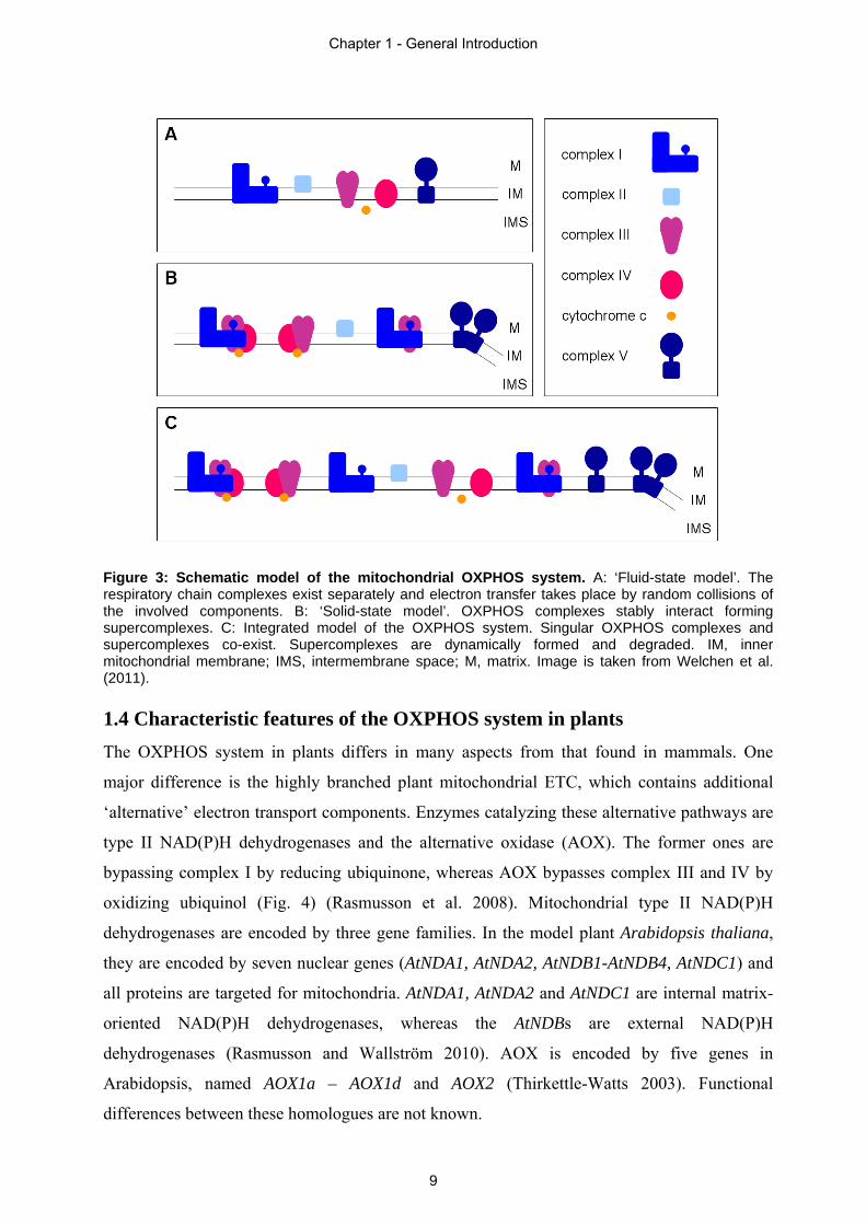
Figure 3: Schematic model of the mitochondrial OXPHOS system. A: ‘Fluid-state model’. The respiratory chain complexes exist separately and electron transfer takes place by random collisions of the involved components. B: ‘Solid-state model’. OXPHOS complexes stably interact forming supercomplexes. C: Integrated model of the OXPHOS system. Singular OXPHOS complexes and supercomplexes co-exist. Supercomplexes are dynamically formed and degraded. IM, inner mitochondrial membrane; IMS, intermembrane space; M, matrix. Image is taken from Welchen et al. (2011). 1.4 Characteristic features of the OXPHOS system in plants
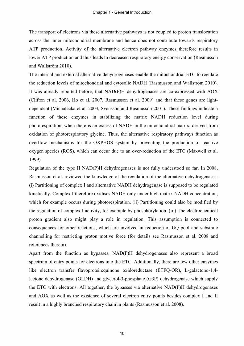
The OXPHOS system in plants differs in many aspects from that found in mammals. One
major difference is the highly branched plant mitochondrial ETC, which contains additional
‘alternative’ electron transport components. Enzymes catalyzing these alternative pathways are
type II NAD(P)H dehydrogenases and the alternative oxidase (AOX). The former ones are
bypassing complex I by reducing ubiquinone, whereas AOX bypasses complex III and IV by
oxidizing ubiquinol (Fig. 4) (Rasmusson et al. 2008). Mitochondrial type II NAD(P)H
dehydrogenases are encoded by three gene families. In the model plant Arabidopsis thaliana,
they are encoded by seven nuclear genes (AtNDA1, AtNDA2, AtNDB1-AtNDB4, AtNDC1) and
all proteins are targeted for mitochondria. AtNDA1, AtNDA2 and AtNDC1 are internal matrix-
oriented NAD(P)H dehydrogenases, whereas the AtNDBs are external NAD(P)H
dehydrogenases (Rasmusson and Wallström 2010). AOX is encoded by five genes in
Arabidopsis, named AOX1a – AOX1d and AOX2 (Thirkettle-Watts 2003). Functional
differences between these homologues are not known.
Chapter 1 - General Introduction
9
The transport of electrons via these alternative pathways is not coupled to proton translocation
across the inner mitochondrial membrane and hence does not contribute towards respiratory
ATP production. Activity of the alternative electron pathway enzymes therefore results in
lower ATP production and thus leads to decreased respiratory energy conservation (Rasmusson
and Wallström 2010).
The internal and external alternative dehydrogenases enable the mitochondrial ETC to regulate
the reduction levels of mitochondrial and cytosolic NADH (Rasmusson and Wallström 2010).
It was already reported before, that NAD(P)H dehydrogenases are co-expressed with AOX
(Clifton et al. 2006, Ho et al. 2007, Rasmusson et al. 2009) and that these genes are light-
dependent (Michalecka et al. 2003, Svensson and Rasmusson 2001). These findings indicate a
function of these enzymes in stabilizing the matrix NADH reduction level during
photorespiration, when there is an excess of NADH in the mitochondrial matrix, derived from
oxidation of photorespiratory glycine. Thus, the alternative respiratory pathways function as
overflow mechanisms for the OXPHOS system by preventing the production of reactive
oxygen species (ROS), which can occur due to an over-reduction of the ETC (Maxwell et al.
1999).
Regulation of the type II NAD(P)H dehydrogenases is not fully understood so far. In 2008,
Rasmusson et al. reviewed the knowledge of the regulation of the alternative dehydrogenases:
(i) Partitioning of complex I and alternative NADH dehydrogenase is supposed to be regulated
kinetically. Complex I therefore oxidises NADH only under high matrix NADH concentration,
which for example occurs during photorespiration. (ii) Partitioning could also be modified by
the regulation of complex I activity, for example by phosphorylation. (iii) The electrochemical
proton gradient also might play a role in regulation. This assumption is connected to
consequences for other reactions, which are involved in reduction of UQ pool and substrate
channelling for restricting proton motive force (for details see Rasmusson et al. 2008 and
references therein).
Apart from the function as bypasses, NAD(P)H dehydrogenases also represent a broad
spectrum of entry points for electrons into the ETC. Additionally, there are few other enzymes
like electron transfer flavoprotein:quinone oxidoreductase (ETFQ-OR), L-galactono-1,4-
lactone dehydrogenase (GLDH) and glycerol-3-phosphate (G3P) dehydrogenase which supply
the ETC with electrons. All together, the bypasses via alternative NAD(P)H dehydrogenases
and AOX as well as the existence of several electron entry points besides complex I and II
result in a highly branched respiratory chain in plants (Rasmusson et al. 2008).
Chapter 1 - General Introduction
10
Figure 4: Respiratory chain complexes and alternative respiratory enzymes in plant mitochondria. The standard complexes are depicted in white, type II NAD(P)H dehydrogenases and AOX are shown in grey. Broken lines represent unclear enzymatic properties. SDH, succinate dehydrogenase (complex II). Image is taken from Rasmusson and Wallström (2010).
Another remarkable difference in the OXPHOS systems of plants and mammals is the presence
of additional protein subunits in respiratory complexes in plants, which introduce side activities
to these complexes.
For example, complex I consists of 14 core subunits in all organisms but contains about 30
extra subunits in eukaryotes (Klodmann and Braun 2011). As reported in Klodmann et al.
(2010), complex I from Arabidopsis thaliana has 13 plant specific subunits. One of these
additional subunits is the GLDH. This enzyme catalyzes the final step of the ascorbate
synthesis pathway and is associated with a smaller version of complex I (Heazlewood et al.
2003, Millar et al. 2003). Pineau et al. (2008) assumed GLDH to be a bifunctional protein
which is not only involved in ascorbate synthesis, but also in the assembly of complex I.
Another group of plant specific subunits in complex I is composed of five structurally related
30 kDa proteins, which show sequence similarity to gamma-type carbonic anhydrases (γCA) of
the archaebacterium Methanosarcina thermophila (also referred to as CAM). Single particle
electron microscopy of complex I from Arabidopsis thaliana, Zea mays and the green alga
Polytomella indicates a plant specific extra domain composed of γCAs that is attached to the
central part of the membrane arm of the complex, protruding into the matrix (Sunderhaus et al.
Chapter 1 - General Introduction
11
2006, Peters et al. 2008). The five carbonic anhydrase proteins are named CA1, CA2, CA3 and
carbonic anhydrases like proteins 1 and 2 (CAL1, CAL2). These two CAL proteins differ from
the γCA of the archaebacteria to a greater extend than CA1 – CA3 (Braun and Zabaleta 2007).
Carbonic anhydrases are zinc containing metallo-enzymes, catalyzing very rapidly the inter-
conversion of CO2 and HCO3-. So far such an activity could not been demonstrated for the
γCAs in plants.
Therefore, the physiological role of the CA extra domain in plant complex I is not completely
understood. Several lines of evidence indeed support a model in which CAs and CALs are
involved in CO2/HCO3- metabolism (reviewed in Klodmann and Braun 2011). Additionally, a
physiological role of the plant specific CA-domain in photorespiration was postulated by Braun
and Zabaleta (2007). In plants, CO2 is an essential substrate for photosynthesis, which becomes
limiting under certain conditions such as closed stomata due to arid environmental conditions.
This rapidly results in low concentration of CO2 in chloroplasts, whereas mitochondria at the
same time produce large amounts of CO2 due to photorespiration. Braun and Zabaleta (2007)
suggested an active CO2 transport system between mitochondria and chloroplasts, which is
based on the CO2/HCO3- conversion at complex I. Bicarbonate is then transported across the
mitochondrial and chloroplast membranes and is again re-converted into CO2 by CAs in the
chloroplast. An analogous mechanism for CO2 concentration is well characterized for
cyanobacteria, termed CO2 concentrating mechanism (CMM) (Price et al. 2008).
As previously reported, also the succinate dehydrogenase (complex II) of Arabidopsis thaliana
consists of four extra plant specific subunits (Eubel et al. 2003, Millar et al. 2004), termed SDH
5 – SDH 8. Huang et al. (2010) reported that 7 of the 8 subunits found in Arabidopsis have
homologues in rice, while SDH 8 is missing. This study reveals that also rice complex II
contains at least three plant specific subunits. In all other organisms, complex II consists of
only four subunits. The functions of the ‘core’-subunits are well established (section 1.2), but
until today the functions of the plant specific subunits are still unknown and none of these
proteins show homology to known functional proteins in any database. Hence, it is not clear if
the extra subunits of complex II in plants also introduce side activities to the complex as it
could be shown for complex I in plants.
Another complex, for which specific side activities in plants were detected, is the cytochrome c
reductase. As reported by Braun et al. (1992), two core subunits of complex III display
mitochondrial processing peptidase (MPP) activity. The major MPP activity, which leads to a
cleavage of the presequences from imported precursor proteins, normally can be found in the
Chapter 1 - General Introduction
12
soluble fraction of fungal and mammalian mitochondria. In plants, this activity is located to
complex III in the inner mitochondrial membrane.
1.5 Approaches used for the investigation of the OXPHOS system
The aim of investigations of the OXPHOS system is to further characterize the physiological
functions and the structures of the complexes and supercomplexes. One important technique
for the investigation of protein complexes is blue native polyacrylamide gel electrophoresis
(BN-PAGE), which is also used extensively in this thesis (for details see section 2.3) (Schägger
and von Jagow 1991). This method allows separation of OXPHOS complexes and stable
supercomplexes on polyacrylamide gels under native conditions. In a first step, mitochondrial
membranes are solubilised by the use of a mild, non-ionic detergent. In most cases this is
dodecyl maltoside (DDM), Triton X100 or digitonin. Working with Arabidopsis and other
plant mitochondria, e.g. from maize, digitonin is the detergent of choice, because it is best
suited to retain complexes with high molecular masses like plant complex I or the I+III2
supercomplex. After solubilization, proteins are mixed with the anionic dye Coomassie blue.
This water-soluble dye has a high affinity to hydrophilic and hydrophobic proteins and
introduces negative charges to the proteins without denaturing them. Since all proteins are now
negatively charged in a uniform fashion, they migrate towards the anode, thereby getting
resolved according to size and not to charge. Coomassie blue addition also prevents protein
aggregation, since all proteins carry negative charges. The separation capacity of BN-PAGE is
best if the gradient of the polyacrylamide gel is chosen according to sample properties, because
proteins get stuck in the gel when they reach their size dependent specific pore-size limit
(Wittig et al. 2006). The size of pores is depending on concentration of acrylamide and
amounts of cross-linkers (e.g. bisacrylamide). An increase in acrylamide concentration results
in a decrease of pore-size and vice versa (Rüchel et al. 1978). Therefore, separation of proteins
with low molecular weights would require high acrylamide concentrations resulting in smaller
pore-sizes.
BN-PAGE is often the first step in various approaches. The most common one is the
combination with sodium dodecyl sulfate polyacrylamide gel electrophoresis (SDS-PAGE).
For this, the protein complexes separated by BN-PAGE are first denatured by β-
mercaptoethanol and SDS. Afterwards, protein complex subunits are resolved in a second gel
dimension according to molecular weight. By this procedure, known as two-dimensional (2D)
BN/SDS-PAGE, the subunit composition of protein complexes and supercomplexes can be
visualized (Wittig et al. 2006).
Chapter 1 - General Introduction
13
Other methods often used in combination with BN-PAGE are: (i) in-gel activity stains (sections
2.1 and 2.4), (ii) native immunoblotting (supplementary material of section 2.4), (iii)
electroelution with subsequent analyses by isoelectric focusing (IEF), sometimes also followed
by SDS-PAGE as a third-dimension (3D BN/IEF/SDS-PAGE) and (iv) a second BN-PAGE
(2D BN/BN-PAGE) (Wittig et al. 2006).
When it becomes necessary to compare the protein complex or subunit composition of different
samples, e.g. mutant lines, blue native difference gel electrophoresis (BN-DIGE) is the method
of choice. For this, the protein fractions of interest are labelled with different fluorescent dyes,
pooled and then separated on a single gel, thereby minimizing gel-to-gel variations. However,
the BN-DIGE technique can be combined with a second dimension SDS-PAGE. Section 2.3
will describe this method in detail.
Besides all these techniques, mainly based on electrophoresis, other methods are available
which enable investigations of the OXPHOS complexes. One powerful technique is single-
particle EM, which is also used in this thesis (sections 2.1 and 2.2). For this procedure
individual protein complexes and supercomplexes are purified, mostly by the use of sucrose
density gradients, and investigated by negative stain or cryo-EM. Thousands of images are
taken, classified and averaged electronically, resulting in an image of the structure of a
complex. Cryo-EM allows depicting internal details of the object whereas the negative stain
reflects the shape of a protein complex. Single-particle EM is able to generate detailed images
of structures and determine the orientation of singular complexes as well as the contact sites of
complexes in supramolecular structures.
Furthermore, cryo-electron tomography (cryo-ET) is used for 3D reconstruction of a sample
from tilted 2D images taken by a CCD-sensor at cryogenic temperatures. This method
generates structural information of complex cellular organizations at subnanometer resolution
without changes due to chemical treatments, staining procedures or microsectioning. In this
thesis cryo-electron tomography is used to investigate the arrangement of supercomplexes
within intact mitochondria (section 2.2).
1.6 Objective of the thesis
The objective of this thesis is the extended characterization of the OXPHOS system in plant
mitochondria. This includes structural and functional analyses of supercomplexes, single
protein complexes and plant specific subunits. Furthermore, a method for comparative analyses
of protein complexes was established.
In the following chapter four investigations on the plant OXPHOS system are presented:
Chapter 1 - General Introduction
14
In section 2.1, a structural characterization of complex I and the I+III2 supercomplex in Zea
mays, which emphasises on the carbonic anhydrase domain, will be presented. In this study,
carbonic anhydrases are explored for the first time in a C4 plant. Additionally, not reported so
far for any plant species, a structural heterogeneity within complex I is displayed.
The supramolecular structure and function of the mitochondrial OXPHOS system in general is
reviewed in section 2.2.
A new approach for the comparative analysis of protein complexes by BN-DIGE is described
in detail in section 2.3. This method is an excellent tool for comparative investigations of the
OXPHOS system.
For the first time, the ratios of OXPHOS complexes in different plant tissues were analyzed
and results will be discussed in section 2.4. So far, only mammalian OXPHOS complexes were
investigated in such an approach and the knowledge related to plants is limited. In this section,
the quantity of the five OXPHOS complexes from different tissues of Arabidopsis thaliana is
defined. This study reveals strong differences in complex abundances, especially with respect
to complexes I and II. Furthermore, it leads to new insights into physiological functions of
complex I.
Supplementary discussions of the thesis as well as an outlook are given in chapter 3.
Chapter 1 - General Introduction
15
Chapter 2
2 Publications and manuscripts
This thesis comprises four manuscripts. The first manuscript ‘A structural investigation of
complex I and I+III2 supercomplex from Zea mays at 11-13 Å resolution: assignment of the
carbonic anhydrase domain and evidence for structural heterogeneity within complex I’ was
published in the scientific journal ‘Biochimica et Biophysica Acta’ (1777, 84-93) in 2008. I
performed the isolation of the maize mitochondria, the gel electrophoresis procedures (1D BN-
PAGE, 2D BN/SDS-PAGE) and the spot picking for protein analyses by mass spectrometry as
well as the purification of complex I and I+III2 supercomplex by sucrose gradient
ultracentrifugation. All figures concerning these parts were designed by myself. Mass
spectrometry was carried out by Prof. Dr. L. Jänsch and single-particle electron microscopy
was done by Dr. N.V. Dudkina. The bulk of the protocol was written by Dr. N.V. Dudkina, I
added the main part of the materials and methods section and was involved in proof reading.
The manuscript was corrected by Prof. Dr. H.-P. Braun and Prof. Dr. E.J. Boekema.
The second manuscript, a review on the ‘Structure and function of mitochondrial
supercomplexes’ was published in the scientific journal ‘Biochimica et Biophysica Acta’
(1797, 664-670) in 2010. Together with Prof. Dr. H.-P. Braun, I was responsible for the
literature research for this review. In cooperation with the co-authors we wrote the manuscript.
Figures in this manuscript concerning single-particle EM and electron tomography were
prepared by Dr. N.V. Dudkina and Dr. R. Kouril.
The third manuscript ‘Comparative analyses of protein complexes by blue native DIGE’ is a
protocol for fluorophore labelling of native protein fractions for separation by blue native
PAGE. I wrote the manuscript, which was subsequently corrected by Prof. Dr. H.-P. Braun.
This manuscript is in press and will be published soon in the book ‘Differential Gel
Electrophoresis’, part of the series ‘Methods in Molecular Biology’ published by Humana
Press.
In the fourth manuscript ‘Complex I - complex II ratio strongly differs in various organs of
Arabidopsis thaliana’ approximately 90% of the research was performed by myself. All
experiments and figures in this manuscript were made by me, with support of the co-authors in
some parts. The complete manuscript was written by myself and subsequent correction were
done by Prof. Dr. H.-P. Braun. At this stage the manuscript is still in preparation.
Chapter 2 - Publications and Manuscripts
16
A structural investigation of complex I and I+ III2 supercomplex fromZea mays at 11–13 Å resolution: Assignment of the carbonic anhydrase
domain and evidence for structural heterogeneity within complex I
Katrin Peters a, Natalya V. Dudkina b, Lothar Jänsch c, Hans-Peter Braun a,⁎, Egbert J. Boekema b
a Institute for Plant Genetics, Faculty of Natural Sciences, Leibniz Universität Hannover, Herrenhäuser Str. 2, D-30419 Hannover, Germanyb Groningen Biomolecular Sciences and Biotechnology Institute, University of Groningen, Nijenborgh 4, 9747 AG Groningen, The Netherlands
c Proteome Research Group, Division of Cell and Immune Biology, Helmholtz Centre for Infection Research, Inhoffenstraβe 7, D-38124 Braunschweig, Germany
Received 21 August 2007; received in revised form 18 October 2007; accepted 19 October 2007Available online 4 November 2007
Abstract
The projection structures of complex I and the I+ III2 supercomplex from the C4 plant Zea mays were determined by electron microscopy andsingle particle image analysis to a resolution of up to 11 Å. Maize complex I has a typical L-shape. Additionally, it has a large hydrophilic extra-domain attached to the centre of the membrane arm on its matrix-exposed side, which previously was described for Arabidopsis and which wasreported to include carbonic anhydrase subunits. A comparison with the X-ray structure of homotrimeric γ-carbonic anhydrase from thearchaebacterium Methanosarcina thermophila indicates that this domain is also composed of a trimer. Mass spectrometry analyses allowed toidentify two different carbonic anhydrase isoforms, suggesting that the γ-carbonic anhydrase domain of maize complex I most likely is aheterotrimer. Statistical analysis indicates that the maize complex I structure is heterogeneous: a less-abundant “type II” particle has a 15 Å shortermembrane arm and an additional small protrusion on the intermembrane-side of the membrane arm if compared to the more abundant “type I”particle. The I+ III2 supercomplex was found to be a rigid structure which did not break down into subcomplexes at the interface between thehydrophilic and the hydrophobic arms of complex I. The complex I moiety of the supercomplex appears to be only of “type I”. This would meanthat the “type II” particles are not involved in the supercomplex formation and, hence, could have a different physiological role.© 2007 Elsevier B.V. All rights reserved.
Keywords: Complex I; Cytochrome c reductase; Carbonic anhydrase; Supercomplex; Electron microscopy, Zea mays
1. Introduction
Complex I is the major entrance point of electrons to therespiratory chain. It catalyses the transfer of two electrons fromNADH to quinone, which is coupled to the translocation of fourprotons across the inner mitochondrial membrane [1–3]. Thesubunit composition of complex I is highly variable depending onthe type of organism. Bovine and human complex I are composedof about 46 different subunits and have a molecular weight ofabout 1 MDa. Complex I of prokaryotes and chloroplasts aresubstantially smaller and composedmostly of 14 subunits that arehomologues of a “core” complex of mitochondrial complex I.They have been defined as the “minimal” enzyme. The remaining
subunits are so-called “accessory” subunits [3]. Complex I con-sists of a hydrophobic membrane arm and a hydrophilic pe-ripheral arm, which protrudes into the matrix. Together they givecomplex I an unique L-shape, as has been revealed at low-resolution by three-dimensional electron microscopy [4–6]. Thecrystal structure of the peripheral arm of complex I from Thermusthermophilus has been solved [7]. The positions of eight subunitsand all redox centres of the enzyme were determined, includingnine iron–sulfur centres.
The main known function of the membrane arm is protontranslocation [8], but the precise functions of the membranedomain are not well understood because of a lack of high-resolution structural data. However, a medium-resolution pro-jection map at 8 Å of complex I from E. coli was recentlyobtained by electron microscopy [9]. It indicates the presence ofabout 60 transmembrane α-helices, both perpendicular to the
Available online at www.sciencedirect.com
Biochimica et Biophysica Acta 1777 (2008) 84–93www.elsevier.com/locate/bbabio
⁎ Corresponding author. Tel.: +49 511 7622674; fax: +49 511 7623608.E-mail address: [email protected] (H.-P. Braun).
0005-2728/$ - see front matter © 2007 Elsevier B.V. All rights reserved.doi:10.1016/j.bbabio.2007.10.012
Chapter 2 - Publications and Manuscripts
17
membrane plane and tilted, which is consistent with secondarystructure predictions. A possible binding site and access channelfor quinone is found at the interface with the peripheral arm.Tentative assignment of individual subunits to the features of themap has been made. The NuoL and NuoM subunits, which wereproposed to be responsible for proton translocation, are localizedat the tip of the membrane arm of complex I. Since this tip is at asubstantial distance to the redox centres of the peripheral arm ofcomplex I, conformational changes most likely play a role in thecoupling between electron transfer and proton pumping.
Complex I can form stable associations with complex III ofthe respiratory chain [10,11]. This interaction is especially stablein plants. An investigation by EM and single particle analysisrevealed a lateral association of dimeric complex III to the tip ofthe membrane part of complex I in Arabidopsis [12]. The func-tional role of the I+ III2 supercomplex so far is unknown.
Complex I of plant mitochondria resembles complex I ofother multicellular organisms but includes some extra subunits[13–15]. As a consequence, its overall molecular mass is slightlylarger than that of complex I of beef [16]. Some of the extrasubunits introduce side-activities into plant complex I. Inprobably all higher eukaryotes, the “acyl carrier protein” of themitochondrial fatty acid biosynthesis pathway is integrated intocomplex I [17,18]. However, occurrence of this protein incomplex I of plants recently has been disputed [19]. Addition-ally, L-galactono-1,4-lactone dehydrogenase (GalLDH), theterminal enzyme of the mitochondrial ascorbate biosynthesispathway, forms part of complex I in plants [20]. Furthermore,plant mitochondria include a group of five structurally similar30 kDa proteins which resemble a γ-type carbonic anhydrase ofthe archaebacterium Methanosarcina thermophila. A structuralcharacterization by single particle electron microscopy ofcomplex I from Arabidopsis and the green alga Polytomellaindicated a plant-specific spherical extra-domain of about 60 Åin diameter, which is attached to the central part of the membranearm of complex I on its matrix face [15]. This spherical domain isproposed to be composed of the γ-carbonic anhydrase sub-units. Although the inner features of the domain could not beresolved it is probably arranged as a trimer of three subunits,because γ-carbonic anhydrase of Methanosarcina thermophilais known to have a trimeric structure [21,22].
The functional role of the complex I integrated carbonicanhydrases in plants is not quite understood. It was speculated thatthey form part of an active CO2 transport system between mito-chondria and chloroplasts for efficient CO2 fixation during photo-synthesis [23]. CO2, one of the main substrates of photosynthesis,is often growth limiting in plants. The CO2 concentration withinchloroplasts especially declines if plants are grown in the presenceof high-light conditions, enabling high rates of CO2 fixation.Furthermore, the CO2 concentration declines at high temperaturedue to Ribulose-1,5-bisphosphate Carboxylase/Oxygenase(RubisCO) kinetics and water solubility of oxygen and CO2. Asa consequence, the Oxygenase side-activity of RubisCO increasesdramatically, giving rise to the formation of phosphoglycolate. Thiscompound cannot be used for the Calvin cycle and is recycled bythe so-called “photorespiration” pathway. Finally, during photo-respiration, large amounts of CO2 are liberated in the mitochondria.
In summary, CO2 concentration in the chloroplasts of plant cellsoften is low. At the same time, the mitochondria produce largeamounts of CO2. Rapid conversion of mitochondrial CO2 intobicarbonate by carbonic anhydrases is speculated to form the basisof an active indirect CO2 transport mechanism between mitochon-dria and chloroplasts. Indeed, genes encoding the complex Iintegrated carbonic anhydrases are down-regulated inArabidopsis,if plants are cultivated in the presence of elevated CO2
concentration [22]. An analogous role of complex I was reportedin the context of a cyanobacterial CO2 concentrating mechanism[24].
A characterization of maize (Zea mays) complex I was ini-tiated to further investigate the physiological role of the mito-chondrial carbonic anhydrases in plants. In contrast to the “C3”plant Arabidopsis, maize is a so-called “C4” plant that usesphosphoenolpyruvate (PEP) for pre-fixation of CO2 in the formof a four-carbon (C4) compound. Pre-fixation is carried out inspecialized cells termed mesophyll cells, which also carry outthe photosynthetic light reactions and water splitting. The finalCO2 fixation by RubisCO takes place in so-called bundle sheathcells, which do not carry out photosynthetic water splitting. C4
metabolism is based on the transfer of C4-compounds frommeso-phyll to bundle sheath cells and liberation of CO2 in bundle sheathcells. As a consequence, the final CO2 fixation byRubisCO is veryefficient and photorespiration is avoided. Therefore, the functionalrole of the mitochondrial carbonic anhydrases might differ be-tweenArabidopsis and maize. However, in certain subtypes of C4
metabolism, which use amitochondrial enzyme for CO2 release inbundle sheath cells, the presence of carbonic anhydrases in mito-chondria might be especially important.
Here, we describe a structural analysis by single particle elec-tron microscopy of maize complex I. It has the same L-shapedform like Arabidopsis complex I, including the extra carbonicanhydrase domain. This domain is, as well as other features, muchbetter resolved in the current projection maps, and comparison tothe high-resolution X-ray structure of the γ-carbonic anhydrasenow shows it to be a trimer. Mass spectrometry was performed toevaluate the composition of the carbonic anhydrase trimer. Inaddition, the structure of the respiratory I+III2 supercomplex wasanalyzed. This supercomplex has a horse-shoe structure, identicalto the one found in Arabidopsis. It appears to be a very stable,rigid structure that allowed the determination of projection mapsat 12 Å resolution. New insights into the complex I–complex IIIinteraction are presented.
2. Materials and methods
2.1. Cultivation of maize seedlings
Green maize seedlings (Zea mays convar saccharata L. “Tasty Sweet” F1)were cultivated in a greenhouse under long-day conditions (16 h light, 8 h dark)at 22 °C for 9 days. Etiolated maize seedlings were cultivated in growthchambers in the absence of light at 22 °C for the same time period.
2.2. Isolation of maize mitochondria
Starting material for organelle preparations were 100 g of green and etiolatedtissue. The material was suspended each in 500 ml of ice-cold “grinding buffer”(0.4 M mannitol, 1.0 mM EGTA, 25.0 mM MOPS, 0.1% [w/v] bovine serum
85K. Peters et al. / Biochimica et Biophysica Acta 1777 (2008) 84–93
Chapter 2 - Publications and Manuscripts
18
albumin [BSA], 15 mM β-mercaptoethanol, and 0.05 mM phenylmethylsulfo-nyl fluoride [PMSF]/KOH, pH 7.8). The cells were disrupted by homogeniza-tion for three periods of 10 s using a Waring blender and then filtered throughtwo layers of muslin. Mitochondria were isolated by differential centrifugationand Percoll density gradient centrifugation as described by Braun et al. [25]. Thethree-step Percoll gradients for density gradient centrifugation contained 14%,26%, and 45% Percoll in 0.8 M mannitol, 2.0 mM EGTA, 20.0 mM KH2PO4/KOH, pH 7.2. After gradient centrifugation (45 min at 70,000 ×g), mitochondriawere isolated from the 26%/45% interphase. To remove the Percoll the purifiedmitochondria were centrifuged three times in “resuspension buffer” (0.4 Mmannitol, 1.0 mM EGTA, 10.0 mM KH2PO4, 0.2 mM PMSF/KOH, pH 7.2) for10 min at 14,500 ×g.
Purities of our organelle preparations were investigated by analyses ofprotein complex compositions using 2D Blue-native/SDS-PAGE (see below)[26]. Mitochondrial fractions included all the known protein complexes of theOXPHOS system but were devoid of plastidic complexes, e.g. the photosystems,the b6f complex and the plastidic ATP synthase complex. The latter two com-plexes are also formed in etioplasts but were absent in mitochondrial fractionsisolated form maize seedlings cultivated in the dark (data not shown). Further-more, the subunit completeness of all OXPHOS complexes was very goodindicating that the purified organelles were isolated in a very intact form.
2.3. Gel electrophoreses procedures and immunoblotting
One-dimensional Blue-native PAGE and two-dimensional Blue-native/SDS-PAGE were carried out as outlined in Heinemeyer et al. [27]. Proteins wereeither visualized by Coomassie blue colloidal staining [28] or blotted ontonitrocellulose filters. Blots were incubated over night with an antiserum directedagainst the C-terminal half of a complex I integrated carbonic anhydrase ofArabidopsis (encoded by locus At1g47260; [22]). Visualization of immune-positive protein spots was performed using biotinylated secondary antibodies,avidin, and horseradish peroxidase (Vectastain ABC kit, Vector laboratories,Burlingame, CA, USA).
2.4. Protein analyses by mass spectrometry
Proteins of interest were cut out of 2D Blue-native/SDS gels and pre-treatedfor mass spectrometry (MS) analyses as described previously in Eubel et al. [11].Selected tryptic peptides were sequenced by Electrospray Ionization MS/MSusing the Q-TOF II mass spectrometer (Micromass, Watres, Milford, MA,USA). Proteins were identified by MASCOT (http://www.matrixscience.com/)using the NCBI protein database.
2.5. Purification of complex I and I+III2 supercomplex from maize bysucrose gradient ultracentrifugation
Isolated mitochondria were solubilized by digitonin (5 mg of detergent permg of mitochondrial protein), and protein complexes were subsequentlyresolved by sucrose gradient ultracentrifugation as previously described byDudkina et al. [12]. Fractions were removed from the gradient from bottom totop. Protein complexes present in individual fractions were resolved by BNPAGE and identified on the basis of their subunit compositions on second geldimensions, which were carried out in the presence of SDS [14]. Fractionsincluding complex I and the I+III2 supercomplex were directly used for EManalysis.
2.6. Electron microscopy and single particle analysis
Selected fractions of the sucrose gradient including the I+ III2 supercomplexand complex I were directly used for electron microscopy. Electron microscopywas performed on a Philips CM12 electron microscope equipped with a slow-scan CCD camera. Data acquisition and single particle analyses includingalignments of projections with multi-reference and non-reference procedures,multivariate statistical analysis and classification, was carried out as outlined byDudkina et al. [12]. Resolution was determined according to Van Heel 1987 [29]by 2σ and 3σ criteria.
The trimeric X-ray structure of γ-carbonic anhydrase (PDB accession number1QRE) from Methanosarcina thermophila [30] and the hydrophilic domain ofcomplex I (PDB accession number 2FUG) from Thermus thermophilus [7] wereused tomodel the carbonic anhydrase domain and the hydrophilic armof complex I.VIS5D software (http://www.ssec.wisc.edu/~billh/vis5d.html) and PyMOL soft-ware were used for visualization. For the modeling of the I+III2 supercomplex weused the X-ray structures of cytochrome bc1 complex (PDB accession number1BGY) from bovine mitochondria [31] and 3D EM model of complex I fromYarrowia lipolytica [6].
3. Results
3.1. Characterization of complex I and the I+III2 supercomplexof maize
Mitochondria from green and etiolated maize seedlings werepurified to investigate the structure of complex I and the I+ III2
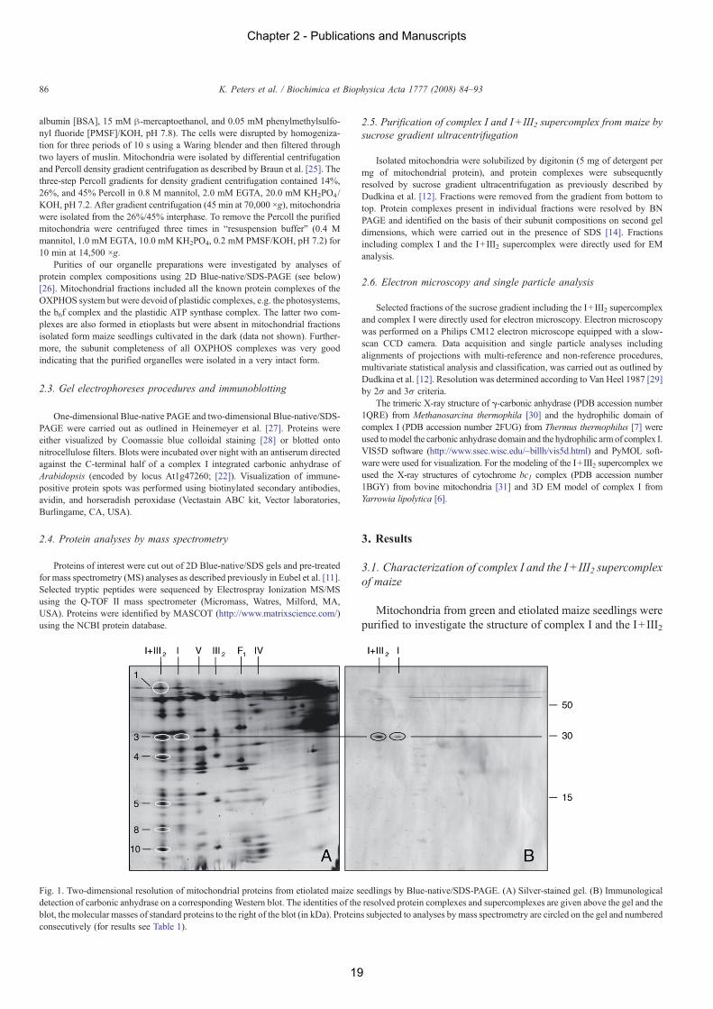
Fig. 1. Two-dimensional resolution of mitochondrial proteins from etiolated maize seedlings by Blue-native/SDS-PAGE. (A) Silver-stained gel. (B) Immunologicaldetection of carbonic anhydrase on a corresponding Western blot. The identities of the resolved protein complexes and supercomplexes are given above the gel and theblot, the molecular masses of standard proteins to the right of the blot (in kDa). Proteins subjected to analyses by mass spectrometry are circled on the gel and numberedconsecutively (for results see Table 1).
86 K. Peters et al. / Biochimica et Biophysica Acta 1777 (2008) 84–93
Chapter 2 - Publications and Manuscripts
19
supercomplex from a C4 plant. The OXPHOS system of maizewas analyzed by 2D Blue-native/SDS-PAGE. On the first geldimension, complex I runs at about 1000 kDa and the I+ III2supercomplex at 1500 kDa (data not shown). Subunits of theOXPHOS complexes were resolved by SDS-PAGE. The sub-unit compositions of the OXPHOS complexes of mitochondriaof etiolated maize seedlings (Fig. 1) resemble the ones of Ar-abidopsis [14]. On the 2D gel, complex III2 is resolved into 9distinct subunits, complex I into N25 and the I+ III2 supercom-plex also into N25 subunits. The latter two complexes probablyinclude several further subunits, which are invisible on the 2Dgels due to overlapping positions. The subunit composition ofcomplex I and the I+ III2 supercomplex of mitochondria ofgreen maize seedlings was indistinguishable from the onesobtained for etiolated seedlings upon analyses by 2D Blue-native PAGE (data not shown).
The presence of carbonic anhydrases within complex I andthe I+ III2 supercomplex of maize was investigated by im-munoblotting using an antibody directed against a complex Iintegrated carbonic anhydrase of Arabidopsis. The antibodyspecifically recognizes an epitope on a protein in the 30 kDarange of both complexes (Fig. 1B). The presence of carbonicanhydrases within complex I and the I+ III2 supercomplex ofmaize was confirmed by mass spectrometry (MS). For thisapproach, the 30 kDa spot and 5 further spots of the I+ III2supercomplex were cut out from a 2D BN/SDS gel, trypsinatedand prepared for MS analysis. Overall, 13 peptides sequenceswere obtained (Table 1), which exactly match peptide sequencesencoded by the rice genome (the complete genome sequence ofmaize currently is not available; rice is the closest relative ofmaize to be completely sequenced). The peptides are part of 8different proteins, four of which belong to complex I (75, 23 and11 kDa subunits and a protein homologous to a complex I in-tegrated carbonic anhydrase of rice), and four of which belong tocomplex III2 (cytochrome c1, the Rieske iron–sulfur protein,14 kDa and 8.2 kDa subunits). Sequence identities between themaize peptides and the corresponding amino acid sequences fromArabidopsis are in the range of 65 to 90% (data not shown).
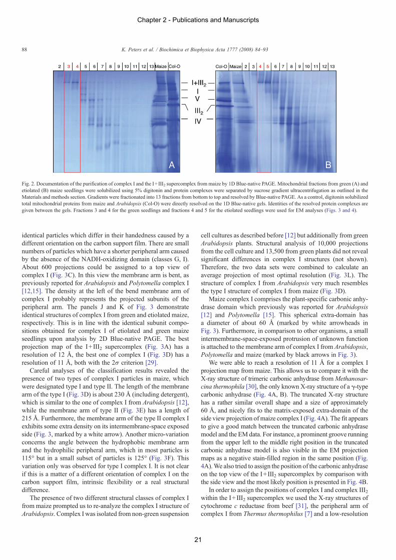
For EM analyses, purified mitochondrial fractions from greenand etiolated seedlings were loaded onto sucrose gradients andprotein complexes were separated by ultracentrifugation. After-wards, gradients were fractionated and small aliquots of allfractions were analysed by 1D Blue-native PAGE to monitor theprotein complex composition of the fractions (Fig. 2). Fractionsclose to the bottom of the gradients included pure complex I andI+ III2 supercomplex. These fractions were selected for furtheranalyses using single particle EM.
3.2. Electron microscopy
Negatively stained electron microscopy specimens of frac-tions 3 and 4 for the green seedlings and fractions 4 and 5 for theetiolated seedlings indicated large numbers of projections ofcomplex I and I+ III2 supercomplex suitable for single particleimage analysis. We analyzed a selected data set of about 28,000projections from green maize, which was grown in the light, anda data set of about 12,000 projections from etiolated maize,which was grown in the dark. An initial analysis by multi-reference alignment, multivariate statistical analysis and classi-fication of the projections indicated that both sets comprised thesame classes of projections with similar numbers of particles.Hence, the two data sets were also combined in one large dataset and analysed together, to get better resolution in the finalprojection maps.
After classification of the separate data sets and combined setof projections, a gallery of different projection maps of singularcomplex I and the I+ III2 supercomplex was obtained (Fig. 3).The I+ III2 supercomplex has one preferable orientation in aspecific top-view position (Fig. 3A). Only 75 views could beassigned to side view positions; the sum of the best 32 pro-jections is shown in Fig. 3B. Due to the very low numbers ofparticles, the resolution of this side view projection is verylimited. The classes shown in parts D–K of Fig. 3 represent sideviews of complex I. All these projections show the membrane-embedded arm in horizontal position and the hydrophilic arm inabout vertical position. The classes D/H and G/I represent
Table 1
Spot no. a (peptide) Identified peptide sequence b Protein identity c Accession no. d (organism)
1 (b) GSGEEIGTYVEK 75 kDa subunit, complex I gi|115454943 (rice)(c) SNYLMNTSIAGLEK 75 kDa subunit, complex I gi|125545494 (rice)
3 (b) LGSTIQGGLR Carbonic anhydrase, complex I gi|115473681 (rice)(c) IPSGEVWVGNPAK Carbonic anhydrase, complex I gi|115473681 (rice)(d) DLVGVAYTEEETK cyt c1, complex III gi|115442085 (rice)(f) DVVSFLSWAAEPEMEER cyt c1, complex III gi|34907202 (rice)
4 (b) LANSVDVASLR Rieske iron-sulfur protein, complex III P49727 (maize)(c) NVTINYPFEK 23 kDa TYKY subunit, complex I gi|115455639 (rice)(d) SINTLFLTEMVR 23 kDa TYKY subunit, complex I gi|115455639 (rice)(e) NQDAGLADLPATVAAVK Rieske iron-sulfur protein, complex III P49727 (maize)
5 (b) QSLGALPLYQR 14 kDa protein, complex III gi|115471095 (rice)8 (b) GFVMEFAENLILR 11 kDa subunit of complex I (At1g67350) gi|115454659 (rice)10 (b) AVVYAISPFQQK 8.2 kDa protein, complex III gi|115466706 (rice)a The spot numbers correspond to the numbers given on Fig. 1.b Peptides were identified by ESI-MS/MS as outlined in the Material and methods section.c Proteins were identified by MASCOT (http://www.matrixscience.com/) using the NCBI protein database.d NCBI protein accession codes of the most similar annotated proteins.
87K. Peters et al. / Biochimica et Biophysica Acta 1777 (2008) 84–93
Chapter 2 - Publications and Manuscripts
20
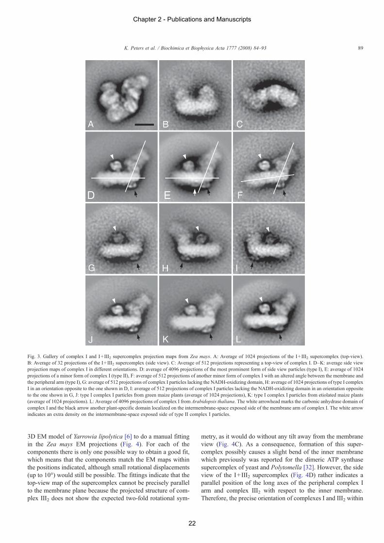
identical particles which differ in their handedness caused by adifferent orientation on the carbon support film. There are smallnumbers of particles which have a shorter peripheral arm causedby the absence of the NADH-oxidizing domain (classes G, I).About 600 projections could be assigned to a top view ofcomplex I (Fig. 3C). In this view the membrane arm is bent, aspreviously reported for Arabidopsis and Polytomella complex I[12,15]. The density at the left of the bend membrane arm ofcomplex I probably represents the projected subunits of theperipheral arm. The panels J and K of Fig. 3 demonstrateidentical structures of complex I from green and etiolated maize,respectively. This is in line with the identical subunit compo-sitions obtained for complex I of etiolated and green maizeseedlings upon analysis by 2D Blue-native PAGE. The bestprojection map of the I+ III2 supercomplex (Fig. 3A) has aresolution of 12 Å, the best one of complex I (Fig. 3D) has aresolution of 11 Å, both with the 2σ criterion [29].
Careful analyses of the classification results revealed thepresence of two types of complex I particles in maize, whichwere designated type I and type II. The length of the membranearm of the type I (Fig. 3D) is about 230 Å (including detergent),which is similar to the one of complex I from Arabidopsis [12],while the membrane arm of type II (Fig. 3E) has a length of215 Å. Furthermore, the membrane arm of the type II complex Iexhibits some extra density on its intermembrane-space exposedside (Fig. 3, marked by a white arrow). Another micro-variationconcerns the angle between the hydrophobic membrane armand the hydrophilic peripheral arm, which in most particles is115° but in a small subset of particles is 125° (Fig. 3F). Thisvariation only was observed for type I complex I. It is not clearif this is a matter of a different orientation of complex I on thecarbon support film, intrinsic flexibility or a real structuraldifference.
The presence of two different structural classes of complex Ifrom maize prompted us to re-analyze the complex I structure ofArabidopsis. Complex I was isolated from non-green suspension
cell cultures as described before [12] but additionally from greenArabidopsis plants. Structural analysis of 10,000 projectionsfrom the cell culture and 13,500 from green plants did not revealsignificant differences in complex I structures (not shown).Therefore, the two data sets were combined to calculate anaverage projection of most optimal resolution (Fig. 3L). Thestructure of complex I from Arabidopsis very much resemblesthe type I structure of complex I from maize (Fig. 3D).
Maize complex I comprises the plant-specific carbonic anhy-drase domain which previously was reported for Arabidopsis[12] and Polytomella [15]. This spherical extra-domain hasa diameter of about 60 Å (marked by white arrowheads inFig. 3). Furthermore, in comparison to other organisms, a smallintermembrane-space-exposed protrusion of unknown functionis attached to the membrane arm of complex I from Arabidopsis,Polytomella and maize (marked by black arrows in Fig. 3).
We were able to reach a resolution of 11 Å for a complex Iprojection map from maize. This allows us to compare it with theX-ray structure of trimeric carbonic anhydrase fromMethanosar-cina thermophila [30], the only known X-ray structure of a γ-typecarbonic anhydrase (Fig. 4A, B). The truncated X-ray structurehas a rather similar overall shape and a size of approximately60 Å, and nicely fits to the matrix-exposed extra-domain of theside view projection ofmaize complex I (Fig. 4A). The fit appearsto give a good match between the truncated carbonic anhydrasemodel and the EMdata. For instance, a prominent groove runningfrom the upper left to the middle right position in the truncatedcarbonic anhydrase model is also visible in the EM projectionmaps as a negative stain-filled region in the same position (Fig.4A).We also tried to assign the position of the carbonic anhydraseon the top view of the I+III2 supercomplex by comparison withthe side view and the most likely position is presented in Fig. 4B.
In order to assign the positions of complex I and complex III2within the I+ III2 supercomplex we used the X-ray structures ofcytochrome c reductase from beef [31], the peripheral arm ofcomplex I from Thermus thermophilus [7] and a low-resolution
Fig. 2. Documentation of the purification of complex I and the I+ III2 supercomplex from maize by 1D Blue-native PAGE. Mitochondrial fractions from green (A) andetiolated (B) maize seedlings were solubilized using 5% digitonin and protein complexes were separated by sucrose gradient ultracentrifugation as outlined in theMaterials and methods section. Gradients were fractionated into 13 fractions from bottom to top and resolved by Blue-native PAGE. As a control, digitonin solubilizedtotal mitochondrial proteins from maize and Arabidopsis (Col-O) were directly resolved on the 1D Blue-native gels. Identities of the resolved protein complexes aregiven between the gels. Fractions 3 and 4 for the green seedlings and fractions 4 and 5 for the etiolated seedlings were used for EM analyses (Figs. 3 and 4).
88 K. Peters et al. / Biochimica et Biophysica Acta 1777 (2008) 84–93
Chapter 2 - Publications and Manuscripts
21
3D EM model of Yarrowia lipolytica [6] to do a manual fittingin the Zea mays EM projections (Fig. 4). For each of thecomponents there is only one possible way to obtain a good fit,which means that the components match the EM maps withinthe positions indicated, although small rotational displacements(up to 10°) would still be possible. The fittings indicate that thetop-view map of the supercomplex cannot be precisely parallelto the membrane plane because the projected structure of com-plex III2 does not show the expected two-fold rotational sym-
metry, as it would do without any tilt away from the membraneview (Fig. 4C). As a consequence, formation of this super-complex possibly causes a slight bend of the inner membranewhich previously was reported for the dimeric ATP synthasesupercomplex of yeast and Polytomella [32]. However, the sideview of the I+ III2 supercomplex (Fig. 4D) rather indicates aparallel position of the long axes of the peripheral complex Iarm and complex III2 with respect to the inner membrane.Therefore, the precise orientation of complexes I and III2 within
Fig. 3. Gallery of complex I and I+ III2 supercomplex projection maps from Zea mays. A: Average of 1024 projections of the I+ III2 supercomplex (top-view).B: Average of 32 projections of the I+ III2 supercomplex (side view). C: Average of 512 projections representing a top-view of complex I. D–K: average side viewprojection maps of complex I in different orientations. D: average of 4096 projections of the most prominent form of side view particles (type I), E: average of 1024projections of a minor form of complex I (type II), F: average of 512 projections of another minor form of complex I with an altered angle between the membrane andthe peripheral arm (type I), G: average of 512 projections of complex I particles lacking the NADH-oxidizing domain, H: average of 1024 projections of type I complexI in an orientation opposite to the one shown in D, I: average of 512 projections of complex I particles lacking the NADH-oxidizing domain in an orientation oppositeto the one shown in G, J: type I complex I particles from green maize plants (average of 1024 projections), K: type I complex I particles from etiolated maize plants(average of 1024 projections). L: Average of 4096 projections of complex I from Arabidopsis thaliana. The white arrowhead marks the carbonic anhydrase domain ofcomplex I and the black arrow another plant-specific domain localized on the intermembrane-space exposed side of the membrane arm of complex I. The white arrowindicates an extra density on the intermembrane-space exposed side of type II complex I particles.
89K. Peters et al. / Biochimica et Biophysica Acta 1777 (2008) 84–93
Chapter 2 - Publications and Manuscripts
22
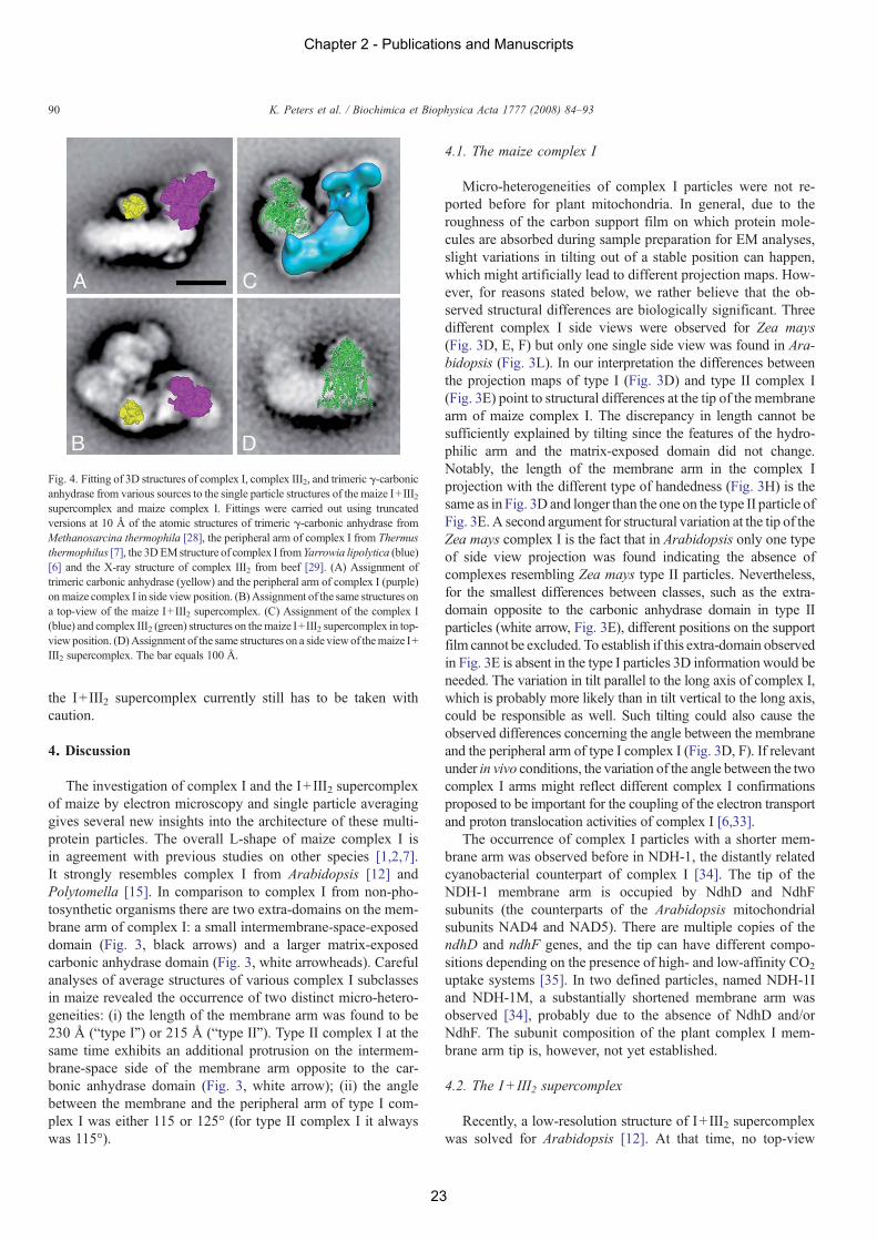
the I+ III2 supercomplex currently still has to be taken withcaution.
4. Discussion
The investigation of complex I and the I+ III2 supercomplexof maize by electron microscopy and single particle averaginggives several new insights into the architecture of these multi-protein particles. The overall L-shape of maize complex I isin agreement with previous studies on other species [1,2,7].It strongly resembles complex I from Arabidopsis [12] andPolytomella [15]. In comparison to complex I from non-pho-tosynthetic organisms there are two extra-domains on the mem-brane arm of complex I: a small intermembrane-space-exposeddomain (Fig. 3, black arrows) and a larger matrix-exposedcarbonic anhydrase domain (Fig. 3, white arrowheads). Carefulanalyses of average structures of various complex I subclassesin maize revealed the occurrence of two distinct micro-hetero-geneities: (i) the length of the membrane arm was found to be230 Å (“type I”) or 215 Å (“type II”). Type II complex I at thesame time exhibits an additional protrusion on the intermem-brane-space side of the membrane arm opposite to the car-bonic anhydrase domain (Fig. 3, white arrow); (ii) the anglebetween the membrane and the peripheral arm of type I com-plex I was either 115 or 125° (for type II complex I it alwayswas 115°).
4.1. The maize complex I
Micro-heterogeneities of complex I particles were not re-ported before for plant mitochondria. In general, due to theroughness of the carbon support film on which protein mole-cules are absorbed during sample preparation for EM analyses,slight variations in tilting out of a stable position can happen,which might artificially lead to different projection maps. How-ever, for reasons stated below, we rather believe that the ob-served structural differences are biologically significant. Threedifferent complex I side views were observed for Zea mays(Fig. 3D, E, F) but only one single side view was found in Ara-bidopsis (Fig. 3L). In our interpretation the differences betweenthe projection maps of type I (Fig. 3D) and type II complex I(Fig. 3E) point to structural differences at the tip of the membranearm of maize complex I. The discrepancy in length cannot besufficiently explained by tilting since the features of the hydro-philic arm and the matrix-exposed domain did not change.Notably, the length of the membrane arm in the complex Iprojection with the different type of handedness (Fig. 3H) is thesame as in Fig. 3D and longer than the one on the type II particle ofFig. 3E. A second argument for structural variation at the tip of theZea mays complex I is the fact that in Arabidopsis only one typeof side view projection was found indicating the absence ofcomplexes resembling Zea mays type II particles. Nevertheless,for the smallest differences between classes, such as the extra-domain opposite to the carbonic anhydrase domain in type IIparticles (white arrow, Fig. 3E), different positions on the supportfilm cannot be excluded. To establish if this extra-domain observedin Fig. 3E is absent in the type I particles 3D information would beneeded. The variation in tilt parallel to the long axis of complex I,which is probably more likely than in tilt vertical to the long axis,could be responsible as well. Such tilting could also cause theobserved differences concerning the angle between the membraneand the peripheral arm of type I complex I (Fig. 3D, F). If relevantunder in vivo conditions, the variation of the angle between the twocomplex I arms might reflect different complex I confirmationsproposed to be important for the coupling of the electron transportand proton translocation activities of complex I [6,33].
The occurrence of complex I particles with a shorter mem-brane arm was observed before in NDH-1, the distantly relatedcyanobacterial counterpart of complex I [34]. The tip of theNDH-1 membrane arm is occupied by NdhD and NdhFsubunits (the counterparts of the Arabidopsis mitochondrialsubunits NAD4 and NAD5). There are multiple copies of thendhD and ndhF genes, and the tip can have different compo-sitions depending on the presence of high- and low-affinity CO2
uptake systems [35]. In two defined particles, named NDH-1Iand NDH-1M, a substantially shortened membrane arm wasobserved [34], probably due to the absence of NdhD and/orNdhF. The subunit composition of the plant complex I mem-brane arm tip is, however, not yet established.
4.2. The I+III2 supercomplex
Recently, a low-resolution structure of I+ III2 supercomplexwas solved for Arabidopsis [12]. At that time, no top-view
Fig. 4. Fitting of 3D structures of complex I, complex III2, and trimeric γ-carbonicanhydrase from various sources to the single particle structures of the maize I+III2supercomplex and maize complex I. Fittings were carried out using truncatedversions at 10 Å of the atomic structures of trimeric γ-carbonic anhydrase fromMethanosarcina thermophila [28], the peripheral arm of complex I from Thermusthermophilus [7], the 3DEMstructure of complex I fromYarrowia lipolytica (blue)[6] and the X-ray structure of complex III2 from beef [29]. (A) Assignment oftrimeric carbonic anhydrase (yellow) and the peripheral arm of complex I (purple)onmaize complex I in side view position. (B)Assignment of the same structures ona top-view of the maize I+III2 supercomplex. (C) Assignment of the complex I(blue) and complex III2 (green) structures on themaize I+III2 supercomplex in top-viewposition. (D)Assignment of the same structures on a side viewof themaize I+III2 supercomplex. The bar equals 100 Å.
90 K. Peters et al. / Biochimica et Biophysica Acta 1777 (2008) 84–93
Chapter 2 - Publications and Manuscripts
23
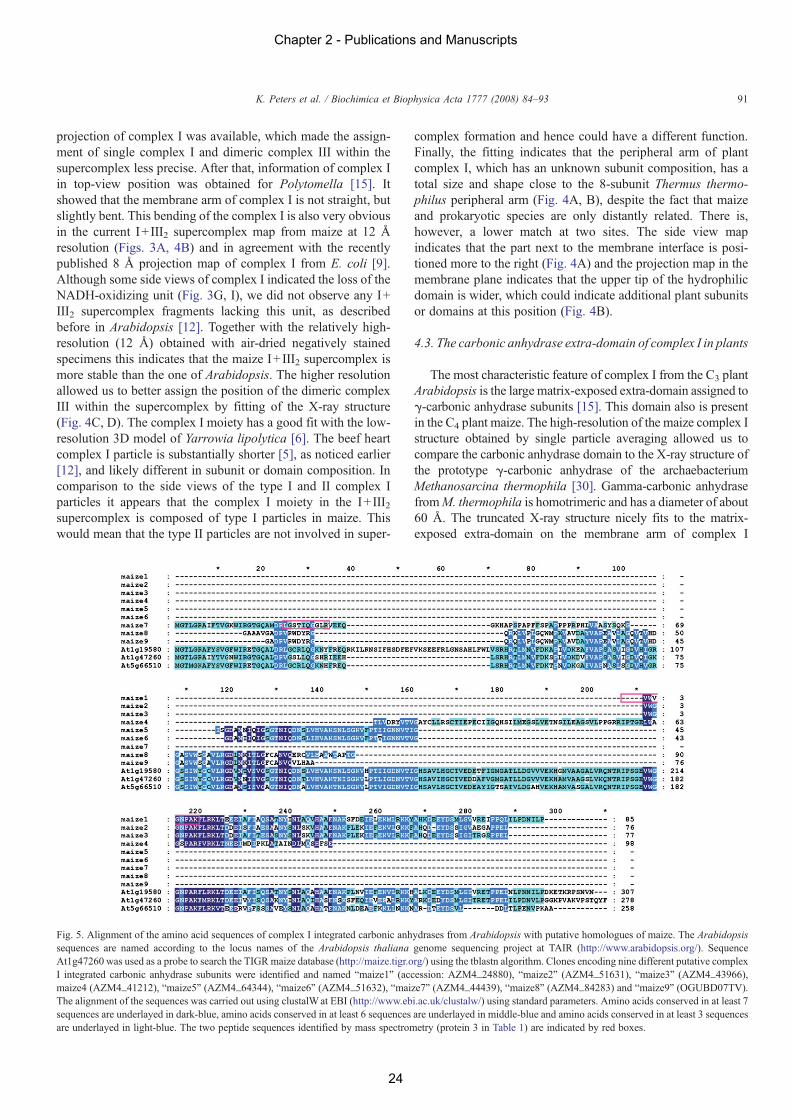
projection of complex I was available, which made the assign-ment of single complex I and dimeric complex III within thesupercomplex less precise. After that, information of complex Iin top-view position was obtained for Polytomella [15]. Itshowed that the membrane arm of complex I is not straight, butslightly bent. This bending of the complex I is also very obviousin the current I+ III2 supercomplex map from maize at 12 Åresolution (Figs. 3A, 4B) and in agreement with the recentlypublished 8 Å projection map of complex I from E. coli [9].Although some side views of complex I indicated the loss of theNADH-oxidizing unit (Fig. 3G, I), we did not observe any I+III2 supercomplex fragments lacking this unit, as describedbefore in Arabidopsis [12]. Together with the relatively high-resolution (12 Å) obtained with air-dried negatively stainedspecimens this indicates that the maize I+ III2 supercomplex ismore stable than the one of Arabidopsis. The higher resolutionallowed us to better assign the position of the dimeric complexIII within the supercomplex by fitting of the X-ray structure(Fig. 4C, D). The complex I moiety has a good fit with the low-resolution 3D model of Yarrowia lipolytica [6]. The beef heartcomplex I particle is substantially shorter [5], as noticed earlier[12], and likely different in subunit or domain composition. Incomparison to the side views of the type I and II complex Iparticles it appears that the complex I moiety in the I+ III2supercomplex is composed of type I particles in maize. Thiswould mean that the type II particles are not involved in super-
complex formation and hence could have a different function.Finally, the fitting indicates that the peripheral arm of plantcomplex I, which has an unknown subunit composition, has atotal size and shape close to the 8-subunit Thermus thermo-philus peripheral arm (Fig. 4A, B), despite the fact that maizeand prokaryotic species are only distantly related. There is,however, a lower match at two sites. The side view mapindicates that the part next to the membrane interface is posi-tioned more to the right (Fig. 4A) and the projection map in themembrane plane indicates that the upper tip of the hydrophilicdomain is wider, which could indicate additional plant subunitsor domains at this position (Fig. 4B).
4.3. The carbonic anhydrase extra-domain of complex I in plants
The most characteristic feature of complex I from the C3 plantArabidopsis is the large matrix-exposed extra-domain assigned toγ-carbonic anhydrase subunits [15]. This domain also is presentin the C4 plant maize. The high-resolution of the maize complex Istructure obtained by single particle averaging allowed us tocompare the carbonic anhydrase domain to the X-ray structure ofthe prototype γ-carbonic anhydrase of the archaebacteriumMethanosarcina thermophila [30]. Gamma-carbonic anhydrasefromM. thermophila is homotrimeric and has a diameter of about60 Å. The truncated X-ray structure nicely fits to the matrix-exposed extra-domain on the membrane arm of complex I
Fig. 5. Alignment of the amino acid sequences of complex I integrated carbonic anhydrases from Arabidopsis with putative homologues of maize. The Arabidopsissequences are named according to the locus names of the Arabidopsis thaliana genome sequencing project at TAIR (http://www.arabidopsis.org/). SequenceAt1g47260 was used as a probe to search the TIGRmaize database (http://maize.tigr.org/) using the tblastn algorithm. Clones encoding nine different putative complexI integrated carbonic anhydrase subunits were identified and named “maize1” (accession: AZM4_24880), “maize2” (AZM4_51631), “maize3” (AZM4_43966),maize4 (AZM4_41212), “maize5” (AZM4_64344), “maize6” (AZM4_51632), “maize7” (AZM4_44439), “maize8” (AZM4_84283) and “maize9” (OGUBD07TV).The alignment of the sequences was carried out using clustalWat EBI (http://www.ebi.ac.uk/clustalw/) using standard parameters. Amino acids conserved in at least 7sequences are underlayed in dark-blue, amino acids conserved in at least 6 sequences are underlayed in middle-blue and amino acids conserved in at least 3 sequencesare underlayed in light-blue. The two peptide sequences identified by mass spectrometry (protein 3 in Table 1) are indicated by red boxes.
91K. Peters et al. / Biochimica et Biophysica Acta 1777 (2008) 84–93
Chapter 2 - Publications and Manuscripts
24
frommaize (Fig. 4A). This indicates that the complex I integratedγ-carbonic anhydrase domain of Zea mays also is a trimer. InArabidopsis, five different carbonic anhydrase subunits formpart of complex I. It currently is not known whether the carbonicanhydrase domains of complex I includes three copies of the sameisoform or different isoforms. Probing the maize genome se-quence database (http://maize.tigr.org/) with the sequence of oneof the carbonic anhydrases of Arabidopsis allowed to identifypartial sequences of nine different putative γ-carbonic anhy-drases. Since themaize genome sequence has not been completed,further homologues might exist. The two peptide sequencesobtained byMS analyses of the 30 kDa subunit of maize complexI (Fig. 1, Table 1) are identical to amino acid stretches of two of themaize carbonic anhydrase isoforms (Fig. 5). Therefore, at leasttwo forms of γ-carbonic anhydrases occur within the maizecomplex I. We conclude that the carbonic anhydrase domain ofplant complex I most likely is heterotrimeric.
The carbonic anhydrase domain seems to be a general featureof plant complex I, since it was now described for the C3 plantArabidopsis, the C4 plant maize and the green alga Polytomella[[12,15], this study]. It was proposed that the complex I in-tegrated carbonic anhydrases might play a role in the context ofa carbon transport system between mitochondria and chlor-oplasts to increase the efficiency of photosynthetic carbon fix-ation. According to this hypothesis, mitochondrial catabolismrepresents an important source of CO2 for photosynthesis. Thehypothesis is supported by a recent study investigating the CO2
uptake/CO2 fixation ratio in isolated protoplasts versus isolatedchloroplasts [36]. The ratio was considerably lower in proto-plasts, which was interpreted to be caused by CO2 supply bymitochondria in protoplasts. The presence of carbonic anhy-drases seems to be of equal importance in mitochondria of C3
and C4 plants but possibly for different reasons. In C3 plants,CO2 transport from mitochondria to plastids might be especiallyimportant during photorespiration, whereas in C4 plants thistransport should be most important in the context of the CO2
liberation step in the bundle sheath cells. The CO2 liberationstep is known to be based on different enzymatic reactions,which take place in different cellular compartments [37]. Inmaize, the conversion of malate into pyruvate and CO2 takesplace in plastids by a NADP+ dependant malic enzyme. In otherplants, this step occurs in mitochondria by the act of a NAD+
dependant malic enzyme. Alternatively, CO2 liberation is car-ried out by a PEP carboxykinase localized in the cytosol ofbundle sheath cells. However, the metabolism of C4 subtypesseems not to be exclusively based on one or the other CO2
liberation reaction, but rather on all of them to varying extends[38]. Possibly the complex I integrated carbonic anhydrases areespecially important in C4 plants with dominating NAD+ malicenzyme dependant CO2 liberation. Further investigations willbe necessary to clarify the precise physiological role of themitochondrial carbonic anhydrases in C3 and C4 plants.
Acknowledgements
We thank Dr. Roman Kouřil and Dr. Wilko Keegstra for theirhelp with the processing and modelling of supercomplex
structures and Dr. Gert Oostergetel for invaluable help withelectron microscopy. We also like to thank Dr. M. Radermacherfor providing Yarrowia lipolytica complex I images. DagmarLewejohann is thanked for expert technical assistance. Researchof our laboratories is supported by the Deutsche Forschungsge-meinschaft (grant Br 1829-7/3).
References
[1] T. Yagi, A. Matsuno-Yagi, The proton-translocating NADH-quinoneoxidoreductase in the respiratory chain: the secret unlocked, Biochemistry42 (2003) 2266–2274.
[2] T. Friedrich, B. Böttcher, The gross structure of the respiratory complex I: alego system, Biochim. Biophys. Acta 1608 (2004) 1–9.
[3] U. Brandt, Energy convertingNADH: quinone oxidoreductase (complex I),Ann. Rev. Biochem. 75 (2006) 69–92.
[4] V. Guenebaut, R. Vincentelli, D. Mills, H. Weiss, K.R. Leonard, Three-dimensional structure of NADH-dehydrogenase from Neurospora crassaby electron microscopy and conical tilt reconstruction, J. Mol. Biol. 265(1997) 409–418.
[5] N. Grigorieff, Three-dimensional structure of bovine NADH: ubiquinoneoxidoreductase (complex I) at 22Å in ice, J.MolBiol. 277 (1998) 1033–1046.
[6] M. Radermacher, T. Ruiz, T. Clason, S. Benjamin, U. Brandt, V. Zickermann,The three-dimensional structure of complex I from Yarrowia lipolytica: ahighly dynamic enzyme, J. Struct. Biol. 154 (2006) 269–279.
[7] L.A. Sazanov, P. Hinchliffe, Structure of the hydrophilic domain of respiratorycomplex I from Thermus thermophilus, Science 311 (2006) 1430–1436.
[8] U. Brandt, Proton-translocation by membrane-bound NADH: ubiquinone-oxidoreductase (complex I) through redox-gated ligand conduction,Biochim. Biophys. Acta 1318 (1997) 79–91.
[9] E.A. Baranova, P.J. Holt, L.A. Sazanov, Projection structure of themembrane domain of Escherichia coli respiratory complex I at 8 Åresolution, J. Mol. Biol. 366 (2007) 140–154.
[10] H. Schägger, K. Pfeiffer, Supercomplexes in the respiratory chains of yeastand mammalian mitochondria, EMBO J. 19 (2000) 1777–1783.
[11] H. Eubel, L. Jänsch, H.-P. Braun, New insights into the respiratory chain ofplant mitochondria supercomplexes and a unique composition of complexII, Plant Physiol. 133 (2003) 274–286.
[12] N.V. Dudkina, H. Eubel, W. Keegstra, E.J. Boekema, H.-P. Braun,Structure of a mitochondrial supercomplex formed by respiratory-chaincomplexes I and III, Proc. Natl. Acad. Sci. U.S.A. 102 (2005) 3225–3229.
[13] J.L. Heazlewood, K.A. Howell, A.H. Millar, Mitochondrial complex Ifrom Arabidopsis and rice: orthologs of mammalian and yeast componentscoupled to plant-specific subunits, Biochim. Biophys. Acta 1604 (2003)159–169.
[14] P. Cardol, F. Vanrobaeys, B. Devreese, J. Van Beeumen, R. Matagne, C.Remacle, Higher plant-like subunit composition of mitochondrialcomplex I from: 31 conserved components among eukaryotes, Biochim.Biophys. Acta 1658 (2004) 212–224.
[15] S. Sunderhaus, N.V. Dudkina, L. Jänsch, J. Klodmann, J. Heinemeyer,M. Perales, E. Zabaleta, E.J. Boekema, H.-P. Braun, Carbonic anhydrasesubunits form a matrix-exposed domain attached to the membrane armof mitochondrial complex I in plants, J. Biol. Chem. 281 (2006)6482–6488.
[16] L. Jänsch, V. Kruft, U.K. Schmitz, H.-P. Braun, Cytochrome c reductasefrom potato does not comprise three core proteins but contains an addi-tional lowmolecular weight subunit, Eur. J. Biochem. 228 (1995) 878–885.
[17] M.J. Runswick, I.M. Fearnley, J.M. Skehel, J.E. Walker, Presence of anacyl carrier protein in NADH:ubiquinone oxidoreductase from bovineheart mitochondria, FEBS Lett. 286 (1991) 121–124.
[18] U. Sackmann, R. Zensen, D. Röhlen, U. Jahnke, H. Weiss, The acyl-carrierprotein in Neurospora crassa mitochondria is a subunit of NADH:ubiquinone reductase (complex I), Eur. J. Biochem. 200 (1991) 463–469.
[19] E.H. Meyer, J.L. Heazlewood, A.H. Millar, Mitochondrial acyl carrierproteins in Arabidopsis thaliana are predominantly soluble matrix proteinsand none can be confirmed as subunits of respiratory Complex I, Plant.Mol. Biol. 64 (2007) 319–327.
92 K. Peters et al. / Biochimica et Biophysica Acta 1777 (2008) 84–93
Chapter 2 - Publications and Manuscripts
25
[20] A.H. Millar, V. Mittova, G. Kiddle, J.L. Heazlewood, C.G. Bartoli, F.L.Theodoulou, C.H. Foyer, Control of ascorbate synthesis by respiration andits implications for stress responses, Plant Physiol. 133 (2003) 443–447.
[21] G. Parisi, M. Perales, M. Fornasari, A. Colaneri, N. Schain, D. Casati, S.Zimmermann, A. Brennicke, A. Araya, J. Ferry, J. Echave, E. Zabaleta,Gamma carbonic anhydrases in plant mitochondria, Plant Mol. Biol. 55(2004) 193–207.
[22] M. Perales, H. Eubel, J. Heinemeyer, A. Colaneri, E. Zabaleta, H.-P.Braun, Disruption of a nuclear gene encoding a mitochondrial gammacarbonic anhydrase reduces complex I and supercomplex I+ III2 levels andalters mitochondrial physiology in Arabidopsis, J. Mol. Biol. 350 (2005)263–277.
[23] H.-P. Braun, E. Zabaleta, Carbonic anhydrase subunits of the mitochon-drial NADH dehydrogenase complex (complex I) in plants, Physiol. Plant.129 (2007) 114–122.
[24] G.D. Price, M.R. Badger, F.J. Woodger, B.M. Long, Advances inunderstanding the cyanobacterial CO2-concentrating-mechanism (CCM):functional components, Ci transporters, diversity, genetic regulation andprospects for engineering into plants, J. Exp. Bot. (in press).
[25] H.-P. Braun, M. Emmermann, V. Kruft, U.K. Schmitz, Cytochrome c1from potato: a protein with a presequence for targeting to the mitochondrialintermembrane space, Mol. Gen. Genet. 231 (1992) 217–225.
[26] N. Hausmann, W.Werhahn, B. Huchzermeyer, H.-P. Braun, J. Papenbrock,How to document the purity of mitochondria prepared from green tissue oftobacco, pea and Arabidopsis thaliana, Phyton 43 (2003) 215–229.
[27] J. Heinemeyer, D. Lewejohann, H.-P. Braun, Blue-native gel electrophor-esis for the characterization of protein complexes in plants, Meth. Mol.Biol. 335 (2007) 343–352.
[28] V. Neuhoff, R. Stamm, I. Pardowitz, N. Arold, W. Ehrhardt, D. Taube,Essential problems in quantification of proteins following colloidalstaining with Coomassie Brilliant Blue dyes in polyacrylamide gels, andtheir solution, Electrophoresis 11 (1990) 101–117.
[29] M. van Heel, Similarity measures between images, Ultramicroscopy 21(1987) 95–100.
[30] T.M. Iverson, B.E. Alber, C. Kisker, J.G. Ferry, D.C. Rees, A closer lookat the active site of gamma-class carbonic anhydrases: high-resolutioncrystallographic studies of the carbonic anhydrase from Methanosarcinathermophila, Biochemistry 39 (2000) 9222–9231.
[31] S. Iwata, J.W. Lee, K. Okada, J.K. Lee,M. Iwata, B. Rasmussen, T.A. Link,S. Ramaswamy, B.K. Jap, Complete structure of the 11-subunit bovinemitochondrial cytochrome bc1 complex, Science 281 (1998) 64–71.
[32] N.V. Dudkina, S. Sunderhaus, H.-P. Braun, E.J. Boekema, Characteriza-tion of dimeric ATP synthase and cristae membrane ultrastructure fromSaccharomyces and Polytomella mitochondria, FEBS Lett. 580 (2006)3427–3432.
[33] B. Böttcher, D. Scheide, M. Hesterberg, L. Nagel-Steger, T. Friedrich, Anovel, enzymatically active conformation of the Escherichia coli NADH:ubiquinone oxidoreductase (complex I), J. Biol. Chem 277 (2002)17970–17977.
[34] A.A. Arteni, P. Zhang, N. Battchikova, T. Ogawa, E.-M. Aro, E.J.Boekema, Structural characterization of NDH-1 complexes of Thermosy-nechococcus elongatus by single particle electron microscopy, Biochim.Biophys. Acta 1757 (2006) 1469–1475.
[35] T. Ogawa, H. Mi, Cyanobacterial NADPH dehydrogenase complexes,Photosynth. Res. 93 (1–3) (2007) 69–77.
[36] K. Riazunnisa, L. Padmavathi, H. Bauwe, A.S. Raghavendra, Markedlylow requirement of added CO2 for photosynthesis by mesophyll proto-plasts of pea (Pisum sativum): possible roles of photorespiratory CO2 andcarbonic anhydrase, Physiol. Plant. 128 (2006) 763–772.
[37] S.v. Caemmerer, R.T. Furbank, The C4 pathway: an efficient CO2 pump,Photosynthesis Research 77 (2003) 191–207.
[38] G.E. Edwards, R.T. Furbank, M.D. Hatch, C.B. Osmond, What does it taketo be C4? Lessons from the evolution of C4 photosynthesis, Plant Physiol.125 (2001) 46–49.
93K. Peters et al. / Biochimica et Biophysica Acta 1777 (2008) 84–93
Chapter 2 - Publications and Manuscripts
26
Review
Structure and function of mitochondrial supercomplexes
Natalya V. Dudkina a, Roman Kouřil a, Katrin Peters b, Hans-Peter Braun b, Egbert J. Boekema a,⁎a Electron microscopy group, Groningen Biomolecular Sciences and Biotechnology Institute, University of Groningen, Nijenborgh 4, 9747 AG Groningen, The Netherlandsb Institute for Plant Genetics, Faculty of Natural Sciences, Universität Hannover, Herrenhäuser Str. 2, D-30419 Hannover, Germany
a b s t r a c ta r t i c l e i n f o
Article history:Received 27 October 2009Received in revised form 14 December 2009Accepted 16 December 2009Available online 28 December 2009
Keywords:Oxidative phosphorylationMitochondriaRespirasomeATP synthaseElectron microscopySupercomplex
The five complexes (complexes I–V) of the oxidative phosphorylation (OXPHOS) system of mitochondria canbe extracted in the form of active supercomplexes. Single-particle electron microscopy has provided 2D and3D data describing the interaction between complexes I and III, among I, III and IV and in a dimeric form ofcomplex V, between two ATP synthase monomers. The stable interactions are called supercomplexes whichalso form higher-ordered oligomers. Cryo-electron tomography provides new insights on how thesesupercomplexes are arranged within intact mitochondria. The structure and function of OXPHOSsupercomplexes are discussed.
© 2009 Elsevier B.V. All rights reserved.
1. Introduction
Mitochondria are intracellular organelles which accommodate intheir inner or cristae membrane large numbers of five oxidativephosphorylation complexes (complexes I–V). The complexes I to IVare oxidoreductases which, with the exception of complex II, coupleelectron transport with translocation of protons across the innermitochondrial membrane (Fig. 1). The generated proton motive forceis used by ATP synthase (complex V) for ATP synthesis from ADP andphosphate. Complex I or NADH dehydrogenase is the first and majorentrance point of electrons to the respiratory chain. It transferselectrons from NADH molecules to a lipophilic quinone designatedubiquinone. Complex II or succinate dehydrogenase transmitselectrons from succinate to ubiquinone and directly connects thecitric acid cycle to the respiratory chain. From the reduced ubiquinoneelectrons can be transferred to complex III or cytochrome c reductasewhich exists in themembrane as a functional dimer. The small proteincytochrome cmediates electron transfer from cytochrome c reductaseto cytochrome c oxidase (complex IV). Finally, electrons aretransferred to molecular oxygen which is reduced to water.
The ideas about the overall organization of these five complexes,which together form the oxidative phosphorylation (OXPHOS)system, have been changing with time. A so-called “fluid-statemodel” is supported by the finding that all individual proteincomplexes of the OXPHOS system can be purified to homogeneity inan enzymatically active form and by lipid dilution experiments
(reviewed in [1,2]). It postulates that the respiratory chain complexesfreely diffuse in membrane and that the electron transfer is based onrandom collisions of the single complexes.
The fluid-state model is challenged by a “solid-state model”whichproposes stable interactions between the OXPHOS complexes withinentities named supercomplexes. This model is now supported by awide range of experimental findings: 1. Supercomplexes can beresolved by blue native polyacrylamide gel electrophoresis (BN-PAGE) [3,4]. 2. Supercomplexes are active as shown by in-gel activitymeasurements within blue native gels [3,5]. 3. Electron microscopy(EM) structures revealed defined interactions of OXPHOS complexeswithin the isolated respiratory supercomplexes [6–10]. 4. Flux controlexperiments [11,12] confirm that the respiratory chain operates asone functional unit. 5. Point mutations in genes encoding one of thesubunits of one OXPHOS complex affect the stability of anothercomplex. Thus, complex III is required to maintain complex I in mouseand human cultured cells mitochondria [13]. Complex IV is necessaryfor the assembly or stability of complex I in mouse fibroblasts [14]. 6.Oxygen uptake by isolated mitochondria of potato correlates with theabundance of supercomplexes [5]. 7. Some supercomplexes needcardiolipin for their formation [15,16]. Until some years ago, the fluid-state model was widely accepted in the field of bioenergetics. Thenewest edition of the Lehninger textbook is the first general textbookthat presents a section on OXPHOS supercomplexes [17].
In many organisms the complexes I, III and IV were found toassociate into specific supercomplexes (reviewed in [18]). Onlycomplex II seems not to form part of any respiratory chainsupercomplexes. The possible explanation for the singular state ofcomplex II is that it is also involved in the citric acid cycle. However, arecent publication on the OXPHOS system ofmousemitochondria also
Biochimica et Biophysica Acta 1797 (2010) 664–670
⁎ Corresponding author.E-mail address: [email protected] (E.J. Boekema).
0005-2728/$ – see front matter © 2009 Elsevier B.V. All rights reserved.doi:10.1016/j.bbabio.2009.12.013
Contents lists available at ScienceDirect
Biochimica et Biophysica Acta
j ourna l homepage: www.e lsev ie r.com/ locate /bbab io
Chapter 2 - Publications and Manuscripts
27
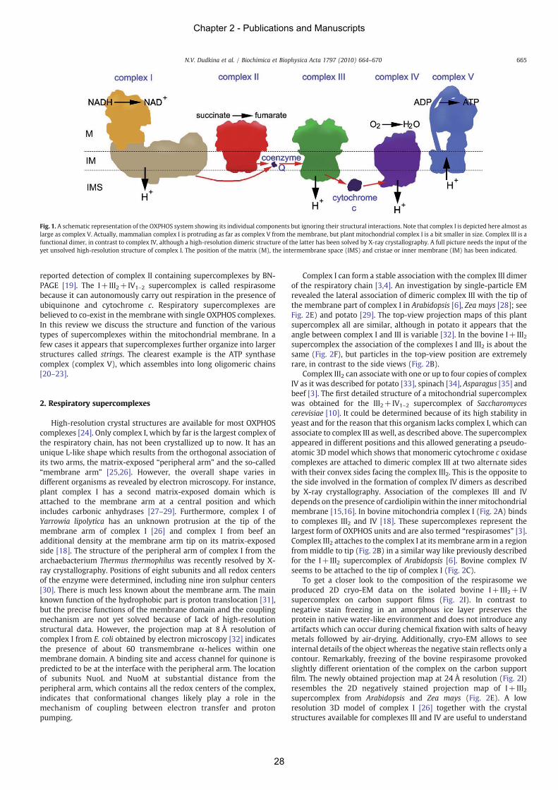
reported detection of complex II containing supercomplexes by BN-PAGE [19]. The I+III2+IV1–2 supercomplex is called respirasomebecause it can autonomously carry out respiration in the presence ofubiquinone and cytochrome c. Respiratory supercomplexes arebelieved to co-exist in the membrane with single OXPHOS complexes.In this review we discuss the structure and function of the varioustypes of supercomplexes within the mitochondrial membrane. In afew cases it appears that supercomplexes further organize into largerstructures called strings. The clearest example is the ATP synthasecomplex (complex V), which assembles into long oligomeric chains[20–23].
2. Respiratory supercomplexes
High-resolution crystal structures are available for most OXPHOScomplexes [24]. Only complex I, which by far is the largest complex ofthe respiratory chain, has not been crystallized up to now. It has anunique L-like shape which results from the orthogonal association ofits two arms, the matrix-exposed “peripheral arm” and the so-called“membrane arm” [25,26]. However, the overall shape varies indifferent organisms as revealed by electron microscopy. For instance,plant complex I has a second matrix-exposed domain which isattached to the membrane arm at a central position and whichincludes carbonic anhydrases [27–29]. Furthermore, complex I ofYarrowia lipolytica has an unknown protrusion at the tip of themembrane arm of complex I [26] and complex I from beef anadditional density at the membrane arm tip on its matrix-exposedside [18]. The structure of the peripheral arm of complex I from thearchaebacterium Thermus thermophilus was recently resolved by X-ray crystallography. Positions of eight subunits and all redox centersof the enzyme were determined, including nine iron sulphur centers[30]. There is much less known about the membrane arm. The mainknown function of the hydrophobic part is proton translocation [31],but the precise functions of the membrane domain and the couplingmechanism are not yet solved because of lack of high-resolutionstructural data. However, the projection map at 8 Å resolution ofcomplex I from E. coli obtained by electron microscopy [32] indicatesthe presence of about 60 transmembrane α-helices within onemembrane domain. A binding site and access channel for quinone ispredicted to be at the interface with the peripheral arm. The locationof subunits NuoL and NuoM at substantial distance from theperipheral arm, which contains all the redox centers of the complex,indicates that conformational changes likely play a role in themechanism of coupling between electron transfer and protonpumping.
Complex I can form a stable association with the complex III dimerof the respiratory chain [3,4]. An investigation by single-particle EMrevealed the lateral association of dimeric complex III with the tip ofthe membrane part of complex I in Arabidopsis [6], Zea mays [28]; seeFig. 2E) and potato [29]. The top-view projection maps of this plantsupercomplex all are similar, although in potato it appears that theangle between complex I and III is variable [32]. In the bovine I+III2supercomplex the association of the complexes I and III2 is about thesame (Fig. 2F), but particles in the top-view position are extremelyrare, in contrast to the side views (Fig. 2B).
Complex III2 can associate with one or up to four copies of complexIV as it was described for potato [33], spinach [34], Asparagus [35] andbeef [3]. The first detailed structure of a mitochondrial supercomplexwas obtained for the III2+IV1–2 supercomplex of Saccharomycescerevisiae [10]. It could be determined because of its high stability inyeast and for the reason that this organism lacks complex I, which canassociate to complex III as well, as described above. The supercomplexappeared in different positions and this allowed generating a pseudo-atomic 3D model which shows that monomeric cytochrome c oxidasecomplexes are attached to dimeric complex III at two alternate sideswith their convex sides facing the complex III2. This is the opposite tothe side involved in the formation of complex IV dimers as describedby X-ray crystallography. Association of the complexes III and IVdepends on the presence of cardiolipinwithin the innermitochondrialmembrane [15,16]. In bovine mitochondria complex I (Fig. 2A) bindsto complexes III2 and IV [18]. These supercomplexes represent thelargest form of OXPHOS units and are also termed “respirasomes” [3].Complex III2 attaches to the complex I at its membrane arm in a regionfrom middle to tip (Fig. 2B) in a similar way like previously describedfor the I+III2 supercomplex of Arabidopsis [6]. Bovine complex IVseems to be attached to the tip of complex I (Fig. 2C).
To get a closer look to the composition of the respirasome weproduced 2D cryo-EM data on the isolated bovine I+III2+IVsupercomplex on carbon support films (Fig. 2I). In contrast tonegative stain freezing in an amorphous ice layer preserves theprotein in native water-like environment and does not introduce anyartifacts which can occur during chemical fixation with salts of heavymetals followed by air-drying. Additionally, cryo-EM allows to seeinternal details of the object whereas the negative stain reflects only acontour. Remarkably, freezing of the bovine respirasome provokedslightly different orientation of the complex on the carbon supportfilm. The newly obtained projection map at 24 Å resolution (Fig. 2I)resembles the 2D negatively stained projection map of I+III2supercomplex from Arabidopsis and Zea mays (Fig. 2E). A lowresolution 3D model of complex I [26] together with the crystalstructures available for complexes III and IV are useful to understand
Fig. 1. A schematic representation of the OXPHOS system showing its individual components but ignoring their structural interactions. Note that complex I is depicted here almost aslarge as complex V. Actually, mammalian complex I is protruding as far as complex V from the membrane, but plant mitochondrial complex I is a bit smaller in size. Complex III is afunctional dimer, in contrast to complex IV, although a high-resolution dimeric structure of the latter has been solved by X-ray crystallography. A full picture needs the input of theyet unsolved high-resolution structure of complex I. The position of the matrix (M), the intermembrane space (IMS) and cristae or inner membrane (IM) has been indicated.
665N.V. Dudkina et al. / Biochimica et Biophysica Acta 1797 (2010) 664–670
Chapter 2 - Publications and Manuscripts
28
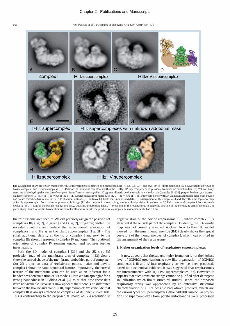
the respirasome architecture. We can precisely assign the positions ofcomplexes III2 (Fig. 2J, in green) and I (Fig. 2J, in yellow) within therevealed structure and deduce the same overall association ofcomplexes I and III2 as in the plant supercomplex (Fig. 2H). Thesmall additional density at the tip of complex I and next to thecomplex III2 should represent a complex IV monomer. The rotationalorientation of complex IV remains unclear and requires furtherinvestigation.
Both the 3D model of complex I [26] and the 2D cryo-EMprojection map of the membrane arm of complex I [32] clearlyshow the curved shape of themembrane-embedded part of complex I.Our 2D projection data of Arabidopsis, potato, maize and bovinecomplex I show the same structural feature. Importantly, the curvedfeature of the membrane arm can be used as an indicator for ahandedness determination of 3D models. Here we can apologize for awrong handedness in Dudkina et al. [6], as at that time these datawere not available. Because it now appears that there is no differencebetween the bovine and plant I+III2 supercomplex, we conclude thatcomplex III is always attached to complex I at its inner curved side.This is contradictory to the proposed 3D model at 32 Å resolution in
negative stain of the bovine respirasome [36], where complex III isattached at the outside part of the complex I. Evidently, the 3D densitymap was not correctly assigned. A closer look to their 3D modelviewed from the inner membrane side (IMS) clearly shows the typicalcurvature of the membrane part of complex I, which was omitted inthe assignment of the respirasome.
3. Higher organization levels of respiratory supercomplexes
It now appears that the supercomplex formation is not the highestlevel of OXPHOS organization. A row-like organization of OXPHOScomplexes I, III and IV into respiratory strings has been proposed,based on biochemical evidence. It was suggested that respirasomesare interconnected with III2+IV4 supercomplexes [37]. However, itappears that such transient strings cannot be purified after detergentsolubilization which limits structural studies. Hence, the proposedrespiratory string was approached by an extensive structuralcharacterization of all its possible breakdown products, which arethe various types of supercomplexes. About 400,000molecular projec-tions of supercomplexes from potato mitochondria were processed
Fig. 2. Examples of EM projection maps of OXPHOS supercomplexes obtained by negative staining (A, B, C, E, F, G, H) and cryo-EM (I, J) plus modelling. (A–C) Averaged side views ofbovine complex I and its supercomplexes. (D) Positions of individual complexes within the I+III2+IV supercomplex or respirasome from bovine mitochondria [18]. Yellow: X-raystructure of the hydrophilic domain of complex I from Thermus thermophilus [30], green: dimeric bovine cytochrome c reductase (complex III) [52], purple: bovine cytochrome coxidase (complex IV) [53]. (E) Top-view of the I+III2 supercomplex from maize [28]. (F, G) Top-views of I+III2 supercomplexes with an unknown additional mass from bovineand potato mitochondria, respectively (N.V. Dudkina, R. Kouřil, J.B. Bultema, E.J. Boekema, unpublished data). (H) Assignment of the complexes I and III2 within the top-view mapof I+III2 supercomplex from maize, as presented in image (E); the complex III dimer is in green in a tilted position; in yellow the 3D EM structure of complex I from Yarrowialipolytica [26]. (I) Map of the bovine respirasome (N.V. Dudkina, unpublished data). (J) Modelling of the respirasome. In beige the position of the membrane arm of complex I, ingreen X-ray structure of the bovine dimeric complex III and in purple the position of a complex IV monomer. Scale bar, 10 nm.
666 N.V. Dudkina et al. / Biochimica et Biophysica Acta 1797 (2010) 664–670
Chapter 2 - Publications and Manuscripts
29
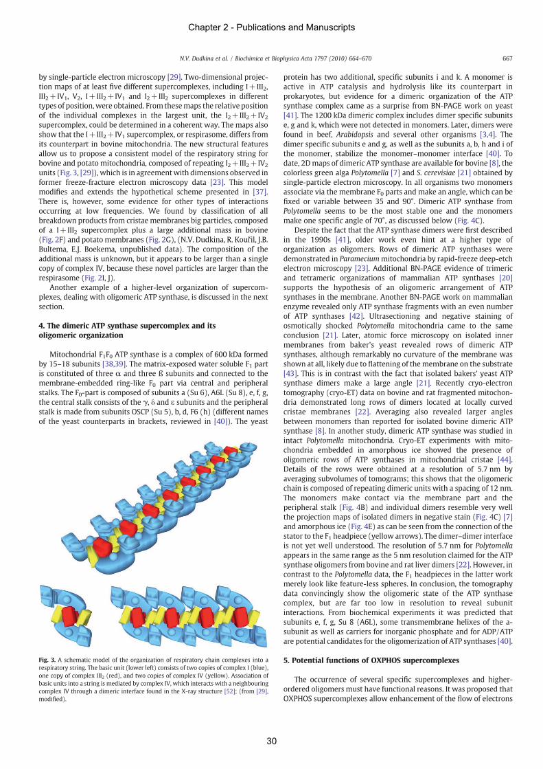
by single-particle electron microscopy [29]. Two-dimensional projec-tion maps of at least five different supercomplexes, including I+III2,III2+IV1, V2, I+III2+IV1 and I2+III2 supercomplexes in differenttypes of position,were obtained. From thesemaps the relative positionof the individual complexes in the largest unit, the I2+III2+IV2
supercomplex, could be determined in a coherent way. The maps alsoshow that the I+III2+IV1 supercomplex, or respirasome, differs fromits counterpart in bovine mitochondria. The new structural featuresallow us to propose a consistent model of the respiratory string forbovine and potato mitochondria, composed of repeating I2+III2+IV2
units (Fig. 3, [29]), which is in agreementwith dimensions observed informer freeze-fracture electron microscopy data [23]. This modelmodifies and extends the hypothetical scheme presented in [37].There is, however, some evidence for other types of interactionsoccurring at low frequencies. We found by classification of allbreakdown products from cristae membranes big particles, composedof a I+III2 supercomplex plus a large additional mass in bovine(Fig. 2F) and potato membranes (Fig. 2G), (N.V. Dudkina, R. Kouřil, J.B.Bultema, E.J. Boekema, unpublished data). The composition of theadditional mass is unknown, but it appears to be larger than a singlecopy of complex IV, because these novel particles are larger than therespirasome (Fig. 2I, J).
Another example of a higher-level organization of supercom-plexes, dealing with oligomeric ATP synthase, is discussed in the nextsection.
4. The dimeric ATP synthase supercomplex and itsoligomeric organization
Mitochondrial F1F0 ATP synthase is a complex of 600 kDa formedby 15–18 subunits [38,39]. The matrix-exposed water soluble F1 partis constituted of three α and three ß subunits and connected to themembrane-embedded ring-like F0 part via central and peripheralstalks. The F0-part is composed of subunits a (Su 6), A6L (Su 8), e, f, g,the central stalk consists of the γ, δ and ε subunits and the peripheralstalk is made from subunits OSCP (Su 5), b, d, F6 (h) (different namesof the yeast counterparts in brackets, reviewed in [40]). The yeast
protein has two additional, specific subunits i and k. A monomer isactive in ATP catalysis and hydrolysis like its counterpart inprokaryotes, but evidence for a dimeric organization of the ATPsynthase complex came as a surprise from BN-PAGE work on yeast[41]. The 1200 kDa dimeric complex includes dimer specific subunitse, g and k, which were not detected in monomers. Later, dimers werefound in beef, Arabidopsis and several other organisms [3,4]. Thedimer specific subunits e and g, as well as the subunits a, b, h and i ofthe monomer, stabilize the monomer–monomer interface [40]. Todate, 2Dmaps of dimeric ATP synthase are available for bovine [8], thecolorless green alga Polytomella [7] and S. cerevisiae [21] obtained bysingle-particle electron microscopy. In all organisms two monomersassociate via the membrane F0 parts and make an angle, which can befixed or variable between 35 and 90°. Dimeric ATP synthase fromPolytomella seems to be the most stable one and the monomersmake one specific angle of 70°, as discussed below (Fig. 4C).
Despite the fact that the ATP synthase dimers were first describedin the 1990s [41], older work even hint at a higher type oforganization as oligomers. Rows of dimeric ATP synthases weredemonstrated in Parameciummitochondria by rapid-freeze deep-etchelectron microscopy [23]. Additional BN-PAGE evidence of trimericand tetrameric organizations of mammalian ATP synthases [20]supports the hypothesis of an oligomeric arrangement of ATPsynthases in the membrane. Another BN-PAGE work on mammalianenzyme revealed only ATP synthase fragments with an even numberof ATP synthases [42]. Ultrasectioning and negative staining ofosmotically shocked Polytomella mitochondria came to the sameconclusion [21]. Later, atomic force microscopy on isolated innermembranes from baker's yeast revealed rows of dimeric ATPsynthases, although remarkably no curvature of the membrane wasshown at all, likely due to flattening of themembrane on the substrate[43]. This is in contrast with the fact that isolated bakers' yeast ATPsynthase dimers make a large angle [21]. Recently cryo-electrontomography (cryo-ET) data on bovine and rat fragmented mitochon-dria demonstrated long rows of dimers located at locally curvedcristae membranes [22]. Averaging also revealed larger anglesbetween monomers than reported for isolated bovine dimeric ATPsynthase [8]. In another study, dimeric ATP synthase was studied inintact Polytomella mitochondria. Cryo-ET experiments with mito-chondria embedded in amorphous ice showed the presence ofoligomeric rows of ATP synthases in mitochondrial cristae [44].Details of the rows were obtained at a resolution of 5.7 nm byaveraging subvolumes of tomograms; this shows that the oligomericchain is composed of repeating dimeric units with a spacing of 12 nm.The monomers make contact via the membrane part and theperipheral stalk (Fig. 4B) and individual dimers resemble very wellthe projection maps of isolated dimers in negative stain (Fig. 4C) [7]and amorphous ice (Fig. 4E) as can be seen from the connection of thestator to the F1 headpiece (yellow arrows). The dimer–dimer interfaceis not yet well understood. The resolution of 5.7 nm for Polytomellaappears in the same range as the 5 nm resolution claimed for the ATPsynthase oligomers from bovine and rat liver dimers [22]. However, incontrast to the Polytomella data, the F1 headpieces in the latter workmerely look like feature-less spheres. In conclusion, the tomographydata convincingly show the oligomeric state of the ATP synthasecomplex, but are far too low in resolution to reveal subunitinteractions. From biochemical experiments it was predicted thatsubunits e, f, g, Su 8 (A6L), some transmembrane helixes of the a-subunit as well as carriers for inorganic phosphate and for ADP/ATPare potential candidates for the oligomerization of ATP synthases [40].
5. Potential functions of OXPHOS supercomplexes
The occurrence of several specific supercomplexes and higher-ordered oligomers must have functional reasons. It was proposed thatOXPHOS supercomplexes allow enhancement of the flow of electrons
Fig. 3. A schematic model of the organization of respiratory chain complexes into arespiratory string. The basic unit (lower left) consists of two copies of complex I (blue),one copy of complex III2 (red), and two copies of complex IV (yellow). Association ofbasic units into a string is mediated by complex IV, which interacts with a neighbouringcomplex IV through a dimeric interface found in the X-ray structure [52]; (from [29],modified).
667N.V. Dudkina et al. / Biochimica et Biophysica Acta 1797 (2010) 664–670
Chapter 2 - Publications and Manuscripts
30
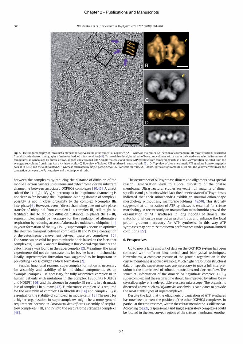
between the complexes by reducing the distance of diffusion of themobile electron carriers ubiquinone and cytochrome c or by substratechanneling between associated OXPHOS complexes [10,45]. A directrole of the I+III2(+IV1–2) supercomplex in ubiquinone-channeling isnot clear so far, because the ubiquinone-binding domain of complex Ipossibly is not in close proximity to the complex I–complex III2interphase [6]. However, even if direct channeling does not take place,transfer of ubiquinol from complex I to complex III2 still might befacilitated due to reduced diffusion distances. In plants the I+III2supercomplex might be necessary for the regulation of alternativerespiration by reducing access of alternative oxidase to ubiquinol [4].In yeast formation of the III2+IV1–2 supercomplex seems to optimizethe electron transport between complexes III and IV by a contractionof the cytochrome c movement between these two complexes [10].The same can be valid for potato mitochondria based on the facts thatcomplexes I, III and IV are rate limiting in flux control experiments andcytochrome cwas found in the supercomplex [2]. Meantime, the sameexperiments did not demonstrate this for bovine heart mitochondria.Finally, supercomplex formation was suggested to be important inpreventing excess oxygen radical formation [2].
Besides functional reasons, supercomplex formation is necessaryfor assembly and stability of its individual components. As anexample, complex I is necessary for fully assembled complex III inhuman patients with mutations in the complex I subunits NDUFS2and NDUFS4 [46] and the absence in complex III results in a dramaticloss of complex I in humans [47]. Furthermore, complex IV is requiredfor the assembly of complex I in fibroblasts [14] and complex III2 isessential for the stability of complex I in mouse cells [13]. The need fora higher organization in supercomplexes might be a more generalrequirement because in Paracoccus denitrificans assembly of respira-tory complexes I, III, and IV into the respirasome stabilizes complex I[48].
The occurrence of ATP synthase dimers and oligomers has a specialreason. Dimerization leads to a local curvature of the cristaemembrane. Ultrastructural studies on yeast null mutants of dimerspecific e and g subunits which lack the dimeric state of ATP synthasesindicated that their mitochondria exhibit an unusual onion-shapemorphology without any membrane foldings [49,50]. This stronglysuggests that dimerization of ATP synthases is essential for cristaemorphology. A recent study on mammalian mitochondria proved theorganization of ATP synthases in long ribbons of dimers. Themitochondrial cristae may act as proton traps and enhance the localproton gradient necessary for ATP synthesis. In this way ATPsynthases may optimize their own performance under proton-limitedconditions [22].
6. Prospectives
Up to now a large amount of data on the OXPHOS system has beencollected with different biochemical and biophysical techniques.Nevertheless, a complete picture of the protein organization in thecristaemembrane is not yet available.Much higher resolution structuraldata on specific supercomplexes are necessary to give a full interpre-tation at the atomic level of subunit interactions and electron flow. Thestructural information of the dimeric ATP synthase complex, I+III2supercomplex and the respirasome should be improved by either X-raycrystallography or single-particle electron microscopy. The organismsdiscussed above, such as Polytomella, are obvious candidates to providethe most stable types of supercomplexes.
Despite the fact that the oligomeric organization of ATP synthaseshas now been proven, the position of the other OXPHOS complexes, inparticular the respirasomes,within the cristaemembrane is still unclear.According to [22], respirasomes and single respiratory complexes couldbe located in the less curved regions of the cristae membrane. Another
Fig. 4. Electron tomography of Polytomella mitochondria reveals the arrangement of oligomeric ATP synthase molecules. (A) Section of a tomogram (3D reconstruction) calculatedfrom dual-axis electron tomography of an ice-embeddedmitochondrion [44]. To reveal fine detail, hundreds of boxed subvolumes with a size as indicated were selected from severaltomograms, as symbolized by purple arrows, aligned and averaged. (B) A single molecule of dimeric ATP synthase from tomography data in a side-view position, selected from theaveraged subvolume from image A at a 4× larger scale. (C) Side-view of isolated ATP synthase in negative stain [7]. (D) Top-view of the same dimeric ATP synthase from tomographydata as in B. (E) Top-view of isolated ATP synthase calculated by single-particle cryo-EM. Bar scale for frame A, 100 nm. Bar scale for frames B–E, 10 nm. The yellow arrows mark theconnection between the F1 headpiece and the peripheral stalk.
668 N.V. Dudkina et al. / Biochimica et Biophysica Acta 1797 (2010) 664–670
Chapter 2 - Publications and Manuscripts
31
possibility would be a localization between the rows of ATP synthases.However, available data suggest that likely the amount of respiratorychain complexes is very limited in comparison to ATP synthases and it isdifficult to detect them at this resolution. A breakthrough is expected byfurther application of cryo-electron tomography, as discussed for ATPsynthase oligomers (Fig. 4). Electron tomography is an emergingtechnique, but still limited in resolution. In addition to 3D averaging andclassification of individual subvolumes improvement in resolution isexpected in the near future by perfecting cryo-electron microscopyhardware and software, and implementing the newgeneration of directelectron detectors, replacing the currently used inefficient slow-scanCCD cameras.
Finally, our understanding of supercomplex organization will alsoincrease by studying the structural aspects of the many naturallyoccurring mutants of the OXPHOS system. Since the first report of acomplex I deficit related to a human mitochondrial disorder in 1988,many other mitochondrial diseases have been associated to structuraland functional defects at the level of this enzyme complex I (see forinstance [46,51]).
Acknowledgements
We thank Mr. Jelle Bultema for discussion. This work wassupported by the Netherlands organization of scientific research(NWO) and by the Deutsche Forschungsgemeinschaft (grant Br 1829-8/1).
References
[1] C.R. Hackenbrock, B. Chazotte, S.S. Gupte, The random collision model and acritical assessment of diffusion and collision in mitochondrial electron transport, J.Bioenerg. Biomembr. 18 (1986) 331–368.
[2] G. Lenaz, M.L. Genova, Structural and functional organization of the mitochondrialrespiratory chain: a dynamic super-assembly, Int. J. Biochem. Cell Biol. 41 (2009)1750–1772.
[3] H. Schägger, K. Pfeiffer, Supercomplexes in the respiratory chains of yeast andmammalian mitochondria, EMBO J. 19 (2000) 1777–1783.
[4] H. Eubel, L. Jänsch, H.-P. Braun, New insights into the respiratory chain of plantmitochondria supercomplexes and a unique compositions of complex II, PlantPhysiol. 133 (2003) 274–286.
[5] H. Eubel, J. Heinemeyer, S. Sunderhaus, H.P. Braun, Respiratory chain super-complexes in plant mitochondria, Plant Physiol. Biochem. 42 (2004) 937–942.
[6] N.V. Dudkina, H. Eubel, W. Keegstra, E.J. Boekema, H.-P. Braun, Structure of amitochondrial supercomplex formed by respiratory-chain complexes I and III,Proc. Natl. Acad. Sci. USA 102 (2005) 3225–3229.
[7] N.V. Dudkina, J. Heinemeyer, W. Keegstra, E.J. Boekema, H.P. Braun, Structure ofdimeric ATP synthase from mitochondria: an angular association of monomersinduces the strong curvature of the inner membrane, FEBS Lett. 579 (2005)5769–5772.
[8] F. Minauro-Sanmiguel, S. Wilkens, J.J. García, Structure of dimeric mitochondrialATP synthase: novel F0 bridging features and the structural basis of mitochondrialcristae biogenesis, Proc. Natl. Acad. Sci. USA 102 (2005) 12356–12358.
[9] E. Schäfer, H. Seelert, N.H. Reifschneider, F. Krause, N.A. Dencher, J. Vonck,Architecture of activemammalian respiratory chain supercomplexes, J. Biol. Chem.281 (2006) 15370–15375.
[10] J. Heinemeyer, H.P. Braun, E.J. Boekema, R. Kouřil, A structural model of thecytochrome C reductase/oxidase supercomplex from yeast mitochondria, J. Biol.Chem. 282 (2007) 12240–12248.
[11] H. Boumans, L.A. Grivell, J.A. Berden, The respiratory chain in yeast behaves as asingle functional unit, J. Biol. Chem. 273 (1998) 4872–4877.
[12] C. Bianchi, M.L. Genova, G.P. Castelli, G. Lenaz, The mitochondrial respiratory chainis partially organized in a supercomplex assembly: kinetic evidence using fluxcontrol analysis, J. Biol. Chem. 279 (2004) 36562–36569.
[13] R. Acín-Pérez, M. Bayona-Bafaluy, P. Fernández-Silva, R. Moreno-Loshuertos, A.Pérez-Martos, C. Bruno, C. Moraes, J. Enríquez, Respiratory complex III is requiredto maintain complex I in mammalian mitochondria, Mol. Cell 13 (2004) 805–815.
[14] F. Diaz, H. Fukui, S. Garcia, C.T. Moraes, Cytochrome c oxidase is required for theassembly/stability of respiratory complex I in mouse fibroblasts, Mol. Cell Biol. 26(2006) 4872–4881.
[15] M. Zhang, E. Mileykovskaya, W. Dowhan, Gluing the respiratory chain together.Cardiolipin is required for supercomplex formation in the inner mitochondrialmembrane, J. Biol. Chem. 277 (2002) 43553–43556.
[16] K. Pfeiffer, V. Gohil, R.A. Stuart, C. Hunte, U. Brandt, M.L. Greenberg, H. Schägger,Cardiolipin stabilizes respiratory chain supercomplexes, J. Biol. Chem. 278 (2003)52873–52880.
[17] D.L. Nelson, M.M. Cox, Lehninger Principles of Biochemistry, Fifth editionW.H.Freeman, New York, 2008.
[18] N.V. Dudkina, S. Sunderhaus, E.J. Boekema, H.P. Braun, The higher level oforganization of the oxidative phosphorylation system: mitochondrial super-complexes, J. Bioenerg. Biomembr. 40 (2008) 419–424.
[19] R. Acín-Pérez, P. Fernández-Silva, M.L. Peleato, A. Pérez-Martos, J.A. Enriquez,Respiratory active mitochondrial supercomplexes, Mol. Cell 32 (2008) 529–539.
[20] F. Krause, N.H. Reifschneider, S. Goto, N.A. Dencher, Active oligomeric ATPsynthases in mammalian mitochondria, Biochem. Biophys. Res. Commun. 329(2005) 583–590.
[21] N.V. Dudkina, S. Sunderhaus, H.P. Braun, E.J. Boekema, Characterization of dimericATP synthase and cristae membrane ultrastructure from Saccharomyces and Po-lytomella mitochondria, FEBS Lett. 580 (2006) 3427–3432.
[22] M. Strauss, G. Hofhaus, R.R. Schröder, W. Kühlbrandt, Dimer ribbons of ATPsynthase shape the inner mitochondrial membrane, EMBO J. 27 (2008)1154–1160.
[23] R.D. Allen, C.C. Schroeder, A.K. Fok, An investigation of mitochondrial inner mem-branes by rapid-freeze deep-etch techniques, J. Cell Biol. 108 (1989) 2233–2240.
[24] P.R. Rich, The molecular machinery of Keilin's respiratory chain, Biochem. Soc.Trans. 31 (2003) 1095–1105.
[25] V. Guenebaut, R. Vincentelli, D. Mills, H. Weiss, K.R. Leonard, Three-dimensionalstructure of NADH-dehydrogenase fromNeurospora crassa by electronmicroscopyand conical tilt reconstruction, J. Mol. Biol. 265 (1997) 409–418.
[26] M. Radermacher, T. Ruiz, T. Clason, S. Benjamin, U. Brandt, V. Zickermann, Thethree-dimensional structure of complex I from Yarrowia lipolytica: a highlydynamic enzyme, J. Struct. Biol. 154 (2006) 269–279.
[27] S. Sunderhaus, N.V. Dudkina, L. Jänsch, J. Klodmann, J. Heinemeyer, M. Perales, E.Zabaleta, E.J. Boekema, H.-P. Braun, Carbonic anhydrase subunits form a matrix-exposed domain attached to the membrane arm of mitochondrial complex I inplants, J. Biol. Chem. 281 (2006) 6482–6488.
[28] K. Peters, N.V. Dudkina, L. Jänsch, H.P. Braun, E.J. Boekema, A structuralinvestigation of complex I and I+III2 supercomplex from Zea mays at 11–13 Åresolution: assignment of the carbonic anhydrase domain and evidence forstructural heterogeneity within complex I, Biochim. Biophys. Acta 1777 (2008)84–93.
[29] J.B. Bultema, H.P. Braun, E.J. Boekema, R. Kouřil, Megacomplex organization of theoxidative phosphorylation system by structural analysis of respiratory super-complexes from potato, Biochim. Biophys. Acta 1767 (2009) 60–67.
[30] L.A. Sazanov, P. Hinchliffe, Structure of the hydrophilic domain of respiratorycomplex I from Thermus thermophilus, Science 311 (2006) 1430–1436.
[31] U. Brandt, Proton-translocation by membrane-bound NADH: ubiquinone-oxidoreductase (complex I) through redox-gated ligand conduction, Biochim.Biophys. Acta 1318 (1997) 79–91.
[32] E.A. Baranova, P.J. Holt, L.A. Sazanov, Projection structure of the membranedomain of Escherichia coli respiratory complex I at 8 Å resolution, J. Mol. Biol. 366(2007) 140–154.
[33] H. Eubel, J. Heinemeyer, H.P. Braun, Identification and characterization ofrespirasomes in potato mitochondria, Plant Physiol. 134 (2004) 1450–1459.
[34] F. Krause, N.H. Reifschneider, D. Vocke, H. Seelert, S. Rexroth, N.A. Dencher,“Respirasome”-like supercomplexes in green leaf mitochondria of spinach, J. Biol.Chem. 279 (2004) 48369–48375.
[35] N.V. Dudkina, J. Heinemeyer, S. Sunderhaus, E.J. Boekema, H.P. Braun, Respiratorychain supercomplexes in the plant mitochondrial membrane, Trends Plant Sci. 11(2006) 232–240.
[36] E. Schäfer, N.A. Dencher, J. Vonck, D.N. Parcej, Three-dimensional structure of therespiratory chain supercomplex I1III2IV1 from bovine heart mitochondria,Biochemistry 46 (2007) 2579–2585.
[37] I. Wittig, R. Carrozzo, F.M. Santorelli, H. Schägger, Supercomplexes and sub-complexes of mitochondrial oxidative phosphorylation, Biochim. Biophys. Acta1757 (2006) 1066–1072.
[38] J.P. Abrahams, A.G. Leslie, R. Lutter, J.E. Walker, Structure at 2.8 Å resolution of F1-ATPase from bovine heart mitochondria, Nature 370 (1994) 621–628.
[39] D. Stock, C. Gibbons, I. Arechaga, A.G. Leslie, J.E. Walker, The rotary mechanism ofATP synthase, Curr. Opin. Struct. Biol. 10 (2000) 672–679.
[40] I. Wittig, H. Schägger, Structural organization of mitochondrial ATP synthase,Biochim. Biophys. Acta 1777 (2008) 592–598.
[41] I. Arnold, K. Pfeiffer, W. Neupert, R.A. Stuart, H. Schägger, Yeast mitochondrialF1F0-ATP synthase exists as a dimer: identification of three dimer-specificsubunits, EMBO J. 17 (1998) 7170–7178.
[42] I. Wittig, H. Schägger, Advantages and limitations of clear-native PAGE,Proteomics 5 (2005) 4338–4346.
[43] N. Buzhynskyy, P. Sens, V. Prima, J.N. Sturgis, S. Scheuring, Rows of ATP synthasedimers in native mitochondrial inner membranes, Biophys. J. 93 (2007) 2870–2876.
[44] N.V. Dudkina, G.T. Oostergetel, D. Lewejohann, H.P. Braun, E.J. Boekema, Row-likeorganization of ATP synthase in intact mitochondria determined by cryo-electrontomography, Biochim. Biophys. Acta. 1797 (2010) 272–277.
[45] H. Schägger, Respiratory chain supercomplexes, IUBMB Life 52 (2001) 119–128.[46] C. Ugalde, R.J.R.J. Janssen, L.P. van den Heuvel, J.A. Smeitink, L.G.J. Nijtmans,
Differences in assembly or stability of complex I and other mitochondrialOXPHOS complexes in inherited complex I deficiency, Hum. Mol. Genet. 13(2004) 659–667.
[47] E.L. Blakely, A.L. Mitchell, N. Fisher, B. Meunier, L.G. Nijtmans, A.M. Schaefer, M.J.Jackson, D.M. Turnbull, R.W. Taylor, A mitochondrial cytochrome b mutationcausing severe respiration chain enzyme deficiency in humans and yeast, FEBS J.14 (2005) 3583–3592.
[48] A. Stroh, O. Anderka, K. Pfeiffer, T. Yagi, M. Finel, B. Ludwig, H. Schägger, Assemblyof respiratory complexes I, III, and IV into NADH oxidase supercomplex stabilizescomplex I in Paracoccus denitrificans, J. Biol. Chem. 279 (2004) 5000–5007.
669N.V. Dudkina et al. / Biochimica et Biophysica Acta 1797 (2010) 664–670
Chapter 2 - Publications and Manuscripts
32
[49] P. Paumard, J. Vaillier, B. Coulary, J. Schaeffer, V. Soubannier, D.M.Mueller, D. Brethes, J.P. di Rago, J. Velours, The ATP synthase is involvedin generating mitochondrial cristae morphology, EMBO J. 21 (2002)221–230.
[50] M.F. Giraud, P. Paumard, V. Soubannier, J. Vaillier, G. Arselin, B. Salin, J. Schaeffer, D.Brethes, P. di Rago, J. Velours, Is there a relationship between the supramolecularorganization of the mitochondrial ATP synthase and the formation of cristae?Biochim. Biophys. Acta 1555 (2002) 174–180.
[51] U. Brandt, Energy converting NADH: quinone oxidoreductase (complex I), Annu.Rev. Biochem. 75 (2006) 69–92.
[52] S. Iwata, J.W. Lee, K. Okada, J.K. Lee, M. Iwata, B. Rasmussen, T.A. Link, S.Ramaswamy, B.K. Jap, Complete structure of the 11-subunit bovine mitochondrialcytochrome bc1 complex, Science 281 (1998) 64–71.
[53] T. Tsukihara, H. Aoyama, E. Yamashita, T. Tomizaki, H. Yamaguchi, K. Shinzawa-Itoh, R. Nakashima, R. Yaono, S. Yoshikawa, The whole structure of the 13-subunitoxidized cytochrome c oxidase at 2.8 Å, Science 272 (1996) 1136–1144.
670 N.V. Dudkina et al. / Biochimica et Biophysica Acta 1797 (2010) 664–670
Chapter 2 - Publications and Manuscripts
33
Comparative Analyses of Protein Complexes by Blue Native DIGE
Katrin Peters1 and Hans-Peter Braun1*
1 Institute for Plant Genetics, Leibniz Universität Hannover, Herrenhäuser Str. 2, D-30419
Hannover, Germany
* Author for correspondance, [email protected]
Summary
Classically, DIGE is carried out on the basis of two-dimensional (2D) IEF / SDS PAGE. This
allows comparative analyses of large protein sets. However, 2D IEF / SDS PAGE only poorly
resolves hydrophobic proteins and is not compatible with native protein characterizations.
Blue native PAGE represents a powerful alternative. Combined with CyDye labeling, blue
native DIGE offers several useful applications like quantitative comparison of protein
complexes of related protein fractions. Here we present a protocol for fluorophore labeling of
native protein fractions for separation by blue native PAGE.
Key words: Blue native PAGE, DIGE labeling, protein complexes, membrane proteins,
mitochondria
Abbreviations: 2D: two-dimensional, BN PAGE: blue native polyacrylamide gel
electrophoresis, BN / SDS PAGE: blue native / sodium dodecyl sulfate polyacrylamide gel
electrophoresis, DIGE: difference gel electrophoresis, DMF: dimethylformamide, IEF / SDS
PAGE: isoelectric focusing / sodium dodecyl sulfate polyacrylamide gel electrophoresis, pI:
isoelectric point, PMSF: phenylmethylsulfonyl fluoride.
Chapter 2 - Publications and Manuscripts
34
1. Introduction
Fluorophore-based difference gel electrophoresis (DIGE) represents an ideal system for
comparative proteomics. Different protein fractions can be co-eletrophoresed on a single gel,
thereby eliminating gel-to-gel variations. If evaluated with special software tools, fluorophore
detection allows exact relative quantification of proteins present in the protein fractions to be
compared.
Classically, DIGE is carried out based on the two-dimensional (2D) IEF / SDS PAGE system
(1). Indeed, using this experimental system, large protein sets are efficiently resolved.
However, 2D IEF / SDS PAGE also has limitations. For instance, hydrophobic proteins are
not well resolved during the IEF gel dimension and proteins with very basic pIs often get lost.
Additionally, 2D IEF / SDS PAGE is not compatible with native enzyme characterizations.
Two-dimenional blue native (BN) / SDS PAGE represents an alternative gel system which
allows overcoming these limitations. Hydrophobic as well as basic proteins are easily
resolved. If applied as a one-dimensional system, BN PAGE is even compatible with in-gel
enzyme activity stainings.
BN PAGE was first published by Hermann Schägger (2). The principle idea was to use
Coomassie Blue not only for protein staining after gel electrophoresis, but also for the
introduction of negative charges into proteins by incubating protein fractions with this
compound before gel electrophoresis. Coomassie Blue is an anionic molecule but in contrast
to SDS does not denature proteins. Furthermore, protein complexes remain stable. If
combined with low-percentage polyacrylamide gels, protein complexes can be resolved by
BN PAGE. In case sample preparation takes place in the presence of mild non-ionic
detergents, even hydrophobic membrane-bound protein complexes can easily be separated. As
with IEF / SDS PAGE, strips of blue native gels can be horizontally transferred onto a second
gel dimension which is carried out in the presence of SDS. Using this experimental set up,
protein complexes are separated into their subunits which form vertical rows of spots on the
resulting 2D gels.
BN PAGE and 2D BN / SDS PAGE can be combined with the DIGE technology (3). BN
DIGE allows systematic and quantitative comparison of protein complexes of related protein
fractions, structural investigation of protein complexes, assignment of protein complexes to
subcellular fractions like organelles, electrophoretic mapping of isoforms of subunits of
protein complexes with respect to a larger proteome and topological investigations (4). So far,
BN DIGE has only been applied in a few studies (4, 5, 6, 7, 8, 9). Indeed, the potential of BN
Chapter 2 - Publications and Manuscripts
35
DIGE was only very recently recognized. Here we present a BN DIGE protocol suitable for
the comparative analyses of protein complexes of biological fractions.
2. Materials
Buffers, solutions and reagents are given in the order of usage according to the methods
protocol. Prepare all solutions freshly using analytical grade chemicals in combination with
pure de-ionized water.
2.1. Preparation of a BN gel
1. Acrylamide solution: 40%, acryl / bisacryl = 32 / 1 (AppliChem, Darmstadt, Germany)
2. Gel buffer BN (6x): 1.5 M amino caproic acid, 150 mM BisTris, pH 7.0 (adjust at 4°C)
3. N,N,N',N'-tetramethylethylenediamine (TEMED), 99% (Sigma, St. Louis, Missouri,
USA)
4. Ammonium persulfate solution (APS), 10% (w/v) Ammonium persulfate
5. Cathode buffer BN (5x): 250 mM Tricine, 75 mM BisTris, 0.1% (w/v) Coomassie G 250
(e.g. Merck, Darmstadt, Germany), pH 7.0 (adust at 4°C)
6. Anode buffer BN (6x): 300 mM BisTris, pH 7.0 (adjust at 4°C)
2.2. Sample preparation for BN DIGE
1. Solubilization buffer, pH 7.4, with digitonin: 30 mM HEPES, 150 mM potassium acetate,
10% (v/v) glycerol, pH 7.4, supplemented with 5% digitonin (see Note 1). Solution
should be briefly boiled for dissolving digitonin. Buffer is stored at 4°C.
2. Solubilization buffer, pH 10: 30 mM HEPES, 150 mM potassium acetate, 10% (v/v)
glycerol, pH 10 (see Note 2).
3. CyDyeTM fluor minimal labeling reagents. Cy2TM, Cy3TM and Cy5TM (GE Healthcare,
Munich, Germany). The fluorophores (400 pmol) are diluted in DMF according to the
manufacturers instructions. Diluted CyDyes are stored at -20°C and should be used within
3 months (see Note 3).
4. Lysine stock solution: 10 mM lysine, stored at 4°C.
Chapter 2 - Publications and Manuscripts
36
5. Blue loading buffer: 750 mM aminocaproic acid, 5% (w/v) Coomassie 250 G, stored at
4°C.
2.3. SDS PAGE for second gel dimension
1. Acrylamide solution: 40%, acryl / bisacryl = 32 / 1 (AppliChem, Darmstadt, Germany)
2. N,N,N',N'-tetramethylethylenediamine (TEMED), 99% (Sigma, St. Louis, Missouri,
USA)
3. Ammonium persulfate solution (APS), 10% (w/v) Ammonium persulfate
4. Gel buffer SDS: 3 M Tris-HCl, 0.3% (w/v) SDS, pH 8.45
5. Gel buffer BN (6x): see 2.1.
6. Cathode buffer SDS: 0.1 M Tris base, 0.1 M Tricine, 0.1% (w/v) SDS, pH 8.25
7. Anode buffer SDS: 0.2 M Tris-HCl, pH 8.9
8. Overlay solution: 1 M Tris-HCl, 0.1% (w/v) SDS, pH 8.45
9. Denaturation solution: 1.0% (w/v) SDS, 1.0% (v/v) β-mercaptoethanol
10. SDS solution: 10% (w/v) SDS
3. Methods
The following protocol is suitable for the comparison of protein complexes of two related
protein fractions. It is based on the usage of the CyDyeTM fluor labeling reagents (GE
Healthcare, Munich, Germany). Both fractions should contain 100 µg protein and should be
labeled with different fluorophores. Afterwards, the samples are combined and loaded onto a
single blue native gel. The BN gel should be prepared before sample preparation.
3.1. Preparation of a BN gel
Best resolution capacity of BN gels is achieved if the electrophoretic separation distance is
greater than 12 cm. The following instructions refer to the Protean II electrophoresis unit
(BioRad, Richmond, Ca, USA; gel dimensions 0.15 x 16 x 20 cm). However, units from other
manufacturers are of comparable suitability for BN-PAGE, e.g. the Hoefer SE-400 or SE-600
gel systems (GE healthcare, Munich, Germany). Usage of gradient gels is recommended,
Chapter 2 - Publications and Manuscripts
37
because molecular masses of protein complexes can vary between 50 kDa and several
thousand of kDa (see Note 4).
1. Prepare a 4.5% separation gel solution by mixing 2.4 ml of Acrylamide solution with 3.5
ml of Gel buffer BN and 15.1 ml ddH2O.
2. Prepare a 16% separation gel solution by mixing 7.4 ml of Acrylamide solution with 3 ml
of Gel buffer BN, 4.6 ml ddH2O and 3.5 ml glycerol.
3. Transfer the two gel solutions into the two chambers of a gradient former and connect the
gradient former via a hose and a needle with the space in-between two glass plates which
are pre-assembled in a gel casting stand. Gradient gels can either be poured from the top
(16% gel solution has to enter the gel sandwich first) or from the bottom (4.5% gel
solution has to enter first). Pouring the gel from the bottom, an adjustable pump (for
example BioRad EconoPump) is needed.
4. Add TEMED and APS to the two gel solutions (95 µl 10% APS / 9.5 µl TEMED to the
4.5% gel solution, 61 µl APS / 6.1 µl TEMED to the 16% gel solution).
5. Pour the gradient gel, leaving space for the stacking gel, and overlay with ddH2O. The gel
should polymerize in about 60 minutes.
6. Pour off the ddH2O.
7. Prepare the stacking gel solution by mixing 1.5 ml of Acrylamide solution, 2.5 ml of Gel
buffer BN and 11 ml ddH2O.
8. Add 65 µl APS and 6.5 µl TEMED and pour the stacking gel around an inserted comb.
The stacking gel should polymerize within 30 minutes.
9. Prepare 1xAnode and 1xCathode buffers BN by diluting the corresponding stock
solutions.
10. Once the polymerization of the stacking gel is finished, carefully remove the comb.
11. Add Cathode and Anode buffers BN to the upper and lower chambers of the gel unit
respectively. Cool down the unit to 4°C.
3.2. Sample preparation for BN-DIGE
Starting material can either be whole cells or isolated organelles. Fractions should be treated
under mild conditions in order to keep proteins in their native conformation (avoid high salt,
ionic detergents, high temperatures, urea etc.). All steps of the sample preparation should be
Chapter 2 - Publications and Manuscripts
38
carried out at 4°C. The BN gel should be prepared before the sample preparation is started
(see 3.1.).
1. Cell or organelle fractions, including 100 µg protein each, are centrifuged at full speed for
10 min at 4°C using an Eppendorf centrifuge.
2. Sedimented material is resuspended in 10 µL of solubilization buffer, pH 7.4, with
digitonin and incubated for 20 min at 4°C (see Note 5).
3. Fractions are again centrifuged at full speed for 20 min at 4°C using an Eppendorf
centrifuge to remove insoluble material.
4. The supernatants containing solubilised proteins and protein complexes are transferred
into new 1.5mL-Eppendorf vessels and supplemented with 10 µL of solubilization buffer,
pH 10, to adjust the pH value of the protein solutions to about 8.5 (see Note 6).
5. Labeling reaction: For CyDyeTM labeling reactions, each sample is labeled with one
CyDye by the addition of 1 µL of diluted CyDye solution to each protein fraction (see
Note 7). Centrifuge the samples briefly and incubate for 30 min on ice in the dark.
6. Stop labeling reaction: Add 1 µL of lysine stock solution and incubate samples for 10 min
in the dark.
7. Combine the two labeled protein fractions in one Eppendorf vessel.
8. Add 2 µL of Blue loading buffer to the combined protein fraction. Sample now is ready
for loading on a BN gel.
3.3. Blue native PAGE for first gel dimension
1. Load combined CyDye-labeled protein sample (see 3.2.) into a well of a BN gel (see 3.1.).
2. Connect the gel unit to a power supply. Start electrophoresis at constant voltage (100V for
45 minutes) and continue at constant current (15 mA for about 11 hours). Electrophoresis
should be carried out at 4°C and in the dark in order to protect the dyes.
3. (optionally:) The Cathode buffer BN, normally containing Coomassie Blue, may be
replaced after half of the electrophoresis run against a Cathode buffer BN lacking
Coomassie Blue (see Note 8).
Chapter 2 - Publications and Manuscripts
39
3.4. SDS PAGE for second gel dimension
BN PAGE can be combined with a second gel dimension, which is carried out in the presence
of SDS, to further separate the protein complexes into their subunits. All published protocols
for SDS PAGE are suitable for combination with BN PAGE, e.g. the system published by
Laemmli (10). However, the Tricine-SDS PAGE system developed by Schägger (11)
generally gives the best resolution. The following instructions refer to this gel system carried
out in the Protean II gel unit (BioRad, Richmond, Ca, USA). As mentioned above, gel
electrophoresis units of other manufacturers are of equal suitability.
1. Cut out a strip of a BN gel and incubate it in the Denaturation solution for 30 minutes at
room temperature.
2. Wash the strip with ddH2O for 30 to 60 seconds (this step is important because -
mercaptoethanol inhibits polymerisation of acrylamide).
3. Place the strip on a glass plate of an electrophoresis unit at the position of the teeth of a
normal gel comb.
4. Assemble the gel electrophoresis unit by adding 1 mm spacers, the second glass plate and
clamps and transfer all into the gel casting stand (the reduced thickness of the gel of the
second gel dimension (1 mm) in comparison to the strip of the first gel dimension (1.5
mm) avoids sliding down of the gel strip between the glass plates in vertical position).
5. Prepare a 16% separation gel solution by mixing 12.4 ml of Acrylamide solution, 10 ml of
Gel buffer SDS, 7.6 ml ddH2O, 100 µl APS, 10 µl TEMED and pour the solution into the
space in-between the two glass plates below the BN gel strip. Leave space for the sample
gel solution for embedding the BN gel strip. Overlay the separation gel with the Overlay
solution. The gel should polymerize in about 60 minutes (see Note 9).
6. Prepare a 10% sample gel solution by mixing 2.5 ml of Acrylamide solution, 3.4 ml of
Gel buffer BN, 1 ml glycerol, 100 µl of SDS solution, 2.9 ml ddH2O, 83 µl APS and 8.3
µl TEMED.
7. Discard the overlay solution and pour the sample gel embedding the BN gel lane. The
casting stand should be held slightly diagonal to avoid air bubbles captured underneath the
BN gel lane (see Note 10).
8. Add the Cathode and Anode buffers SDS to the upper and lower chambers of the gel unit.
9. Connect the gel unit to a power supply. Carry out electrophoresis at 30 mA per gel over
night. The gel run should take place in the dark to protect the fluorophores.
Chapter 2 - Publications and Manuscripts
40
3.5. Visualization of CyDyeTM -labeled protein
After completion of gel electrophoresis, fluorophors can be detected in 1D BN or 2D BN/SDS
gels using a fluorescence scanner (e.g. the Typhoon fluorescence scanner from GE
Healthcare) (Figure 1). Keep gels in the dark before starting the scanning procedure. Using
the CyeDye fluorophores, gels should be scanned at 50-100-µm resolution at the appropriate
excitation wavelengths (488 nm for Cy2TM, 532 nm for Cy3TM and 633 nm for Cy5TM).
Digital gel images can be visualized using the Image Quant analysis software (GE
Healthcare). Quantification of relative differences of individual protein abundances can be
carried out using specific software such as Delta 2-D (Decodon, Greifswald, Germany) or
DeCyder TM (GE Healthcare).
4. Notes
1. Digitonin is necessary for the solubilization of membrane-bound protein complexes of
cellular or organellar fractions. Other non-ionic detergents (e.g. dodecylmaltoside and
Triton X-100) can be used but should be applied in the presence of modified buffer
systems; see Wittig et al. (12).
2. The pH value of the buffer must be >9. Normally, it is at about pH 10 without
adjustment. Therefore, the buffer can be directly used.
3. Always use CyDyes from the same reaction kit diluted with dimethylformamide
(DMF) of one batch to assure comparative labeling conditions. Consume diluted
CyDyes within three months.
4. If very large protein complexes (>3 MDa) have to be resolved, the acrylamide gradient
gel of the BN gel dimension can be substituted by a 2.5% agarose gel prepared in Gel
buffer BN (13).
5. The resulting detergent-to-protein ratio is 5:1 (w/w).
6. A pH value of about 8.5 is a prerequisite for efficient labeling. The pH value of the
protein solution can be easily controlled using pH test strips.
7. Additionally, a third fraction containing a mixture of both protein fractions (50µg
protein each) can be labeled with a third CyDye as internal standard.
8. Coomassie Blue might quench the fluorescence signal of the CyDyes. If the cathode
buffer is substituted by the same buffer lacking Coomassie Blue after half of the
electrophoresis run, the background of the resulting BN gel is much clearer.
Chapter 2 - Publications and Manuscripts
41
9. Concerning the Tricine-SDS PAGE system for the second gel dimension, Schägger
and von Jagow (11) originally proposed a two-step separation gel consisting of a large
16% phase and a smaller 10% phase (called “spacer gel”). The advantage of this
slightly more complicated gel system is a better resolution of large protein subunits.
The two gel solutions are poured one upon the other (glycerol is added to the 16% gel
solution to avoid mixing with the 10% gel solution, for details see Schägger and von
Jagow (11).
10. The transfer of the strip of BN gel onto a second gel dimension is proposed to be
carried out by embedding the gel lane by the sample gel of the second dimension. This
procedure ensures optimal physical contact between the BN gel lane and the SDS gel
of the second dimension. However, BN gel strips can also be fixed onto a pre-poured
SDS gel using agarose as usually carried out for 2D IEF / SDS PAGE. Physical
contact of the gels might not be optimal, but the time period between the end of the
first and the start of the second electrophoresis is shortened.
Acknowledgements
The authors would like to thank Christina Rode, Institute for Plant Genetics, Leibniz
Universität Hannover, for critical reading of the manuscript.
Chapter 2 - Publications and Manuscripts
42
References
1. O’Farrell P.Z. and Goodman H.M. (1976) Resolution of simian virus 40 proteins in
whole cell extracts by two-dimensional electrophoresis: heterogeneity of the major
capsid protein. Cell. 9: 289-298.
2. Schägger H. and von Jagow G. (1991) Blue native electrophoresis for isolation of
membrane protein complexes in enzymatically active form. Anal. Biochem. 199: 223-
231.
3. Perales M., Eubel H., Heinemeyer J., Colaneri A., Zabaleta E., Braun H.P. (2005)
Disruption of a nuclear gene encoding a mitochondrial gamma carbonic anhydrase
reduces complex I and supercomplex I+III2 levels and alters mitochondrial physiology
in Arabidopsis. J Mol Biol 350: 263-277.
4. Heinemeyer J., Scheibe B., Schmitz U.K., and Braun H.P. (2009) Blue native DIGE as
a tool for comparative analyses of protein complexes. J Prot 72: 539-544.
5. Hedman E., Widén C., Asadi A., Dinnetz I., Schröder W.P., Gustafsson J.A.,
Wikström A.C. (2006) Proteomic identification of glucocorticoid receptor interacting
proteins. Proteomics 6: 3114-3126.
6. Gillardon F., Rist W., Kussmaul L., Vogel J., Berg M., Danzer K., Kraut N., Hengerer
B. (2007) Proteomic and functional alterations in brain mitochondria from Tg2576
mice occur before amyloid plaque deposition. Proteomics 7: 605-616.
7. Reisinger V. and Eichacker L.A. (2007) How to analyze protein complexes by 2D
blue native SDS-PAGE. Proteomics 7: 6-16.
8. Dani D. and Dencher N.A. (2008) Native-DIGE: a new look at the mitochondrial
membrane proteome. Biotechnol J. 3: 817-822.
9. Dani D. Shimokawa I., Komatsu T., Higami Y., Warnken U., Schokraie E., Schnölzer
M., Krause F., Sugawa M.D., Dencher N.A. (2010) Modulation of oxidative
Chapter 2 - Publications and Manuscripts
43
10. Laemmli, U.K. (1970) Cleavage of structural proteins during the assembly of the head
of bacteriophage T4. Nature 227, 680-685.
11. Schägger, H. and von Jagow, G. (1987) Tricine-sodium dodecyl sulfate-
polyacrylamide gel electrophoresis for the separation of proteins in the range from 1 to
100 kDa. Anal. Biochem. 166, 368-379.
12. Wittig I., Braun H.-P., Schägger, H. (2006) Blue-native PAGE. Nature Protocols 1:
418–428.
13. Henderson, N.S., Nijtmans, L.G., Lindsay, J.G., Lamantea, E., Zeviani, M. and Holt,
I.J. (2000) Separation of intact pyruvate dehydrogenase complex using blue native
agarose gel electrophoresis. Electrophoresis 21, 2925-2931.
14. Wittig I., Karas M., Schägger H. (2007) High resolution clear native electrophoresis
for in-gel functional assays and fluorescence studies of membrane protein complexes.
Mol Cell Proteomics 6: 1215–1225.
Chapter 2 - Publications and Manuscripts
44
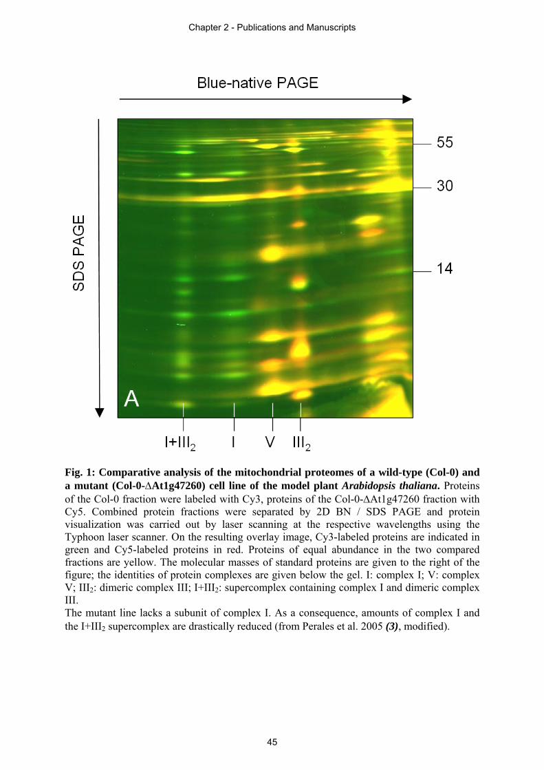
Fig. 1: Comparative analysis of the mitochondrial proteomes of a wild-type (Col-0) and a mutant (Col-0-At1g47260) cell line of the model plant Arabidopsis thaliana. Proteins of the Col-0 fraction were labeled with Cy3, proteins of the Col-0-At1g47260 fraction with Cy5. Combined protein fractions were separated by 2D BN / SDS PAGE and protein visualization was carried out by laser scanning at the respective wavelengths using the Typhoon laser scanner. On the resulting overlay image, Cy3-labeled proteins are indicated in green and Cy5-labeled proteins in red. Proteins of equal abundance in the two compared fractions are yellow. The molecular masses of standard proteins are given to the right of the figure; the identities of protein complexes are given below the gel. I: complex I; V: complex V; III2: dimeric complex III; I+III2: supercomplex containing complex I and dimeric complex III. The mutant line lacks a subunit of complex I. As a consequence, amounts of complex I and the I+III2 supercomplex are drastically reduced (from Perales et al. 2005 (3), modified).
Chapter 2 - Publications and Manuscripts
45
Complex I - complex II ratio strongly differs in various organs of
Arabidopsis thaliana.
Katrin Peters1, Markus Nießen2, Christoph Peterhänsel2, Bettina Späth3, Angela Hölzle3,
Stefan Binder3, Anita Marchfelder3, Hans-Peter Braun1*
1 Institute of Plant Genetics, Faculty of Natural Sciences, Leibniz Universität Hannover,
30419 Hannover, Germany
2 Institute of Botany, Faculty of Natural Sciences, Leibniz Universität Hannover, 30419
Hannover, Germany
3 Molekulare Botanik, Universität Ulm, 89081 Ulm, Germany
* Corresponding author: Tel.: +49 511 7622674; fax: +49 511 7623608
E-mail address: [email protected]
Abbreviations: OXPHOS, oxidative phosphorylation; BN-PAGE, blue native polyacrylamide
gel electrophoresis
Key words: oxidative phosphorylation, respiratory chain, OXPHOS complexes, mitochondria,
stoichiometry
Running title: Ratio of OXPHOS complexes in Arabidopsis
Chapter 2 - Publications and Manuscripts
46
Abstract
In most studies, amounts of the protein complexes of the oxidative phosphorylation
(OXPHOS) system in different organs or tissues are quantified on the basis of isolated
mitochondrial fractions. However, efficiencies of mitochondrial isolations might differ with
respect to tissue type due to varying efficiencies of cell disruption during organelle isolation
procedures or due to tissue-specific properties of organelles. Here we report an
immunological investigation on the ratio of the five OXPHOS complexes in different tissues
of Arabidopsis thaliana which is based on total protein fractions isolated from five
Arabidopsis organs (leaves, stems, flowers, roots and seeds) and from callus. Antibodies were
generated against one surface exposed subunit of each of the five OXPHOS complexes and
used for systematic immunoblotting experiments. Amounts of all complexes are highest in
flowers (likewise with respect to organ fresh weight or total protein content of the flower
fraction). Relative amounts of protein complexes in all other fractions were determined with
respect to their amounts in flowers. Our investigation reveals high relative amounts of
complex I in green organs (leaves and stems) but much lower amounts in non-green organs
(roots, callus tissue). In contrast, complex II only is represented by low relative amounts in
green organs but by significantly higher amounts in non-green organs, especially in seeds. In
fact, the complex I – complex II ratio differs by factor 37 between callus and leaf, indicating
drastic differences in electron entry into the respiratory chain in these two fractions. Results
are confirmed by in gel activity measurements for the protein complexes of the OXPHOS
system and comparative 2D blue native / SDS PAGE analyses using isolated mitochondria.
We suggest that complex I has an especially important role in the context of photosynthesis
which might be due to its indirect involvement in photorespiration and its numerous
enzymatic side activities in plants.
Chapter 2 - Publications and Manuscripts
47
Introduction
Mitochondria represent an important site for ATP production in most eukaryotic cells.
Generation of ATP is carried out by the oxidative phosphorylation (OXPHOS) system, which
consists of five multi-protein complexes (complex I – V) in the inner mitochondrial
membrane as well as cytochrome c and ubiquinone (Hatefi 1985). Complexes I to IV form
part of the electron transport chain (ETC) also termed respiratory chain. This chain catalyzes
NADH oxidation by reduction of oxygen to water. The transport of electrons by complex I, III
and IV is coupled to the translocation of protons from the matrix to the intermembrane space.
The resulting proton gradient is used by the ATP-synthase (complex V) to produce ATP. The
OXPHOS system in plants differs from the one in most mammals because some of its
respiratory chain complexes have several extra protein subunits which introduce additional
functions into these complexes. For example, complex I from plants includes more than 10
extra subunits (Klodmann and Braun 2011; Klodmann et al. 2010). Some of these proteins
resemble known enzymes like gamma-type carbonic anhydrases (γ-CA) or L-galactone-1,4-
lactone dehydrogenase (GLDH).
Furthermore, the plant OXPHOS system is especially multifaceted because it involves
additional ‘alternative’ oxidoreductases. As a consequence, respiratory electron transport is
highly branched in plants. Enzymes catalyzing these alternative pathways are type II
NAD(P)H dehydrogenases and the alternative oxidase (AOX). Former ones can bypass
complex I, whereas AOX bypasses complex III and IV (Rasmusson et al. 2008). The transport
of electrons via these alternative pathways is not coupled with proton translocation across the
inner mitochondrial membrane and therefore not directly involved in respiratory ATP
production. The activity of the alternative electron pathway enzymes therefore results in a
lower ATP production of the respiratory system and thus leads to a decrease of respiratory
energy conservation (Rasmusson and Wallström 2010). This especially is important under
high light conditions due to excess formation of reduction equivalents within chloroplasts (by
photosynthesis) and mitochondria (by glycine to serine conversion in the context of
photorespiration) (Rasmusson et al. 2008). In fact, expression of the genes encoding AOX and
alternative NAD(P)H dehydrogenases is light regulated (Rasmusson and Escobar 2007).
Thus, the alternative respiratory pathways are considered to be the basis for an overflow
protection mechanism for the OXPHOS system which prevents production of reactive oxygen
species (ROS) due to over-reduction of the ETC in the light. Besides the alternative
Chapter 2 - Publications and Manuscripts
48
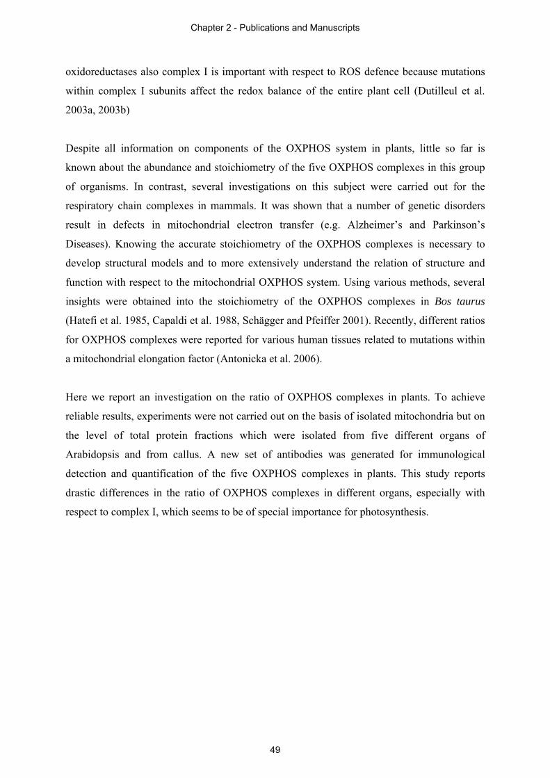
oxidoreductases also complex I is important with respect to ROS defence because mutations
within complex I subunits affect the redox balance of the entire plant cell (Dutilleul et al.
2003a, 2003b)
Despite all information on components of the OXPHOS system in plants, little so far is
known about the abundance and stoichiometry of the five OXPHOS complexes in this group
of organisms. In contrast, several investigations on this subject were carried out for the
respiratory chain complexes in mammals. It was shown that a number of genetic disorders
result in defects in mitochondrial electron transfer (e.g. Alzheimer’s and Parkinson’s
Diseases). Knowing the accurate stoichiometry of the OXPHOS complexes is necessary to
develop structural models and to more extensively understand the relation of structure and
function with respect to the mitochondrial OXPHOS system. Using various methods, several
insights were obtained into the stoichiometry of the OXPHOS complexes in Bos taurus
(Hatefi et al. 1985, Capaldi et al. 1988, Schägger and Pfeiffer 2001). Recently, different ratios
for OXPHOS complexes were reported for various human tissues related to mutations within
a mitochondrial elongation factor (Antonicka et al. 2006).
Here we report an investigation on the ratio of OXPHOS complexes in plants. To achieve
reliable results, experiments were not carried out on the basis of isolated mitochondria but on
the level of total protein fractions which were isolated from five different organs of
Arabidopsis and from callus. A new set of antibodies was generated for immunological
detection and quantification of the five OXPHOS complexes in plants. This study reports
drastic differences in the ratio of OXPHOS complexes in different organs, especially with
respect to complex I, which seems to be of special importance for photosynthesis.
Chapter 2 - Publications and Manuscripts
49
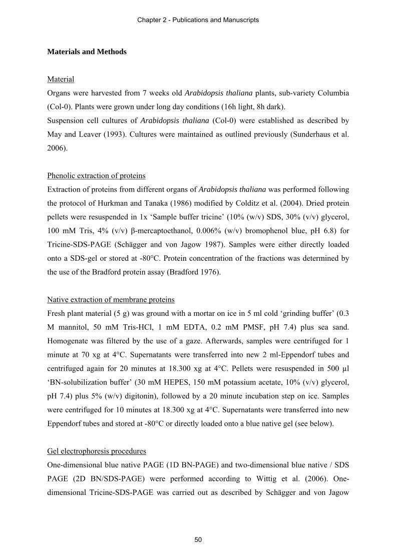
Materials and Methods
Material
Organs were harvested from 7 weeks old Arabidopsis thaliana plants, sub-variety Columbia
(Col-0). Plants were grown under long day conditions (16h light, 8h dark).
Suspension cell cultures of Arabidopsis thaliana (Col-0) were established as described by
May and Leaver (1993). Cultures were maintained as outlined previously (Sunderhaus et al.
2006).
Phenolic extraction of proteins
Extraction of proteins from different organs of Arabidopsis thaliana was performed following
the protocol of Hurkman and Tanaka (1986) modified by Colditz et al. (2004). Dried protein
pellets were resuspended in 1x ‘Sample buffer tricine’ (10% (w/v) SDS, 30% (v/v) glycerol,
100 mM Tris, 4% (v/v) β-mercaptoethanol, 0.006% (w/v) bromophenol blue, pH 6.8) for
Tricine-SDS-PAGE (Schägger and von Jagow 1987). Samples were either directly loaded
onto a SDS-gel or stored at -80°C. Protein concentration of the fractions was determined by
the use of the Bradford protein assay (Bradford 1976).
Native extraction of membrane proteins
Fresh plant material (5 g) was ground with a mortar on ice in 5 ml cold ‘grinding buffer’ (0.3
M mannitol, 50 mM Tris-HCl, 1 mM EDTA, 0.2 mM PMSF, pH 7.4) plus sea sand.
Homogenate was filtered by the use of a gaze. Afterwards, samples were centrifuged for 1
minute at 70 xg at 4°C. Supernatants were transferred into new 2 ml-Eppendorf tubes and
centrifuged again for 20 minutes at 18.300 xg at 4°C. Pellets were resuspended in 500 µl
‘BN-solubilization buffer’ (30 mM HEPES, 150 mM potassium acetate, 10% (v/v) glycerol,
pH 7.4) plus 5% (w/v) digitonin), followed by a 20 minute incubation step on ice. Samples
were centrifuged for 10 minutes at 18.300 xg at 4°C. Supernatants were transferred into new
Eppendorf tubes and stored at -80°C or directly loaded onto a blue native gel (see below).
Gel electrophoresis procedures
One-dimensional blue native PAGE (1D BN-PAGE) and two-dimensional blue native / SDS
PAGE (2D BN/SDS-PAGE) were performed according to Wittig et al. (2006). One-
dimensional Tricine-SDS-PAGE was carried out as described by Schägger and von Jagow
Chapter 2 - Publications and Manuscripts
50
(1987). Solubilization of mitochondrial protein was performed using digitonin at a
concentration of 5 g/g mitochondrial protein (Eubel et al. 2003).
Western blotting
Proteins separated on polyacrylamide gels were blotted onto nitrocellulose membranes for
antibody staining using the Trans Blot Cell from BioRad (Munich, Germany). Blotting was
carried out as described by Kruft et al. (2001). Immunohistochemical stains were performed
by using the VectaStain ABC Kit (Vector Laboratories, Burlingame, CA, USA) according to
manufacturer’s instructions.
For quantitative Western blotting experiments proteins were separated by SDS-PAGE using
precast gels (Mini-Protean TGX™ 10% Tris-HCl, Bio-Rad, Munich, Germany). After
separation, proteins were transferred on a nitrocellulose membrane using a liquid
electroblotting apparatus (Mini-Protean Tetra Cell, Bio-Rad, Munich, Germany). The
membrane was afterwards blocked in TTBS buffer (20 mM Tris-HCl pH 7.5, 50 mM NaCl,
0.05% Tween 20) with 1% BSA, and then incubated overnight in TTBS with an antibody
directed against the 51-kDa subunit (complex I), the SDH 1-1 subunit (complex II), the alpha
subunit of the mitochondrial processing peptidase (alpha MPP; complex III), the COX2
subunit (complex IV) or the beta subunit of complex V. After washing with TTBS, the
membrane was incubated with a secondary monoclonal antibody directed against rabbit
immunoglobulins. This secondary antibody is coupled to horseradish peroxidase (HRP) (Goat
anti-Rabbit IgG HRP conjugate, Millipore, Billerica, MA, USA). HRP finally converts the
‘Lumi-LightPLUS Western Blotting Substrate’ (Roche, Mannheim, Germany) into a
fluorescence-emitting form. Light signals were recorded with the Lumi-Imager (Roche,
Mannheim, Germany). Quantification of proteins was carried out using the AIDA Image
Analyser Software (Raytest Isotopenmessgeräte GmbH, Straubenhardt, Germany).
Production of antibodies
Antibodies directed against the 51-kDa subunit (complex I), the SDH 1-1 subunit (complex
II), the alpha subunit MPP subunit (complex III), the COX2 subunit (complex IV) and the
beta subunit of complex V were produced as polyclonal peptide specific antibodies by
Eurogentec (Seraing, Belgium) (for details see Supp. Tab. 1). The antibody against the 51-
kDa subunit (complex I) was additionally produced as polyclonal anti-protein antibody by
Eurogentec (Seraing, Belgium). For this approach, the 51-kDa subunit was over-expressed in
E. coli, purified from inclusion bodies and separated by 1D Tricine-SDS-PAGE.
Chapter 2 - Publications and Manuscripts
51
Gel staining procedures
Polyacrylamide gels were stained with Coomassie colloidal (Neuhoff et al. 1988, Neuhoff et
al. 1990). In gel enzyme activity stains for mitochondrial respiratory chain complexes I, II and
IV were carried out as described in Zerbetto et al. (1997) and Jung et al. (2000).
Chapter 2 - Publications and Manuscripts
52
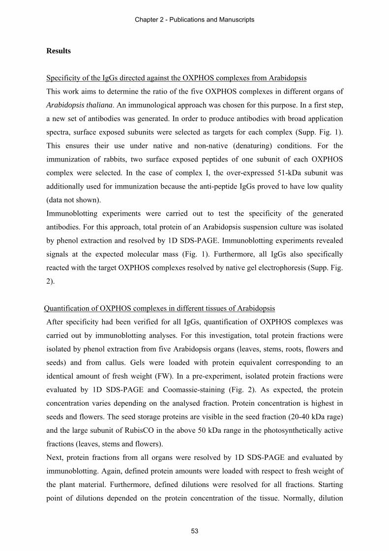
Results
Specificity of the IgGs directed against the OXPHOS complexes from Arabidopsis
This work aims to determine the ratio of the five OXPHOS complexes in different organs of
Arabidopsis thaliana. An immunological approach was chosen for this purpose. In a first step,
a new set of antibodies was generated. In order to produce antibodies with broad application
spectra, surface exposed subunits were selected as targets for each complex (Supp. Fig. 1).
This ensures their use under native and non-native (denaturing) conditions. For the
immunization of rabbits, two surface exposed peptides of one subunit of each OXPHOS
complex were selected. In the case of complex I, the over-expressed 51-kDa subunit was
additionally used for immunization because the anti-peptide IgGs proved to have low quality
(data not shown).
Immunoblotting experiments were carried out to test the specificity of the generated
antibodies. For this approach, total protein of an Arabidopsis suspension culture was isolated
by phenol extraction and resolved by 1D SDS-PAGE. Immunoblotting experiments revealed
signals at the expected molecular mass (Fig. 1). Furthermore, all IgGs also specifically
reacted with the target OXPHOS complexes resolved by native gel electrophoresis (Supp. Fig.
2).
Quantification of OXPHOS complexes in different tissues of Arabidopsis
After specificity had been verified for all IgGs, quantification of OXPHOS complexes was
carried out by immunoblotting analyses. For this investigation, total protein fractions were
isolated by phenol extraction from five Arabidopsis organs (leaves, stems, roots, flowers and
seeds) and from callus. Gels were loaded with protein equivalent corresponding to an
identical amount of fresh weight (FW). In a pre-experiment, isolated protein fractions were
evaluated by 1D SDS-PAGE and Coomassie-staining (Fig. 2). As expected, the protein
concentration varies depending on the analysed fraction. Protein concentration is highest in
seeds and flowers. The seed storage proteins are visible in the seed fraction (20-40 kDa rage)
and the large subunit of RubisCO in the above 50 kDa range in the photosynthetically active
fractions (leaves, stems and flowers).
Next, protein fractions from all organs were resolved by 1D SDS-PAGE and evaluated by
immunoblotting. Again, defined protein amounts were loaded with respect to fresh weight of
the plant material. Furthermore, defined dilutions were resolved for all fractions. Starting
point of dilutions depended on the protein concentration of the tissue. Normally, dilution
Chapter 2 - Publications and Manuscripts
53
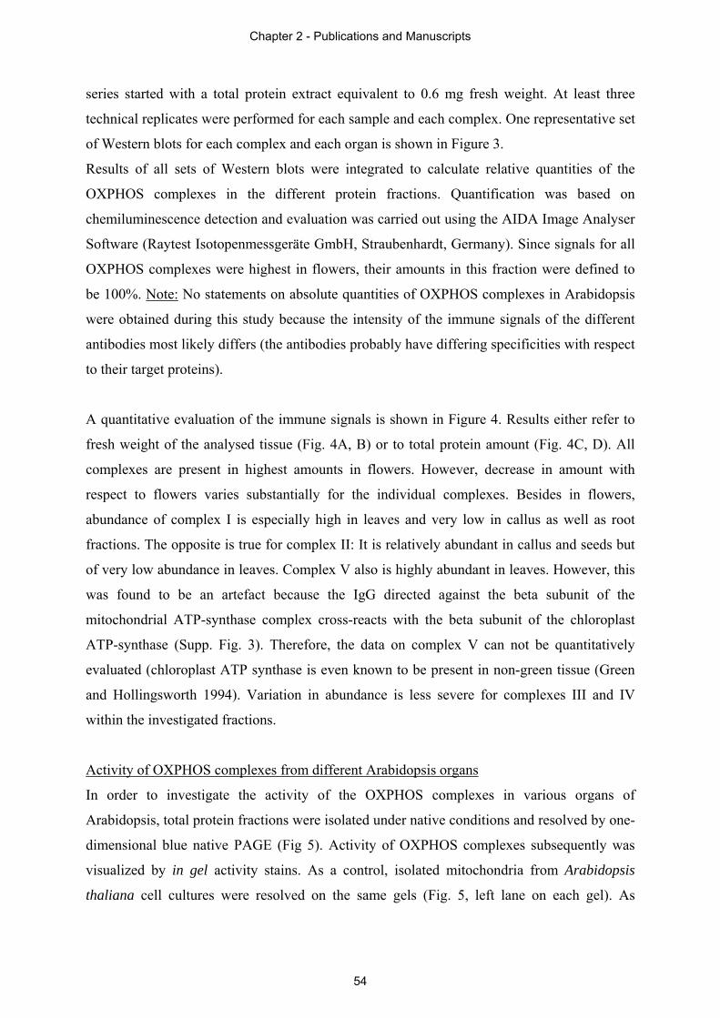
series started with a total protein extract equivalent to 0.6 mg fresh weight. At least three
technical replicates were performed for each sample and each complex. One representative set
of Western blots for each complex and each organ is shown in Figure 3.
Results of all sets of Western blots were integrated to calculate relative quantities of the
OXPHOS complexes in the different protein fractions. Quantification was based on
chemiluminescence detection and evaluation was carried out using the AIDA Image Analyser
Software (Raytest Isotopenmessgeräte GmbH, Straubenhardt, Germany). Since signals for all
OXPHOS complexes were highest in flowers, their amounts in this fraction were defined to
be 100%. Note: No statements on absolute quantities of OXPHOS complexes in Arabidopsis
were obtained during this study because the intensity of the immune signals of the different
antibodies most likely differs (the antibodies probably have differing specificities with respect
to their target proteins).
A quantitative evaluation of the immune signals is shown in Figure 4. Results either refer to
fresh weight of the analysed tissue (Fig. 4A, B) or to total protein amount (Fig. 4C, D). All
complexes are present in highest amounts in flowers. However, decrease in amount with
respect to flowers varies substantially for the individual complexes. Besides in flowers,
abundance of complex I is especially high in leaves and very low in callus as well as root
fractions. The opposite is true for complex II: It is relatively abundant in callus and seeds but
of very low abundance in leaves. Complex V also is highly abundant in leaves. However, this
was found to be an artefact because the IgG directed against the beta subunit of the
mitochondrial ATP-synthase complex cross-reacts with the beta subunit of the chloroplast
ATP-synthase (Supp. Fig. 3). Therefore, the data on complex V can not be quantitatively
evaluated (chloroplast ATP synthase is even known to be present in non-green tissue (Green
and Hollingsworth 1994). Variation in abundance is less severe for complexes III and IV
within the investigated fractions.
Activity of OXPHOS complexes from different Arabidopsis organs
In order to investigate the activity of the OXPHOS complexes in various organs of
Arabidopsis, total protein fractions were isolated under native conditions and resolved by one-
dimensional blue native PAGE (Fig 5). Activity of OXPHOS complexes subsequently was
visualized by in gel activity stains. As a control, isolated mitochondria from Arabidopsis
thaliana cell cultures were resolved on the same gels (Fig. 5, left lane on each gel). As
Chapter 2 - Publications and Manuscripts
54
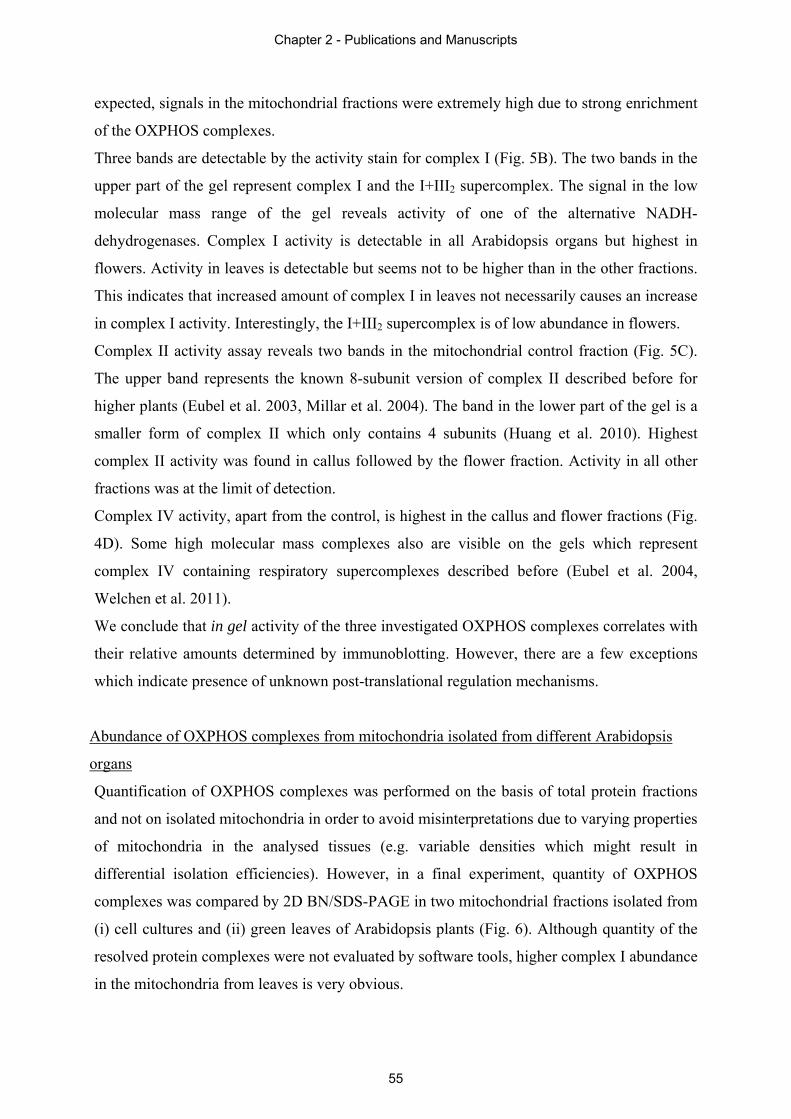
expected, signals in the mitochondrial fractions were extremely high due to strong enrichment
of the OXPHOS complexes.
Three bands are detectable by the activity stain for complex I (Fig. 5B). The two bands in the
upper part of the gel represent complex I and the I+III2 supercomplex. The signal in the low
molecular mass range of the gel reveals activity of one of the alternative NADH-
dehydrogenases. Complex I activity is detectable in all Arabidopsis organs but highest in
flowers. Activity in leaves is detectable but seems not to be higher than in the other fractions.
This indicates that increased amount of complex I in leaves not necessarily causes an increase
in complex I activity. Interestingly, the I+III2 supercomplex is of low abundance in flowers.
Complex II activity assay reveals two bands in the mitochondrial control fraction (Fig. 5C).
The upper band represents the known 8-subunit version of complex II described before for
higher plants (Eubel et al. 2003, Millar et al. 2004). The band in the lower part of the gel is a
smaller form of complex II which only contains 4 subunits (Huang et al. 2010). Highest
complex II activity was found in callus followed by the flower fraction. Activity in all other
fractions was at the limit of detection.
Complex IV activity, apart from the control, is highest in the callus and flower fractions (Fig.
4D). Some high molecular mass complexes also are visible on the gels which represent
complex IV containing respiratory supercomplexes described before (Eubel et al. 2004,
Welchen et al. 2011).
We conclude that in gel activity of the three investigated OXPHOS complexes correlates with
their relative amounts determined by immunoblotting. However, there are a few exceptions
which indicate presence of unknown post-translational regulation mechanisms.
Abundance of OXPHOS complexes from mitochondria isolated from different Arabidopsis
organs
Quantification of OXPHOS complexes was performed on the basis of total protein fractions
and not on isolated mitochondria in order to avoid misinterpretations due to varying properties
of mitochondria in the analysed tissues (e.g. variable densities which might result in
differential isolation efficiencies). However, in a final experiment, quantity of OXPHOS
complexes was compared by 2D BN/SDS-PAGE in two mitochondrial fractions isolated from
(i) cell cultures and (ii) green leaves of Arabidopsis plants (Fig. 6). Although quantity of the
resolved protein complexes were not evaluated by software tools, higher complex I abundance
in the mitochondria from leaves is very obvious.
Chapter 2 - Publications and Manuscripts
55
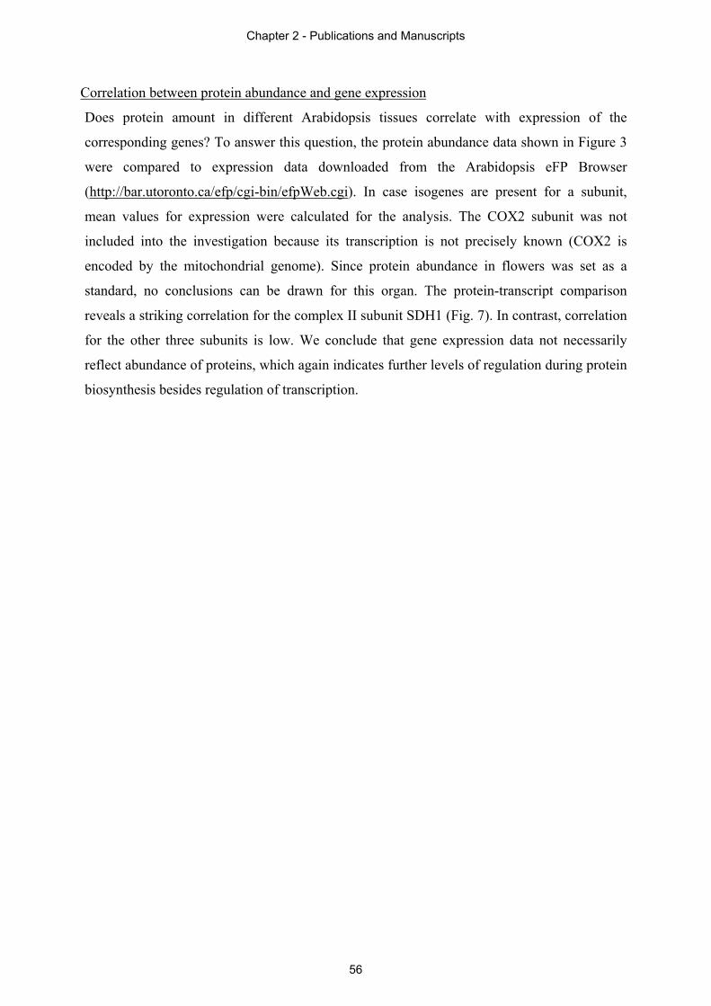
Correlation between protein abundance and gene expression
Does protein amount in different Arabidopsis tissues correlate with expression of the
corresponding genes? To answer this question, the protein abundance data shown in Figure 3
were compared to expression data downloaded from the Arabidopsis eFP Browser
(http://bar.utoronto.ca/efp/cgi-bin/efpWeb.cgi). In case isogenes are present for a subunit,
mean values for expression were calculated for the analysis. The COX2 subunit was not
included into the investigation because its transcription is not precisely known (COX2 is
encoded by the mitochondrial genome). Since protein abundance in flowers was set as a
standard, no conclusions can be drawn for this organ. The protein-transcript comparison
reveals a striking correlation for the complex II subunit SDH1 (Fig. 7). In contrast, correlation
for the other three subunits is low. We conclude that gene expression data not necessarily
reflect abundance of proteins, which again indicates further levels of regulation during protein
biosynthesis besides regulation of transcription.
Chapter 2 - Publications and Manuscripts
56
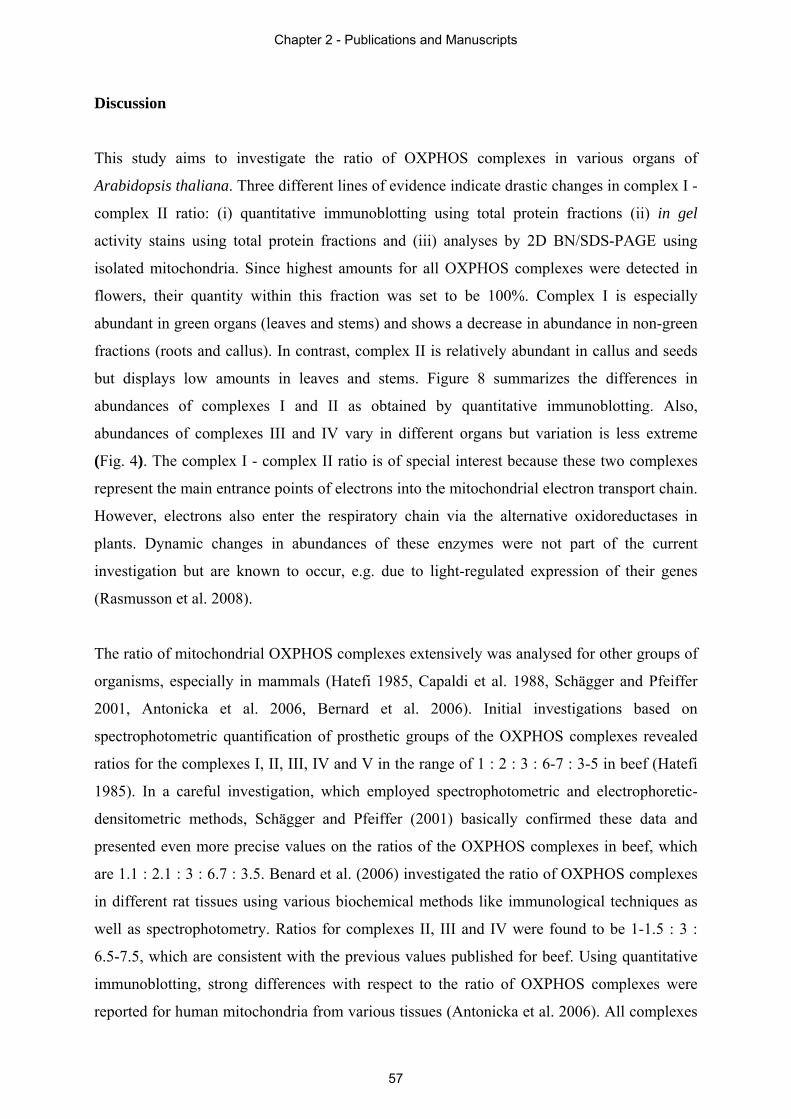
Discussion
This study aims to investigate the ratio of OXPHOS complexes in various organs of
Arabidopsis thaliana. Three different lines of evidence indicate drastic changes in complex I -
complex II ratio: (i) quantitative immunoblotting using total protein fractions (ii) in gel
activity stains using total protein fractions and (iii) analyses by 2D BN/SDS-PAGE using
isolated mitochondria. Since highest amounts for all OXPHOS complexes were detected in
flowers, their quantity within this fraction was set to be 100%. Complex I is especially
abundant in green organs (leaves and stems) and shows a decrease in abundance in non-green
fractions (roots and callus). In contrast, complex II is relatively abundant in callus and seeds
but displays low amounts in leaves and stems. Figure 8 summarizes the differences in
abundances of complexes I and II as obtained by quantitative immunoblotting. Also,
abundances of complexes III and IV vary in different organs but variation is less extreme
(Fig. 4). The complex I - complex II ratio is of special interest because these two complexes
represent the main entrance points of electrons into the mitochondrial electron transport chain.
However, electrons also enter the respiratory chain via the alternative oxidoreductases in
plants. Dynamic changes in abundances of these enzymes were not part of the current
investigation but are known to occur, e.g. due to light-regulated expression of their genes
(Rasmusson et al. 2008).
The ratio of mitochondrial OXPHOS complexes extensively was analysed for other groups of
organisms, especially in mammals (Hatefi 1985, Capaldi et al. 1988, Schägger and Pfeiffer
2001, Antonicka et al. 2006, Bernard et al. 2006). Initial investigations based on
spectrophotometric quantification of prosthetic groups of the OXPHOS complexes revealed
ratios for the complexes I, II, III, IV and V in the range of 1 : 2 : 3 : 6-7 : 3-5 in beef (Hatefi
1985). In a careful investigation, which employed spectrophotometric and electrophoretic-
densitometric methods, Schägger and Pfeiffer (2001) basically confirmed these data and
presented even more precise values on the ratios of the OXPHOS complexes in beef, which
are 1.1 : 2.1 : 3 : 6.7 : 3.5. Benard et al. (2006) investigated the ratio of OXPHOS complexes
in different rat tissues using various biochemical methods like immunological techniques as
well as spectrophotometry. Ratios for complexes II, III and IV were found to be 1-1.5 : 3 :
6.5-7.5, which are consistent with the previous values published for beef. Using quantitative
immunoblotting, strong differences with respect to the ratio of OXPHOS complexes were
reported for human mitochondria from various tissues (Antonicka et al. 2006). All complexes
Chapter 2 - Publications and Manuscripts
57
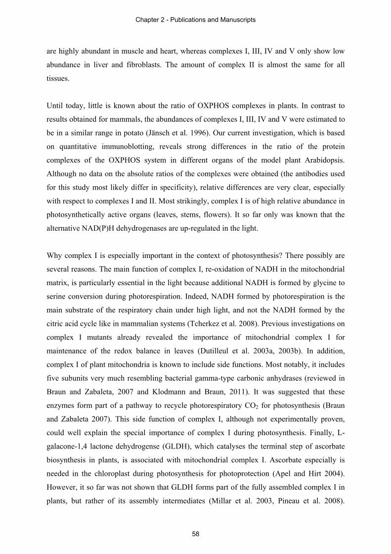
are highly abundant in muscle and heart, whereas complexes I, III, IV and V only show low
abundance in liver and fibroblasts. The amount of complex II is almost the same for all
tissues.
Until today, little is known about the ratio of OXPHOS complexes in plants. In contrast to
results obtained for mammals, the abundances of complexes I, III, IV and V were estimated to
be in a similar range in potato (Jänsch et al. 1996). Our current investigation, which is based
on quantitative immunoblotting, reveals strong differences in the ratio of the protein
complexes of the OXPHOS system in different organs of the model plant Arabidopsis.
Although no data on the absolute ratios of the complexes were obtained (the antibodies used
for this study most likely differ in specificity), relative differences are very clear, especially
with respect to complexes I and II. Most strikingly, complex I is of high relative abundance in
photosynthetically active organs (leaves, stems, flowers). It so far only was known that the
alternative NAD(P)H dehydrogenases are up-regulated in the light.
Why complex I is especially important in the context of photosynthesis? There possibly are
several reasons. The main function of complex I, re-oxidation of NADH in the mitochondrial
matrix, is particularly essential in the light because additional NADH is formed by glycine to
serine conversion during photorespiration. Indeed, NADH formed by photorespiration is the
main substrate of the respiratory chain under high light, and not the NADH formed by the
citric acid cycle like in mammalian systems (Tcherkez et al. 2008). Previous investigations on
complex I mutants already revealed the importance of mitochondrial complex I for
maintenance of the redox balance in leaves (Dutilleul et al. 2003a, 2003b). In addition,
complex I of plant mitochondria is known to include side functions. Most notably, it includes
five subunits very much resembling bacterial gamma-type carbonic anhydrases (reviewed in
Braun and Zabaleta, 2007 and Klodmann and Braun, 2011). It was suggested that these
enzymes form part of a pathway to recycle photorespiratory CO2 for photosynthesis (Braun
and Zabaleta 2007). This side function of complex I, although not experimentally proven,
could well explain the special importance of complex I during photosynthesis. Finally, L-
galacone-1,4 lactone dehydrogense (GLDH), which catalyses the terminal step of ascorbate
biosynthesis in plants, is associated with mitochondrial complex I. Ascorbate especially is
needed in the chloroplast during photosynthesis for photoprotection (Apel and Hirt 2004).
However, it so far was not shown that GLDH forms part of the fully assembled complex I in
plants, but rather of its assembly intermediates (Millar et al. 2003, Pineau et al. 2008).
Chapter 2 - Publications and Manuscripts
58
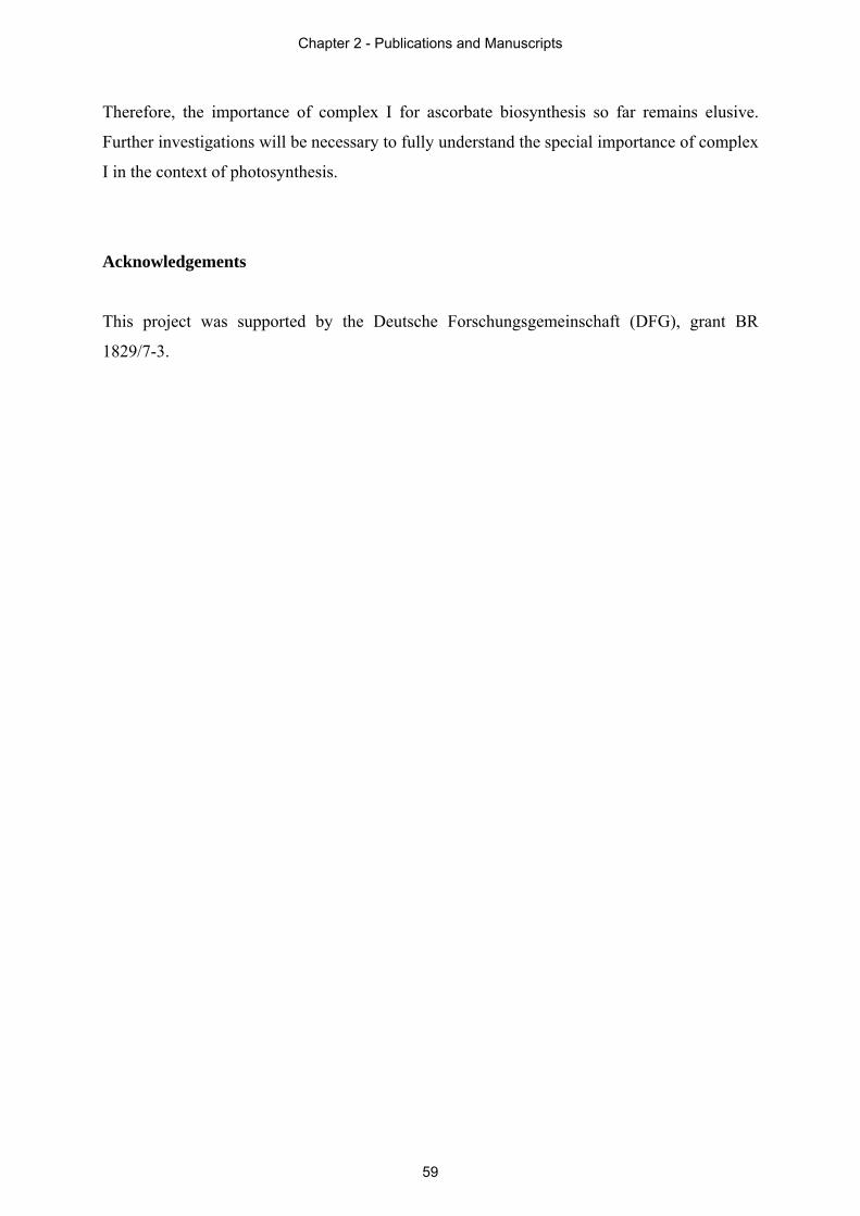
Therefore, the importance of complex I for ascorbate biosynthesis so far remains elusive.
Further investigations will be necessary to fully understand the special importance of complex
I in the context of photosynthesis.
Acknowledgements
This project was supported by the Deutsche Forschungsgemeinschaft (DFG), grant BR
1829/7-3.
Chapter 2 - Publications and Manuscripts
59
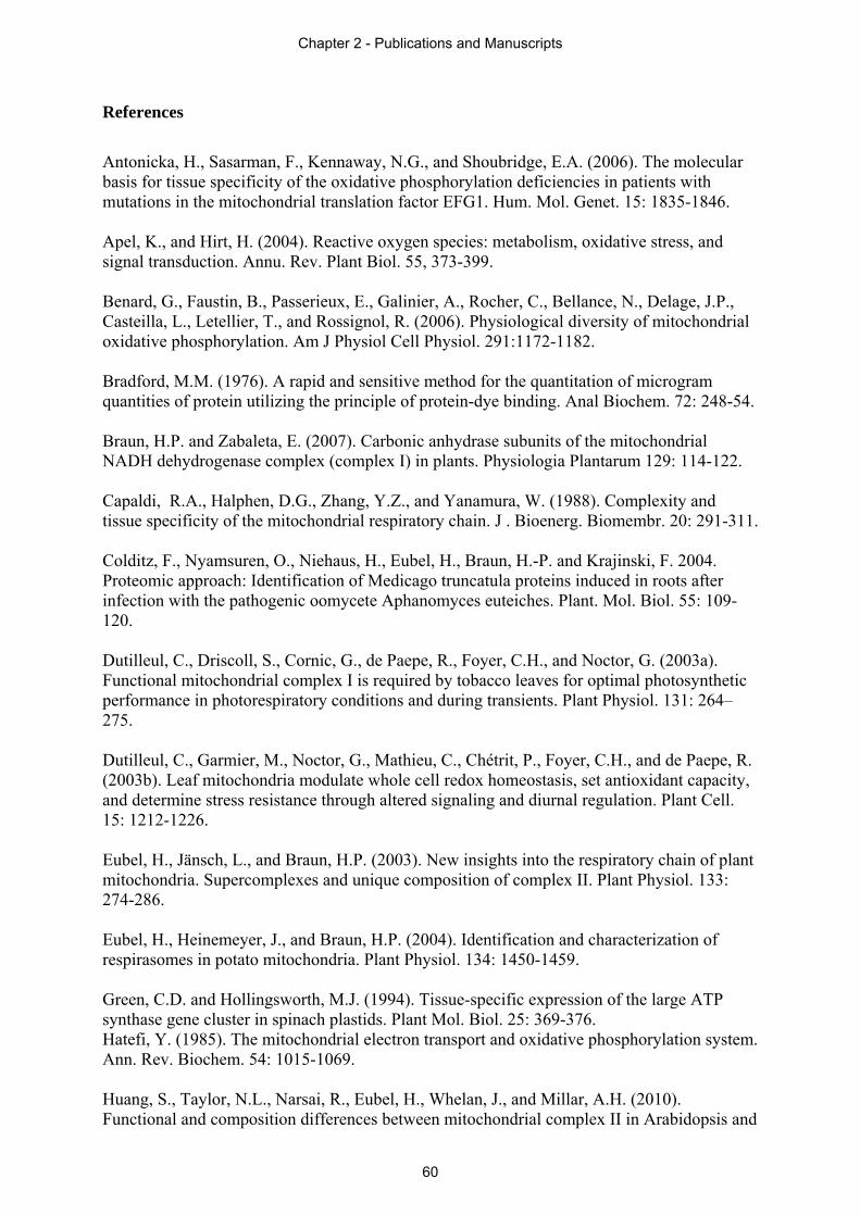
References
Antonicka, H., Sasarman, F., Kennaway, N.G., and Shoubridge, E.A. (2006). The molecular basis for tissue specificity of the oxidative phosphorylation deficiencies in patients with mutations in the mitochondrial translation factor EFG1. Hum. Mol. Genet. 15: 1835-1846. Apel, K., and Hirt, H. (2004). Reactive oxygen species: metabolism, oxidative stress, and signal transduction. Annu. Rev. Plant Biol. 55, 373-399. Benard, G., Faustin, B., Passerieux, E., Galinier, A., Rocher, C., Bellance, N., Delage, J.P., Casteilla, L., Letellier, T., and Rossignol, R. (2006). Physiological diversity of mitochondrial oxidative phosphorylation. Am J Physiol Cell Physiol. 291:1172-1182. Bradford, M.M. (1976). A rapid and sensitive method for the quantitation of microgram quantities of protein utilizing the principle of protein-dye binding. Anal Biochem. 72: 248-54. Braun, H.P. and Zabaleta, E. (2007). Carbonic anhydrase subunits of the mitochondrial NADH dehydrogenase complex (complex I) in plants. Physiologia Plantarum 129: 114-122. Capaldi, R.A., Halphen, D.G., Zhang, Y.Z., and Yanamura, W. (1988). Complexity and tissue specificity of the mitochondrial respiratory chain. J . Bioenerg. Biomembr. 20: 291-311. Colditz, F., Nyamsuren, O., Niehaus, H., Eubel, H., Braun, H.-P. and Krajinski, F. 2004. Proteomic approach: Identification of Medicago truncatula proteins induced in roots after infection with the pathogenic oomycete Aphanomyces euteiches. Plant. Mol. Biol. 55: 109-120. Dutilleul, C., Driscoll, S., Cornic, G., de Paepe, R., Foyer, C.H., and Noctor, G. (2003a). Functional mitochondrial complex I is required by tobacco leaves for optimal photosynthetic performance in photorespiratory conditions and during transients. Plant Physiol. 131: 264–275. Dutilleul, C., Garmier, M., Noctor, G., Mathieu, C., Chétrit, P., Foyer, C.H., and de Paepe, R. (2003b). Leaf mitochondria modulate whole cell redox homeostasis, set antioxidant capacity, and determine stress resistance through altered signaling and diurnal regulation. Plant Cell. 15: 1212-1226. Eubel, H., Jänsch, L., and Braun, H.P. (2003). New insights into the respiratory chain of plant mitochondria. Supercomplexes and unique composition of complex II. Plant Physiol. 133: 274-286. Eubel, H., Heinemeyer, J., and Braun, H.P. (2004). Identification and characterization of respirasomes in potato mitochondria. Plant Physiol. 134: 1450-1459. Green, C.D. and Hollingsworth, M.J. (1994). Tissue-specific expression of the large ATP synthase gene cluster in spinach plastids. Plant Mol. Biol. 25: 369-376. Hatefi, Y. (1985). The mitochondrial electron transport and oxidative phosphorylation system. Ann. Rev. Biochem. 54: 1015-1069. Huang, S., Taylor, N.L., Narsai, R., Eubel, H., Whelan, J., and Millar, A.H. (2010). Functional and composition differences between mitochondrial complex II in Arabidopsis and
Chapter 2 - Publications and Manuscripts
60
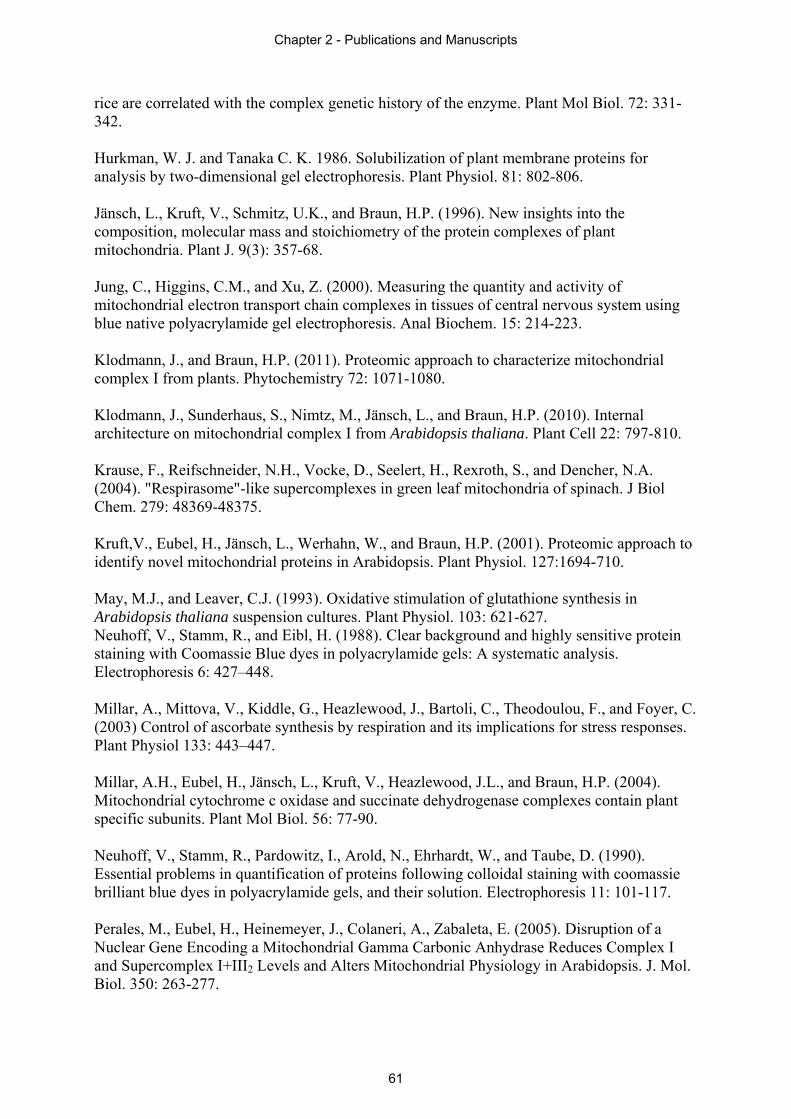
rice are correlated with the complex genetic history of the enzyme. Plant Mol Biol. 72: 331-342. Hurkman, W. J. and Tanaka C. K. 1986. Solubilization of plant membrane proteins for analysis by two-dimensional gel electrophoresis. Plant Physiol. 81: 802-806. Jänsch, L., Kruft, V., Schmitz, U.K., and Braun, H.P. (1996). New insights into the composition, molecular mass and stoichiometry of the protein complexes of plant mitochondria. Plant J. 9(3): 357-68. Jung, C., Higgins, C.M., and Xu, Z. (2000). Measuring the quantity and activity of mitochondrial electron transport chain complexes in tissues of central nervous system using blue native polyacrylamide gel electrophoresis. Anal Biochem. 15: 214-223. Klodmann, J., and Braun, H.P. (2011). Proteomic approach to characterize mitochondrial complex I from plants. Phytochemistry 72: 1071-1080. Klodmann, J., Sunderhaus, S., Nimtz, M., Jänsch, L., and Braun, H.P. (2010). Internal architecture on mitochondrial complex I from Arabidopsis thaliana. Plant Cell 22: 797-810. Krause, F., Reifschneider, N.H., Vocke, D., Seelert, H., Rexroth, S., and Dencher, N.A. (2004). "Respirasome"-like supercomplexes in green leaf mitochondria of spinach. J Biol Chem. 279: 48369-48375. Kruft,V., Eubel, H., Jänsch, L., Werhahn, W., and Braun, H.P. (2001). Proteomic approach to identify novel mitochondrial proteins in Arabidopsis. Plant Physiol. 127:1694-710. May, M.J., and Leaver, C.J. (1993). Oxidative stimulation of glutathione synthesis in Arabidopsis thaliana suspension cultures. Plant Physiol. 103: 621-627. Neuhoff, V., Stamm, R., and Eibl, H. (1988). Clear background and highly sensitive protein staining with Coomassie Blue dyes in polyacrylamide gels: A systematic analysis. Electrophoresis 6: 427–448. Millar, A., Mittova, V., Kiddle, G., Heazlewood, J., Bartoli, C., Theodoulou, F., and Foyer, C. (2003) Control of ascorbate synthesis by respiration and its implications for stress responses. Plant Physiol 133: 443–447. Millar, A.H., Eubel, H., Jänsch, L., Kruft, V., Heazlewood, J.L., and Braun, H.P. (2004). Mitochondrial cytochrome c oxidase and succinate dehydrogenase complexes contain plant specific subunits. Plant Mol Biol. 56: 77-90. Neuhoff, V., Stamm, R., Pardowitz, I., Arold, N., Ehrhardt, W., and Taube, D. (1990). Essential problems in quantification of proteins following colloidal staining with coomassie brilliant blue dyes in polyacrylamide gels, and their solution. Electrophoresis 11: 101-117. Perales, M., Eubel, H., Heinemeyer, J., Colaneri, A., Zabaleta, E. (2005). Disruption of a Nuclear Gene Encoding a Mitochondrial Gamma Carbonic Anhydrase Reduces Complex I and Supercomplex I+III2 Levels and Alters Mitochondrial Physiology in Arabidopsis. J. Mol. Biol. 350: 263-277.
Chapter 2 - Publications and Manuscripts
61
Pineau, B., Layoune, O., Danon, A., and De, P. (2008) L-galactono-1,4-lactone dehydrogenase is required for the accumulation of plant respiratory complex I. J Biol Chem 283: 32500–32505. Rasmusson, A.G., and Escobar, M.A. (2007). Light and diurnal regulation of plant respiratory gene expression. Physiol. Plant. 129: 57-67. Rasmusson, A.G., Geisler, D.A., and Møller, I.M. (2008). The multiplicity of dehydrogenases in the electron transport chain of plant mitochondria. Mitochondrion 8: 47-60. Rasmusson, A.G., and Wallström, S.V. (2010). Involvement of mitochondria in the control of plant cell NAD(P)H reduction levels. Biochem. Soc. Trans. 38: 661-666. Schägger, H. and von Jagow, G. (1987) Tricine-sodium dodecyl sulfate-polyacrylamide gel electrophoresis for the separation of proteins in the range from 1 to 100 kDa. Anal. Biochem. 166, 368-379. Schägger, H. and Pfeiffer, K. (2000). Supercomplexes in the respiratory chains of yeast and mammalian mitochondria. EMBO J. 19: 1777-1783. Schägger, H., and Pfeiffer, K. (2001). The ratio of oxidative phosphorylation complexes I-V in bovine heart mitochondria and the composition of respiratory chain supercomplexes. J Biol Chem. 276: 37861-37867. Sunderhaus, S., Dudkina, N., Jänsch, L., Klodmann, J., Heinemeyer, J., Perales, M., Zabaleta, E., Boekema, E., and Braun, H.P. (2006). Carbonic anhydrases subunits form a matrix-exposed domain attached to the membrane arm of complex I in plants. J. Biol. Chem. 281: 6482-6488. Tcherkez, G., Bligny, R., Gout, E., Mathé, M., and Cornic, G. (2008). Respiratory metabolism of illuminated leaves depends on CO2 and O2 conditions. Proc. Natl. Acad. Sci. 105: 797-802. Welchen, E., Klodmann, J., and Braun, H.P. (2011). Biogenesis and Supramolecular Organization of the Oxidative Phosphorylation System in Plants. Plant Mitochondria, Advances in Plant Biology 1: 327-355. Wittig, I., Braun, H.P., and Schägger, H. (2006). Blue native PAGE. Nat. Protoc. 1: 418-428. Zerbetto, E., Vergani, L., and Dabbeni-Sala, F. (1997). Quantification of muscle mitochondrial oxidative phosphorylation enzymes via histochemical staining of blue native polyacrylamide gels. Electrophoresis 18: 2059-2064.
Chapter 2 - Publications and Manuscripts
62
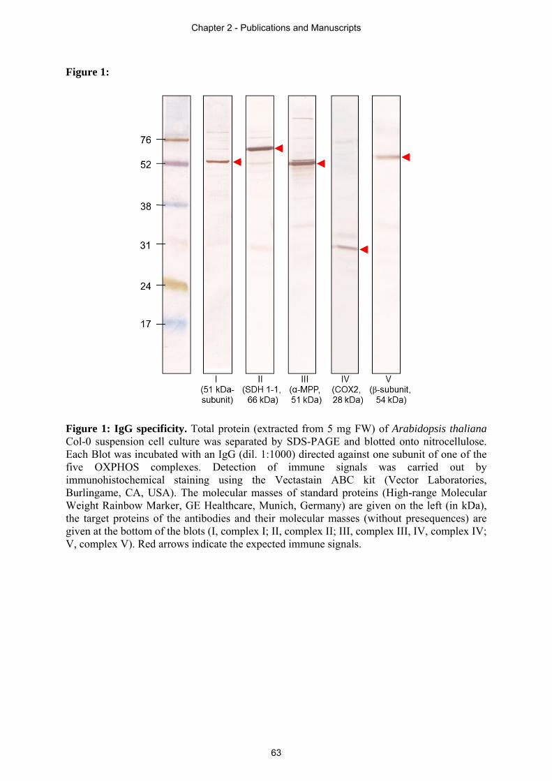
Figure 1:
Figure 1: IgG specificity. Total protein (extracted from 5 mg FW) of Arabidopsis thaliana Col-0 suspension cell culture was separated by SDS-PAGE and blotted onto nitrocellulose. Each Blot was incubated with an IgG (dil. 1:1000) directed against one subunit of one of the five OXPHOS complexes. Detection of immune signals was carried out by immunohistochemical staining using the Vectastain ABC kit (Vector Laboratories, Burlingame, CA, USA). The molecular masses of standard proteins (High-range Molecular Weight Rainbow Marker, GE Healthcare, Munich, Germany) are given on the left (in kDa), the target proteins of the antibodies and their molecular masses (without presequences) are given at the bottom of the blots (I, complex I; II, complex II; III, complex III, IV, complex IV; V, complex V). Red arrows indicate the expected immune signals.
Chapter 2 - Publications and Manuscripts
63
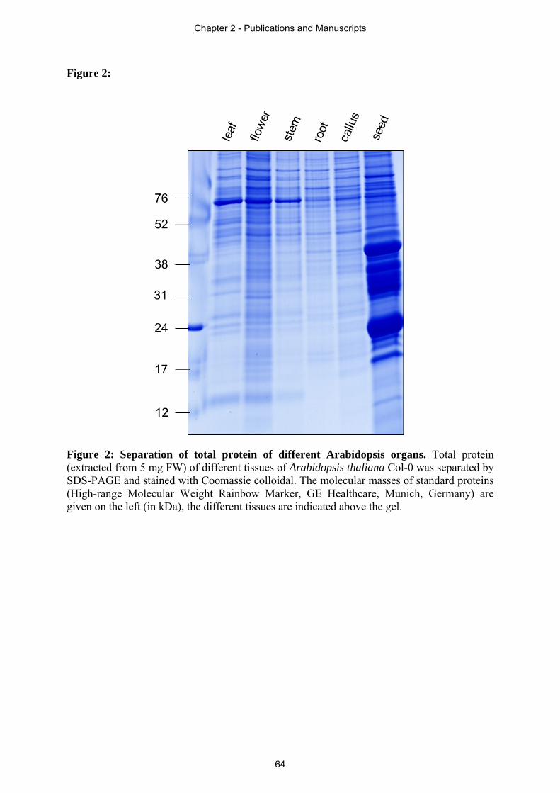
Figure 2:
Figure 2: Separation of total protein of different Arabidopsis organs. Total protein (extracted from 5 mg FW) of different tissues of Arabidopsis thaliana Col-0 was separated by SDS-PAGE and stained with Coomassie colloidal. The molecular masses of standard proteins (High-range Molecular Weight Rainbow Marker, GE Healthcare, Munich, Germany) are given on the left (in kDa), the different tissues are indicated above the gel.
Chapter 2 - Publications and Manuscripts
64
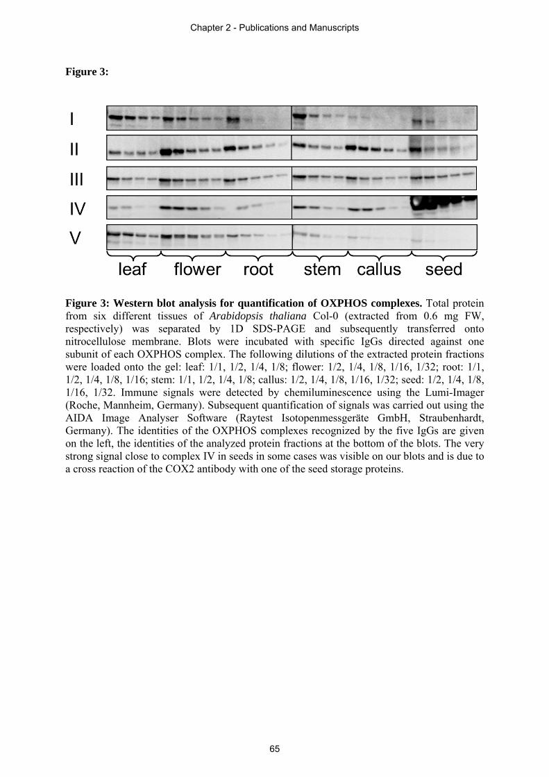
Figure 3:
Figure 3: Western blot analysis for quantification of OXPHOS complexes. Total protein from six different tissues of Arabidopsis thaliana Col-0 (extracted from 0.6 mg FW, respectively) was separated by 1D SDS-PAGE and subsequently transferred onto nitrocellulose membrane. Blots were incubated with specific IgGs directed against one subunit of each OXPHOS complex. The following dilutions of the extracted protein fractions were loaded onto the gel: leaf: 1/1, 1/2, 1/4, 1/8; flower: 1/2, 1/4, 1/8, 1/16, 1/32; root: 1/1, 1/2, 1/4, 1/8, 1/16; stem: 1/1, 1/2, 1/4, 1/8; callus: 1/2, 1/4, 1/8, 1/16, 1/32; seed: 1/2, 1/4, 1/8, 1/16, 1/32. Immune signals were detected by chemiluminescence using the Lumi-Imager (Roche, Mannheim, Germany). Subsequent quantification of signals was carried out using the AIDA Image Analyser Software (Raytest Isotopenmessgeräte GmbH, Straubenhardt, Germany). The identities of the OXPHOS complexes recognized by the five IgGs are given on the left, the identities of the analyzed protein fractions at the bottom of the blots. The very strong signal close to complex IV in seeds in some cases was visible on our blots and is due to a cross reaction of the COX2 antibody with one of the seed storage proteins.
Chapter 2 - Publications and Manuscripts
65
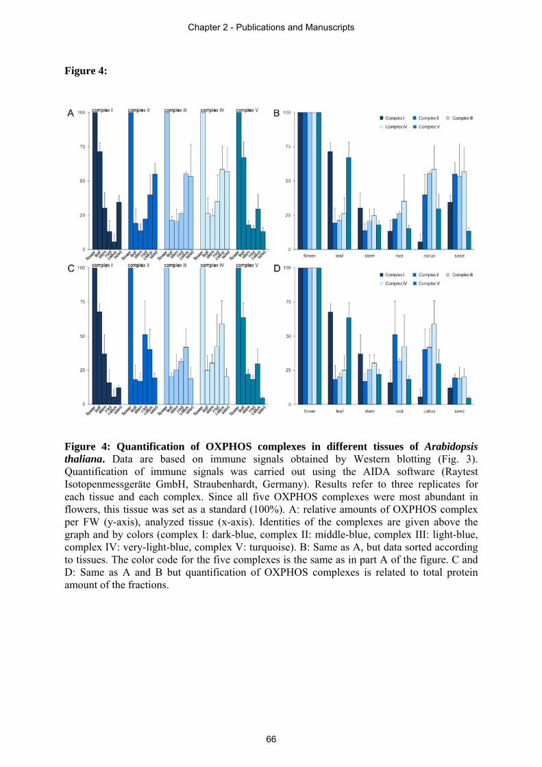
Figure 4:
Figure 4: Quantification of OXPHOS complexes in different tissues of Arabidopsis thaliana. Data are based on immune signals obtained by Western blotting (Fig. 3). Quantification of immune signals was carried out using the AIDA software (Raytest Isotopenmessgeräte GmbH, Straubenhardt, Germany). Results refer to three replicates for each tissue and each complex. Since all five OXPHOS complexes were most abundant in flowers, this tissue was set as a standard (100%). A: relative amounts of OXPHOS complex per FW (y-axis), analyzed tissue (x-axis). Identities of the complexes are given above the graph and by colors (complex I: dark-blue, complex II: middle-blue, complex III: light-blue, complex IV: very-light-blue, complex V: turquoise). B: Same as A, but data sorted according to tissues. The color code for the five complexes is the same as in part A of the figure. C and D: Same as A and B but quantification of OXPHOS complexes is related to total protein amount of the fractions.
Chapter 2 - Publications and Manuscripts
66
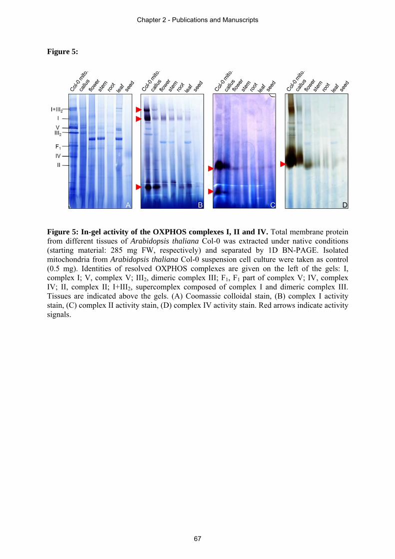
Figure 5:
Figure 5: In-gel activity of the OXPHOS complexes I, II and IV. Total membrane protein from different tissues of Arabidopsis thaliana Col-0 was extracted under native conditions (starting material: 285 mg FW, respectively) and separated by 1D BN-PAGE. Isolated mitochondria from Arabidopsis thaliana Col-0 suspension cell culture were taken as control (0.5 mg). Identities of resolved OXPHOS complexes are given on the left of the gels: I, complex I; V, complex V; III2, dimeric complex III; F1, F1 part of complex V; IV, complex IV; II, complex II; I+III2, supercomplex composed of complex I and dimeric complex III. Tissues are indicated above the gels. (A) Coomassie colloidal stain, (B) complex I activity stain, (C) complex II activity stain, (D) complex IV activity stain. Red arrows indicate activity signals.
Chapter 2 - Publications and Manuscripts
67
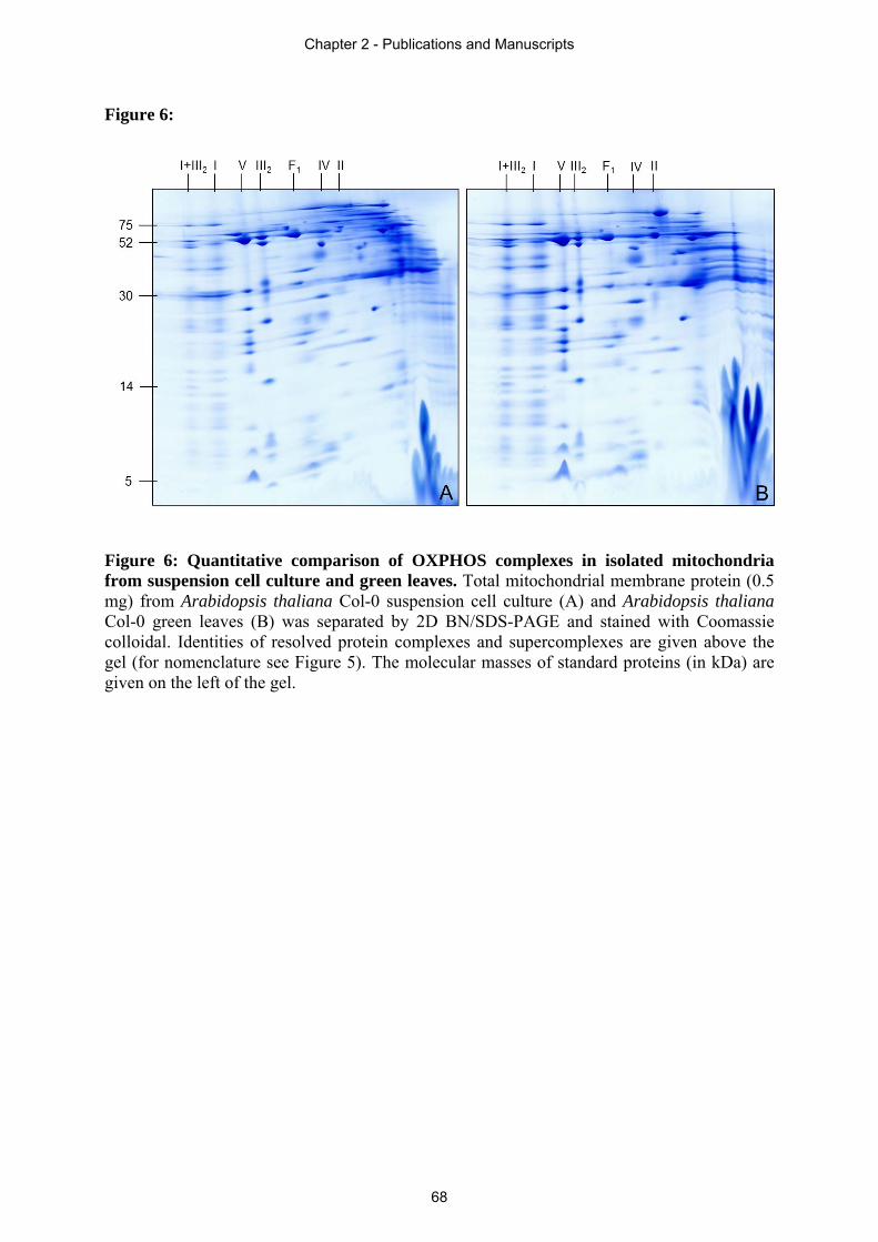
Figure 6:
Figure 6: Quantitative comparison of OXPHOS complexes in isolated mitochondria from suspension cell culture and green leaves. Total mitochondrial membrane protein (0.5 mg) from Arabidopsis thaliana Col-0 suspension cell culture (A) and Arabidopsis thaliana Col-0 green leaves (B) was separated by 2D BN/SDS-PAGE and stained with Coomassie colloidal. Identities of resolved protein complexes and supercomplexes are given above the gel (for nomenclature see Figure 5). The molecular masses of standard proteins (in kDa) are given on the left of the gel.
Chapter 2 - Publications and Manuscripts
68
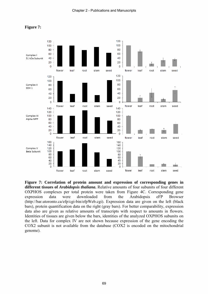
Figure 7:
Figure 7: Correlation of protein amount and expression of corresponding genes in different tissues of Arabidopsis thaliana. Relative amounts of four subunits of four different OXPHOS complexes per total protein were taken from Figure 4C. Corresponding gene expression data were downloaded from the Arabidopsis eFP Browser (http://bar.utoronto.ca/efp/cgi-bin/efpWeb.cgi). Expression data are given on the left (black bars), protein quantification data on the right (gray bars). For better comparability, expression data also are given as relative amounts of transcripts with respect to amounts in flowers. Identities of tissues are given below the bars, identities of the analyzed OXPHOS subunits on the left. Data for complex IV are not shown because expression of the gene encoding the COX2 subunit is not available from the database (COX2 is encoded on the mitochondrial genome).
Chapter 2 - Publications and Manuscripts
69
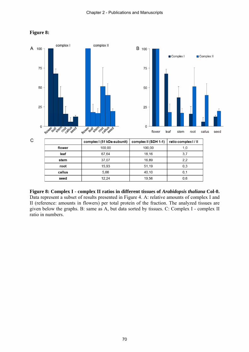
Figure 8:
Figure 8: Complex I - complex II ratios in different tissues of Arabidopsis thaliana Col-0. Data represent a subset of results presented in Figure 4. A: relative amounts of complex I and II (reference: amounts in flowers) per total protein of the fraction. The analyzed tissues are given below the graphs. B: same as A, but data sorted by tissues. C: Complex I - complex II ratio in numbers.
Chapter 2 - Publications and Manuscripts
70
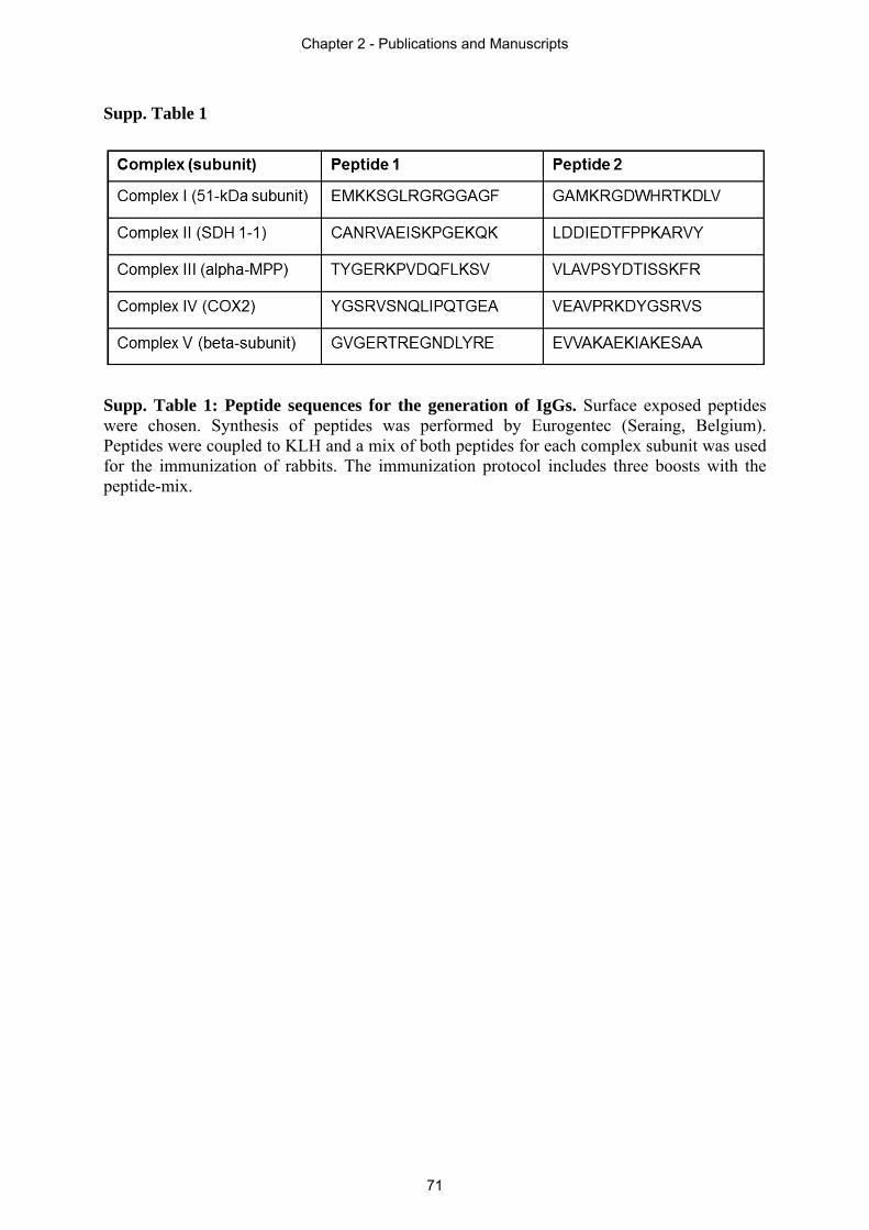
Supp. Table 1
Supp. Table 1: Peptide sequences for the generation of IgGs. Surface exposed peptides were chosen. Synthesis of peptides was performed by Eurogentec (Seraing, Belgium). Peptides were coupled to KLH and a mix of both peptides for each complex subunit was used for the immunization of rabbits. The immunization protocol includes three boosts with the peptide-mix.
Chapter 2 - Publications and Manuscripts
71
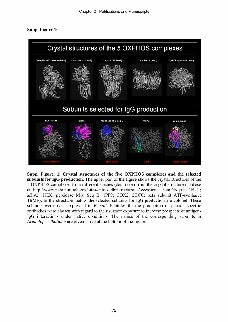
Supp. Figure 1:
Supp. Figure. 1: Crystal structures of the five OXPHOS complexes and the selected subunits for IgG production. The upper part of the figure shows the crystal structures of the 5 OXPHOS complexes from different species (data taken from the crystal structure database at http://www.ncbi.nlm.nih.gov/sites/entrez?db=structure. Accessions: NuoF/Nqo1: 2FUG; sdhA: 1NEK; peptidase M16 Seq B: 1PP9; COX2: 2OCC; beta subunit ATP-synthase: 1BMF). In the structures below the selected subunits for IgG production are colored. These subunits were over- expressed in E. coli. Peptides for the production of peptide specific antibodies were chosen with regard to their surface exposure to increase prospects of antigen-IgG interactions under native conditions. The names of the corresponding subunits in Arabidopsis thaliana are given in red at the bottom of the figure.
Chapter 2 - Publications and Manuscripts
72
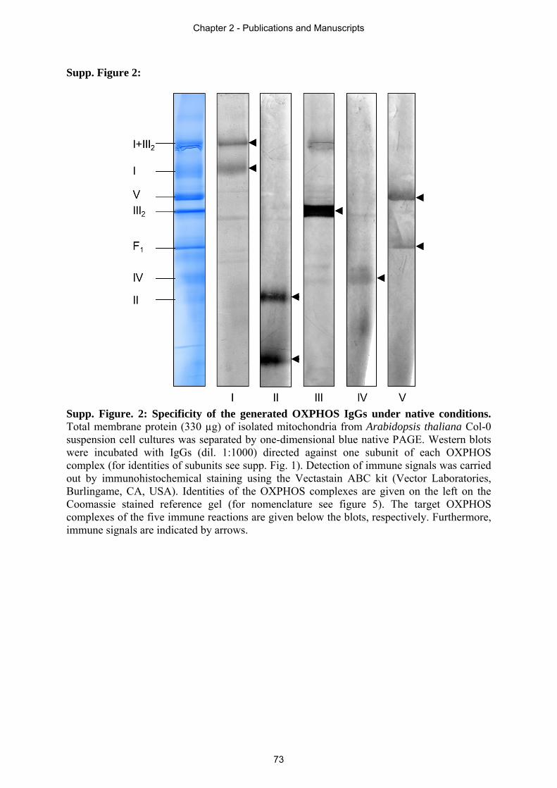
Supp. Figure 2:
Supp. Figure. 2: Specificity of the generated OXPHOS IgGs under native conditions. Total membrane protein (330 µg) of isolated mitochondria from Arabidopsis thaliana Col-0 suspension cell cultures was separated by one-dimensional blue native PAGE. Western blots were incubated with IgGs (dil. 1:1000) directed against one subunit of each OXPHOS complex (for identities of subunits see supp. Fig. 1). Detection of immune signals was carried out by immunohistochemical staining using the Vectastain ABC kit (Vector Laboratories, Burlingame, CA, USA). Identities of the OXPHOS complexes are given on the left on the Coomassie stained reference gel (for nomenclature see figure 5). The target OXPHOS complexes of the five immune reactions are given below the blots, respectively. Furthermore, immune signals are indicated by arrows.
Chapter 2 - Publications and Manuscripts
73
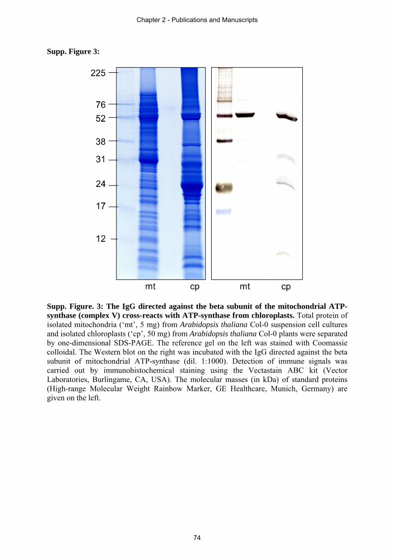
Supp. Figure 3:
Supp. Figure. 3: The IgG directed against the beta subunit of the mitochondrial ATP-synthase (complex V) cross-reacts with ATP-synthase from chloroplasts. Total protein of isolated mitochondria (‘mt’, 5 mg) from Arabidopsis thaliana Col-0 suspension cell cultures and isolated chloroplasts (‘cp’, 50 mg) from Arabidopsis thaliana Col-0 plants were separated by one-dimensional SDS-PAGE. The reference gel on the left was stained with Coomassie colloidal. The Western blot on the right was incubated with the IgG directed against the beta subunit of mitochondrial ATP-synthase (dil. 1:1000). Detection of immune signals was carried out by immunohistochemical staining using the Vectastain ABC kit (Vector Laboratories, Burlingame, CA, USA). The molecular masses (in kDa) of standard proteins (High-range Molecular Weight Rainbow Marker, GE Healthcare, Munich, Germany) are given on the left.
Chapter 2 - Publications and Manuscripts
74
Chapter 3
3 Supplementary discussion and outlook
In this supplementary discussion, the structure as well as possible functions of OXPHOS
complexes and supercomplexes will be discussed against the background of a highly branched
electron transport chain in plants. In the very end of this thesis, the outlook will present
promising approaches, which were beyond of the scope of this thesis and which might
contribute towards a more profound understanding of the plant OXPHOS system.
3.1 The highly branched electron transport chain of plant mitochondria
A deeper knowledge of the mitochondrial respiratory chain is of crucial importance since it is
known from mammals that disorders concerning this part are leading to severe diseases, e.g.
Alzheimer’s and Parkinson’s disease in humans. Also for plants, the recent state of knowledge
shows that the mitochondrial OXPHOS system has more functions besides electron transfer
and oxidative phosphorylation. From this point of view, it is very important to get a complete
characterization of the OXPHOS system with all its structural and functional specialties in
plants, because this will result in a more detailed understanding of the interaction patterns of
different pathways in plants under defined conditions. In contrast to other organisms, the
electron transport chain in plants is highly branched, due to the existence of alternative
oxidoreductases (type II NAD(P)H dehydrogenases, AOX) and other enzymes (GLDH,
ETFQ-OR, G3P) acting as additional electron entry points into the respiratory chain.
In their investigation of supercomplexes in plant mitochondria, Eubel et al. (2003) were able to
show that neither AOX nor the alternative NAD(P)H dehydrogenases form part of any
supercomplex. However, they hypothesized that formation of the I+III2 supercomplex might
play a role in regulation of alternative respiration in a way that the I+III2 supercomplex reduces
access of AOX to its substrate ubiquinol by channelling electrons directly from complex I via
UQ to complex III2 within the supercomplex. Following the hypothesis of Eubel et al. (2003)
would mean that the more I+III2 supercomplex formation exists, the less AOX activity should
occur and vice versa. Indeed, there are several lines of evidence indicating the existence of a
direct electron channelling: (i) flux control experiments (Lenaz and Genova 2009), (ii) X-ray
structure of the matrix arm of complex I (Sazanov and Hinchliffe 2006) and single-particle EM
data for the I+III2 supercomplex (Dudkina et al. 2005), which display a close proximity of the
ubiquinone reduction and ubiquinol oxidation sites in the supercomplex. Even if direct electron
channelling does not occur in the supercomplex, the transfer of ubiquinol from complex I to
complex III2 might be favoured because of potentially lower diffusion distances. For yeast, it
Chapter 3 - Supplementary Discussion and Outlook
75
was reported by Heinemeyer et al. (2007) that formation of the III2+IV1-2 supercomplex
optimizes electron transport between these two complexes. This was also shown for potato
mitochondria by Lenaz and Genova (2009), who also suggested that supercomplex formation
might limit the generation of ROS.
In 2010, Sunderhaus et al. initiated a study on the supramolecular structure of the OXPHOS
system in the thermogenic tissue of Arum maculatum, which is known to produce large
amounts of AOX for heat production during pollination in the spadix. Accordingly, the electron
channelling assumption of Eubel et al. (2003) would predict low amounts of I+III2
supercomplex. In contrast to this, Sunderhaus et al. (2010) reported that the respiratory chain in
Arum maculatum differs from the one in Arabidopsis thaliana in a way that no monomeric
complex I could be detected. Complex I is only present in the I+III2 supercomplex.
Furthermore, supercomplexes containing complex IV could be shown for Arum maculatum,
even though only in low abundances, but not for Arabidopsis. As already reported by Eubel et
al. (2003), also Sunderhaus et al. (2010) were able to show that AOX does not form part of any
OXPHOS complex or supercomplex and no connection between the amounts of I+III2
supercomplex and AOX activity was detected. Hence, they concluded, that supercomplex
formation does not affect the activity of alternative respiration. This might also invalidate the
assumption that electrons are directly channelled, via UQ, between complex I and complex III2
within the supercomplex.
Thus, not only the occurrence of electron channelling is disputable, but the question if
OXPHOS complexes and alternative respiratory enzymes are using common UQ pools also
remains unsolved. Dudkina et al. (2005) reported that there might be an UQ pool in the
complex III2-containing supercomplex. On the other hand, Rasmusson et al. (2008) pointed out
that the use of different UQ pools seems more reliable for the case that there is no direct
channelling of electrons within supercomplex formation.
Although there is no evidence that the I+III2 supercomplex regulates alternative respiration, it
is known that complex I is involved in maintaining the redox balance during photosynthesis.
Dutilleul et al. (2003a) investigated a cytoplasmic male-sterile Nicotiana sylvestris mutant
(CMSII) which lacks functional complex I. They were able to show that the CMSII mutant has
an increased alternative respiration via NADH dehydrogenases and AOX as well as modified
abundances and activities of several antioxidant enzymes. This study led the authors to the
statement that the loss of functional complex I induces antioxidant crosstalk within the cell and
readjustment of organelle metabolism in order to maintain the cellular redox balance. Hence, it
can be deduced that complex I plays an important role for the redox balance under regular
Chapter 3 - Supplementary Discussion and Outlook
76
photosynthetic conditions and that the absence of complex I forces the cell to modulate
enzymatic activity in order to keep the redox balance stable.
Such an effect could also be demonstrated by Dutilleul et al. (2003b). By investigating the role
of mitochondrial complex I in leaves using the same CMSII mutant the following conclusions
were drawn: (i) Complex I plays an important role in photorespiratory metabolism in vivo. (ii)
Mitochondrial complex I is required for optimal photosynthetic performance. (iii) Complex I is
necessary to avoid redox disruption of photosynthesis when leaf mitochondria have to oxidize
simultaneously respiratory and photorespiratory substrates. Both studies (Dutilleul et al. 2003a,
Dutilleul et al. 2003b) revealed a necessity of mitochondrial complex I for the maintenance of
an appropriate redox balance in leaves. This is especially true during phases of adjustment of
the photosynthetic system like transitions or decreased intercellular CO2 that occur during the
onset of drought.
These findings are in line with the data reported in section 2.4, in which the ratio of OXPHOS
complexes was investigated in different tissues of Arabidopsis thaliana. Complex I was found
to be highly abundant in green organs (leaves and stems) but less abundant in non-green tissue
(callus and seeds). This led to the suggestion that complex I might have further light-dependent
side functions besides its participation in electron transport and proton translocation. This is
supported by the findings of Dutilleul et al. (2003a, 2003b) on the importance of complex I in
processes of photosynthesis, photorespiration and maintenance of cell redox balance.
Since evidence was adduced for complex I being involved in maintaining the cell redox
balance, the upcoming question again is: What about the potential role of the I+III2
supercomplex in this regulatory mechanism? The hypothesized function of this supercomplex,
the regulation of alternative respiration, could not be proven so far, but there are several hints
which indicate a role of this supercomplex in redox balance.
(i) First, the I+III2 supercomplex is very abundant in higher plants like Arabidopsis thaliana
(Dudkina et al. 2005), Zea mays (Peters et al. 2008), Solanum tuberosum (Bultema et al. 2009)
and Arum maculatum (Sunderhaus et al. 2010) but of lower abundance in mammalian
mitochondria (Eubel et al. 2004b). In mammals and yeast supercomplexes containing complex
IV (I+III+IV in various amounts of the individual complexes) are mainly existent (Dudkina et
al. 2008), whereas in plants these larger supercomplexes are rarely detectable (Eubel et al.
2004a, Eubel et al. 2004b, Krause et al. 2004, Dudkina et al. 2006a, Sunderhaus et al. 2010).
The fact that the I+III2 supercomplex mainly exists in plants, together with the knowledge that
the alternative oxidoreductases (alternative NAD(P)H dehydrogenases and AOX) are plant
specific lead to the assumption that, indeed, the I+III2 supercomplex might play a role in the
Chapter 3 - Supplementary Discussion and Outlook
77
context of alternative respiration. Although it could not be proven so far that the supercomplex
regulates alternative respiration, an interaction of alternative oxidoreductases and the I+III2
supercomplex would be perfectly in line with the study of Sunderhaus et al. (2010). The
authors were able to show that in Arum maculatum spadix mitochondria, which are known to
have an extremely high rate of alternative respiration, complex I is completely absent in its
monomeric form but is existent in the I+III2 supercomplex.
(ii) Furthermore, photosynthesis always comes along with photorespiration. Especially under
high light conditions, there is excess formation of reduction equivalents within chloroplasts (by
photosynthesis) and mitochondria (by glycine to serine conversion in the context of
photorespiration). If the previous hypothesized direct electron channelling between complex I
and complex III2 within the supercomplex exists, this would result in a higher respiration rate
due to a faster electron transfer because of the reduced diffusion distance between both
complexes (Dudkina et al. 2005). This would explain the high amounts of I+III2 supercomplex
formation in plant mitochondria in contrast to other organisms which do not carry out
photosynthesis. Additionally, this assumption is supported by the finding that mitochondria
from green leaves, where most of the photosynthesis in plants takes place and therefore high
amounts of reduction equivalents are present, contain clearly more I+III2 supercomplex than
mitochondria isolated from non-green callus tissue (section 2.4).
(iii) Further evidence was given by Lenaz and Genova (2009), who suggested a possible role of
supercomplexes in limiting ROS formation. Complex I is known to be one of the major sources
for ROS formation in mitochondria. There is evidence that ROS are generated due to the
reaction of complex I bound FMN with oxygen (Galkin and Brandt 2005). Hence, Lenaz and
Genova (2009) hypothesized that FMN only becomes exposed to oxygen when complex I is
dissociated from complex III2. In their study they also suggest that complex I might undergo
conformational changes when associated with other complexes and thereby avoid FMN to react
with oxygen.
Taking this knowledge into account, conclusions can be drawn which are going beyond the
statements of Dutilleul et al. (2003a, 2003b). The investigated CMSII mutant not only lacks
functional complex I but also the I+III2 supercomplex (Pineau et al. 2005). Since complex I in
its monomeric form is a major source of ROS formation, as mentioned above, it makes sense
that up to 90% of complex I is associated with dimeric complex III forming a supercomplex
structure, which is known to limit ROS generation. However, in the CMSII mutant complex I is
absent and hence, one major source of ROS is discarded. The higher abundance of alternative
oxidoreductases, which was determined by Dutilleul et al. (2003a) in the CMSII mutant, is due
Chapter 3 - Supplementary Discussion and Outlook
78
to an increase of NADH, which occurs because of the lack of complex I and the I+III2
supercomplex, which normally oxidises the NADH. Concerning NADH oxidation, again the
I+III2 supercomplex seems to be more important than complex I because oxidation of NADH
within the supercomplex structure is supposed to be enhanced due to reduced diffusion
distances between the single complexes (Dudkina et al. 2005). Therefore, it is questionable if
the contribution to optimized photosynthesis (including photorespiratory metabolism) and cell
redox balance really depends on complex I or if it is more likely the I+III2 supercomplex which
is responsible for these regulatory mechanisms.
(iv) As a last point, further evidence is given by a study of Perales et al. (2005) in which a
complex I mutant lacking CA2 (ΔAt1g47260) was investigated. As already mentioned in
section 1.4, CAs are suggested to be involved in CO2 distribution between mitochondria and
chloroplasts. This process is especially important under conditions resulting in high
photorespiration rates. Therefore, CAs can also be considered to play a role in optimizing
photosynthetic processes. Perales et al. (2005) were able to show that lack of CA2 results in a
serious decrease of complex I abundance. Additionally, also the I+III2 supercomplex is
extremely decreased. If this supercomplex has another important functional role besides the
hypothesized ones, remaining complex I amounts in the CA2 mutant should be existent in the
supercomplex in order to keep its function. Since this is obviously not the case in this mutant, it
can be assumed that the I+III2 supercomplex is most probably somehow involved in optimizing
photosynthetic processes and stabilizing cell redox balance.
All this evidence indicates a functional role of the I+III2 supercomplex in processes related to
alternative respiration, optimizing photosynthetic processes, stabilizing cell redox balance and
preventing ROS formation. Nevertheless, more investigations on the potential functions of this
supercomplex are necessary.
However, besides the previous suggested function of supercomplex formation in the regulation
of alternative respiration, the necessity of supercomplexes for the assembly and stability of
individual components was determined. Evidence exist that complex I is necessary for the
presence of fully assembled complex III in human mitochondria (Ugalde et al. 2004). Other
studies reveal that an absence of assembled complex III results in a loss of complex I in human
mitochondria confirming a structural dependence of these two complexes (Blakely et al. 2005).
And also in mouse cells complex III was found to be responsible for the stability of complex I
(Acín-Pérez et al. 2004). Furthermore, it was reported that complex IV is important for
complex I assembly in fibroblasts (Diaz et al. 2006). These functional roles of supercomplexes
Chapter 3 - Supplementary Discussion and Outlook
79
would also explain why activity of the I+III2 supercomplex is also detectable in non-
photosynthetically active tissues like callus (section 2.4).
3.2 Outlook
This thesis could reveal new insights into structure and function of the respiratory chain in
plants. In order to achieve a complete characterization of the OXPHOS system, a substantial
investment of additional work is required. Many questions concerning structure and function of
the respiratory chain complexes still remain unanswered. Especially in plants this topic is much
more complicated than in other organisms due to the plant specific extra subunits, which are
present in most respiratory complexes. Individual complexes participate in electron transport
and oxidative phosphorylation but additionally fulfil supplementary functions, like complex I,
which was shown to be involved in whole cell redox balance. The functions of the
supercomplexes are not entirely solved so far and also the formation of supercomplexes to
higher oligomeric structures needs further investigation. In the following section, some
approaches will be outlined, which could contribute to a deeper understanding of the OXPHOS
system.
Structural investigations of respiratory chain complexes were done by X-ray crystallography.
Unfortunately, this was performed only for complexes from bacteria (Escherichia coli,
Thermus thermophilus), yeast (Yarrowia lipolytica) and mammals (Bos taurus, Sus scrofa) but
not for plants. Crystallization of complex I, which by far is the largest of the respiratory
complexes, required the most effort. The first crystal structure of the entire bacterial complex I
from Thermus thermophilus was published by Efremov et al. (2010) and shortly after, the
structure of the entire mitochondrial enzyme from Yarrowia lipolytica was presented by Hunte
et al. (2010). In both studies, the resolution of the membrane arm was too low to display the
assignment of subunits. Recently, Efremov and Sazanov (2011) published the structure of
complex I membrane domain from E. coli with a higher resolution and were able to identify
subunits.
X-ray crystallography produces detailed information about structure and also indirectly about
function of the respiratory chain complexes. However, a cross-species transfer of these data is
difficult because of the significant differences in OXPHOS systems between plants and other
organisms (plant specific subunits, highly branched electron transfer system). Since crystal
structures from non-plant species can only provide hints for their plant counterparts, the need
for other strategies enabling the investigation of the OXPHOS system in plants is obvious.
Chapter 3 - Supplementary Discussion and Outlook
80
Thanks to progress in methodology, also plant OXPHOS complexes have become accessible to
structural characterization. For example, Klodmann et al. (2010) used a biochemical approach
to assign subunits to plant complex I. Furthermore, the progress in EM techniques enables
generation of high quality structural data, resulting in new findings about structures and
possible functions of plant respiratory chain complexes and supercomplexes. By the use of
methods like cryo-ET, it also became possible to reveal the overall structure of the OXPHOS
system in mitochondrial membranes (section 2.2). Due to the progress in EM techniques and
biochemical methods to purify organelles and individual protein complexes, it will become
possible to obtain more new, very detailed, structural data of the plant OXPHOS system in the
near future. Recently, Dudkina et al. (2011) were able to investigate the interaction of the
complexes I, III and IV within the bovine respirasome using cryo-ET. For the analysis of
supercomplexes it could be beneficial to use plant species, which are known to possess very
stable supercomplexes. The colourless green alga Polytomella, for example, contains unusually
stable dimeric ATP synthase complex and was already used for the investigation of the row-
like organization of this supercomplex in intact mitochondria by cryo-ET (Dudkina et al.
2010b).
Besides the structural analysis of the OXPHOS system, it is also important to figure out the
functions of the individual complexes and supercomplexes. As already discussed in section 3.1,
plant OXPHOS complexes are involved in several processes of vital importance for the plant
cell, like complex I which is involved in maintaining the redox balance during photosynthesis.
In this thesis, the investigation of the ETC was based on proteomics. For a complete
understanding of the functions of OXPHOS complexes, it might be beneficial to also undertake
transcriptomic studies. Proteome and transcriptome are highly dynamic, changing with time,
requirements and stresses. Integration of information from these two fields will lead to a more
complete understanding of the living organism. Analysing the transcriptome under defined
conditions as well as in specific tissues will reveal differences in the expression of the genes of
the individual OXPHOS complexes. This in turn, on the one hand, might lead to suggestions of
possible side functions of those higher expressed complexes besides electron transport. On the
other hand, since it is known that the expression level of a gene does not necessarily correlate
with the amount of protein, comparison of the expression profiles of the OXPHOS complexes
with the proteomic data will indicate the rate of regulation of the complexes. This regulation
either takes place on the level of transcripts (transcriptional processes associated with nuclear
or organelle genomes, post-transcriptional processing, RNA-editing) or on the protein level
(post-translational modifications, modification of proteins modifying enzymatic functions).
Chapter 3 - Supplementary Discussion and Outlook
81
However, expression profiling in combination with proteomic studies can be beneficial to
figure out potential differences in gene expression, protein amounts as well as regulation of the
five OXPHOS complexes under defined conditions.
As already discussed in section 3.1, plant OXPHOS complexes differ in their abundances
regarding different tissues. Thereby, complex I was found to be highly abundant in green
organs (section 2.4), which is in line with its contribution in maintaining cell redox balance
during photosynthesis (Dutilleul et al. 2003a, Dutilleul et al. 2003b). Hence, it will be very
interesting to also have a look at the other complexes, since complex II behaves inversely to
complex I and shows higher amounts in non-green tissue. This suggests a role of complex II in
this type of tissue. Indeed, Gleason et al. (2011) reported that complex II contributes to
localized ROS production in mitochondria, which regulates plant stress and defense response.
This is perfectly in line with the high amounts of complex II detected in callus (section 2.4)
since it is known that this tissue is heavily stress exposed (Halliwell 2003). And also for the
other respiratory chain complexes, tissue specific investigations might reveal new insights into
physiological functions.
Another powerful tool for physiological studies is the investigation of knock-out mutants.
Plenty of information about functions of complex I is achieved by analyses of knock-out
mutants (e.g. Dutilleul et al. 2003a, Dutilleul et al. 2003b, Perales et al. 2005). Today, lots of
Arabidopsis thaliana knock-out mutants are commercially available. Therefore, this is a
promising approach for future investigations of the physiological role of individual OXPHOS
complexes under defined conditions. Since there is evidence that complex I is involved in
photosynthetic processes, analyses of complex I mutants in combination with different CO2
concentrations (high CO2, low CO2) are inevitable. In this context, also the I+III2 supercomplex
should be examined more carefully due to its suggested potential role in regulation of
alternative respiration as well as the possible relation to processes which are involved in
stabilizing cell redox balance (section 3.1).
Chapter 3 - Supplementary Discussion and Outlook
82
References
Acín-Pérez, R., Bayona-Bafaluy, M.P., Fernández-Silva, P., Moreno-Loshuertos, R., Pérez-
Martos, A., Bruno, C., Moraes, C.T. and Enríquez, J.A. (2004) Respiratory complex III is
required to maintain complex I in mammalian mitochondria. Mol. Cell 13: 805–815.
Berry, E.A., Guergova-Kuras, M., Huang, L.S. and Crofts, A.R. (2000) Structure and function
of cytochrome bc complexes. Annu Rev Biochem 69: 1005–1075.
Blakely, E.L., Mitchell, A.L., Fisher, N., Meunier, B., Nijtmans, L.G., Schaefer, A.M.,
Jackson, M.J., Turnbull, D.M. and Taylor, R.W. (2005) A mitochondrial cytochrome b
mutation causing severe respiratory chain enzyme deficiency in humans and yeast. FEBS J
272: 3583–3592.
Brandt, U. (2006) Energy converting NADH:quinone oxidoreductase (complex I). Annu. Rev.
Biochem 75: 69–92.
Braun, H.-P. and Schmitz, U.K. (1999) The protein-import apparatus of plant mitochondria.
Planta 209: 267–274.
Braun, H.-P. and Zabaleta, E. (2007) Carbonic anhydrase subunits of the mitochondrial NADH
dehydrogenase complex (complex I) in plants. Physiol Plant 129: 114–122.
Braun, H.-P., Emmermann, M., Kruft, V. and Schmitz, U.K. (1992) The general mitochondrial
processing peptidase from potato is an integral part of cytochrome c reductase of the
respiratory chain. EMBO J 11: 3219–3227.
Bultema, J., Braun, H.-P., Boekema, E. and Kouril, R. (2009) Megacomplex organization of
the oxidative phosphorylation system by structural analysis of respiratory supercomplexes from
potato. Biochim Biophys Acta 1787: 60–67.
Carrie, C., Murcha, M.W. and Whelan, J. (2010) An in silico analysis of the mitochondrial
protein import apparatus of plants. BMC Plant Biol 10: 249.
Clifton, R., Millar, A.H. and Whelan, J. (2006) Alternative oxidases in Arabidopsis: a
comparative analysis of differential expression in the gene family provides new insights into
function of non-phosphorylating bypasses. Biochim. Biophys. Acta 1757: 730–741.
Davies, K.M., Strauss, M., Daum, B., Kief, J.H., Osiewacz, H.D., Rycovska, A., Zickermann,
V. and Kuhlbrandt, W. (2011) Macromolecular organization of ATP synthase and complex I in
whole mitochondria. Proceedings of the National Academy of Sciences 108: 14121–14126.
References
83
Diaz, F., Fukui, H., Garcia, S. and Moraes, C.T. (2006) Cytochrome c oxidase is required for
the assembly/stability of respiratory complex I in mouse fibroblasts. Mol. Cell. Biol 26: 4872–
4881.
Dudkina, N.V., Eubel, H., Keegstra, W., Boekema, E.J. and Braun, H.-P. (2005) Structure of a
mitochondrial supercomplex formed by respiratory-chain complexes I and III. Proc. Natl.
Acad. Sci. U.S.A 102: 3225–3229.
Dudkina, N.V., Heinemeyer, J., Sunderhaus, S., Boekema, E.J. and Braun, H.-P. (2006a)
Respiratory chain supercomplexes in the plant mitochondrial membrane. Trends in Plant
Science 11: 232–240.
Dudkina, N.V., Kouril, R., Peters, K., Braun, H.-P. and Boekema, E.J. (2010a) Structure and
function of mitochondrial supercomplexes. Biochim. Biophys. Acta 1797: 664–670.
Dudkina, N.V., Kudryashev, M., Stahlberg, H. and Boekema, E.J. (2011) Interaction of
complexes I, III, and IV within the bovine respirasome by single particle cryoelectron
tomography. Proceedings of the National Academy of Sciences.
Dudkina, N.V., Oostergetel, G.T., Lewejohann, D., Braun, H.-P. and Boekema, E.J. (2010b)
Row-like organization of ATP synthase in intact mitochondria determined by cryo-electron
tomography. Biochimica et Biophysica Acta (BBA) - Bioenergetics 1797: 272–277.
Dudkina, N.V., Sunderhaus, S., Boekema, E.J. and Braun, H.-P. (2008) The higher level of
organization of the oxidative phosphorylation system: mitochondrial supercomplexes. J
Bioenerg Biomembr 40: 419–424.
Dutilleul, C., Driscoll, S., Cornic, G., Paepe, R. de, Foyer, C.H. and Noctor, G. (2003b)
Functional mitochondrial complex I is required by tobacco leaves for optimal photosynthetic
performance in photorespiratory conditions and during transients. Plant Physiol 131: 264–275.
Dutilleul, C., Garmier, M., Noctor, G., Mathieu, C., Chétrit, P., Foyer, C.H. and Paepe, R. de
(2003a) Leaf mitochondria modulate whole cell redox homeostasis, set antioxidant capacity,
and determine stress resistance through altered signaling and diurnal regulation. Plant Cell 15:
1212–1226.
Efremov, R.G. and Sazanov, L.A. (2011) Structure of the membrane domain of respiratory
complex I. Nature 476: 414–420.
Efremov, R.G., Baradaran, R. and Sazanov, L.A. (2010) The architecture of respiratory
complex I. Nature 465: 441–445.
References
84
Eubel, H. (2004a) Identification and Characterization of Respirasomes in Potato Mitochondria.
Plant Physiol 134: 1450–1459.
Eubel, H., Heinemeyer, J., Sunderhaus, S. and Braun, H.-P. (2004b) Respiratory chain
supercomplexes in plant mitochondria. Plant Physiology and Biochemistry 42: 937–942.
Eubel, H., Jänsch, L. and Braun, H.-P. (2003) New insights into the respiratory chain of plant
mitochondria. Supercomplexes and a unique composition of complex II. Plant Physiol 133:
274–286.
Fowler, L.R. and Hatefi, Y. (1961) Reconstitution of the electron transport system. III.
Reconstitution of DPNH oxidase, succinic oxidase, and DPNH, succinic oxidase. Biochem
Biophys Res Commun 5: 203–208.
Fowler, L.R. and Richardson, S.H. (1963) Studies on the electron transfer system. L. On the
mechanism of reconstitution of the mitochondrial electron transfer system. J Biol Chem 238:
456–463.
Galkin, A. and Brandt, U. (2005) Superoxide radical formation by pure complex I
(NADH:ubiquinone oxidoreductase) from Yarrowia lipolytica. J. Biol. Chem 280: 30129–
30135.
Gleason, C., Huang, S., Thatcher, L.F., Foley, R.C., Anderson, C.R., Carroll, A.J., Millar, A.H.
and Singh, K.B. (2011) Mitochondrial complex II has a key role in mitochondrial-derived
reactive oxygen species influence on plant stress gene regulation and defense. Proc. Natl. Acad.
Sci. U.S.A 108: 10768–10773.
Gray, M.W., Burger, G. and Lang, B.F. (2001) The origin and early evolution of mitochondria.
Genome Biol 2: 1018.1 - 1018.5.
Hackenbrock, C.R., Chazotte, B. and Gupte, S.S. (1986) The random collision model and a
critical assessment of diffusion and collision in mitochondrial electron transport. J Bioenerg
Biomembr 18: 331–368.
Halliwell, B. (2003) Oxidative stress in cell culture: an under-appreciated problem? FEBS Lett
540: 3–6.
Hatefi, Y. (1985) The Mitochondrial Electron Transport and Oxidative Phosphorylation
System. Annu. Rev. Biochem 54: 1015–1069.
References
85
Hatefi, Y., Haavik, A.G., Fowler, L.R. and Griffiths, D.E. (1962) Studies on the electron
transfer system. XLII. Reconstitution of the electron transfer system. J Biol Chem 237: 2661–
2669.
Hatefi, Y., Jurtshuk, P. and Haavik, A.G. (1961) Studies on the electron transport system.
XXXI. DPNH-cytochrome c reductase II. Biochim Biophys Acta 52: 119–129.
Heazlewood, J., Howell, K. and Millar, A. (2003) Mitochondrial complex I from Arabidopsis
and rice: orthologs of mammalian and fungal components coupled with plant-specific subunits.
Biochim Biophys Acta 1604: 159–169.
Heinemeyer, J., Braun, H.-P., Boekema, E.J. and Kouril, R. (2007) A structural model of the
cytochrome C reductase/oxidase supercomplex from yeast mitochondria. J. Biol. Chem 282:
12240–12248.
Ho, L.H.M., Giraud, E., Lister, R., Thirkettle-Watts, D., Low, J., Clifton, R., Howell, K.A.,
Carrie, C., Donald, T. and Whelan, J. (2007) Characterization of the regulatory and expression
context of an alternative oxidase gene provides insights into cyanide-insensitive respiration
during growth and development. Plant Physiol 143: 1519–1533.
Huang, S., Taylor, N.L., Narsai, R., Eubel, H., Whelan, J. and Millar, A.H. (2010) Functional
and composition differences between mitochondrial complex II in Arabidopsis and rice are
correlated with the complex genetic history of the enzyme. Plant Mol. Biol 72: 331–342.
Hunte, C., Zickermann, V. and Brandt, U. (2010) Functional modules and structural basis of
conformational coupling in mitochondrial complex I. Science 329: 448–451.
Klodmann, J. and Braun, H.-P. (2011) Proteomic approach to characterize mitochondrial
complex I from plants. Phytochemistry 72: 1071–1080.
Klodmann, J., Sunderhaus, S., Nimtz, M., Jansch, L. and Braun, H.-P. (2010) Internal
architecture of mitochondrial complex I from Arabidopsis thaliana. Plant Cell 22: 797–810.
Krause, F., Reifschneider, N.H., Vocke, D., Seelert, H., Rexroth, S. and Dencher, N.A. (2004)
"Respirasome"-like supercomplexes in green leaf mitochondria of spinach. J. Biol. Chem 279:
48369–48375.
Lenaz, G. and Genova, M.L. (2009) Structural and functional organization of the mitochondrial
respiratory chain: a dynamic super-assembly. Int. J. Biochem. Cell Biol 41: 1750–1772.
Logan, D.C. (2006) The mitochondrial compartment. Journal of Experimental Botany 57:
1225–1243.
References
86
Mannella, C.A. (2006) Structure and dynamics of the mitochondrial inner membrane cristae.
Biochimica et Biophysica Acta (BBA) - Molecular Cell Research 1763: 542–548.
Mannella, C.A. and Tedeschi, H. (1987) Importance of the mitochondrial outer membrane
channel as a model biological channel. J. Bioenerg. Biomembr 19: 305–308.
Mannella, C.A., Marko, M. and Buttle, K. (1997) Reconsidering mitochondrial structure: new
views of an old organelle. Trends Biochem Sci 22: 37–38.
Maxwell, D.P., Wang, Y. and McIntosh, L. (1999) The alternative oxidase lowers
mitochondrial reactive oxygen production in plant cells. Proc. Natl. Acad. Sci. U.S.A 96:
8271–8276.
Michalecka, A.M., Svensson, A.S., Johansson, F.I., Agius, S.C., Johanson, U., Brennicke, A.,
Binder, S. and Rasmusson, A.G. (2003) Arabidopsis genes encoding mitochondrial type II
NAD(P)H dehydrogenases have different evolutionary origin and show distinct responses to
light. Plant Physiol 133: 642–652.
Millar, A., Mittova, V., Kiddle, G., Heazlewood, J., Bartoli, C., Theodoulou, F. and Foyer, C.
(2003) Control of ascorbate synthesis by respiration and its implications for stress responses.
Plant Physiol 133: 443–447.
Millar, A.H., Eubel, H., Jänsch, L., Kruft, V., Heazlewood, J.L. and Braun, H.-P. (2004)
Mitochondrial cytochrome c oxidase and succinate dehydrogenase complexes contain plant
specific subunits. Plant Mol. Biol 56: 77–90.
Mitchell, P. (1961) Coupling of phosphorylation to electron and hydrogen transfer by a chemi-
osmotic type of mechanism. Nature 191: 144–148.
Pangborn, M. (1942) Isolation and purification of a serologically active phospholipid from beef
heart. J Biol Chem: 247–256.
Perales, M., Eubel, H., Heinemeyer, J., Colaneri, A., Zabaleta, E. and Braun, H.-P. (2005)
Disruption of a Nuclear Gene Encoding a Mitochondrial Gamma Carbonic Anhydrase Reduces
Complex I and Supercomplex I+III2 Levels and Alters Mitochondrial Physiology in
Arabidopsis. Journal of Molecular Biology 350: 263–277.
Peters, K., Dudkina, N.V., Jänsch, L., Braun, H.-P. and Boekema, E.J. (2008) A structural
investigation of complex I and I+III2 supercomplex from Zea mays at 11-13 Ǻ resolution:
assignment of the carbonic anhydrase domain and evidence for structural heterogeneity within
complex I. Biochim. Biophys. Acta 1777: 84–93.
References
87
Pineau, B., Layoune, O., Danon, A. and De, P. (2008) L-galactono-1,4-lactone dehydrogenase
is required for the accumulation of plant respiratory complex I. J Biol Chem 283: 32500–
32505.
Pineau, B., Mathieu, C., Gérard-Hirne, C., Paepe, R. de and Chétrit, P. (2005) Targeting the
NAD7 subunit to mitochondria restores a functional complex I and a wild type phenotype in
the Nicotiana sylvestris CMS II mutant lacking nad7. J. Biol. Chem 280: 25994–26001.
Price, G., Badger, M., Woodger, F. and Long, B. (2008) Advances in understanding the
cyanobacterial CO2-concentrating-mechanism (CCM): functional components, Ci transporters,
diversity, genetic regulation and prospects for engineering into plants. J Exp Bot 59: 1441–
1461.
Rasmusson, A.G. and Wallström, S.V. (2010) Involvement of mitochondria in the control of
plant cell NAD(P)H reduction levels. Biochem. Soc. Trans 38: 661–666.
Rasmusson, A.G., Fernie, A.R. and van Dongen, J.T. (2009) Alternative oxidase: a defence
against metabolic fluctuations? Physiol Plant 137: 371–382.
Rasmusson, A.G., Geisler, D.A. and Møller, I.M. (2008) The multiplicity of dehydrogenases in
the electron transport chain of plant mitochondria. Mitochondrion 8: 47–60.
Rüchel R., Steere, R.L. and Erbe, E.F. (1978) Transmission-electron microscopic observations
of freeze-etched polyacrylamide gels. J Chromatogr 166: 563–575.
Sagan, L. (1967) On the origin of mitosing cells. J Theor Biol 14: 255–274.
Sazanov, L.A. and Hinchliffe, P. (2006) Structure of the hydrophilic domain of respiratory
complex I from Thermus thermophilus. Science 311: 1430–1436.
Schägger, H. (2002) Respiratory chain supercomplexes of mitochondria and bacteria.
Biochimica et Biophysica Acta (BBA) - Bioenergetics 1555: 154–159.
Schägger, H. and Jagow, G. von (1991) Blue native electrophoresis for isolation of membrane
protein complexes in enzymatically active form. Anal. Biochem 199: 223–231.
Scheffler, I.E. (1998) Molecular genetics of succinate:quinone oxidoreductase in eukaryotes.
Prog Nucleic Acid Res Mol Biol 60: 267–315.
Schlame, M., Rua, D. and Greenberg, M. (2000) The biosynthesis and functional role of
cardiolipin. ., 39 (2000), pp. 257–288. Progr. Lipid Res. 39: 257–288.
Stock, D., Gibbons, C., Arechaga, I., Leslie, A.G. and Walker, J.E. (2000) The rotary
mechanism of ATP synthase. Curr. Opin. Struct. Biol 10: 672–679.
References
88
Strauss, M., Hofhaus, G., Schröder, R.R. and Kühlbrandt, W. (2008) Dimer ribbons of ATP
synthase shape the inner mitochondrial membrane. EMBO J 27: 1154–1160.
Sun, F., Huo, X., Zhai, Y., Wang, A., Xu, J., Su, D., Bartlam, M. and Rao, Z. (2005) Crystal
structure of mitochondrial respiratory membrane protein complex II. Cell 121: 1043–1057.
Sunderhaus, S., Dudkina, N., Jansch, L., Klodmann, J., Heinemeyer, J., Perales, M., Zabaleta,
E., Boekema, E. and Braun, H.-P. (2006) Carbonic anhydrase subunits form a matrix-exposed
domain attached to the membrane arm of mitochondrial complex I in plants. J Biol Chem 281:
6482–6488.
Sunderhaus, S., Klodmann, J., Lenz, C. and Braun, H.-P. (2010) Supramolecular structure of
the OXPHOS system in highly thermogenic tissue of Arum maculatum. Plant Physiol. Biochem
48: 265–272.
Svensson, A.S. and Rasmusson, A.G. (2001) Light-dependent gene expression for proteins in
the respiratory chain of potato leaves. Plant J 28: 73–82.
Thirkettle-Watts, D. (2003) Analysis of the Alternative Oxidase Promoters from Soybean.
Plant Physiol 133: 1158–1169.
Tsukihara, T., Aoyama, H., Yamashita, E., Tomizaki, T., Yamaguchi, H., Shinzawa-Itoh, K.,
Nakashima, R., Yaono, R. and Yoshikawa, S. (1996) The whole structure of the 13-subunit
oxidized cytochrome c oxidase at 2.8 A. Science 272: 1136–1144.
Ugalde, C., Janssen, R.J.R.J., van den Heuvel, L.P., Smeitink, J.A.M. and Nijtmans, L.G.J.
(2004) Differences in assembly or stability of complex I and other mitochondrial OXPHOS
complexes in inherited complex I deficiency. Hum. Mol. Genet 13: 659–667.
Vander Heiden, M.G. (2001) Bcl-xL Promotes the Open Configuration of the Voltage-
dependent Anion Channel and Metabolite Passage through the Outer Mitochondrial Membrane.
Journal of Biological Chemistry 276: 19414–19419.
Welchen, E., Klodmann, J. and Braun, H.-P. (2011) Biogenesis and supermolecular
organization of the oxidative phosphorylation system in plants. In Plant Mitochondria. Edited
by Kempken, F. pp. 327–355. Springer Science+Business Media LLC, New York, NY.
Wittig, I., Braun, H.-P. and Schägger, H. (2006) Blue native PAGE. Nat Protoc 1: 418–428.
Yagi, T., Yano, T., Di Bernado, S. and Matsuno-Yagi, A. (1998) Procaryotic complex I (NDH-
1), an overview. Biochim. Biophys. Acta 1364: 125–133.
References
89
Yankovskaya, V., Horsefield, R., Törnroth, S., Luna-Chavez, C., Miyoshi, H., Léger, C.,
Byrne, B., Cecchini, G. and Iwata, S. (2003) Architecture of succinate dehydrogenase and
reactive oxygen species generation. Science 299: 700–704.
References
90

Curriculum vitae
Name: Katrin Peters
Date of Birth: 16.03.1982
Place of Birth: Uelzen, Germany
Citizenship: Citizen of Germany
Marital Status: unmarried
SCHOOL EDUCATION
1988 – 1992 Primary school in Wieren
1992 – 1994 Orientierungsstufe in Bad Bodenteich
1994 – 2001 Secondary school ‘Herzog-Ernst-Gymnasium’ in Uelzen
Qualification: ‘Abitur’
ACADEMIC EDUCATION
Oct. 2001 – Sept. 2002 Student at the Universität Hildesheim,
major: Lehramt Grund-, Haupt- und Realschule
subjects: Biology, English, Mathematics
Oct. 2002 – Oct. 2005 Student at the Leibniz Universität Hannover,
major: Plant Biotechnology
Oct. 2005 Bachelor-degree in Plant Biotechnolgy (B.Sc.)
Thesis: “Untersuchungen zur Apoptose in Zellkulturen von
Arabidopsis thaliana.“
(‘Investigation of apoptosis in cell cultures of Arabidopsis
thaliana.’)
Grade: ‘excellent’
Oct. 2005 – Sept. 2007 Student at the Leibniz Universität Hannover,
major: Plant Biotechnology
Sept. 2007 Master-degree in Plant Biotechnology (M.Sc.)
91
Thesis: “Untersuchungen zum mitochondrialen NADH-
Dehydrogenase-Komplex aus Mais“
(‘Investigation of the mitochondrial NADH-dehydrogenase-
complex in maize.’)
Grade: ‘with honours’
Since Oct. 2007 PhD student at the Leibniz Universität Hannover, Institute of
Plant Genetics, Department Plant Proteomics, Prof. Dr. Hans-
Peter Braun
92
Publications
Journal research papers and manuscripts:
Peters, K., Dukina, N.V., Jänsch, L., Braun, H.-P. and Boekema, E.J. (2008): A structural investigation of complex I and I+III2 supercomplex from Zea mays at 11-13 Å resolution: assignment of the carbonic anhydrase domain and evidence for structural heterogeneity within complex I. Biochim. Biophys. Acta (Bioenergetics) 1777: 84-93. Dudkina, N.V., Kouřil, R., Peters, K., Braun, H.-P. and Boekema, E.J. (2010): Structure and function of mitochondrial supercomplexes. Biochim. Biophys. Acta (Bioenergetics) 1797: 664-670. Peters, K. and Braun, H.-P. (2011): Comparative analyses of protein complexes by blue native DIGE. Methods Mol. Biol. In press. Peters, K., Nießen, M., Peterhänsel, C., Späth, B., Marchfelder, A., Hölzle, A., Binder, S. and Braun, H.-P.: Complex I - complex II ratio strongly differs in various organs of Arabidopsis thaliana. In preparation.
Conference Contributions:
Posters
Peters, K., Dudkina, N.V., Boekema, E.J. and Braun, H.-P.: Characterization of mitochondrial complex I and supercomplex I+III2 from Zea mays. International Conference for Plant Mitochondrial Biology (ICPMB), 2007, Nara, Japan. Peters, K., Späth, B., Hölzle, A., Binder, S., Marchfelder, A. and Braun, H.-P.: Immune depletion approach for in depth analysis of the plant mitochondrial proteome. International Conference for Plant Mitochondrial Biology (ICPMB), 2009, Lake Tahoe, USA. Peters, K., Klodmann, J., Sunderhaus, S. and Braun, H.-P.: Physiological role of mitochondrial gamma carbonic anhydrases in the CO2 metabolism of plant cells. 24th New Phytologist Symposium: Plant respiration and climate change: scaling from mitochondria to the globe, 2010, Oxford, UK. Peters, K., Nießen, M., Peterhänsel, C. and Braun, H.-P.: Quantification of OXPHOS complexes in different tissues of Arabidopsis thaliana. International Conference for Plant Mitochondrial Biology (ICPMB), 2011, Hohenroda, Germany.
93
Danksagung
Lieber Hans-Peter, an dieser Stelle möchte ich mich als erstes bei dir dafür bedanken, dass du mir die Möglichkeit gegeben hast in deiner Abteilung zu promovieren. Vielen Dank für die großartige Betreuung und Unterstützung und danke auch dafür, dass du dir immer Zeit für mich genommen hast und mich motiviert hast, wenn es mal nicht so gut lief. Vielen Dank auch dafür, dass du mir die Teilnahme an internationalen Konferenzen ermöglicht hast, die mich nicht nur wissenschaftlich voran gebracht haben, sondern die mir auch privat sehr viel Freude bereitet haben und an die ich mich noch lange und gerne zurück erinnern werde. Der gesamten AG Braun möchte ich ganz herzlich für die schöne Zeit danken. Ohne das freundschaftliche Arbeitsklima und eure stete Hilfsbereitschaft hätte die Arbeit hier mit Sicherheit nicht so viel Spaß gemacht. Bei meinen lieben Bürokollegen Christina, Jenni und Christof möchte ich mich für die schöne Zeit in unserem „Doktoranden-Kinderzimmer“ bedanken. Ich kann mir kaum vorstellen, dass es da draußen noch irgendwo eine Bürogemeinschaft gibt, die auf so engem Raum so viel Spaß hat. Liebe Steffi, liebe Jenni, bei euch möchte ich mich vor allem für eure Freundschaft und eure Unterstützung bedanken und dafür, dass ihr bei Problemen jedweder Art immer für mich da gewesen seid. Liebe Dagmar, vielen Dank für deine stetige Unterstützung im Labor, für die vielen wertvollen Tipps und für alles, was du mir in all den Jahren im Labor beigebracht hast. Ohne deine Hilfe und deine souveräne Art wäre der ein oder andere Versuch mit Sicherheit schief gegangen. Bedanken möchte ich mich auch bei Christoph Peterhänsel sowie bei der gesamten CP-Arbeitsgruppe dafür, dass ihr mich so freundlich bei euch im Labor aufgenommen habt und für die vielen Tipps und Diskussionen. Lieber Markus, vielen Dank für deine Freundschaft und vor allem für deine unermüdliche Bereitschaft mir jederzeit mit Rat und Tat zur Seite zu stehen und mich immer wieder aufs Neue zu motivieren. Die Welt hat nicht genug Lakritz um das wieder gut zu machen! Liebe Steffi, lieber Holger, lieber Markus, vielen Dank für die vielen Tipps und die Unterstützung während der Schlussphase dieser Arbeit. Liebe Mama, lieber Papa, lieber Mirko, vielen Dank für eure Liebe und euer Vertrauen in mich. Ihr habt mich zu dem gemacht, was ich bin und ohne euch hätte ich das hier wohl nicht geschafft! Ihr seid die Besten! Liebe Omek, vielen Dank für deine Unterstützung und dafür, dass heute alle diese Seiten lesen können. Du bist die weltbeste Omi, die man sich wünschen kann! Lieber Kurt, ich bin glücklich, dass du da bist und freue mich auf alles was jetzt kommt!
94
Erklärung
Hierdurch erkläre ich, dass ich meine Dissertation mit dem Titel „Characterization of the
OXPHOS system in plant mitochondria“ selbständig verfasst und die benutzten Hilfsmittel und
Quellen sowie gegebenenfalls die zu Hilfeleistungen herangezogenen Institutionen vollständig
angegeben habe.
Die Dissertation wurde nicht schon als Masterarbeit, Diplomarbeit oder andere Prüfungsarbeit
verwendet.
Hannover, den 14.10.2011
95