Embed Size (px)
Citation preview
Organs and Cells of Immune
System
By
Prof. Dr. Batool Hassan Al-Ghurabi
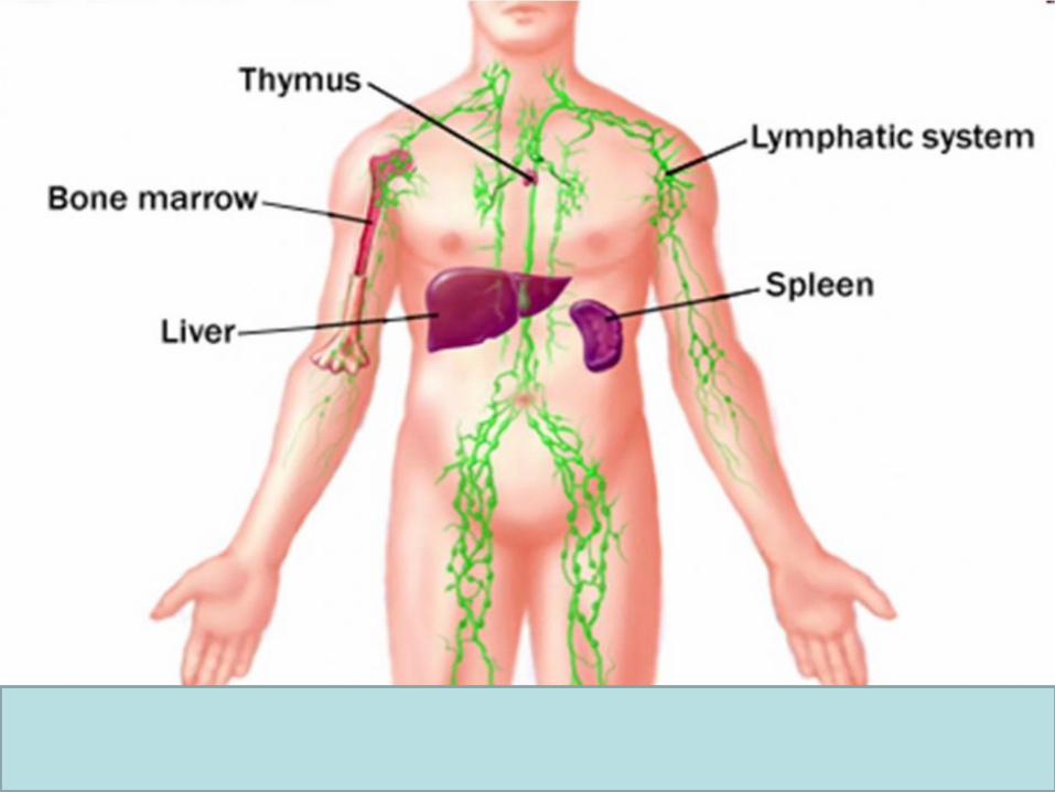
Organs and Cells of Immune System
Organs concerned with immune reactions are
called lymphoid organs. They contain
lymphoid cells.
Lymphoid organs are of 2 types.
1. Primary lymphoid organs
2. Secondary lymphoid organs
1. Primary lymphoid organs
Are the major site of lymphopoiesis. The lymphoid cells
proliferate, differentiate and mature in to immune
competent cells in the absence of antigenic stimulation. The
primary lymphoid organs are large at birth and they atrophy
with age progression; major primary lymphoid organs are
1. Thymus (site of T-cell maturation in human)
2. Bone marrow (site of B cell maturation in human)
3. Bursa of fabricious (site of B-cell maturation in bird)
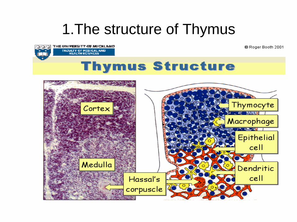
1.The structure of Thymus
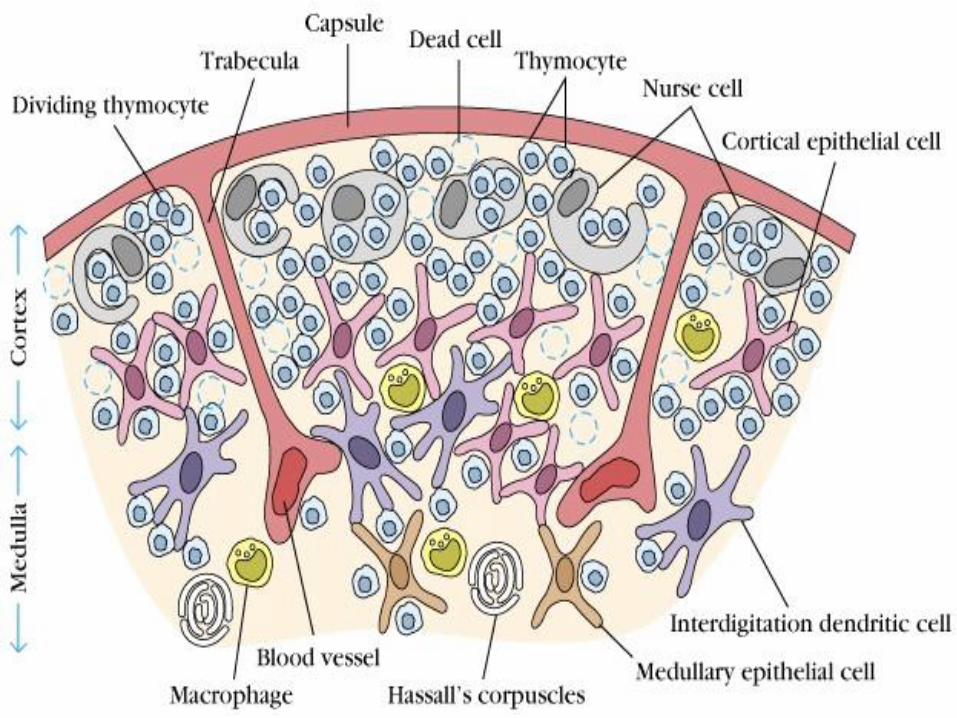
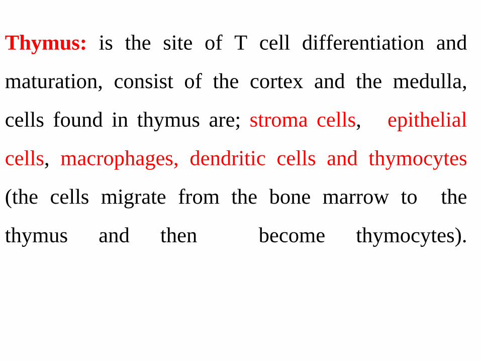
Thymus: is the site of T cell differentiation and
maturation, consist of the cortex and the medulla,
cells found in thymus are; stroma cells, epithelial
cells, macrophages, dendritic cells and thymocytes
(the cells migrate from the bone marrow to the
thymus and then become thymocytes).
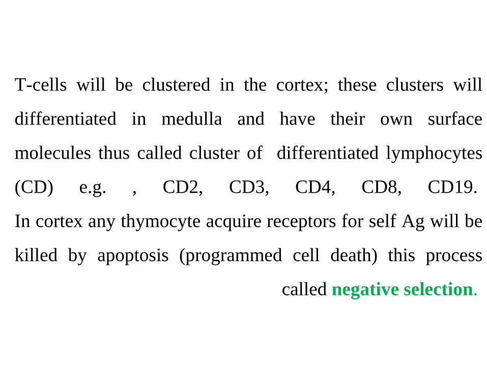
T-cells will be clustered in the cortex; these clusters will
differentiated in medulla and have their own surface
molecules thus called cluster of differentiated lymphocytes
(CD) e.g. , CD2, CD3, CD4, CD8, CD19.
In cortex any thymocyte acquire receptors for self Ag will be
killed by apoptosis (programmed cell death) this process
called negative selection.
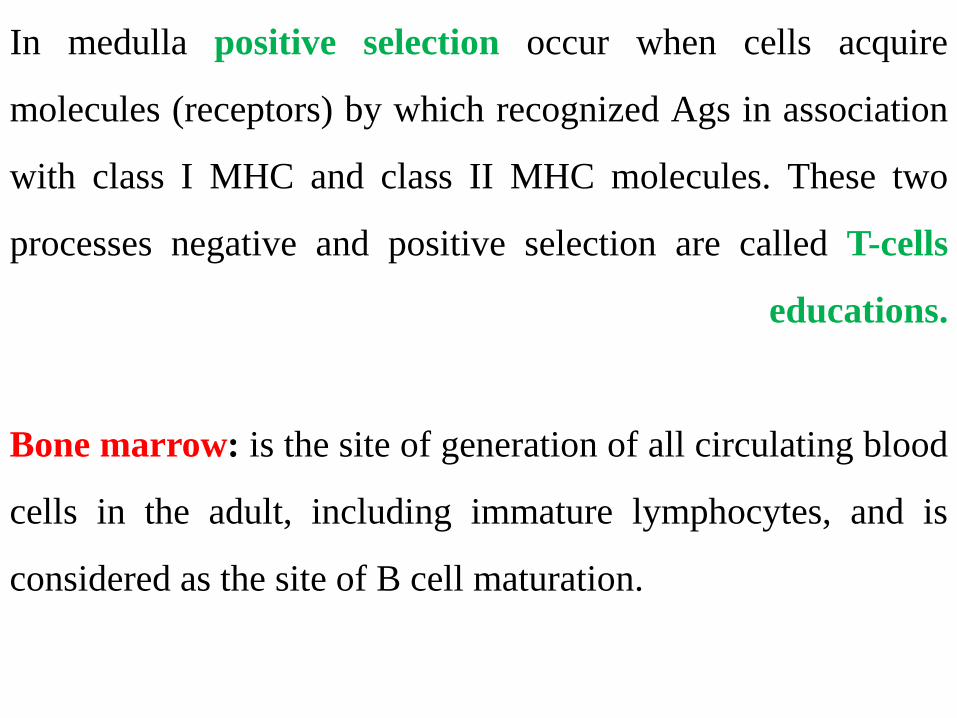
In medulla positive selection occur when cells acquire
molecules (receptors) by which recognized Ags in association
with class I MHC and class II MHC molecules. These two
processes negative and positive selection are called T-cells
educations.
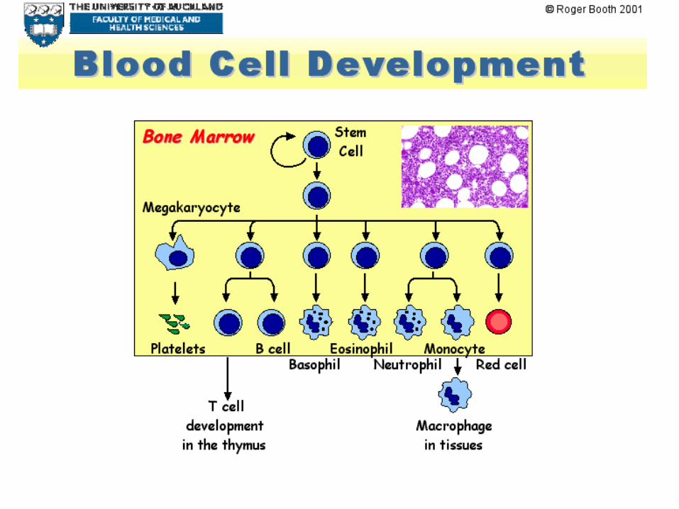
Bone marrow: is the site of generation of all circulating blood
cells in the adult, including immature lymphocytes, and is
considered as the site of B cell maturation.
2. Secondary lymphoid organs
Lymphocytes are made functional in the secondary
lymphoid organs. The secondary lymphoid organs are small
and poorly developed at birth and they grow progressively
with age. The secondary lymphoid organs include:
1. Lymph nodes
2. Spleen
3. Mucosal associated lymphoid tissues (MALT), such as gut-
associated lymphoid tissue (GALT).
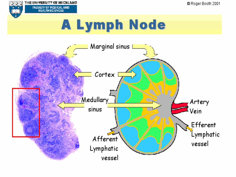
Lymph nodes: are the organs in which immune responses to
lymphoid-borne antigens are initiated, they have many
functions.
- Filter and eliminate foreign antigens.
-Site of immune response.
-Site of lymphocytes residence and source of recirculation
cells.
The spleen: is the major site of immune responses to blood-
borne antigens
-Site of immune cell residence.
-Site of immune response.
-Produce some active substances, such as complement.
-Filtration.
Mucosal associated lymphoid tissues (MALT)
The MALT of the gastrointestinal and respiratory tracts is
colonized by lymphocytes and antigen presenting cell that
initiate immune responses to ingested and inhaled antigens.
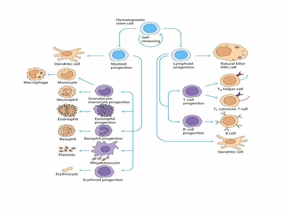
Cells of the immune system
Stem cells of the immune system originated from the
yolk sack in the first six weeks of gestation, after that liver
take this function, then bone marrow will be responsible for
originate and proliferate stem cell under some hormones and
enzymes.
Stem cells include lymphoid series (T, B and NK cells) and
myeloid series (RBCs, monocytes and granulocytes).
A. Lymphocytes
-Lymphocytes are mononucleate, nongranular leukocytes of
lymphoid tissue participating in immunity.
-They are found in blood, lymph and lymphoid tissue such as
spleen, lymph nodes, tonsils and peyer’s patches.
-They are spherical or oval in shape and arise from
haemopoetic stem cells.
1. T-Lymphocytes
- represent about 70% of the total lymphocyte population.
- all T cells express CD3 on their surfaces, along with T cell
receptors (TCRs) which recognize specific antigens presented
in an MHC I or MHC II molecule.
-There are different types of T cells:
1. T-initiator or inducer: have CD+4
2. T-helper: have CD+4, which have two types T-helper1 (Th1)
and T-helper2 (Th2).
3. Tdh (delay hypersensitivity): have CD+4.
4. T-cytotoxic: have CD+8, kill viral infected cells and tumor
cells .
5. T-Memory: have CD+4 and CD+8, play a role in secondary
immune response.
Maturation and development of T-cell
- The first stage in development is the arrangement of the
functional T-cell receptor (β-TCR) to avoid death by
apoptosis (programmed cell death).
• - The developing T-lymphocytes will acquire α and β T-cell
receptor (TCR).
-CD+4 and CD+8 molecules define the effecter function
and the MHC or (HLA) restriction of T-cells.
Mode of killing of T-lymphocytes:-
-Direct by cell to cell action (cytotoxic cell).
-Indirect by cytokines secretion (helper cell).
2. B-lymphocytes
Lymphocyte that matures in bursa of fabricious or bone
marrow and that responsible for humoral immunity is called B
lymphocytes.
Mode of killing: - By specific immunoglobulins.
B-cell development:-
-pro-B-cell: contain CD45 and CD19.
-Pre-B-cell: contain intra cytoplasmic μ chain.
-Immature B-cell: have surface IgM only.
-Mature B-cell: have surface IgM and IgD.
When B-cell activated it will differentiated in to plasma cell
and secrete Abs (immunoglobulin).
3. Natural killer cells (NK cells)
They form the third population of lymphocytes. The NK
cells have 2 or 3 large granules in the cytoplasm. Hence
they are also called large granular
lymphocytes. They destroy the cancer cells and cells
infected with virus, do not need antibody for activity, are
activated by interferon and interleukin-2.
Mode of killing:-
Kill by Ab dependent cell- mediated cytotoxicity
(ADCC). Antibodies bind to organisms via their Fab
region. NK cells, attach via FC receptors, and kill these
organisms not by phagocytosis but by release of toxic
substances called perforins that found in their granules.
B. Macrophages
Macrophages are large mononuclear phagocytic cells
derived from monocytes. Macrophages are concentrated in
lymph nodes, spleen, bone marrow and liver.
C. Eosinophils
They are acidophilic leucocytes and are called eosinophils
because eosin (acid dye) stains the granules of the
cytoplasm of these cells . Granules are rich in hydrolytic
enzymes.
D. Basophils
The cytoplasm of these cells containing granules that stains
with basic dyes. The basophilic granules contain heparin,
histamine, serotonin, platelet activating factor.
E. Neutrophils
Neutrophils form the major part of the white blood cell.
They are motile, short lived cells with multilobed nucleus.
Major function of the neutrophil is phagocytosis.

Complement system By
Prof. Dr. Batool Hassan Al-Ghurabi
Complement system is a part of the immune
system that helps or complements the
ability of antibodies and phagocytic cells to
clear pathogens from an organism.
It is part of the innate and adaptive immune
system.
- The complement system consists of a number
of small proteins (30 proteins) found in the
blood. In general synthesized by the liver
(hepatocytes).
- Many components are precursors (pro-
proteins) which are functionally inactive until
proteolytic cleavage, which removes an
inhibitory fragment and exposes the active site.
When stimulated by one of several triggers,
proteases in the system cleave specific proteins
to activation cascade of further cleavages.
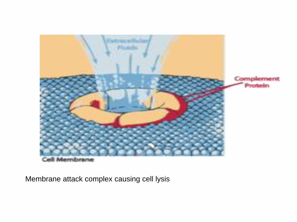
The end-result of this activation cascade is
formation of membrane attack complex (MAC).
Complement Activation
There are three separate pathways which activate the
complement system:
1. classical pathway: activated by antibody-antigen
complexes ( immune complexes) on pathogen surfaces.
2. mannose-binding lectin pathway: activated when
mannose-binding lectin binds to the carbohydrate molecule
mannose on pathogen surfaces.
3. alternative pathway: C3 reacts directly with pathogen
surfaces
-All three of these pathways act to generate the enzyme C3
convertase.
-This cleaves C3 into two parts (C3a and C3b) and activates
the rest of the cascade.
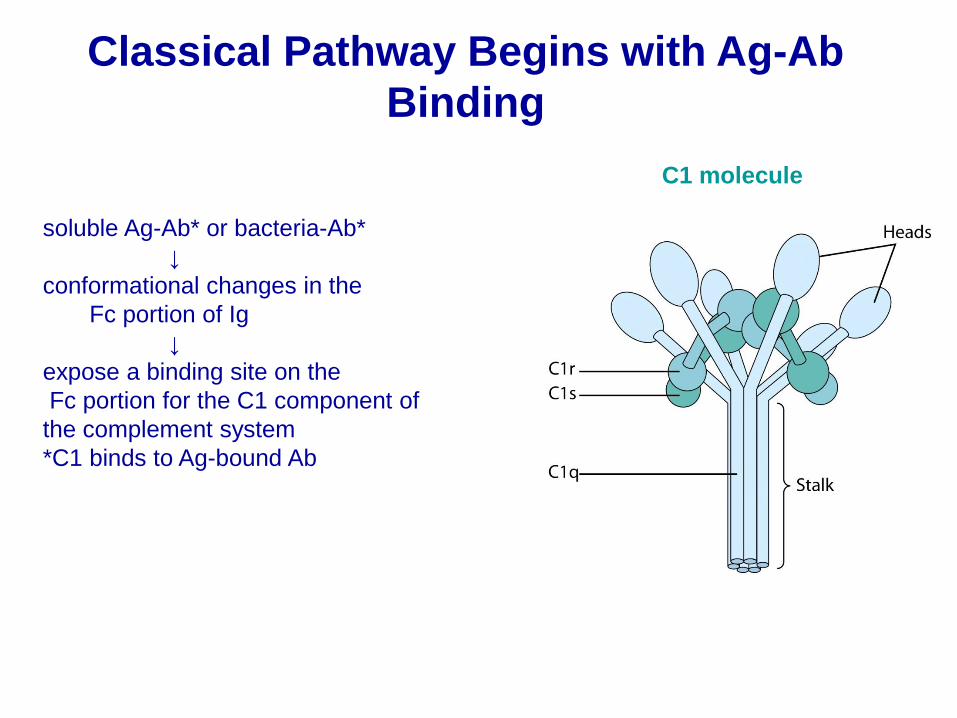
Classical Pathway Begins with Ag-Ab
Binding
soluble Ag-Ab* or bacteria-Ab*
↓
conformational changes in the
Fc portion of Ig
↓
expose a binding site on the
Fc portion for the C1 component of
the complement system
*C1 binds to Ag-bound Ab
C1 molecule
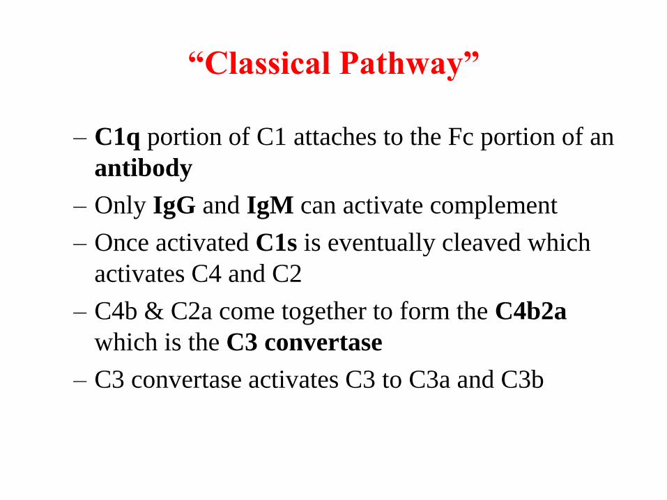
“Classical Pathway”
– C1q portion of C1 attaches to the Fc portion of an
antibody
– Only IgG and IgM can activate complement
– Once activated C1s is eventually cleaved which
activates C4 and C2
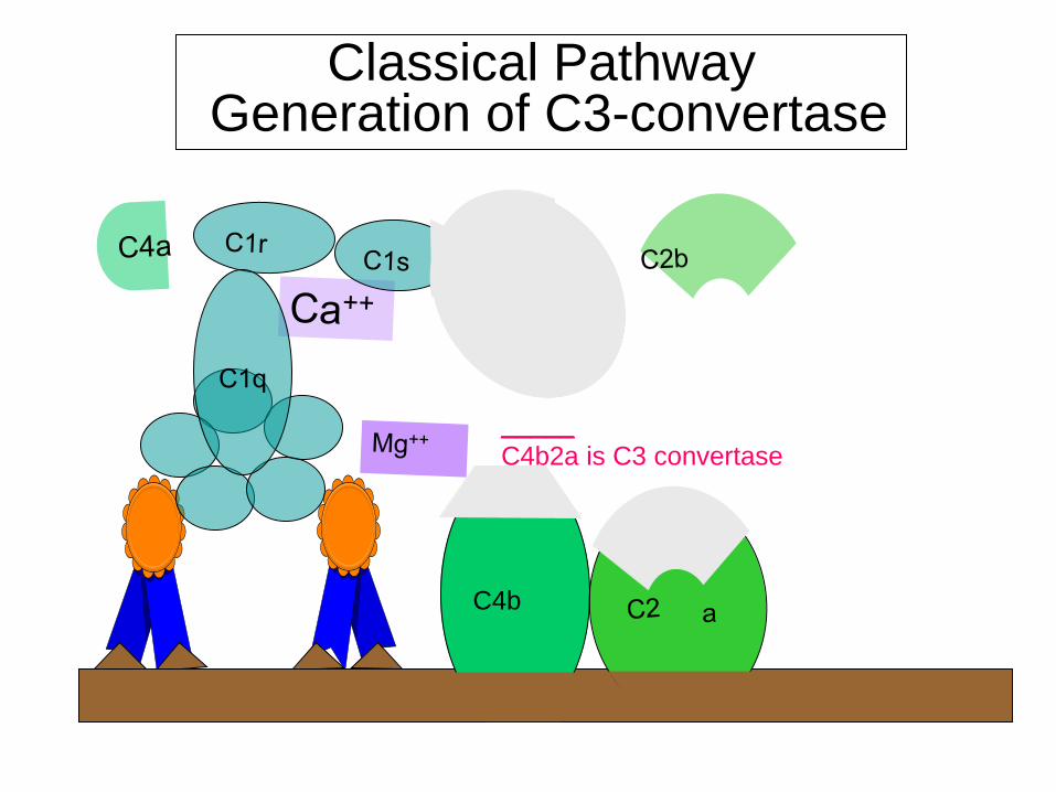
– C4b & C2a come together to form the C4b2a
which is the C3 convertase
– C3 convertase activates C3 to C3a and C3b
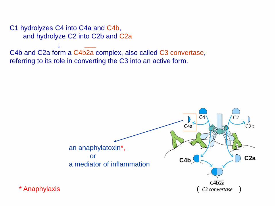
C1 hydrolyzes C4 into C4a and C4b,
and hydrolyze C2 into C2b and C2a
↓ ___
C4b and C2a form a C4b2a complex, also called C3 convertase,
referring to its role in converting the C3 into an active form.
C4b C2a
( )
an anaphylatoxin*,
or
a mediator of inflammation
* Anaphylaxis
____
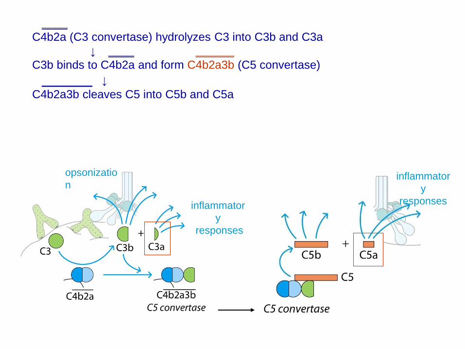
C4b2a (C3 convertase) hydrolyzes C3 into C3b and C3a
↓ ____ ______
C3b binds to C4b2a and form C4b2a3b (C5 convertase)
_______ ↓
C4b2a3b cleaves C5 into C5b and C5a
inflammator
y
responses inflammator
y
responses
opsonizatio
n

Components of the Classical Pathway
C4
C3
C1 complex
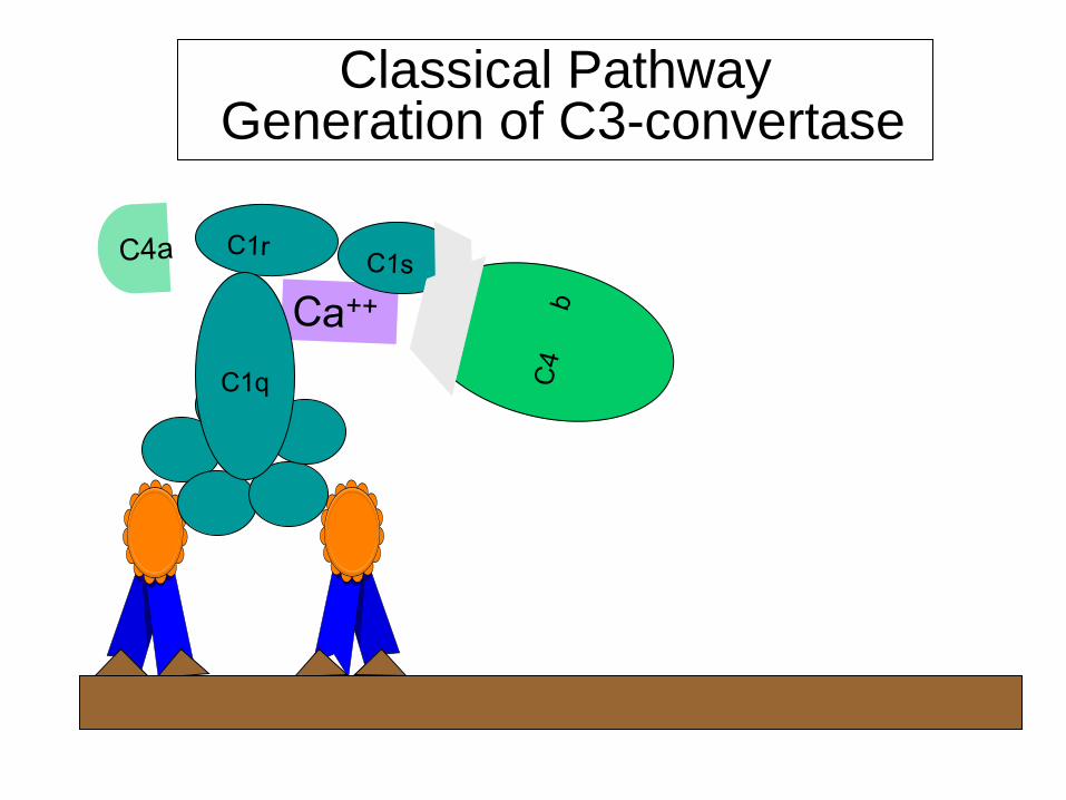
Classical Pathway Generation of C3-convertase
Classical Pathway Generation of C3-convertase
C4b
_____
C4b2a is C3 convertase
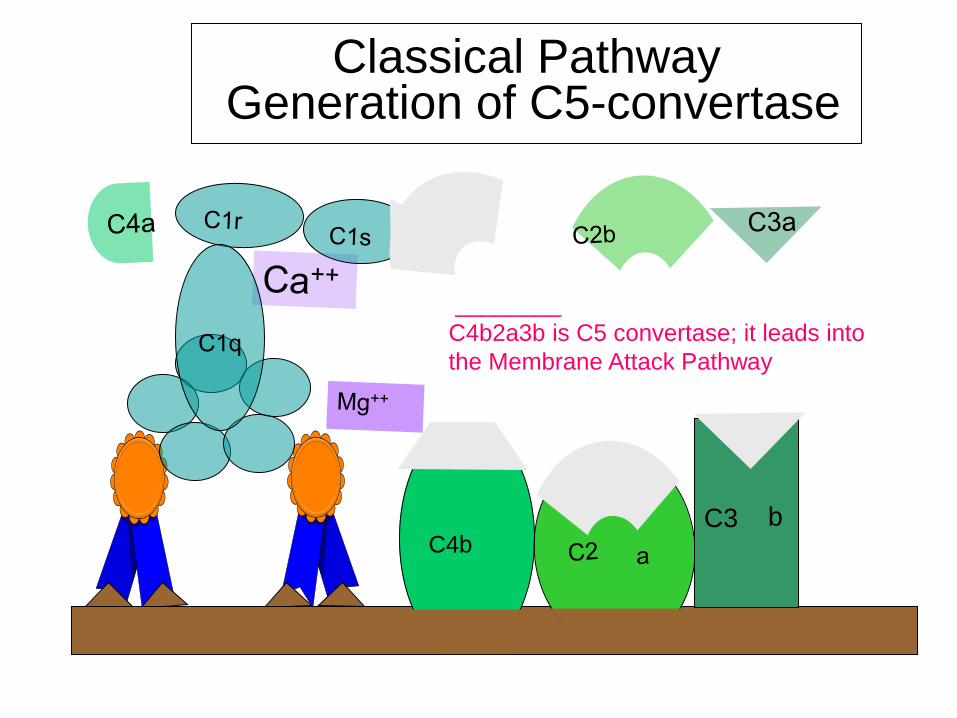
Classical Pathway Generation of C5-convertase
C4b
C3
b
________
C4b2a3b is C5 convertase; it leads into
the Membrane Attack Pathway
C1q C2 C4
2a 2b 4b 4
a
C3-convertase
C3
C3a C3b
C5-Convertase
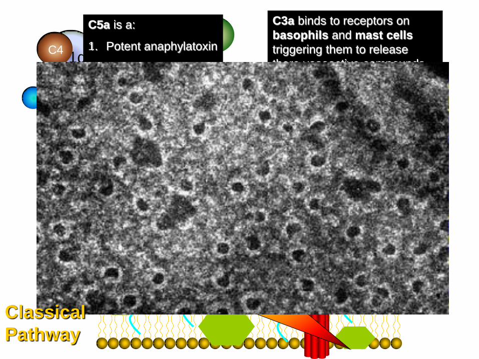
C3a binds to receptors on
basophils and mast cells
triggering them to release
there vasoactive compounds
(enhances vasodilation and
vasopermeability) -
ANAPHYLATOXIN
C5
C5a C5b
C5a is a:
1. Potent anaphylatoxin
2. Chemoattractant for
neutrophils
C6
C7 C8
C9
Classical
Pathway
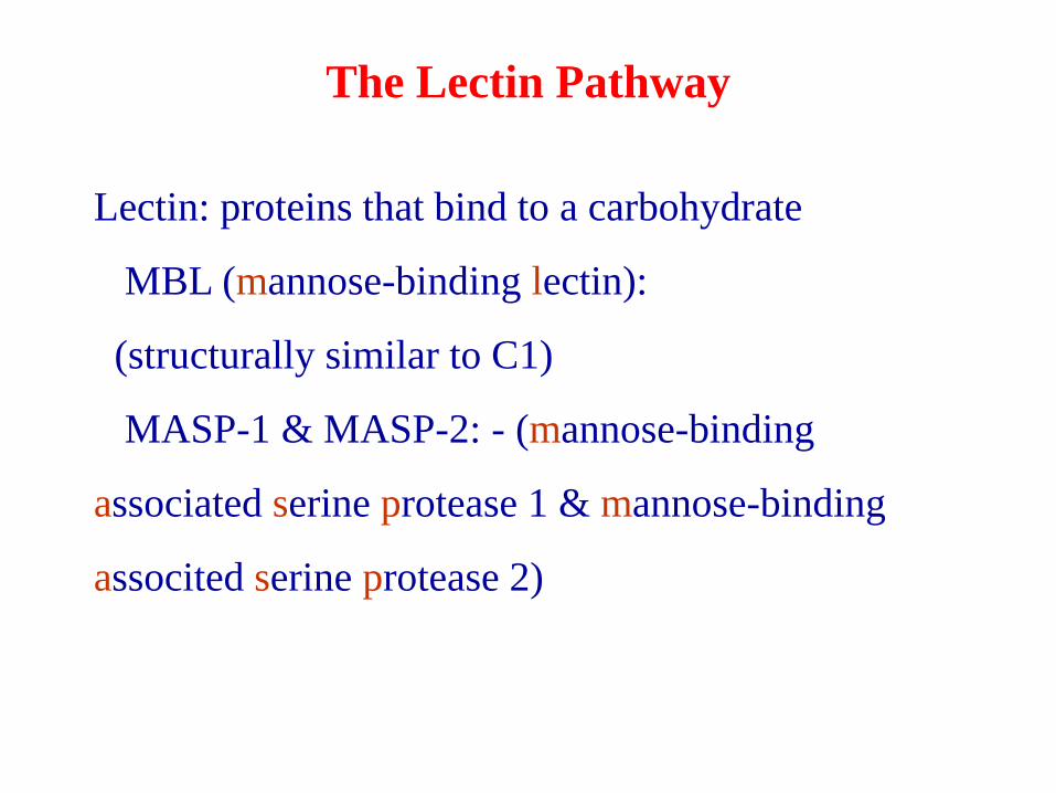
The Lectin Pathway
Lectin: proteins that bind to a carbohydrate
MBL (mannose-binding lectin):
(structurally similar to C1)
MASP-1 & MASP-2: - (mannose-binding
associated serine protease 1 & mannose-binding
associted serine protease 2)
- MBL is induced during inflammatory responses.
- After MBL binds to the surface of a microbe, MBL-associated serine
proteases-1 ( MASP-1) and MASP-2, bind to MBL.
- The MBL-MASP-1/2 complex mimics the activity of C1, and causes
cleavage and activation of C4 and C2.
- Thus, the lectin pathway is Ab-independent. It is an important innate
defense mechanism comparable to the alternative pathway, but utilizing
the elements of the classical pathway, except for the C1 proteins.
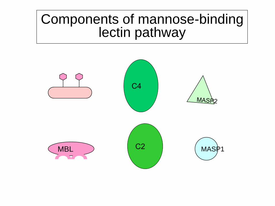
Components of mannose-binding lectin pathway
MBL MASP1
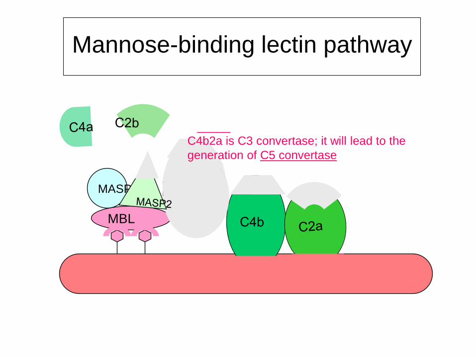
Mannose-binding lectin pathway
MBL
_____
C4b2a is C3 convertase; it will lead to the
generation of C5 convertase
MASP1
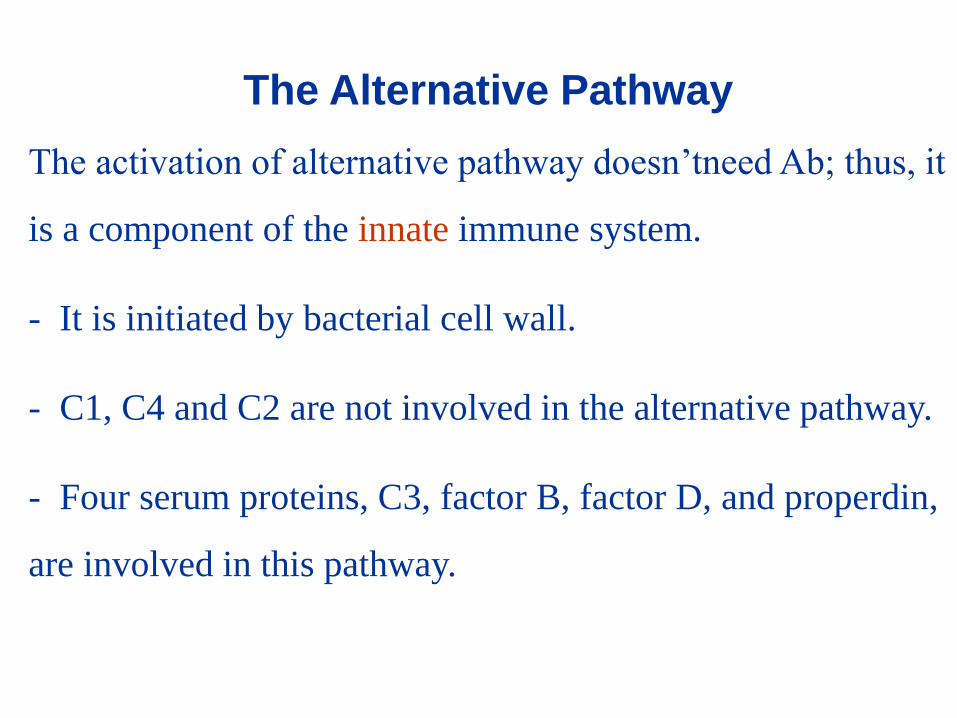
The Alternative Pathway
The activation of alternative pathway doesn’tneed Ab; thus, it
is a component of the innate immune system.
- It is initiated by bacterial cell wall.
- C1, C4 and C2 are not involved in the alternative pathway.
- Four serum proteins, C3, factor B, factor D, and properdin,
are involved in this pathway.
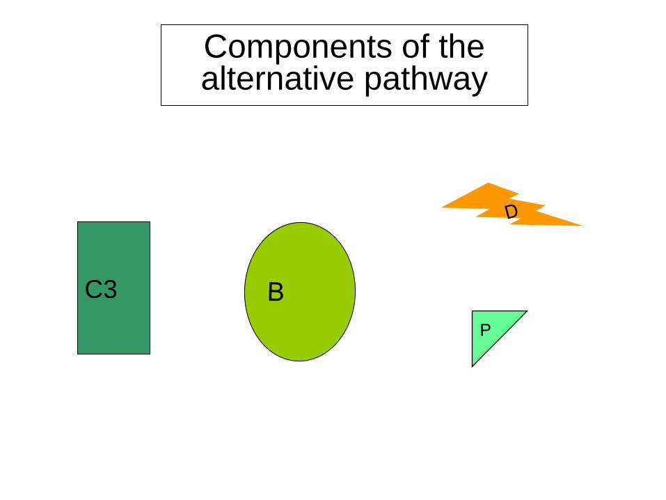
Components of the alternative pathway
C3
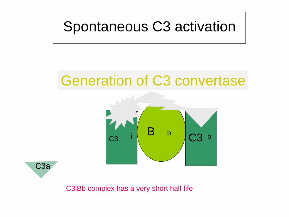
Spontaneous C3 activation
C3
i
Generation of C3 convertase
C3iBb complex has a very short half life
b
C3
b
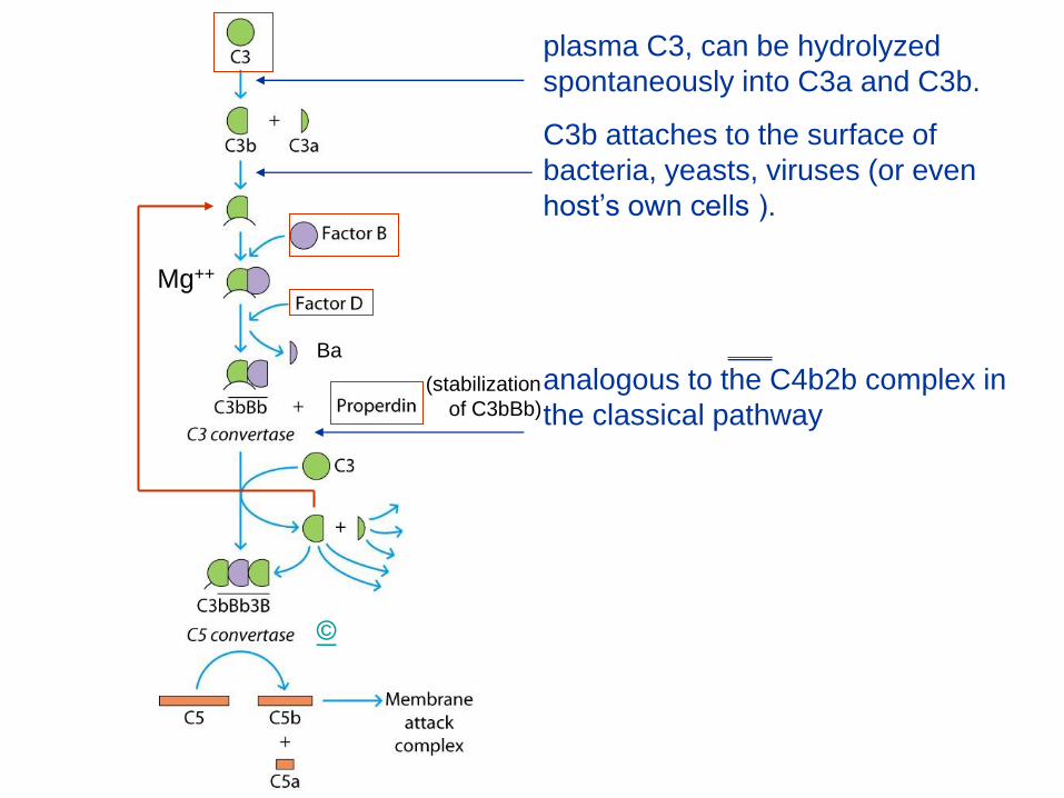
plasma C3, can be hydrolyzed
spontaneously into C3a and C3b.
C3b attaches to the surface of
bacteria, yeasts, viruses (or even
host’s own cells ).
___
analogous to the C4b2b complex in
the classical pathway
Ba
(stabilization
of C3bBb)
Mg++
©
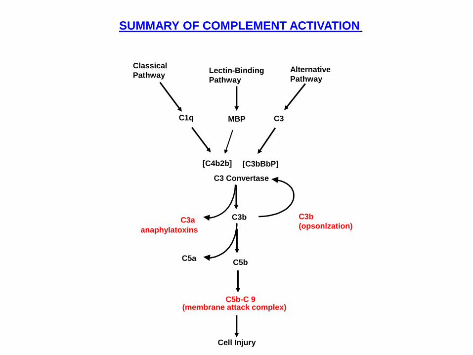
SUMMARY OF COMPLEMENT ACTIVATION
Classical
Pathway Lectin-Binding
Pathway
Alternative
Pathway
MBP C3 C1q
[C4b2b] [C3bBbP]
C3b
(opsonlzation)
C5b-C 9
C5b C5a
(membrane attack complex)
Cell Injury
C3b
anaphylatoxins
C3a
C3 Convertase
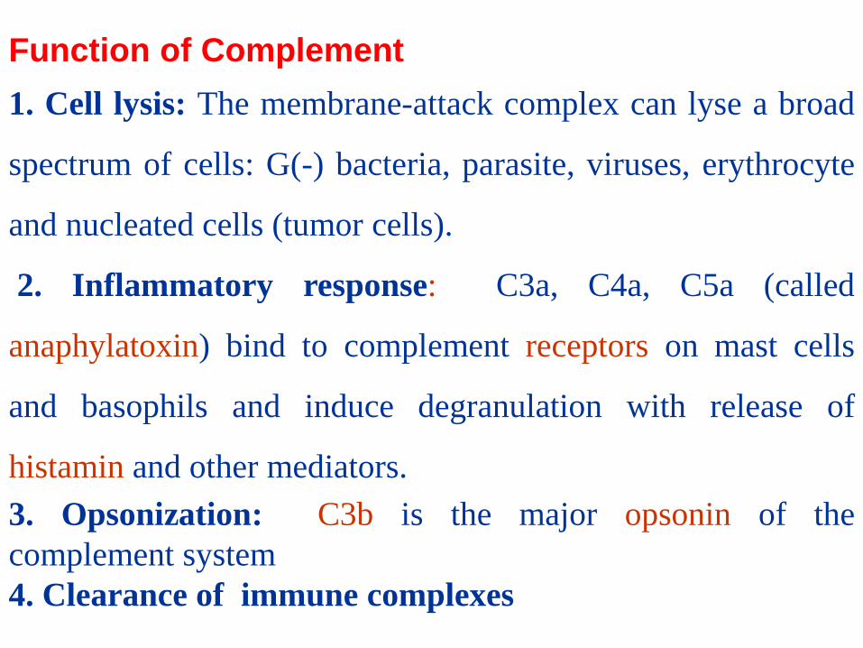
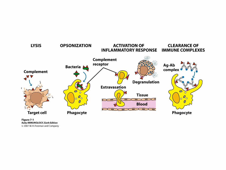
Function of Complement
1. Cell lysis: The membrane-attack complex can lyse a broad
spectrum of cells: G(-) bacteria, parasite, viruses, erythrocyte
and nucleated cells (tumor cells).
2. Inflammatory response: C3a, C4a, C5a (called
anaphylatoxin) bind to complement receptors on mast cells
and basophils and induce degranulation with release of
histamin and other mediators.
3. Opsonization: C3b is the major opsonin of the
complement system
4. Clearance of immune complexes
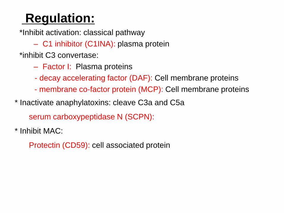
Regulation: *Inhibit activation: classical pathway
– C1 inhibitor (C1INA): plasma protein
*inhibit C3 convertase:
– Factor I: Plasma proteins
- decay accelerating factor (DAF): Cell membrane proteins
- membrane co-factor protein (MCP): Cell membrane proteins
* Inactivate anaphylatoxins: cleave C3a and C5a
serum carboxypeptidase N (SCPN):
* Inhibit MAC:
Protectin (CD59): cell associated protein
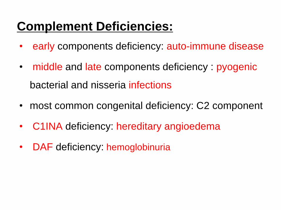
Complement Deficiencies:
• early components deficiency: auto-immune disease
• middle and late components deficiency : pyogenic
bacterial and nisseria infections
• most common congenital deficiency: C2 component
• C1INA deficiency: hereditary angioedema
• DAF deficiency: hemoglobinuria