Embed Size (px)
Citation preview
COMMUNICATION Atsushi Shishido et al .
Oriented collagen films with high Young's modulus by
self-assembly on micrometer grooved polydimethylsiloxane
Materials Advancesrsc.li/materials-advances
ISSN 2633-5409
Volume 2
Number 21
7 November 2021
Pages 6735–7090
6984 | Mater. Adv., 2021, 2, 6984–6987 © 2021 The Author(s). Published by the Royal Society of Chemistry
Cite this: Mater. Adv., 2021,
2, 6984
Oriented collagen films with high Young’smodulus by self-assembly on micrometer groovedpolydimethylsiloxane†
Miho Aizawa,ab Hirona Nakamura,ac Kohsuke Matsumoto,ac Takahiro Ogumaa andAtsushi Shishido *ac
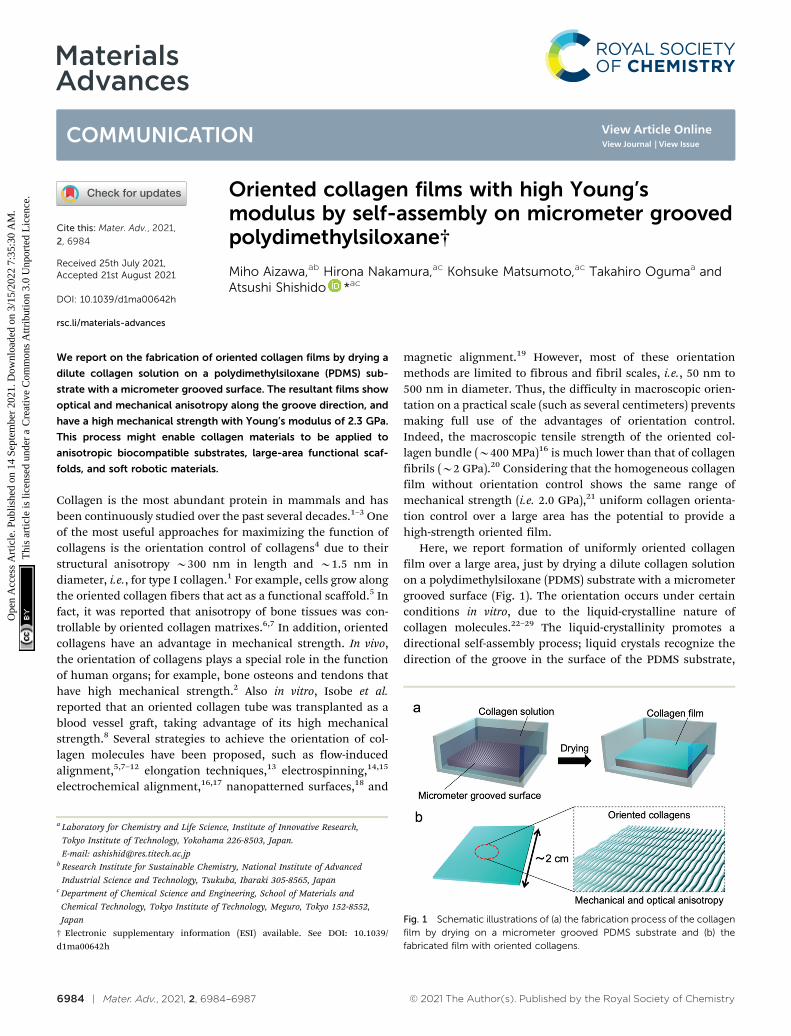
We report on the fabrication of oriented collagen films by drying a
dilute collagen solution on a polydimethylsiloxane (PDMS) sub-
strate with a micrometer grooved surface. The resultant films show
optical and mechanical anisotropy along the groove direction, and
have a high mechanical strength with Young’s modulus of 2.3 GPa.
This process might enable collagen materials to be applied to
anisotropic biocompatible substrates, large-area functional scaf-
folds, and soft robotic materials.
Collagen is the most abundant protein in mammals and hasbeen continuously studied over the past several decades.1–3 Oneof the most useful approaches for maximizing the function ofcollagens is the orientation control of collagens4 due to theirstructural anisotropy B300 nm in length and B1.5 nm indiameter, i.e., for type I collagen.1 For example, cells grow alongthe oriented collagen fibers that act as a functional scaffold.5 Infact, it was reported that anisotropy of bone tissues was con-trollable by oriented collagen matrixes.6,7 In addition, orientedcollagens have an advantage in mechanical strength. In vivo,the orientation of collagens plays a special role in the functionof human organs; for example, bone osteons and tendons thathave high mechanical strength.2 Also in vitro, Isobe et al.reported that an oriented collagen tube was transplanted as ablood vessel graft, taking advantage of its high mechanicalstrength.8 Several strategies to achieve the orientation of col-lagen molecules have been proposed, such as flow-inducedalignment,5,7–12 elongation techniques,13 electrospinning,14,15
electrochemical alignment,16,17 nanopatterned surfaces,18 and
magnetic alignment.19 However, most of these orientationmethods are limited to fibrous and fibril scales, i.e., 50 nm to500 nm in diameter. Thus, the difficulty in macroscopic orien-tation on a practical scale (such as several centimeters) preventsmaking full use of the advantages of orientation control.Indeed, the macroscopic tensile strength of the oriented col-lagen bundle (B400 MPa)16 is much lower than that of collagenfibrils (B2 GPa).20 Considering that the homogeneous collagenfilm without orientation control shows the same range ofmechanical strength (i.e. 2.0 GPa),21 uniform collagen orienta-tion control over a large area has the potential to provide ahigh-strength oriented film.
Here, we report formation of uniformly oriented collagenfilm over a large area, just by drying a dilute collagen solutionon a polydimethylsiloxane (PDMS) substrate with a micrometergrooved surface (Fig. 1). The orientation occurs under certainconditions in vitro, due to the liquid-crystalline nature ofcollagen molecules.22–29 The liquid-crystallinity promotes adirectional self-assembly process; liquid crystals recognize thedirection of the groove in the surface of the PDMS substrate,
Fig. 1 Schematic illustrations of (a) the fabrication process of the collagenfilm by drying on a micrometer grooved PDMS substrate and (b) thefabricated film with oriented collagens.
a Laboratory for Chemistry and Life Science, Institute of Innovative Research,
Tokyo Institute of Technology, Yokohama 226-8503, Japan.
E-mail: [email protected] Research Institute for Sustainable Chemistry, National Institute of Advanced
Industrial Science and Technology, Tsukuba, Ibaraki 305-8565, Japanc Department of Chemical Science and Engineering, School of Materials and
Chemical Technology, Tokyo Institute of Technology, Meguro, Tokyo 152-8552,
Japan
† Electronic supplementary information (ESI) available. See DOI: 10.1039/d1ma00642h
Received 25th July 2021,Accepted 21st August 2021
DOI: 10.1039/d1ma00642h
rsc.li/materials-advances
MaterialsAdvances
COMMUNICATION
Ope
n A
cces
s A
rtic
le. P
ublis
hed
on 1
4 Se
ptem
ber
2021
. Dow
nloa
ded
on 3
/15/
2022
7:3
5:30
AM
. T
his
artic
le is
lice
nsed
und
er a
Cre
ativ
e C
omm
ons
Attr
ibut
ion
3.0
Unp
orte
d L
icen
ce.
View Article OnlineView Journal | View Issue
© 2021 The Author(s). Published by the Royal Society of Chemistry Mater. Adv., 2021, 2, 6984–6987 | 6985
and this macroscopically aligns collagens. We successfullyobserved that the fabricated collagen film exhibited opticaland mechanical anisotropy depending on the orientation direc-tion. Furthermore, a subsequent thermal cross-linking reactionmade the collagen film insoluble in water, maintaining themechanical anisotropy of the collagen films, which successfullyachieved high mechanical strength with Young’s modulus of2.3 GPa. Hence, this process has great advantages to practicalapplications due to its simplicity, efficient orientation in a largearea, high mechanical strength, and water resistance.
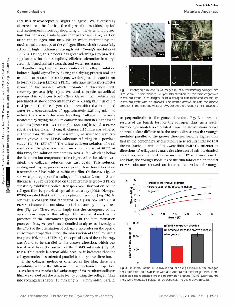
Hypothesizing that the concentration of a collagen solutioninduced liquid-crystallinity during the drying process and theresultant orientation of collagens, we designed an experimentto form a collagen film on a PDMS substrate with a micrometergroove in the surface, which promotes a directional self-assembly process (Fig. 1(a)). We used a pepsin solubilizedporcine skin collagen type-I (Nitta Gelatin Inc.), which waspurchased at stock concentrations of B5.0 mg mL�1 in diluteHCl (pH B 3.1). The collagen solution was diluted with distilledwater to a concentration of approximately 1.25 mg mL�1 toreduce the viscosity for easy handling. Collagen films werefabricated by drying the dilute collagen solution in a handmadeglass box (size: 3 cm� 3 cm; height: 12.5 mm) in which a PDMSsubstrate (size: 2 cm � 2 cm; thickness: 1.25 mm) was adheredat the bottom. To direct self-assembly, we inscribed a micro-meter groove in the PDMS substrate referring to a perviousstudy (Fig. S1, ESI†).30,31 The dilute collagen solution of 4 mlwas cast in the glass box placed on a hotplate set at 38 1C. Atthis time, the solution temperature was 35 1C, which is belowthe denaturation temperature of collagen. After the solvent wasdried, the collagen solution was cast again. This solutioncasting and drying process was repeated four times to obtainfreestanding films with a sufficient film thickness. Fig. 2ashows a photograph of a collagen film (size: 2 cm � 2 cm;thickness: 20 mm) fabricated on the micrometer grooved PDMSsubstrate, exhibiting optical transparency. Observation of thecollagen film by polarized optical microscopy (POM: OlympusBX50) revealed that the film has optical anisotropy (Fig. 2b). Incontrast, a collagen film fabricated in a glass box with a flatPDMS substrate did not show optical anisotropy in any direc-tion (Fig. 2c). These results imply that the emergence of theoptical anisotropy in the collagen film was attributed to thepresence of the micrometer grooves in the film formationprocess. Thus, we performed detailed analyses to investigatethe effect of the orientation of collagen molecules on the opticalanisotropic properties. From the observation of the film with atest plate (Olympus U-TP530), the optical axis of the anisotropywas found to be parallel to the groove direction, which wastransferred from the surface of the PDMS substrate (Fig. S2,ESI†). This result is remarkable because it indicates that thecollagen molecules oriented parallel to the groove direction.
If the collagen molecules oriented in the film, there is apossibility to show the difference in the mechanical properties.To evaluate the mechanical anisotropy of the resultant collagenfilm, we carried out the tensile test by cutting the collagen filmsinto rectangular shapes (15 mm length � 5 mm width) parallel
or perpendicular to the groove direction. Fig. 3 shows theresults of the tensile test for the collagen films. As a result,the Young’s modulus calculated from the stress–strain curvesshowed a clear difference in the tensile directions; the Young’smodulus parallel to the groove direction became higher thanthat in the perpendicular direction. These results indicate thatthe mechanical directionalities were linked with the orientationdirections of collagens because the direction of this mechanicalanisotropy was identical to the results of POM observation. Inaddition, the Young’s modulus of the film fabricated on the flatPDMS substrate showed an intermediate value of Young’s
Fig. 2 Photograph (a) and POM images (b) of a freestanding collagen film(size: 2 cm � 2 cm; thickness: 20 mm) fabricated on the micrometer groovedPDMS substrate. POM images (c) of a collagen film fabricated on the flatPDMS substrate with no grooves. The orange arrows indicate the groovedirection in the film. The white arrows denote the direction of the polarizers.
Fig. 3 (a) Stress–strain (S–S) curves and (b) Young’s moduli of the collagenfilms fabricated on a substrate with and without micrometer grooves. In thecollagen films fabricated on the micrometer grooved PDMS substrate, thefilms were elongated parallel or perpendicular to the groove direction.
Communication Materials Advances
Ope
n A
cces
s A
rtic
le. P
ublis
hed
on 1
4 Se
ptem
ber
2021
. Dow
nloa
ded
on 3
/15/
2022
7:3
5:30
AM
. T
his
artic
le is
lice
nsed
und
er a
Cre
ativ
e C
omm
ons
Attr
ibut
ion
3.0
Unp
orte
d L
icen
ce.
View Article Online
6986 | Mater. Adv., 2021, 2, 6984–6987 © 2021 The Author(s). Published by the Royal Society of Chemistry
modulus, also supporting that the mechanical anisotropy wasderived from the molecular orientation (Fig. 3b). It is importantto note that the fabrication process of the collagen film enablesnot only fabricating the oriented collagen film but also provid-ing the mechanical strength anisotropy.
The orientation of collagen molecules achieved in the pre-sent system can be rationalized by assuming that liquid-crystallinity of the collagen is the main factor. Collagen exhibitsa lyotropic liquid crystalline state as the solution is concen-trated, depending on the pH and the type of collagen.26,27
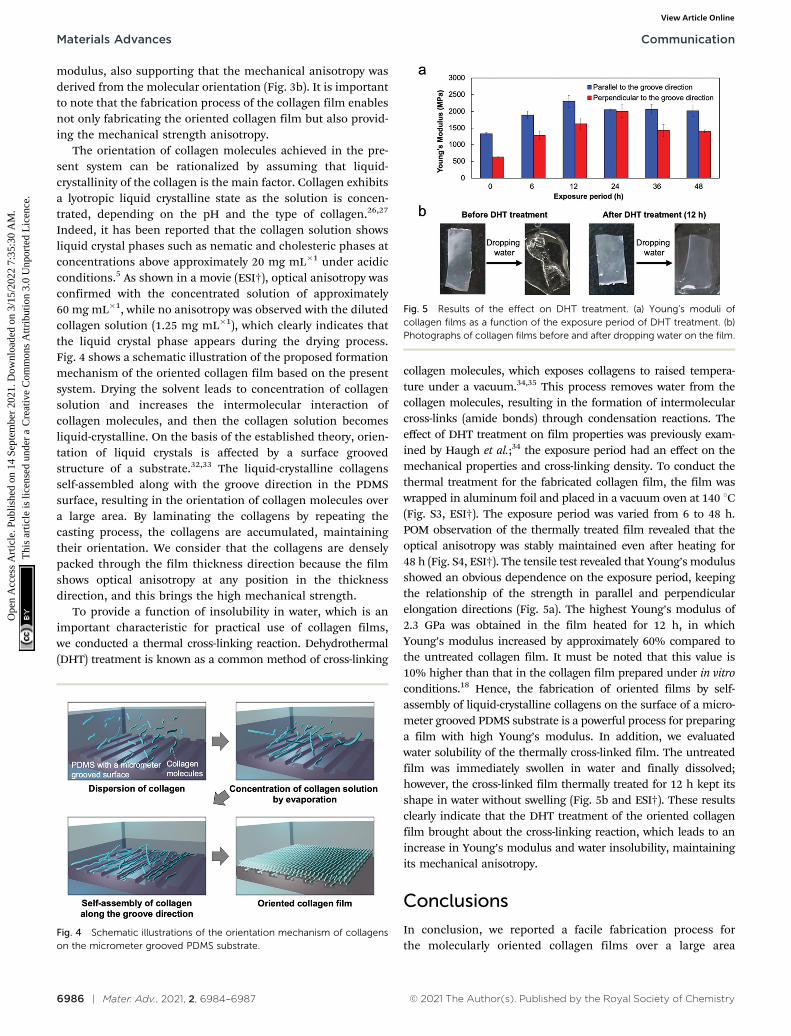
Indeed, it has been reported that the collagen solution showsliquid crystal phases such as nematic and cholesteric phases atconcentrations above approximately 20 mg mL�1 under acidicconditions.5 As shown in a movie (ESI†), optical anisotropy wasconfirmed with the concentrated solution of approximately60 mg mL�1, while no anisotropy was observed with the dilutedcollagen solution (1.25 mg mL�1), which clearly indicates thatthe liquid crystal phase appears during the drying process.Fig. 4 shows a schematic illustration of the proposed formationmechanism of the oriented collagen film based on the presentsystem. Drying the solvent leads to concentration of collagensolution and increases the intermolecular interaction ofcollagen molecules, and then the collagen solution becomesliquid-crystalline. On the basis of the established theory, orien-tation of liquid crystals is affected by a surface groovedstructure of a substrate.32,33 The liquid-crystalline collagensself-assembled along with the groove direction in the PDMSsurface, resulting in the orientation of collagen molecules overa large area. By laminating the collagens by repeating thecasting process, the collagens are accumulated, maintainingtheir orientation. We consider that the collagens are denselypacked through the film thickness direction because the filmshows optical anisotropy at any position in the thicknessdirection, and this brings the high mechanical strength.
To provide a function of insolubility in water, which is animportant characteristic for practical use of collagen films,we conducted a thermal cross-linking reaction. Dehydrothermal(DHT) treatment is known as a common method of cross-linking
collagen molecules, which exposes collagens to raised tempera-ture under a vacuum.34,35 This process removes water from thecollagen molecules, resulting in the formation of intermolecularcross-links (amide bonds) through condensation reactions. Theeffect of DHT treatment on film properties was previously exam-ined by Haugh et al.;34 the exposure period had an effect on themechanical properties and cross-linking density. To conduct thethermal treatment for the fabricated collagen film, the film waswrapped in aluminum foil and placed in a vacuum oven at 140 1C(Fig. S3, ESI†). The exposure period was varied from 6 to 48 h.POM observation of the thermally treated film revealed that theoptical anisotropy was stably maintained even after heating for48 h (Fig. S4, ESI†). The tensile test revealed that Young’s modulusshowed an obvious dependence on the exposure period, keepingthe relationship of the strength in parallel and perpendicularelongation directions (Fig. 5a). The highest Young’s modulus of2.3 GPa was obtained in the film heated for 12 h, in whichYoung’s modulus increased by approximately 60% compared tothe untreated collagen film. It must be noted that this value is10% higher than that in the collagen film prepared under in vitroconditions.18 Hence, the fabrication of oriented films by self-assembly of liquid-crystalline collagens on the surface of a micro-meter grooved PDMS substrate is a powerful process for preparinga film with high Young’s modulus. In addition, we evaluatedwater solubility of the thermally cross-linked film. The untreatedfilm was immediately swollen in water and finally dissolved;however, the cross-linked film thermally treated for 12 h kept itsshape in water without swelling (Fig. 5b and ESI†). These resultsclearly indicate that the DHT treatment of the oriented collagenfilm brought about the cross-linking reaction, which leads to anincrease in Young’s modulus and water insolubility, maintainingits mechanical anisotropy.
Conclusions
In conclusion, we reported a facile fabrication process forthe molecularly oriented collagen films over a large area
Fig. 4 Schematic illustrations of the orientation mechanism of collagenson the micrometer grooved PDMS substrate.
Fig. 5 Results of the effect on DHT treatment. (a) Young’s moduli ofcollagen films as a function of the exposure period of DHT treatment. (b)Photographs of collagen films before and after dropping water on the film.
Materials Advances Communication
Ope
n A
cces
s A
rtic
le. P
ublis
hed
on 1
4 Se
ptem
ber
2021
. Dow
nloa
ded
on 3
/15/
2022
7:3
5:30
AM
. T
his
artic
le is
lice
nsed
und
er a
Cre
ativ
e C
omm
ons
Attr
ibut
ion
3.0
Unp
orte
d L
icen
ce.
View Article Online
© 2021 The Author(s). Published by the Royal Society of Chemistry Mater. Adv., 2021, 2, 6984–6987 | 6987
(2 cm � 2 cm). The key strategy for this achievement is thedirected self-assembly of liquid-crystalline collagen moleculesduring the drying process on the surface of the micrometergrooved PDMS substrate. The fabricated freestanding film showedoptical and mechanical anisotropy due to the orientation ofcollagens. In addition, the thermal cross-linking reaction madethe film water-insoluble and increased the mechanical strength,keeping its anisotropy of the strength in parallel and perpendi-cular elongation directions. To the best of our knowledge, this isthe simplest example of fabricating oriented collagen film over alarge area. This process is expected to widely open the use of thecollagen materials for practical applications such as biocompati-ble substrates with optical and mechanical anisotropy, large-areafunctional scaffolds for accelerating the development of researchin cell biology, and soft robotic materials.
Conflicts of interest
There are no conflicts to declare.
Acknowledgements
This work was supported by a Grant-in Aid for Scientific Researchon Innovative Areas ‘‘Molecular Engine’’ (JSPS KAKENHI grantnumber JP18H05422). This work was supported by JST CRESTgrant number JPMJCR18I4, Japan. This work was supported byJSPS KAKENHI grant number JP20J14875. This work was per-formed under the Cooperative Research Program of ‘‘NetworkJoint Research Center for Materials and Devices’’. This work wasperformed under the Research Program of ‘‘Dynamic Alliance forOpen Innovation Bridging Human, Environment and Materials’’in ‘‘Network Joint Research Center for Materials and Devices’’.
Notes and references
1 M. D. Shoulders and R. T. Raines, Annu. Rev. Biochem., 2009,78, 929–958.
2 D. J. S. Hulmes, J. Struct. Biol., 2002, 137, 2–10.3 C. Sanchez, H. Arribart and M. M. Giraud-Guille,
Nat. Mater., 2005, 4, 277–288.4 A. Sorushanova, L. M. Delgado, Z. Wu, N. Shologu, A. Kshirsagar,
R. Raghunath, A. M. Mullen, Y. Bayan, A. Pandit, M. Raghunathand D. I. Zeugolis, Adv. Mater., 2019, 31, 1801651.
5 J. E. Kirkwood and G. G. Fuller, Langmuir, 2009, 25,3200–3206.
6 A. Matsugaki, G. Aramoto, T. Ninomiya, H. Sawada, S. Hataand T. Nakano, Biomaterials, 2015, 37, 134–143.
7 A. Matsugaki, Y. Isobe, T. Saku and T. Nakano, J. Biomed.Mater. Res., Part A, 2015, 103, 489–499.
8 Y. Isobe, T. Kosaka, G. Kuwahara, H. Mikami, T. Saku andS. Kodama, Materials, 2012, 5, 501–511.
9 N. Saeidi, E. A. Sander and J. W. Ruberti, Biomaterials, 2009,30, 6581–6592.
10 O. F. A. Gutierrez and A. D. Rey, Langmuir, 2016, 32,11799–11812.
11 Y. Tanaka, K. Baba, T. J. Ducan, A. Kubota, T. Asahi,A. J. Quantock, M. Yamato, T. Okano and K. Mishida,Biomaterials, 2011, 32, 3358–3366.
12 B. Lanfer, U. Freudenberg, R. Zimmermann, D. Stamov,V. Korber and C. Werner, Biomaterials, 2008, 29, 3888–3895.
13 K. Poole, K. Khairy, J. Friedrichs, C. Franz, D. A. Cisneros,J. Howard and D. Mueller, J. Mol. Biol., 2005, 349, 380–386.
14 J. A. Matthews, G. E. Wnek, D. G. Simpson and G. L. Bowlin,Biomacromolecules, 2002, 3, 232–238.
15 T. J. Sill and H. A. von Recum, Biomaterials, 2008, 29, 1989–2006.16 X. Cheng, U. A. Gurkan, C. J. Dehen, M. P. Tate,
H. W. Hillhouse, G. J. Simpson and O. Akkus, Biomaterials,2008, 29, 3278–3288.
17 J. A. Paten, S. M. Siadat, M. E. Susilo, E. N. Ismail,J. L. Stoner., J. P. Rothstein and J. W. Ruberti, ACS Nano,2016, 10, 5027–5040.
18 F. A. Denis, A. Pallandre, B. Nysten, A. M. Jonas andC. C. Dupont-Gillain, Small, 2005, 10, 984–991.
19 J. Torbet, M. Malbouyres, N. Builles, V. Justin, M. Roulet,O. Damour, A. Oldberg, F. Ruggiero and D. J. S. Hulmes,Biomaterials, 2007, 28, 4268–4276.
20 M. Minary-Jolandan and M.-F. Yu, Biomacromolecules, 2009,10, 2565–2570.
21 S. Moreno, M. Baniasadi, S. Mohammed, I. Mejia, Y. Chen,M. A. Quevedo-Lopez, N. Kumar, S. Dimitrijevich andM. Minary-Jolandan, Adv. Electron. Mater., 2015, 1, 1500154.
22 M.-M. Giraud-Guille, J. Mol. Biol., 1992, 224, 861–873.23 L. Besseau and M.-M. Giraud-Guille, J. Mol. Biol., 1995, 251,
197–202.24 M.-M. Giraud-Guille, L. Besseau and R. Martin, J. Biomech.,
2003, 36, 1571–1579.25 G. Mosser, A. Anglo, C. Helary, Y. Bouligand and M.-M.
Giraud-Guille, Matrix Biol., 2006, 25, 3–13.26 F. Gobeaux, E. Belamie, G. Mosser, P. Davidson, P. Panine
and M.-M. Giraud-Guille, Langmuir, 2007, 23, 6411–6417.27 A. D. Rey, Soft Matter, 2010, 6, 3402–3429.28 P. De Sa Peixoto, A. Deniset-Besseau, M. Schmutz, A. Anglo,
C. Illoul, M.-C. Schanne-Klein and G. Mosser, Soft Matter,2013, 9, 11241–11248.
29 S. Zhu, Q. Yuan, T. Yin, J. You, Z. Gu, S. Xiong and Y. Hu,J. Mater. Chem. B, 2018, 6, 2650–2676.
30 N. Akamatsu, W. Tashiro, K. Saito, J. Mamiya, M. kinoshita,T. Ikeda, J. Takeya, S. Fujikawa, A. Priimagi and A. Shishido,Sci. Rep., 2014, 4, 5377.
31 R. Taguchi, N. Akamatsu, K. Kuwahara, K. Tokumitsu,Y. Kobayashi, M. Kishino, K. Yaegashi, J. Takeya andA. Shishido, Adv. Mater. Interfaces, 2021, 8, 2001662.
32 G. P. Bryan-Brown, C. V. Brown, I. C. Sage and V. C. Hui,Nature, 1998, 392, 365–367.
33 X. T. Li, A. Natansohn and P. Rochon, Appl. Phys. Lett., 1999,74, 3791–3793.
34 M. G. Haugh, M. J. Jaasma and F. J. O’Brien, J. Biomed.Mater. Res., Part A, 2008, 89, 363–369.
35 K. Adamiak and A. Sionkowska, Int. J. Biol. Macromol., 2020,161, 550–560.
Communication Materials Advances
Ope
n A
cces
s A
rtic
le. P
ublis
hed
on 1
4 Se
ptem
ber
2021
. Dow
nloa
ded
on 3
/15/
2022
7:3
5:30
AM
. T
his
artic
le is
lice
nsed
und
er a
Cre
ativ
e C
omm
ons
Attr
ibut
ion
3.0
Unp
orte
d L
icen
ce.
View Article Online