Embed Size (px)
Citation preview
Origin and significance of clay-coated fractures in mudrock fragments
of the SAFOD borehole (Parkfield, California)
Anja M. Schleicher,1,2 Ben A. van der Pluijm,1 John G. Solum,3 and Laurence N. Warr4
Received 4 April 2006; revised 26 June 2006; accepted 12 July 2006; published 24 August 2006.
[1] The clay mineralogy and texture of rock fragmentsfrom the SAFOD borehole at 3067 m and 3436 m measureddepth (MD) was investigated by electron microscopy (SEM,TEM) and X-ray-diffraction (XRD). The washed andultrasonically cleaned samples show slickenfiber striationsand thin films of Ca-K bearing smectite that are formed onpolished fault surfaces, along freshly opened fractures andwithin adjacent mineralized veins. The cation compositionand hydration behavior of these films differ from the Na-montmorillonite of the fresh bentonite drilling mud,although there is more similarity with circulated mudrecovered from 3479 m MD. We propose that these thinfilm smectite precipitates formed by natural nucleation andcrystal growth during fault creep, probably associated withthe shallow circulation of low temperature aqueous fluidsalong this shallow portion of the San Andreas Fault.Citation: Schleicher, A. M., B. A. van der Pluijm, J. G. Solum,
and L. N. Warr (2006), Origin and significance of clay-coated
fractures in mudrock fragments of the SAFOD borehole
(Parkfield, California), Geophys. Res. Lett., 33, L16313,
doi:10.1029/2006GL026505.
1. Introduction
[2] The San Andreas Fault Observatory at Depth(SAFOD) provides an ideal opportunity to investigate thenature of fault-related clay mineral assemblages along anactive portion of a plate boundary [Wintsch et al., 1995;Scholz, 1999; Zoback, 2000; Hickman et al., 2004]. Theoccurrence of low layer charged smectitic clays are partic-ularly important because of their ability to swell, exchangecations, and transform at low temperatures [Ahn and Peacor,1986; Cuadros and Altaner, 1998]. In addition to theirreactive behavior, the combination of a high water contentand very low permeability gives the swelling clays theirlubricating properties. The occurrence of smectite-bearingfault rock has, therefore, been suggested to contribute to theweakening of faults and is possibly a controlling factor ingoverning seismic stick-slip versus creep mechanisms [Wu etal., 1975;Chester et al., 1993;Morrow et al., 2000;Warr andCox, 2001; Bedrosian et al., 2004]. Although swelling claymineral assemblages have been described along the surface
trace of the SanAndreas Fault [e.g.,Wuet al., 1975] and alongexhumed portions of the fault system [e.g., Solum et al.,2003], our knowledge on the depth distribution of theseminerals along the fault zone is limited.[3] In this contribution we report the first occurrence of
natural smectite-coatings at ca. 3 km depth along polishedfractures and mineralized veins within mudrock fragmentsrecovered from the SAFOD drill hole (stars in Figure 1a).Based on a detailed microscopy, microchemical and diffrac-tion study of the well-washed rock fragments, we have beenable to distinguish between the natural smectite and thatintroduced via the drilling mud.
2. Samples and Methods
[4] Rock fragments were collected from a spot-core of aclay-rich shear zone at 3067 m MD (Figure 1a). This part ofthe bore hole represents a potentially active section of thefault zone, although it is not considered to contain the mainfault trace [Zoback et al., 2005; Ellsworth et al., 2005]. Thegrayish-black, fine-grained fragments are up to 10 mm inaverage length and show distinct polished surfaces withvisible striations. Other rock samples with similar polishedsurfaces were collected at 3436 m MD from inside a corecatcher after a failed coring attempt. The rock fragmentswere carefully washed and ultrasonically cleaned to removethe bentonite drill-mud from the rock surfaces. Bentonitewas used as a drilling fluid during rotary drilling and duringthe failed coring attempt, whereas a KCl-brine was usedduring the coring run at 3067 m MD. The nature of rocksurfaces and freshly opened fractures were then investigatedusing scanning electron microscopy after carbon coating.[5] The fracture-coating and vein filling minerals were
investigated by high-resolution transmission electron mi-croscopy (HRTEM) and analytical electron microscopy(AEM) following the analytical procedure outlined in Warrand Nieto [1998]. Representative rock chip samples werevacuum impregnated with L.R. White resin to prevent acollapse of the smectite interlayers following the procedureof Kim et al. [1995]. Small copper washers (1 mm diameter)were glued onto the prepared thin section, then ion milledand carbon coated. HRTEM-AEM work was undertakenusing a Philips CM12 scanning-transmission electron mi-croscope (STEM) with a Kevex Quantum solid-state detec-tor (120 kV/20 mA). This was combined with lattice-fringeimaging and selected area electron diffraction pattern(SAED) study. Qualitative AEM analyses of fixed octahe-dral and exchangeable cations were obtained using theintensity of dispersed X-rays obtained under consistentmeasurement conditions.[6] X-ray diffraction (XRD) study was undertaken on
minerals sequentially extracted from polished and striated
GEOPHYSICAL RESEARCH LETTERS, VOL. 33, L16313, doi:10.1029/2006GL026505, 2006ClickHere
for
FullArticle
1Department of Geological Sciences, University of Michigan, AnnArbor, Michigan, USA.
2Also at Universitat Wurzburg, Geologisches Institut, Wurzburg,Germany.
3Earthquake Hazards Team, U.S. Geological Survey, Menlo Park,California, USA.
4Centre de Geochimie de la Surface, L’Universite Louis Pasteur/CNRS,Strasbourg, France.
Copyright 2006 by the American Geophysical Union.0094-8276/06/2006GL026505$05.00
L16313 1 of 5
fracture surfaces by ultrasonic dispersion in a solution bath.The extracted fraction was dried and analyzed using aSiemens-Bruker D5000 theta-theta diffractometer operatingat 40 kV and 30 mA (Cu-Ka radiation). Other material wasscratched directly from the fracture surface and prepared asoriented smear mounts. All samples were studied under air-dried, ethylene glycol and water saturated conditions, usingthe same analytical settings and procedures.[7] For comparison, two drill-mud samples were investi-
gated. A fresh Na-montmorillonite used for drilling (MI-Gel) and a used drill-mud sample from 3679 m MD. Thesesamples were prepared and analyzed by the same methodsused for the fracture coatings.
3. Results
3.1. Scanning and Transmission Electron Microscopy
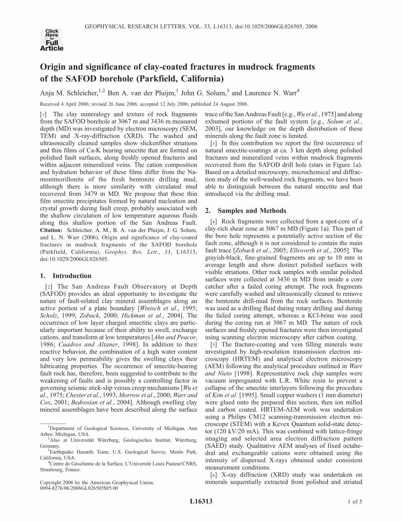
[8] The SEM images in Figures 1b and 1c show surfaceslickenfibres and an ultra thin film mineral coating thatcharacterizes the polished fracture surfaces. The energydispersive spectroscopy (EDS) patterns of these surfacecoatings reveal alumino-silicate minerals with a diversecation content of Na, K, Ca, Mg, and Fe (Figure 1b). Porespaces beneath the fracture surface are partly filled withneocrystallized pyrite that shows well developed cubiccrystal forms and no signs of crystal damage. Large irregulargrains of detrital illite and chlorite with curved and damagedparticle shapes are surrounded by a compactional fabric.These minerals lie perpendicular to the polished fracturesand contain notably higher concentrations of K, Mg and Fe.Authigenic fibrous to columnar Ca-alumino-silicate miner-als were detected in the pores of some rock-chip fracturesthat are likely zeolite minerals (laumontite)[see Solum and
van der Pluijm, 2004; J. G. Solum et al., Mineralogicalcharacterization of protolith and fault rocks from theSAFOD main hole, submitted to Geophysical ResearchLetters, 2006, hereinafter referred to as Solum et al.,submitted manuscript, 2006]. Similar cations can be ob-served in all SEM and TEM analyses by EDS (see inset inFigure 2a) when comparing the elemental composition of thesmectitic coatings with the fresh and used drilling mud.However, the drilling muds are best recognized by theoccurrence of Na, Cl and S. Distinct differences also occurin the Si-Al ratio of the natural and contaminating smectitephases whereby the fracture surface clays are characterizedby lower ratios than the Si enriched drill mud.[9] A smectite phase with a similar elemental composi-
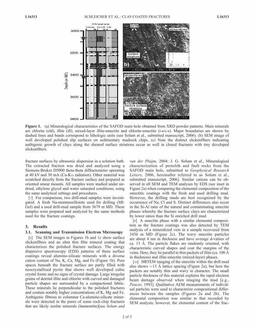
tion as the fracture coatings was also detected by TEManalysis of a mineralized vein in a sample recovered from3436 m MD (Figure 2c). The wavy smectite particlesare about 4 nm in thickness and have average d-values ofca. 13 A. The particle flakes are randomly oriented, withcharacteristic curved shapes and coat the margins of theveins. Here, they lie parallel to thin packets of illite (ca. 100 Ain thickness) and illite-smectite (mixed-layer) phases.[10] HRTEM imaging of the smectite within the drill mud
also shows �13 A lattice spacing (Figure 2a), but here thepackets are notably thin and wavy in character. The smallparticle thickness of this material explains the rapid electronbeam damage observed when imaging the mud [e.g.,Peacor, 1993]. Qualitative AEM measurements of individ-ual particles were used to characterize compositional differ-ences between the samples (Figures 2a and 2b). Theelemental composition was similar to that recorded bySEM analysis; however, the elemental content of the frac-
Figure 1. (a) Mineralogical characteristics of the SAFOD main hole obtained from XRD powder patterns. Main mineralsare chlorite (chl), illite (ill), mixed-layer illite-smectite and chlorite-smectite (i-s/c-s). Major boundaries are shown bydashed lines and bands correspond to lithologic units (see Solum et al., submitted manuscript, 2006). (b) SEM image ofwell developed polished slip surfaces on sedimentary mudrock chips. (c) Note the distinct slickenfibers indicatingauthigenic growth of clays along the sheared surface striations occur as well in closed fractures with tiny developedslickenfibers.
L16313 SCHLEICHER ET AL.: CLAY-COATED FRACTURES L16313
2 of 5
ture coating smectite is much less variable than the drill-mud smectite.[11] Si-Al plots of all smectites follow linear trends with
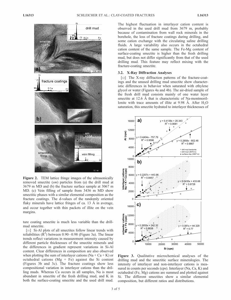
reliabilities (R2) between 0.90–0.98 (Figure 3a). The lineartrends reflect variations in measurement intensity caused bydifferent particle thicknesses of the smectite minerals andthe differences in gradient represent variations in Si-Alcontent. Clear differences in composition are also observedwhen plotting the sum of interlayer cations (Na + Ca + K) oroctahedral cations (Mg + Fe) against the Si content(Figures 3b and 3c). The fracture coatings show lesscompositional variation in interlayer cations than the dril-ling muds. Whereas Ca occurs in all samples, Na is mostabundant in smectite of the fresh drilling mud, and K inboth the surface-coating smectite and the used drill mud.
The highest fluctuation in interlayer cation content isobserved in the used drill mud from 3679 m, probablybecause of contamination from wall rock minerals in theborehole, the loss of fracture coatings during drilling, andsome cation exchange with the circulating saline drillingfluids. A large variability also occurs in the octahedralcation content of the same sample. The Fe-Mg content ofsurface-coating smectite is higher than the fresh drillingmud, but does not differ significantly from that of the useddrilling mud. This feature may reflect mixing with thefracture-coating smectite.
3.2. X-Ray Diffraction Analyses
[12] The X-ray diffraction patterns of the fracture-coat-ings and the unused drilling mud smectite show character-istic differences in behavior when saturated with ethyleneglycol or water (Figures 4a and 4b). The air-dried sample ofthe fresh drill mud consists mainly of one water layersmectite at 12.6 A that is characteristic of Na-montmoril-lonite with trace amounts of illite at 9.98 A. After H2Osaturation, this smectite hydrated to interlayer thicknesses of
Figure 2. TEM lattice fringe images of the ultrasonicallyremoved smectite (sm) particles from (a) the drill mud at3679 m MD and (b) the fracture surface sample at 3067 mMD. (c) Vein filling of sample from 3436 m MD showsmectitic phases with a similar elemental composition as thefracture coatings. The d-values of the randomly orientedflaky minerals have lattice fringes of ca. 13 A in average,
and occur together with thin packets of illite on the veinmargins.
Figure 3. Qualitative microchemical analyses of thedrilling mud and the smectitic surface mineralogies. Theintensity of interlayer and non-interlayer cations is mea-sured in counts per seconds (cps). Interlayer (Na, Ca, K) andoctahedral (Fe, Mg) cations are summed and plotted againstSi. The different smectites show a similar elementalcomposition, but different ratios and distributions.
L16313 SCHLEICHER ET AL.: CLAY-COATED FRACTURES L16313
3 of 5
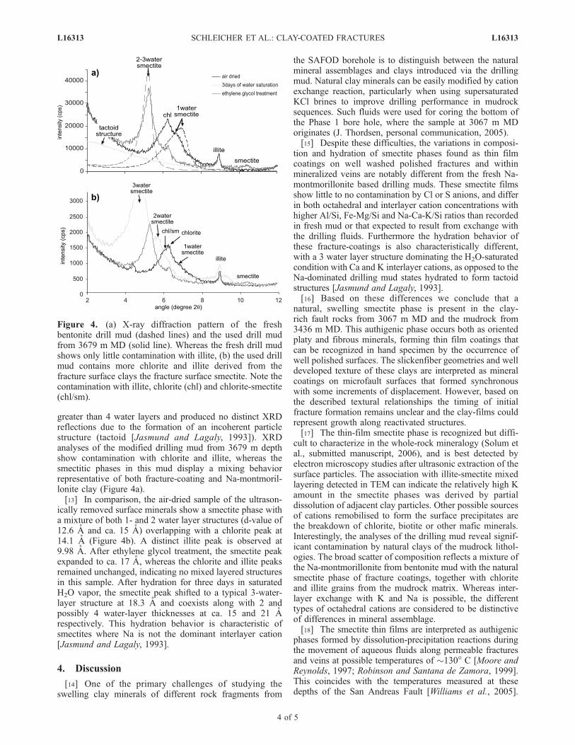
greater than 4 water layers and produced no distinct XRDreflections due to the formation of an incoherent particlestructure (tactoid [Jasmund and Lagaly, 1993]). XRDanalyses of the modified drilling mud from 3679 m depthshow contamination with chlorite and illite, whereas thesmectitic phases in this mud display a mixing behaviorrepresentative of both fracture-coating and Na-montmoril-lonite clay (Figure 4a).[13] In comparison, the air-dried sample of the ultrason-
ically removed surface minerals show a smectite phase witha mixture of both 1- and 2 water layer structures (d-value of12.6 A and ca. 15 A) overlapping with a chlorite peak at14.1 A (Figure 4b). A distinct illite peak is observed at9.98 A. After ethylene glycol treatment, the smectite peakexpanded to ca. 17 A, whereas the chlorite and illite peaksremained unchanged, indicating no mixed layered structuresin this sample. After hydration for three days in saturatedH2O vapor, the smectite peak shifted to a typical 3-water-layer structure at 18.3 A and coexists along with 2 andpossibly 4 water-layer thicknesses at ca. 15 and 21 Arespectively. This hydration behavior is characteristic ofsmectites where Na is not the dominant interlayer cation[Jasmund and Lagaly, 1993].
4. Discussion
[14] One of the primary challenges of studying theswelling clay minerals of different rock fragments from
the SAFOD borehole is to distinguish between the naturalmineral assemblages and clays introduced via the drillingmud. Natural clay minerals can be easily modified by cationexchange reaction, particularly when using supersaturatedKCl brines to improve drilling performance in mudrocksequences. Such fluids were used for coring the bottom ofthe Phase 1 bore hole, where the sample at 3067 m MDoriginates (J. Thordsen, personal communication, 2005).[15] Despite these difficulties, the variations in composi-
tion and hydration of smectite phases found as thin filmcoatings on well washed polished fractures and withinmineralized veins are notably different from the fresh Na-montmorillonite based drilling muds. These smectite filmsshow little to no contamination by Cl or S anions, and differin both octahedral and interlayer cation concentrations withhigher Al/Si, Fe-Mg/Si and Na-Ca-K/Si ratios than recordedin fresh mud or that expected to result from exchange withthe drilling fluids. Furthermore the hydration behavior ofthese fracture-coatings is also characteristically different,with a 3 water layer structure dominating the H2O-saturatedcondition with Ca and K interlayer cations, as opposed to theNa-dominated drilling mud states hydrated to form tactoidstructures [Jasmund and Lagaly, 1993].[16] Based on these differences we conclude that a
natural, swelling smectite phase is present in the clay-rich fault rocks from 3067 m MD and the mudrock from3436 m MD. This authigenic phase occurs both as orientedplaty and fibrous minerals, forming thin film coatings thatcan be recognized in hand specimen by the occurrence ofwell polished surfaces. The slickenfiber geometries and welldeveloped texture of these clays are interpreted as mineralcoatings on microfault surfaces that formed synchronouswith some increments of displacement. However, based onthe described textural relationships the timing of initialfracture formation remains unclear and the clay-films couldrepresent growth along reactivated structures.[17] The thin-film smectite phase is recognized but diffi-
cult to characterize in the whole-rock mineralogy (Solum etal., submitted manuscript, 2006), and is best detected byelectron microscopy studies after ultrasonic extraction of thesurface particles. The association with illite-smectite mixedlayering detected in TEM can indicate the relatively high Kamount in the smectite phases was derived by partialdissolution of adjacent clay particles. Other possible sourcesof cations remobilised to form the surface precipitates arethe breakdown of chlorite, biotite or other mafic minerals.Interestingly, the analyses of the drilling mud reveal signif-icant contamination by natural clays of the mudrock lithol-ogies. The broad scatter of composition reflects a mixture ofthe Na-montmorillonite from bentonite mud with the naturalsmectite phase of fracture coatings, together with chloriteand illite grains from the mudrock matrix. Whereas inter-layer exchange with K and Na is possible, the differenttypes of octahedral cations are considered to be distinctiveof differences in mineral assemblage.[18] The smectite thin films are interpreted as authigenic
phases formed by dissolution-precipitation reactions duringthe movement of aqueous fluids along permeable fracturesand veins at possible temperatures of �130� C [Moore andReynolds, 1997; Robinson and Santana de Zamora, 1999].This coincides with the temperatures measured at thesedepths of the San Andreas Fault [Williams et al., 2005].
Figure 4. (a) X-ray diffraction pattern of the freshbentonite drill mud (dashed lines) and the used drill mudfrom 3679 m MD (solid line). Whereas the fresh drill mudshows only little contamination with illite, (b) the used drillmud contains more chlorite and illite derived from thefracture surface clays the fracture surface smectite. Note thecontamination with illite, chlorite (chl) and chlorite-smectite(chl/sm).
L16313 SCHLEICHER ET AL.: CLAY-COATED FRACTURES L16313
4 of 5
As no direct replacement of detrital grains was observed,precipitation from migrating fluids is the likely mechanismfor deposition on slip surfaces. This is consistent with thesuggestion of fluid-driven seismicity and the occurrence ofmicroearthquakes and creep in the low resistivity zone atParkfield [Unsworth et al., 1997], although elevated fluidpressures have yet to be observed in the SAFOD hole[Zoback et al., 2005].[19] In the vicinity of SAFOD, the San Andreas Fault is
moving through a combination of aseismic creep andrepeating micro-earthquakes [Hickman et al., 2004]. Thesewell oriented smectite coating and slickenfibre geometriesare interpreted as precipitation associated with slow creepmotion in this segment of the fault zone and, thus, representrelatively long-term displacements, as nucleation and crystalgrowth are generally envisaged to be a slower process thanthe duration of seismic slip events.
[20] Acknowledgments. The study was funded through the USNational Science Foundation (EAR-0345985). We thank Steve Hickmanand Mark Zoback for SAFOD coordination and for providing samples,Sarah Draper for help in sample collection and Sara Tourscher for helpfuldiscussions and sharing complementary chemical results. We thank as wellan anonymous reviewer and Jim Evans for their helpful comments.
ReferencesAhn, J. H., and D. R. Peacor (1986), Transmission and analytical electronmicroscopy of the smectite to illite transition, Clays Clay Miner., 34,165–179.
Bedrosian, P. A., M. J. Unsworth, G. D. Egbert, and C. H. Thuerber (2004),Geophysical images of creeping segment of the San Andreas Fault: Im-plications for the role of crustal fluids in the earthquake process, Tecto-nophysics, 385, 137–158.
Chester, F., J. P. Evans, and R. L. Biegel (1993), Internal structure andweakening mechanisms of the San Andreas Fault, J. Geophys. Res.,98, 771–786.
Cuadros, J., and S. P. Altander (1998), Characterization of mixed-layer I-Sfrom bentonites using chemical, and X-ray methods: Constraints on thesmectite-to-illite transformation mechanism, Am. Mineral., 83, 762–774.
Ellsworth, W., et al. (2005), Observing the San Andreas Fault at depth, EosTrans. AGU, 86(52), Fall Meet. Suppl., Abstract T24B-04.
Hickman, S., M. Zoback, and W. Ellsworth (2004), Introduction tospecial section: Preparing for the San Andreas Fault Observatory atDepth, Geophys. Res. Lett., 31, L12S01, doi:10.1029/2004GL020688.
Jasmund, K., and G. Lagaly (1993), Tonminerale und Tone. Struktur,Eigenschaften, Anwendungen und Einsatz in Industrie und Umwelt,490 pp., Springer, New York.
Kim, J. W., D. R. Peacor, D. Tessier, and F. Elsass (1995), A technique formaintaining texture and permanent expansion of smectite interlayers forTEM observations, Clays and Clay Minerals, 43, 51–57.
Moore, D. M., and R. C. Reynolds (1997), X-ray Diffraction and theIdentification and Analysis of Clay Minerals, 378 pp., Oxford Univ.Press, New York.
Morrow, C. A., D. E. Moore, and D. A. Lockner (2000), The effect ofmineral bond strength and adsorbed water on fault gouge frictionalstrength, Geophys. Res. Lett., 27, 815–818.
Peacor, D. R. (1993), Analytical electron microscopy: X-ray analysis,in Minerals and Reactions at the Atomic Scale: Transmission ElectronMicroscopy, vol. 27, Reviews in Mineralogy, Mineral. Soc. of Am., Wa-shington, D. C.
Robinson, D., and A. Santana de Zamora (1999), The smectite to chloritetransition in the Chipilapa geothermal system, El Salvador, Am. Mineral.,84, 607–619.
Scholz, C. H. (1999), Evidence for a strong San Andreas fault, Geology,28(2), 163–166.
Solum, J. G., and B. A. van der Pluijm (2004), Phyllosilicate mineralassemblages of the SAFOD Pilot Hole and comparison with an exhumedsegment of the San Andreas Fault System, Geophys. Res. Lett., 31,L15S19, doi:10.1029/2004GL019909.
Solum, J. G., B. A. van der Pluijm, D. R. Peacor, and L. N. Warr (2003),Influence of phyllosilicate mineral assemblages, fabrics, and fluids on thebehavior of the Punchbowl fault, southern California, J. Geophys. Res.,108(B5), 2233, doi:10.1029/2002JB001858.
Unsworth, M. J., P. E. Malin, G. D. Egbert, and J. R. Booker(1997), Internal structure of the San Andreas Fault at Parkfield, Cali-fornia, Geology, 25(4), 359–362.
Warr, L. N., and F. Nieto (1998), Crystallite thickness and defect density ofphyllosilicates in low-temperature metamorphic pelites: ATEM and XRDstudy of clay-minerals crystallinity-index standards, Can. Mineral., 36,1453–1474.
Warr, L. N., and S. Cox (2001), Clay mineral transformations and weak-ening mechanisms along the Alpine Fault, New Zealand, J. Geol. Soc.London, Spec. Publ., 186, 85–101.
Williams, C. F., M. A. D’Alessio, F. V. Grubb, and S. P. Galanis (2005),Heat flow in the SAFOD main hole, Eos Trans. AGU, 86(52), Fall Meet.Suppl., Abstract T23E-07.
Wintsch, R. P., R. Christoffersen, and A. K. Kronenberg (1995), Fluid-rockreaction weakening of fault zones, J. Geophys. Res., 100, 13,021–13,032.
Wu, F. T., L. Blatter, and H. Robertson (1975), Clay gouges in the SanAndreas Fault system and their possible implications, Pure Appl. Geo-phys., 113, 87–96.
Zoback, M. D. (2000), Strength of the San Andreas, Nature, 405, 31–32.Zoback, M. D., S. Hickman, and B. Ellsworth (2005), Drilling, sampling,and measurements in the San Andreas Fault Zone at seismogenic depth,Eos Trans. AGU, 86(52), Fall Meet. Suppl., Abstract T23E-01.
�����������������������A. M. Schleicher and B. A. van der Pluijm, Department of Geological
Sciences, University of Michigan, 4534b C. C. Little Building, 1100 N.University Ave., Ann Arbor, MI 48109–1005, USA. ([email protected])J. G. Solum, Earthquake Hazards Team, U.S. Geological Survey, 345
Middlefield Road, MS 977, Menlo Park, CA 94025, USA.L. N. Warr, Centre de Geochimie de la Surface, l’universite Louis
Pasteur/CNRS, 1 rue Blessig, F-67084 Strasbourg, France.
L16313 SCHLEICHER ET AL.: CLAY-COATED FRACTURES L16313
5 of 5