Embed Size (px)
Citation preview
Origin of Microglia: Current Conceptsand Past Controversies
Florent Ginhoux1 and Marco Prinz2,3
1Singapore Immunology Network (SIgN), Agency for Science, Technology and Research (A�STAR),Singapore 138648
2Institute of Neuropathology, University of Freiburg, 79106 Freiburg, Germany3BIOSS Centre for Biological Signalling Studies, University of Freiburg, 79106 Freiburg, Germany
Correspondence: [email protected]; [email protected]
Microglia are the resident macrophages of the central nervous system (CNS), which sit inclose proximity to neural structures and are intimately involved in brain homeostasis. Themicroglial population also plays fundamental roles during neuronal expansion and differen-tiation, as well as in the perinatal establishment of synaptic circuits. Any change in thenormal brain environment results in microglial activation, which can be detrimental if notappropriately regulated. Aberrant microglial function has been linked to the development ofseveral neurological and psychiatric diseases. However, microglia also possess potent im-munoregulatory and regenerative capacities, making them attractive targets for therapeuticmanipulation. Such rationale manipulations will, however, require in-depth knowledge oftheir origins and the molecular mechanisms underlying their homeostasis. Here, we discussthe latest advances in our understanding of the origin, differentiation, and homeostasis ofmicroglial cells and their myelomonocytic relatives in the CNS.
Microglia are the resident macrophages ofthe central nervous system (CNS), which
are uniformly distributed throughout the brainand spinal cord with increased densities inneuronal nuclei, including the Substantia nigrain the midbrain (Lawson et al. 1990; Perry1998). They belong to the nonneuronal glialcell compartment and their function is crucialto maintenance of the CNS in both health anddisease (Ransohoff and Perry 2009; Perry et al.2010; Ransohoff and Cardona 2010; Prinz andPriller 2014).
Two key functional features define microglia:immune defense and maintenance of CNS ho-meostasis. As part of the innate immune system,microglia constantly sample their environment,scanning and surveying for signals of externaldanger (Davalos et al. 2005; Nimmerjahn et al.2005; Lehnardt 2010), such as those from invad-ing pathogens, or internal danger signals gener-ated locally bydamaged ordying cells (Bessiset al.2007; Hanisch and Kettenmann 2007). Detectionof such signals initiates a program of microglialresponses that aim to resolve the injury, protect
Editors: Ben A. Barres, Marc R. Freeman, and Beth Stevens
Additional Perspectives on Glia available at www.cshperspectives.org
Copyright # 2015 Cold Spring Harbor Laboratory Press; all rights reserved; doi: 10.1101/cshperspect.a020537
Cite this article as Cold Spring Harb Perspect Biol 2015;7:a020537
1
on February 21, 2022 - Published by Cold Spring Harbor Laboratory Press http://cshperspectives.cshlp.org/Downloaded from
the CNS from the effects of the inflammation,andsupporttissuerepairandremodeling(Ming-hetti and Levi 1998; Goldmann and Prinz 2013).
Microglia are also emerging as crucial con-tributors to brain homeostasis through controlof neuronal proliferation and differentiation,as well as influencing formation of synapticconnections (Lawson et al. 1990; Perry 1998;Hughes 2012; Blank and Prinz 2013). Recentimaging studies revealed dynamic interactionsbetween microglia and synaptic connections inthe healthy brain, which contributed to themodification and elimination of synaptic struc-tures (Perry et al. 2010; Tremblay et al. 2010;Bialas and Stevens 2013). In the prenatal brain,microglia regulate the wiring of forebrain cir-cuits, controlling the growth of dopaminergicaxons in the forebrain and the laminar posi-tioning of subsets of neocortical interneurons(Squarzoni et al. 2014). In the postnatal brain,microglia-mediated synaptic pruning is similar-ly required for the remodeling of neural circuits(Paolicelli et al. 2011; Schafer et al. 2012). Insummary, microglia occupy a central positionin defense and maintenance of the CNS and, as aconsequence, are a key target for the treatmentof neurological and psychiatric disorders.
Although microglia have been studied fordecades, a long history of experimental mis-interpretation meant that their true origins re-mained debated until recently. Although weknew that microglial progenitors invaded thebrain rudiment at very early stages of embryonicdevelopment (Alliot et al. 1999; Ransohoff andPerry 2009), it has now been established thatmicroglia arise from yolk sac (YS)-primitivemacrophages, which persist in the CNS intoadulthood (Davalos et al. 2005; Nimmerjahnet al. 2005; Ginhoux et al. 2010, 2013; Kierdorfand Prinz 2013; Kierdorf et al. 2013a). Moreover,early embryonic brain colonization by microgliais conserved across vertebrate species, implyingthat it is essential for early brain development(Herbomel et al. 2001; Bessis et al. 2007; Hanischand Kettenmann 2007; Verney et al. 2010; Schle-gelmilch et al. 2011; Swinnen et al. 2013). In thisreview, we will present the latest findings in thefield of microglial ontogeny, which provide newinsights into their roles in health and disease.
HISTORICAL PERSPECTIVES ONMACROPHAGES AND MICROGLIA
In 1969, the original phagocyte classificationsystems based on the work of Metchnikoff andEhrlich, and later Aschoff (Aschoff 1924; Min-ghetti and Levi 1998; Gordon and Taylor 2005;Kaufmann 2008), were superseded by the con-cept of the mononuclear phagocyte system(MPS) (van Furth and Cohn 1968; van Furthet al. 1972), a term that is still used today (Geiss-mann et al. 2010). The MPS today includescirculating monocytes in the bloodstream(“inflammatory” or Ly-6Cþ CCR2þCX3CR1lo
monocytes and “patrolling” or Ly-6C2CCR22
CX3CR1hi monocytes in mouse), as well as den-dritic cells and macrophages from both lym-phoid and nonlymphoid organs. In this context,macrophages are the resident phagocytic cellsin lymphoid tissues (spleen, lymph nodes) andnonlymphoid tissues, such as the brain (microg-lia), liver (Kupffer cells), lung (alveolar macro-phages), bone (osteoclasts), kidney (kidneymacrophages), and skin (Langerhans cells). Ofnote, epidermal Langerhans cells are a uniquecell population in the sense that, although aris-ing from macrophage progenitors, they acquireunique dendritic cell features on final differen-tiation compared with other macrophage pop-ulations (Ginhoux and Merad 2010; Hoeffelet al. 2012). At these sites, macrophages con-tribute to steady-state tissue homeostasis viathe clearance of apoptotic cells and the produc-tion of growth factors, but, on infection, theybecome activated to phagocytose pathogensand produce inflammatory cytokines, as theyare equipped with a broad range of pathogen-recognition receptors (Gordon 2002). Althoughthe MPS has been a useful framework for con-sidering phagocyte biology, it also led to theassumption that all tissue macrophages are iden-tical in origin and function. However, this as-sumption has been challenged in recent years,especially in the case of microglia (Ginhoux et al.2010; Hoeffel et al. 2012; Schulz et al. 2012).
It has taken more than 150 years of researchfor microglia to be formally recognized as a sep-arate and specialized macrophage population inthe CNS with distinct developmental origins.
F. Ginhoux and M. Prinz
2 Cite this article as Cold Spring Harb Perspect Biol 2015;7:a020537
on February 21, 2022 - Published by Cold Spring Harbor Laboratory Press http://cshperspectives.cshlp.org/Downloaded from
del Rıo-Hortega was the first to clearly identify asmall population of phagocytic, migratory cellswithin the CNS, which he proposed were ofmesodermal origin (del Rıo-Hortega 1932).Some years later, he introduced the term “mi-croglial cell,” and refined his description ofthese cells as the nonneuronal, nonastrocyticelement of the CNS, distinct from neurectoder-mal oligodendroglia and oligodendrocytes (delRıo-Hortega 1939), which constitute the mac-roglia. Despite some controversies on microgliaorigin from either the mesoderm or the ecto-derm in the history of microglial study (Rezaieand Male 2002; Chan et al. 2007; Ginhoux et al.2013), several investigators followed del Rıo-Hortega’s hypothesis and presented evidencesupporting a mesodermal origin of microgliain light of their morphological and phenotypicsimilarities with macrophages, first by couplinglight/electron microscopy and immunohisto-chemistry, which allowed identification of par-allel morphological features of macrophagesand microglia at various stages of development(Murabe and Sano 1982, 1983), and, second, byshowing that microglial cells were recognized byantisera raised against monocyte/macrophageantigens (Hume et al. 1983; Perry et al. 1985).
MARKERS OF MICROGLIA
Because of their mesodermal origin, microgliashare many features with other myeloid celltypes in the body. Observations of the pheno-typic similarities between circulating mono-cytes, tissue macrophages, and microglia werefirst reported �30 years ago in immunohisto-chemical studies, which showed microglial ex-pression of macrophage markers, including F4/80, Fc receptor, and CD11b, in mouse (Perryet al. 1985) and, later, in human (Akiyama andMcGeer 1990).
As the tissue-resident macrophage of theCNS, murine microglia have since been con-firmed to express multiple macrophage mark-ers, including the colony-stimulating factor(CSF)-1 receptor (CSF-1R, CD115), the integ-rin CD11b, the surface glycoproteins F4/80, theinhibitory immune receptor CD200R, the sur-face enzyme tyrosine-protein phosphatase non-
receptor-type substrate or CD172a, the fractal-kine receptor CX3CR1, and the calcium-bindingprotein lba-1 (Prinz and Mildner 2011). How-ever, few markers, which are specific to micro-glia, have been identified. CD39 (ectonucleo-side triphosphate diphosphohydrolase) geneexpression has recently been proposed to enabledistinction of microglia from their peripheralrelatives (Butovsky et al. 2012). Recent studiesalso reported that microglia have a unique tran-scriptomic signature, which distinguishes themfrom other CNS cells and peripheral macro-phages or monocytes (Gautier et al. 2012; Chiuet al. 2013), and express a unique cluster oftranscripts encoding proteins for sensing en-dogenous ligands and microbes, defined as thesensome (Hickman et al. 2013). Microglia alsoexpress lower levels of the panhematopoieticmarker CD45 compared with tissue macro-phages, which also permits their discriminationfrom monocytes in the bloodstream, whereasthe hemoglobin scavenger receptor CD163 en-ables distinction from perivascular macrophag-es in the steady state (Dijkstra et al. 1985; Serratset al. 2010).
THE YS ORIGIN OF MICROGLIA
The evident phenotypic similarities betweenmicroglia and other macrophage populationsled to ready acceptance of the notion of theirmyeloid origin, although the true identity ofmicroglial progenitors remained controversialuntil recently. Initial studies described the pres-ence of microglial cells during early develop-ment, suggesting that microglia arise from em-bryonic progenitors. These progenitors werefirst proposed by del Rıo-Hortega to be menin-geal macrophages infiltrating the brain duringearly embryonic development. However, themajority of the scientific community at thattime, including del Rıo-Hortega himself, be-lieved that microglia could also be derivedfrom blood monocytes. Monocytes are indeedrecruited to the neonatal and adult brain, inthe latter case, most often under inflammatoryconditions, where they can differentiate into mi-croglia-like cells. These observations long sup-ported the prevailing viewpoint that blood-
Origin of Microglia
Cite this article as Cold Spring Harb Perspect Biol 2015;7:a020537 3
on February 21, 2022 - Published by Cold Spring Harbor Laboratory Press http://cshperspectives.cshlp.org/Downloaded from
circulating monocytes represented microglialprogenitors, replacing those seeding the brainduring embryonic development. In fact, untilrecently, the most consensual hypothesis wasthat embryonic and perinatal hematopoieticwaves of microglial recruitment and differenti-ation occurred in the CNS.
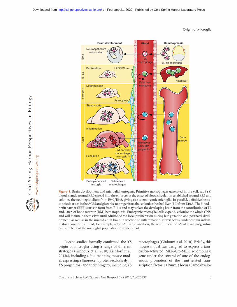
When del Rıo-Hortega first described mi-croglia, he also noted their presence in early de-velopment and proposed that, near this time,they might initially arise from mesodermal cellsof the pia mater, the innermost layer of themeninges (the membranes surrounding theCNS). He reported the “migration of embryoniccorpuscles from the pia into the nerve centres,”but simultaneously proposed that “microgliamay eventually arise from other related ele-ments, chiefly the blood mononuclears,” basedon the similarities in morphology and phago-cytic activity between microglia and monocytes(del Rıo-Hortega 1939). These two statementswere the founding of the “origin of microglia”controversy that was to last for the next 50 years.Later, the observation that the brain rudimentalready contains microglia at E9.5 of the 20 d ofmurine embryonic development (Alliot et al.1991, 1999) forced developmental neuroscien-tists to delve more deeply into the complex sub-ject of embryonic hematopoiesis. Our currentunderstanding of the multifaceted process ofembryonic hematopoiesis was extensively re-viewed (Cumano and Godin 2007; Orkin andZon 2008). The appearance of microglia in theneuroepithelium at E9.5 days suggested thattheir precursors might originate from the YS(Fig. 1). The murine embryonic YS producesearly primitive macrophages and erythrocytesas part of the process of “primitive hemato-poiesis,” occurring from E8.5, as opposed tothe generation of definitive hematopoieticstem cells (HSCs), which occurs in the aorta–gonad–mesonephros (AGM) region of the em-bryo around E10.5. These AGM-derived HSCsthen migrate to the fetal liver (FL) and bonemarrow (BM) and differentiate therein into alllineages, including monocytes, macrophages,and lymphocytes, which are generated as partof “definitive hematopoiesis” (Bertrand et al.2005; Cumano and Godin 2007). In addition,
from E8.25, multilineage erythromyeloid pro-genitors (EMP) and lymphomyeloid progeni-tors also emerge in the YS as a “second wave,”which is already considered as part of the defin-itive hematopoietic wave but called the transientdefinitive stage. The contribution of such pro-genitors to macrophage populations via a fetalmonocyte intermediate remain to be investigat-ed. HSC-derived myeloid cells, such as mono-cytes, are produced abundantly in the FL onlyfrom E12.5/E13.5 (Fig. 1), days after the initialcolonization of the brain rudiment by YS mac-rophages at E9.5. Of interest, a population ofmaternally derived macrophages can be foundin the YS of the embryo as early as E7.5. Thispopulation, however, subsequently decreases innumber, becomes almost undetectable at E9.0,and is later absent in the embryo (Bertrand et al.2005; Kierdorf et al. 2013a).
At E8.5–9.0, the first immature macrophag-es are found in the YS (Takahashi et al. 1996;Lichanska and Hume 2000), and they developthrough a nonmonocytic pathway (Takahashiet al. 1996; Lichanska and Hume 2000). Thefirst macrophage-like cells with an amoeboidshape appear in the rodent neuroepitheliumat a similar time point (Ashwell 1990; Ashwelland Waite 1991; Chan et al. 2007) and weresuggested to be the precursors of microglialcells (Alliot et al. 1999). A clear requirementfor the circulatory system for brain coloniza-tion by YS macrophages was determined usingE9.5–10 Ncx-12/2 embryos, which have nofunctional blood circulation (Koushik et al.2001), and were found to lack microglial pro-genitors, as well as other fetal macrophages, de-spite normal YS hematopoiesis (Ginhoux et al.2010). At E13.5, when the FL is already the pri-mary hematopoietic organ and the main site ofHSC expansion and differentiation (Lichanskaand Hume 2000), microglial precursors can bedetected in significant numbers within the lin-ing of the fourth ventricle (Chan et al. 2007). Asa result of the finding that tissues, such as theCNS, contain YS-derived macrophages, but notHSC or maternal macrophages, it became rea-sonable to believe that microglia originate fromYS macrophages rather than from HSC in theFL or BM.
F. Ginhoux and M. Prinz
4 Cite this article as Cold Spring Harb Perspect Biol 2015;7:a020537
on February 21, 2022 - Published by Cold Spring Harbor Laboratory Press http://cshperspectives.cshlp.org/Downloaded from
Recent studies formally confirmed the YSorigin of microglia using a range of differentstrategies (Ginhoux et al. 2010; Kierdorf et al.2013a), including a fate-mapping mouse mod-el, expressing a fluorescent protein exclusively inYS progenitors and their progeny, including YS
macrophages (Ginhoux et al. 2010). Briefly, thismouse model was designed to express a tam-oxifen-activated MER-Cre-MER recombinasegene under the control of one of the endog-enous promoters of the runt-related tran-scription factor 1 (Runx1) locus (Samokhvalov
Monocyte/other BMprogenitor?
Bonemarrow
Fetal liverFetal livermonocyte
YSmacrophage YS blood islands
Blood
Neuroepitheliumcolonization
Proliferation
Differentiation
Astrocytes
Steady state
Inflammation
Resolution
Embryo-derivedmicroglia
BM-derivedmacrophages
BM-derivedmacrophage(microglia?)
Pericytes
E9.
5E
13.5
New
born
Adu
lt
Brain development Hematopoiesis
Figure 1. Brain development and microglial ontogeny. Primitive macrophages generated in the yolk sac (YS)blood islands around E8.0 spread into the embryos at the onset of blood circulation established around E8.5 andcolonize the neuroepithelium from E9.0/E9.5, giving rise to embryonic microglia. In parallel, definitive hema-topoiesis arises in the AGM and gives rise to progenitors that colonize the fetal liver (FL) from E10.5. The blood–brain barrier (BBB) starts to form from E13.5 and may isolate the developing brain from the contribution of FLand, later, of bone marrow (BM) hematopoiesis. Embryonic microglial cells expand, colonize the whole CNS,and will maintain themselves until adulthood via local proliferation during late gestation and postnatal devel-opment, as well as in the injured adult brain in reaction to inflammation. Nevertheless, under certain inflam-matory conditions found, for example, after BM transplantation, the recruitment of BM-derived progenitorscan supplement the microglial population to some extent.
Origin of Microglia
Cite this article as Cold Spring Harb Perspect Biol 2015;7:a020537 5
on February 21, 2022 - Published by Cold Spring Harbor Laboratory Press http://cshperspectives.cshlp.org/Downloaded from
et al. 2007). When crossed with a Cre-reportermouse strain, recombination can be induced inembryos by a single injection of 4-hydroxyta-moxifen (40OHT) into pregnant females. Activerecombination in these knockin mice occurs ina short time frame, which does not exceed 12-hpostinjection, and leads to irreversible expres-sion of fluorescent protein in Runx1þ cells andtheir progeny (Samokhvalov et al. 2007). Al-though both YS and FL hematopoietic progen-itors express Runx1, YS progenitors are the onlyRunx1þ cells present at E7.5, and, so, injectionof 40OHT at this time specifically and irrevers-ibly tags YS progenitors and their progeny, butnot FL-derived progeny. In contrast, injection oftamoxifen at E8.5 or later will favor the taggingof AGM-derived hematopoietic progenitorsand not the YS progenitors (North et al. 1999;Samokhvalov et al. 2007). Thus, if microgliawere predominantly derived from YS-taggedprogenitors, they should express enhanced yel-low fluorescent protein (eYFP) in the adult CNSwhen 40OHT is injected at E7.25 and not atE8.5. Strikingly, the relative number of taggedmicroglia in mice injected at E7.25 was muchgreater than that of blood monocytes or othercirculating leukocytes (Ginhoux et al. 2010). Incontrast, the relative number of tagged micro-glia in mice injected from E8.0 onward de-creased dramatically, reaching undetectable lev-els in mice injected as close as E8.5, whereas therelative number of eYFPþ leukocytes, includingmonocytes, increased progressively in adultblood. Further confirmation of the YS originof microglia later came from another study us-ing myeloid-specific CSF-1R-Cre mice (Schulzet al. 2012). Importantly, this study also high-lighted further differences between primitiveand definitive hematopoiesis, showing that thelatter relies on the transcription factor myelo-blastosis (MYB), whereas YS-derived macro-phages are MYB independent, but PU.1 depen-dent (Schulz et al. 2012), as described earlier(Sumner et al. 2000). This is in contrast to aprevious study, which reported that mice withnull mutations in PU.1 had normal numbers ofCsf-1rþ phagocytes at E11.5 (Lichanska et al.1999). This further underlines the fact thatYS-derived macrophages constitute an inde-
pendent lineage, distinct from the progeny ofdefinitive HSCs.
In an another study, we further character-ized the early YS progenitor that gives rise tomicroglia in the brain; we observed c-kitþ line-age2 progenitor cells within the YS that have theability to differentiate into CX3CR1þmicrogliain vitro, as well as in vivo (Kierdorf et al. 2013a).These cells also generated Ter119þ erythrocytesand, thus, represent a common erythromyeloidprogenitor (EMP) in the YS. Subsequently,these uncommitted EMPs disappear, and im-mature F4/80þCX3CR12 and F4/80þCX3
CR1þ macrophages develop and seed the sur-face of the developing brain at E9.0 (Kierdorfet al. 2013a).
A similar pattern of events may occur inhumans. In human fetuses, microglia-like cellswith a range of morphologies can be detectedas early as 13 wk of estimated gestational age(Hutchins et al. 1990). However, it appearsthat maturation of the microglial compartmentis ongoing throughout the majority of gesta-tion. Colonization of the spinal cord begins ataround 9 wk, the major influx and distributionof microglia commences at about 16 wk, andramified microglia take up to 22 wk to becomewidely distributed within the intermediate zone(Rezaie and Male 1999; Rezaie et al. 2005). Infact, it is only close to term, at 35 wk, that well-differentiated microglial populations can be de-tected within the developing human brain (Esiriet al. 1991; Rezaie and Male 2002; Rezaie et al.2005; Verney et al. 2010).
Importantly, microglial origin is uniqueamong the wide spectrum of tissue macrophagepopulations (Ginhoux and Jung 2014; Prinzand Priller 2014). Microglia arise predominant-ly from YS-derived macrophages (Fig. 1) (Gin-houx et al. 2010; Kierdorf et al. 2013a), whereasLangerhans cells originate mainly from FL-derived monocytes, but retain a detectable YS-derived macrophage (MF) component (Hoeffelet al. 2012). In contrast, alveolar macrophagesappear to derive mostly from FL-derived mono-cytes with minimal lasting contribution fromYS-derived macrophages (Guilliams et al. 2013;G Hoeffel, J Chen, Y Lavin et al., in prep.).The origin of other tissue-resident macrophage
F. Ginhoux and M. Prinz
6 Cite this article as Cold Spring Harb Perspect Biol 2015;7:a020537
on February 21, 2022 - Published by Cold Spring Harbor Laboratory Press http://cshperspectives.cshlp.org/Downloaded from
populations in the adult remains to be inves-tigated.
FACTORS DETERMINING THEDEVELOPMENT AND HOMEOSTASISOF MICROGLIA
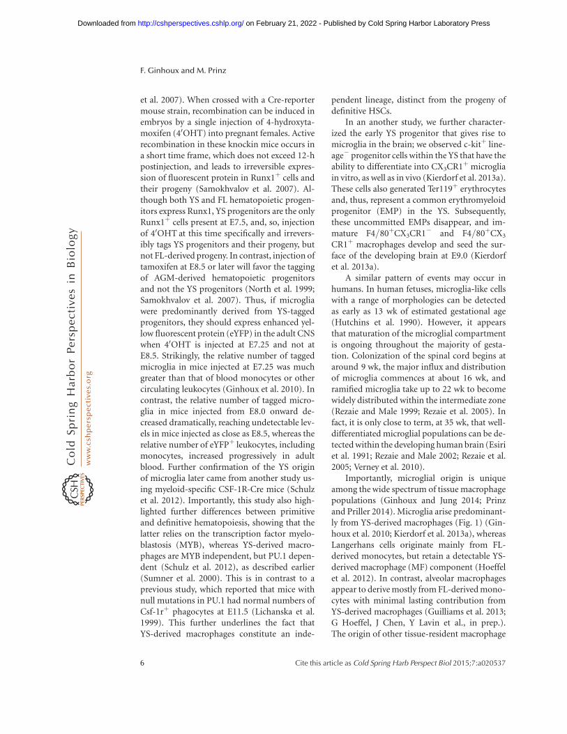
The transcriptional program that controls mi-croglial differentiation is only partially under-stood (Kierdorf and Prinz 2013). A dramaticreduction in numbers of tissue macrophages,including microglia, occurs in mice that lackthe CSF-1R (Dai et al. 2002; Ginhoux et al.2010; Erblich et al. 2011) and in Csf-1op/op mu-tant mice (Yoshida et al. 1990; Wegiel et al. 1998),which have a natural null mutation in the Csf-1gene. These studies clearly establish the impor-tance of CSF-1 and its receptor in macrophagehomeostasis in vivo (Fig. 2) (Pixley and Stanley2004), although the precise role of CSF-1 and itsreceptor during microglial lineage commitmentremains controversial. One hypothesis is thatCSF-1 drives the microglial differentiation ofphagocytic YS macrophages entering the em-bryo (Metcalf 1985), whereas an alternative the-ory is that CSF-1 provides a survival signal forthe differentiating macrophages and that thesesurviving cells then respond to an intrinsic de-velopmental program to become mature micro-glia (Lagasse and Weissman 1997). In favor ofthe latter hypothesis, macrophages are detectedin the YS and the brain rudiment in E10.5Csf-1r2/2 mice, but not in embryos at E12.5(Ginhoux et al. 2010; Hoeffel et al. 2012).
Interestingly, the microglial population ismore profoundly affected by the absence of theCSF-1R than in the absence of its ligand CSF-1(Ginhoux et al. 2010), which suggested the pos-sibility of a second ligand for CSF-1R that waslater identified as interleukin-34 (IL-34) (Linet al. 2008). In vitro IL-34 binds the CSF-1R atdifferent regions than CSF-1 and with higheraffinity (Chihara et al. 2010); IL-34 is alsomore highly conserved in mammalian and avianspecies than CSF-1, suggestive of an importantrole in macrophage homeostasis (Garceau et al.2010). Recently, two groups generated IL-34knockout (KO) mice (Greter et al. 2012; Wanget al. 2012) and reported that, in the brain, de-
ficiency of IL-34 led to a significant decrease inmicroglial cell numbers. Interestingly, IL-34possesses a spatiotemporal expression patternthat differs from that of CSF-1, permitting com-plementary activation of the CSF-1R in bothembryonic and adult tissues (Wei et al. 2010).Of note, an alternate receptor for IL-34 was re-cently identified: the receptor-type protein ty-rosine phosphatase (PTP)-z, which is a cell-sur-face chondroitin sulfate proteoglycan primarilyexpressed on neural progenitors and glial cells(Nandi et al. 2013), suggesting that IL-34 mayhave a wider repertoire of effects within the CNSthan previously appreciated. A comparable re-duction in microglial cell numbers was also re-ported in mice deficient for an adaptor proteinfor CSF-1R, DAP12 (Otero et al. 2009). DAP12contains an immunoreceptor tyrosine-basedactivation motif (ITAM) in its cytoplasmic do-main and is highly expressed in natural killer(NK) and myeloid cells. In vitro, DAP12 con-trols proliferation and survival of macrophagesstimulated with CSF-1, whereas in vivo in oldermice deficient in DAP12 fewer microglia arepresent, in particular, regions of the CNS, whichsuggests a role for DAP12 in the long-termhomeostasis of microglia. Humans with muta-tions in the DAP12 gene develop Nasu–Hakoladisease, characterized by bone cysts and frac-tures and psychotic symptoms leading to severeneurodegeneration and encephalopathy (Palo-neva et al. 2000).
The transcription factor PU.1, expressed ex-clusively in hematopoietic cells, is also involvedin microglial development (Fig. 2). The Pu.1(Sfpi-1) gene is a member of the Ets family oftranscription factors (Rosenbauer and Tenen2007), and its disruption leads to multiple he-matopoietic abnormalities, including a lack ofmature B cells and macrophages (McKercheret al. 1996). In fact, PU.1-deficient mice arenot only devoid of circulating monocytes andtissue macrophages (McKercher et al. 1996),but also parenchymal microglia in the brain(Beers et al. 2006). Similar data have been ob-tained in zebrafish PU.1 mutants that show acomplete loss of brain macrophages (Herbomelet al. 2001). In addition, interferon regulatoryfactor (IRF)-8 has recently been found to regu-
Origin of Microglia
Cite this article as Cold Spring Harb Perspect Biol 2015;7:a020537 7
on February 21, 2022 - Published by Cold Spring Harbor Laboratory Press http://cshperspectives.cshlp.org/Downloaded from
late the transcriptional programing of micro-glial development (Kierdorf et al. 2013a). IRF-8 is a heterodimeric partner of PU.1 with knownroles in the development of B cells and myeloidcells in the BM (Holtschke et al. 1996). We re-cently found that YS-derived F4/80þCX3CR1þ
macrophages were dependent on the presence ofIRF-8 for theirearly development, whereas othermyeloid transcription factors, including MYB,ID2, BATF3, and KLF4, were redundant (Fig. 2)(Kierdorf et al. 2013a). Consequently, microglialdensity was significantly reduced in adult micelacking IRF-8 (Kierdorf et al. 2013a). Moreover,it now seems that IRF-8 may have a role duringthe activation of adult microglia (Horiuchi et al.2012; Masuda et al. 2012; Minten et al. 2012).
THE ADULT MICROGLIAL POPULATION:SELF-RENEWAL RATHER THANREPLENISHMENT BY THE BLOOD
During Homeostasis
Within the first week after birth, the microglialpopulation expands so dramatically (Alliot et al.
1999; Tambuyzer et al. 2009) that it was pre-sumed that the proliferation of embryonic mi-croglial cells alone could not account for such asteep increase in numbers, and so there mustbe a fresh influx of cells from another compart-ment. As initially suggested by Del Rio-Ortega,blood monocytes were believed to invade theCNS in the perinatal period and give rise tomicroglia, replacing the embryonic microglialcells. Several studies supported this hypothesis,notably an early report (Ling 1976) in whichround, amoeboid, phagocytic cells were seenin rat corpus callosum during the first few daysof life and then disappeared coincident withthe appearance of ramified microglia. However,this view has been radically revised in recentyears; unequivocal evidence from fate-mapp-ing mouse models revealed that microglia arenot BM-derived under homeostatic conditions,but originate from the embryonic YS (Ginhouxet al. 2010). Furthermore, new myeloid-specificgene-targeting approaches that focused on thechemokine receptor CX3CR1 have, for the firsttime, enabled the study of the kinetics of truehomeostatic microglial turnover without the
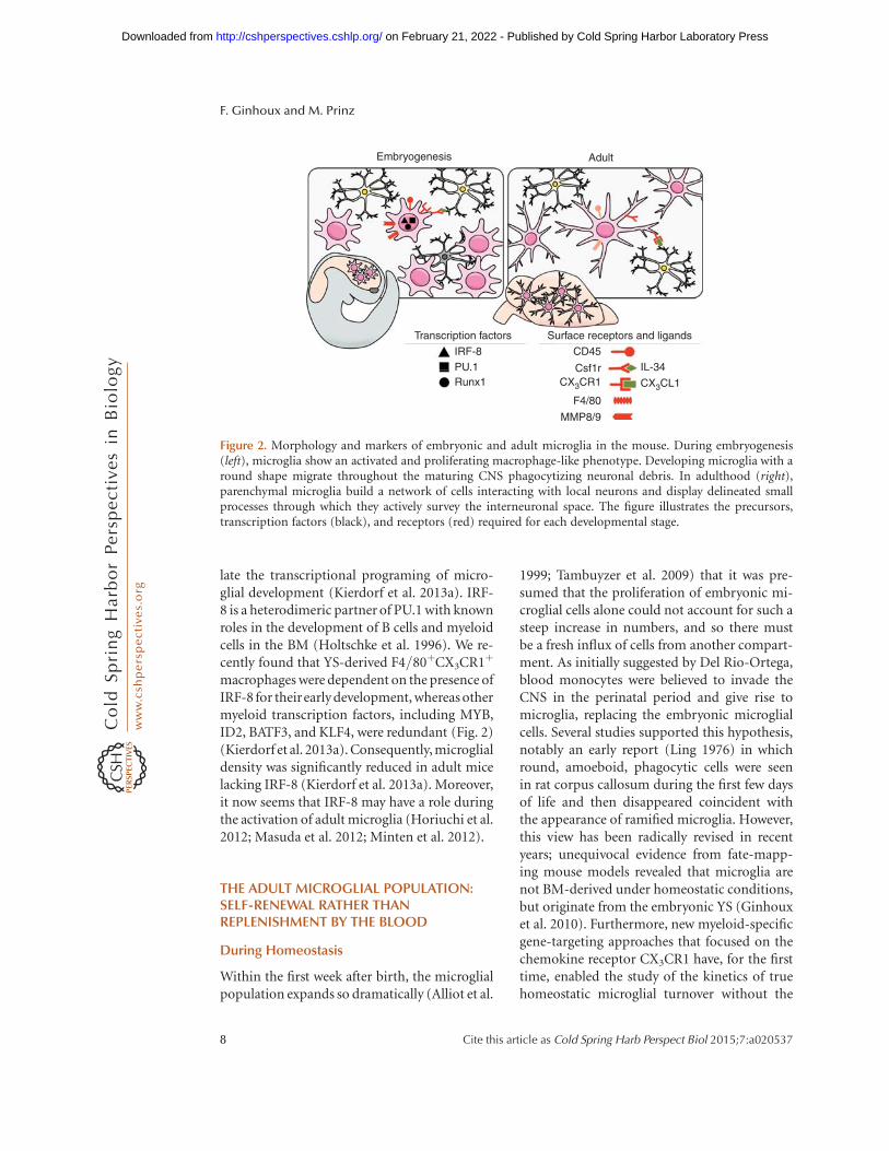
IRF-8IL-34
CD45
Csf1rCX3CR1 CX3CL1
F4/80
MMP8/9
PU.1Runx1
Transcription factors Surface receptors and ligands
AdultEmbryogenesis
Figure 2. Morphology and markers of embryonic and adult microglia in the mouse. During embryogenesis(left), microglia show an activated and proliferating macrophage-like phenotype. Developing microglia with around shape migrate throughout the maturing CNS phagocytizing neuronal debris. In adulthood (right),parenchymal microglia build a network of cells interacting with local neurons and display delineated smallprocesses through which they actively survey the interneuronal space. The figure illustrates the precursors,transcription factors (black), and receptors (red) required for each developmental stage.
F. Ginhoux and M. Prinz
8 Cite this article as Cold Spring Harb Perspect Biol 2015;7:a020537
on February 21, 2022 - Published by Cold Spring Harbor Laboratory Press http://cshperspectives.cshlp.org/Downloaded from
need for irradiation or chemotherapy (Gold-mann et al. 2013; Yona et al. 2013). Using thistechnique, microglia were found to be long livedwith labeled cells traceable for several months,which further argues against replacement byblood cells. In contrast, short-lived circulatingLy-6Chi and Ly-6Clo monocytes were quicklyreplaced by their nonlabeled progeny (Gold-mann et al. 2013; Yona et al. 2013).
Additional evidence for the lack of signifi-cant contribution of monocytes or other BM-derived progenitors to the adult microglial poolcame from prolonged experiments performedin parabiotic mice, in which two adult congenicmice undergo surgery to physically link theircirculatory systems. In reality, even after up to12 months of parabiosis, although monocytesin the blood of the parabionts originate fromboth animals, microglia remained totally of hostorigin, clearly illustrating the absence of contri-bution of monocytes or BM-derived cells to theCNS microglial population (Ajami et al. 2007,2011; Ginhoux et al. 2010; Hashimoto et al.2013).
Taken together, these new genetic approach-es helped to firmly establish the major featuresof microglial population, namely, that they arelong lived in vivo and not replaced by peripheralcells from the circulation, but are able to per-form context-dependent self-renewal to ensurepopulation maintenance.
During Disease
One of the most pressing questions in the fieldof microglial research during recent years hasbeen whether “BM-derived microglia” exist inthe adult brain and, if so, whether they are func-tional. The answer could have profound clinicalimplications because it determines whether itmight be possible to use peripheral microglialprecursors as carriers for neuroprotective or im-mune-modulatory genes into the diseased CNSto treat conditions, such as amyotrophic lateralsclerosis (ALS), Alzheimer’s disease (AD), andParkinson’s disease (PD) (Prinz et al. 2011;Prinz and Priller 2014).
The first seminal cell transplantation exper-iments in rats showed that, following BM trans-
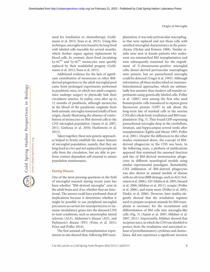
plantation, it was only perivascular macrophag-es that were replaced and not those cells withramified microglial characteristics in the paren-chyma (Hickey and Kimura 1988). Similar re-sults were seen in female patients who under-went sex-mismatched BM transplantation andwere subsequently examined for the engraft-ment of Y-chromosome-positive microglialcells; donor-derived perivascular macrophageswere present, but no parenchymal microgliacould be detected (Unger et al. 1993). Althoughinformative, all these studies relied on immuno-histochemical approaches, which are substan-tially less sensitive than modern cell transfer ex-periments using genetically labeled cells. Prilleret al. (2001) were among the first who usedhematopoietic cells transduced to express greenfluorescent protein (GFP) to ask about thelong-term fate of myeloid cells in the murineCNS after whole body irradiation and BM trans-plantation (Fig. 3). They found GFP-expressingparenchymal microglia deep in the cerebellum,striatum, and hippocampus several weeks aftertransplantation (Eglitis and Mezey 1997; Prilleret al. 2001). Despite the differences to the otherstudies mentioned above, the concept of BM-derived phagocytes in the CNS was born. Inthe following years, a plethora of publicationsappeared that examined the assumed functionand fate of BM-derived mononuclear phago-cytes in different neurological models usingsimilar experimental paradigms. Remarkably,CNS infiltration of BM-derived phagocyteswas also shown in animal models of diseasewith no obvious BBB damage, such as ALS (Sol-omon et al. 2006), AD (Malm et al. 2005; Simardet al. 2006; Mildner et al. 2011), scrapie (Prilleret al. 2006), and many more (Priller et al. 2001;Djukic et al. 2006). However, later studies ele-gantly showed that the irradiation regimenused to prepare recipient animals for BM trans-plants is necessary for the recruitment anddifferentiation of BM cells into microglia-likecells (Fig. 3) (Ajami et al. 2007; Mildner et al.2007, 2011). Importantly, Mildner showed thatrecipient mice, in which the CNS was shielded toprotect from the irradiation and associated re-lease of proinflammatory cytokines and chemo-kines, did not experience a significant invasion
Origin of Microglia
Cite this article as Cold Spring Harb Perspect Biol 2015;7:a020537 9
on February 21, 2022 - Published by Cold Spring Harbor Laboratory Press http://cshperspectives.cshlp.org/Downloaded from
of BM-derived cells into the brain in contrast tothe unshielded mice (Mildner et al. 2007). Be-yond the irradiation issue, these data also sug-gest that microglial engraftment from the bloodrequires preconditioning of the CNS that likelydisrupts the BBB. Additional clarity came fromexperiments in parabiotic mice, which enabledthe study of the turnover of hematopoieticcells for prolonged periods without the needfor irradiation (Ajami et al. 2007). Ajami usedsuch mice to show that, in contrast to irradiatedand transplanted mice, there was no microglialprogenitor recruitment from the circulationin either denervation or CNS neurodegenerativedisease. Intriguingly, if just one parabiont wasirradiated, no further contribution from theother parabiont occurred, in apparent contra-diction to the results of Mildner. However,Ajami further clarified that, although irradia-tion is required for donor cells to engraft, it isnot sufficient; another important but often over-looked requirement is the artificial and con-comitant introduction of a critical numberof donor BM cells into the blood circulation(where they are not normally found). This, inconjunction with the inflammation of the BBBcaused by irradiation, creates the unique non-physiological situation that is required for theBM-to-microglia pathway to prevail (Diserbo
et al. 2002; Li et al. 2004; Capotondo et al.2012). Taking this work further, the same grouprecently used a similar approach, combiningparabiosis and myeloablation, to show that re-cruited monocytes do not persist in the CNSand, therefore, even under these specific condi-tions, do not stably contribute to the residentmicroglial pool (Ajami et al. 2011). However,recruited short-lived monocytes are essentialdrivers of disease severity in multiple sclerosis(MS) and the experimental mouse model ofautoimmune encephalomyelitis (EAE) (Kinget al. 2009; Mildner et al. 2009). In conclusion,BM-derived microglia can engraft into the dis-eased brain and become an integral part of thecellular network in the CNS only under specificnonphysiological conditions. These specificconditions include irradiation and chemother-apeutic regimes, for example, the applicationof myeloablating agents, which all (1) alter theintegrity of the BBB, and (2) induce local pro-duction of myeloattracting chemokines, suchas CCL2 (Boettcher et al. 2008; Lampron et al.2012; Kierdorf et al. 2013b). Taken together,these data also clearly indicate that BM-derivednonmonocytic cells are able to permanentlyengraft to the diseased brain, whereas short-lived monocytes are only transiently recruitedto the CNS.
Endothelialcells
Bon
e m
arro
w c
ells
CCL2
CCR2
B
A
Conditioned ordiseased
Healthy
YS-derivedmicrogliaMacrophageprecursors
Figure 3. Formation of BM-derived microglia in the adult mouse brain. Postnatal BM-derived microglia formonly under defined host conditions in the CNS. BM cells (left) are released into the bloodstream in a chemokinereceptor (CCR)-2-dependent fashion and may enter the conditioned CNS. Local conditioning of the CNS canoccur via irradiation and neurodegeneration, which lead to both disruption of the BBB and induction ofchemokines, such as CCL2, thus, allowing engraftment of BM-derived macrophages. (A) YS-derived microglia(green) perform self-renewal by undergoing proliferation (indicated by an arrow), and (B) BM-derived phago-cyte (purple).
F. Ginhoux and M. Prinz
10 Cite this article as Cold Spring Harb Perspect Biol 2015;7:a020537
on February 21, 2022 - Published by Cold Spring Harbor Laboratory Press http://cshperspectives.cshlp.org/Downloaded from
CONCLUSION
Altogether, these seminal studies establishedthat microglia arise from embryonic hemato-poietic precursors that seed the CNS before birthand, more importantly, before the onset of BMhematopoiesis. It is now accepted that microgliaderive from unique embryonic precursors, theYS macrophages, which are not found in theBM as predicted earlier by the founder of themicroglial field, Pıo del Rıo-Hortega. Thisknowledge has far-reaching implications for theunderstanding of microglial functions in CNSdevelopment. First, the conservation of primi-tive macrophages and their YS derivation, boththroughout evolution and across diverse spe-cies, suggests that microglia play an importantphysiological role in the development of theCNS. Furthermore, microglial cells are presentduring all stages of brain development, includ-ing the early prenatal stages of neuronal cir-cuit building, as well as the postnatal stage ofsynapse elimination. This implies a functionalniche for microglia in the development of neu-ronal circuits of the brain and proposes intrigu-ing possibilities regarding the integrated devel-opment of the neural and immune systems.
ACKNOWLEDGMENTS
We apologize to all colleagues whose work waswas not cited owing to space constraints. Wethank Dr. L. Robinson for critical review andediting of the manuscript. F.G. is supportedby a Singapore Immunology Network coregrant. M.P. is supported by the Federal Ministryof Education and Research (BMBF)-fundedcompetence network of multiple sclerosis(KKNMS), the Gemeinnutzige Hertie-Stiftung(GHST), the Fritz Thyssen Stiftung, the compe-tence network of neurodegenerative disorders(KNDD), and the The Deutsche Forschungs-gemeinschaft (SFB 992, FOR1336).
REFERENCES
Ajami B, Bennett JL, Krieger C, Tetzlaff W, Rossi FM. 2007.Local self-renewal can sustain CNS microglia mainte-nance and function throughout adult life. Nat Neurosci10: 1538–1543.
Ajami B, Bennett JL, Krieger C, McNagny KM, Rossi FM.2011. Infiltrating monocytes trigger EAE progression, butdo not contribute to the resident microglia pool. NatNeurosci 14: 1142–1149.
Akiyama H, McGeer PL. 1990. Brain microglia constitutive-ly express b-2 integrins. J Neuroimmunol 30: 81–93.
Alliot F, Lecain E, Grima B, Pessac B. 1991. Microglial pro-genitors with a high proliferative potential in the embry-onic and adult mouse brain. Proc Natl Acad Sci 88: 1541–1545.
Alliot F, Godin I, Pessac B. 1999. Microglia derive fromprogenitors, originating from the yolk sac, and whichproliferate in the brain. Brain Res Dev Brain Res 117:145–152.
Aschoff L. 1924. Das retikuloendotheliale system [The retic-uloendothelial system]. Erg Inn Med Kinderheilk 26: S1–S117.
Ashwell K. 1990. Microglia and cell death in the developingmouse cerebellum. Brain Res Dev Brain Res 55: 219–230.
Ashwell KW, Waite PM. 1991. Cell death in the developingtrigeminal nuclear complex of the rat. Brain Res DevBrain Res 63: 291–295.
Beers DR, Henkel JS, Xiao Q, Zhao W, Wang J, Yen AA,Siklos L, McKercher SR, Appel SH. 2006. Wild-type mi-croglia extend survival in PU.1 knockout mice with fa-milial amyotrophic lateral sclerosis. Proc Natl Acad Sci103: 16021–16026.
Bertrand JY, Jalil A, Klaine M, Jung S, Cumano A, Godin I.2005. Three pathways to mature macrophages in the earlymouse yolk sac. Blood 106: 3004–3011.
Bessis A, Bechade C, Bernard D, Roumier A. 2007. Micro-glial control of neuronal death and synaptic properties.Glia 55: 233–238.
Bialas AR, Stevens B. 2013. TGF-b signaling regulates neu-ronal C1q expression and developmental synaptic refine-ment. Nat Neurosci 16: 1773–1782.
Blank T, Prinz M. 2013. Microglia as modulators of cogni-tion and neuropsychiatric disorders. Glia 61: 62–70.
Boettcher C, Ulbricht E, Helmlinger D, Mack AF, Reichen-bach A, Wiedemann P, Wagner HJ, Seeliger MW, Bring-mann A, Priller J. 2008. Long-term engraftment ofsystemically transplanted, gene-modified bone marrow-derived cells in the adult mouse retina. Br J Ophthalmol92: 272–275.
Butovsky O, Siddiqui S, Gabriely G, Lanser AJ, Dake B,Murugaiyan G, Doykan CE, Wu PM, Gali RR, Iyer LK,et al. 2012. Modulating inflammatory monocytes with aunique microRNA gene signature ameliorates murineALS. J Clin Invest 122: 3063–3087.
Capotondo A, Milazzo R, Politi LS, Quattrini A, Palini A,Plati T, Merella S, Nonis A, di Serio C, Montini E, et al.2012. Brain conditioning is instrumental for successfulmicroglia reconstitution following hematopoietic stemcell transplantation. Proc Natl Acad Sci 109: 15018–15023.
Chan WY, Kohsaka S, Rezaie P. 2007. The origin and celllineage of microglia: New concepts. Brain Res Rev 53:344–354.
Chihara T, Suzu S, Hassan R, Chutiwitoonchai N, HiyoshiM, Motoyoshi K, Kimura F, Okada S. 2010. IL-34 andM-CSF share the receptor Fms but are not identical in
Origin of Microglia
Cite this article as Cold Spring Harb Perspect Biol 2015;7:a020537 11
on February 21, 2022 - Published by Cold Spring Harbor Laboratory Press http://cshperspectives.cshlp.org/Downloaded from
biological activity and signal activation. Cell Death Differ17: 1917–1927.
Chiu IM, Morimoto ETA, Goodarzi H, Liao JT, O’Keeffe S,Phatnani HP, Muratet M, Carroll MC, Levy S, Tavazoie S,et al. 2013. A neurodegeneration-specific gene-expres-sion signature of acutely isolated microglia from anamyotrophic lateral sclerosis mouse model. Cell Rep 4:385–401.
Cumano A, Godin I. 2007. Ontogeny of the hematopoieticsystem. Annu Rev Immunol 25: 745–785.
Dai X-M, Ryan GR, Hapel AJ, Dominguez MG, Russell RG,Kapp S, Sylvestre V, Stanley ER. 2002. Targeted disruptionof the mouse colony-stimulating factor 1 receptor generesults in osteopetrosis, mononuclear phagocyte defi-ciency, increased primitive progenitor cell frequencies,and reproductive defects. Blood 99: 111–120.
Davalos D, Grutzendler J, Yang G, Kim JV, Zuo Y, Jung S,Littman DR, Dustin ML, Gan WB. 2005. ATP mediatesrapid microglial response to local brain injury in vivo.Nat Neurosci 8: 752–758.
del Rıo-Hortega P. 1932. Microglia. In Cytology and cellularpathology of the nervous system (ed. Penfield W), Vol. 2,pp. 483–534. P.B. Hoeber, New York.
del Rıo-Hortega P. 1939. The microglia. Lancet 233: 1023–1026.
Dijkstra CD, Dopp EA, Joling P, Kraal G. 1985. The hetero-geneity of mononuclear phagocytes in lymphoid organs:Distinct macrophage subpopulations in the rat recog-nized by monoclonal antibodies ED1, ED2 and ED3.Immunology 54: 589–599.
Diserbo M, Agin A, Lamproglou I, Mauris J, Staali F, MultonE, Amourette C. 2002. Blood-brain barrier permeabilityafter g whole-body irradiation: An in vivo microdialysisstudy. Can J Physiol Pharmacol 80: 670–678.
Djukic M, Mildner A, Schmidt H, Czesnik D, Bruck W,Priller J, Nau R, Prinz M. 2006. Circulating monocytesengraft in the brain, differentiate into microglia and con-tribute to the pathology following meningitis in mice.Brain 129: 2394–2403.
Eglitis MA, Mezey E. 1997. Hematopoietic cells differentiateinto both microglia and macroglia in the brains of adultmice. Proc Natl Acad Sci 94: 4080–4085.
Erblich B, Zhu L, Etgen AM, Dobrenis K, Pollard JW. 2011.Absence of colony stimulation factor-1 receptor results inloss of microglia, disrupted brain development and ol-factory deficits. PloS ONE 6: e26317.
Esiri MM, Izzi al MS, Reading MC. 1991. Macrophages,microglial cells, and HLA-DR antigens in fetal and infantbrain. J Clin Pathol 44: 102–106.
Garceau V, Smith J, Paton IR, Davey M, Fares MA, Sester DP,Burt DW, Hume DA. 2010. Pivotal Advance: Avian colo-ny-stimulating factor 1 (CSF-1), interleukin-34 (IL-34),and CSF-1 receptor genes and gene products. J LeukocBiol 87: 753–764.
Gautier EL, Shay T, Miller J, Greter M, Jakubzick C, Ivanov S,Helft J, Chow A, Elpek KG, Gordonov S, et al. 2012. Gene-expression profiles and transcriptional regulatory path-ways that underlie the identity and diversity of mousetissue macrophages. Nat Immunol 13: 1118–1128.
Geissmann F, Gordon S, Hume DA, Mowat AM, RandolphGJ. 2010. Unravelling mononuclear phagocyte heteroge-neity. Nat Rev Immunol 10: 453–460.
Ginhoux F, Jung S. 2014. Monocytes and macrophages: De-velopmental pathways and tissue homeostasis. Nat RevImmunol 14: 392–404.
Ginhoux F, Merad M. 2010. Ontogeny and homeostasis ofLangerhans cells. Immunol Cell Biol 88: 387–392.
Ginhoux F, Greter M, Leboeuf M, Nandi S, See P, Gokhan S,Mehler MF, Conway SJ, Ng LG, Stanley ER, et al. 2010.Fate mapping analysis reveals that adult microglia derivefrom primitive macrophages. Science 330: 841–845.
Ginhoux F, Lim S, Hoeffel G, Low D, Huber T. 2013. Originand differentiation of microglia. Front Cell Neurosci 7: 45.
Goldmann T, Prinz M. 2013. Role of microglia in CNS au-toimmunity. Clin Dev Immunol 2013: 208093–208098.
Goldmann T, Wieghofer P, Muller PF, Wolf Y, Varol D, YonaS, Brendecke SM, Kierdorf K, Staszewski O, Datta M, etal. 2013. A new type of microglia gene targeting showsTAK1 to be pivotal in CNS autoimmune inflammation.Nat Neurosci 16: 1618–1626.
Gordon S. 2002. Pattern recognition receptors: Doubling upfor the innate immune response. Cell 111: 927–930.
Gordon S, Taylor PR. 2005. Monocyte and macrophage het-erogeneity. Nat Rev Immunol 5: 953–964.
Greter M, Lelios I, Pelczar P, Hoeffel G, Price J, Leboeuf M,Kundig TM, Frei K, Ginhoux F, Merad M, et al. 2012.Stroma-derived interleukin-34 controls the developmentand maintenance of langerhans cells and the mainte-nance of microglia. Immunity 37: 1050–1060.
Guilliams M, De Kleer I, Henri S, Post S, Vanhoutte L, DePrijck S, Deswarte K, Malissen B, Hammad H, LambrechtBN. 2013. Alveolar macrophages develop from fetalmonocytes that differentiate into long-lived cells in thefirst week of life via GM-CSF. J Exp Med 210: 1977–1992.
Hanisch U-K, Kettenmann H. 2007. Microglia: Active sen-sor and versatile effector cells in the normal and patho-logic brain. Nat Neurosci 10: 1387–1394.
Hashimoto D, Chow A, Noizat C, Teo P, Beasley MB,Leboeuf M, Becker CD, See P, Price J, Lucas D, et al.2013. Tissue-resident macrophages self-maintain locallythroughout adult life with minimal contribution fromcirculating monocytes. Immunity 38: 792–804.
Herbomel P, Thisse B, Thisse C. 2001. Zebrafish early mac-rophages colonize cephalic mesenchyme and developingbrain, retina, and epidermis through a M-CSF receptor-dependent invasive process. Dev Biol 238: 274–288.
Hickey WF, Kimura H. 1988. Perivascular microglial cells ofthe CNS are bone marrow-derived and present antigen invivo. Science 239: 290–292.
Hickman SE, Kingery ND, Ohsumi TK, Borowsky ML,Wang L-C, Means TK, Khoury El J. 2013. The microglialsensome revealed by direct RNA sequencing. Nat Neuro-sci 16: 1896–1905.
Hoeffel G, Wang Y, Greter M, See P, Teo P, Malleret B, Le-boeuf M, Low D, Oller G, Almeida F, et al. 2012. AdultLangerhans cells derive predominantly from embryonicfetal liver monocytes with a minor contribution of yolksac–derived macrophages. J Exp Med 209: 1167–1181.
Holtschke T, Lohler J, Kanno Y, Fehr T, Giese N, RosenbauerF, Lou J, Knobeloch KP, Gabriele L, Waring JF, et al. 1996.
F. Ginhoux and M. Prinz
12 Cite this article as Cold Spring Harb Perspect Biol 2015;7:a020537
on February 21, 2022 - Published by Cold Spring Harbor Laboratory Press http://cshperspectives.cshlp.org/Downloaded from
Immunodeficiency and chronic myelogenous leukemia-like syndrome in mice with a targeted mutation of theICSBP gene. Cell 87: 307–317.
Horiuchi M, Wakayama K, Itoh A, Kawai K, Pleasure D,Ozato K, Itoh T. 2012. Interferon regulatory factor 8/interferon consensus sequence binding protein is a crit-ical transcription factor for the physiological phenotypeof microglia. J Neuroinflamm 9: 227.
Hughes V. 2012. Microglia: The constant gardeners. Nature485: 570–572 .
Hume DA, Perry VH, Gordon S. 1983. Immunohistochem-ical localization of a macrophage-specific antigen in de-veloping mouse retina: Phagocytosis of dying neuronsand differentiation of microglial cells to form a regulararray in the plexiform layers. J Cell Biol 97: 253–257.
Hutchins KD, Dickson DW, Rashbaum WK, Lyman WD.1990. Localization of morphologically distinct microglialpopulations in the developing human fetal brain: Impli-cations for ontogeny. Brain Res Dev Brain Res 55: 95–102.
Kaufmann SHE. 2008. Immunology’s foundation: The 100-year anniversary of the Nobel Prize to Paul Ehrlich andElie Metchnikoff. Nat Immunol 9: 705–712.
Kierdorf K, Prinz M. 2013. Factors regulating microglia ac-tivation. Front Cell Neurosci 7: 44.
Kierdorf K, Erny D, Goldmann T, Sander V, Schulz C, Per-diguero EG, Wieghofer P, Heinrich A, Riemke P, HolscherC, et al. 2013a. Microglia emerge from erythromyeloidprecursors via Pu.1- and Irf-8-dependent pathways. NatNeurosci 16: 273–280.
Kierdorf K, Katzmarski N, Haas CA, Prinz M. 2013b. Bonemarrow cell recruitment to the brain in the absence ofirradiation or parabiosis bias. PloS ONE 8: e58544.
King IL, Dickendesher TL, Segal BM. 2009. CirculatingLy-6Cþ myeloid precursors migrate to the CNS andplay a pathogenic role during autoimmune demyelinat-ing disease. Blood 113: 3190–3197.
Koushik SV, Wang J, Rogers R, Moskophidis D, Lambert NA,Creazzo TL, Conway SJ. 2001. Targeted inactivation ofthe sodium-calcium exchanger (Ncx1) results in thelack of a heartbeat and abnormal myofibrillar organiza-tion. FASEB J 15: 1209–1211.
Lagasse E, Weissman IL. 1997. Enforced expression of Bcl-2in monocytes rescues macrophages and partially reversesosteopetrosis in op/op mice. Cell 89: 1021–1031.
Lampron A, Lessard M, Rivest S. 2012. Effects of myeloa-blation, peripheral chimerism, and whole-body irradia-tion on the entry of bone marrow-derived cells into thebrain. Cell Transplant 21: 1149–1159.
Lawson LJ, Perry VH, Dri P, Gordon S. 1990. Heterogeneityin the distribution and morphology of microglia in thenormal adult mouse brain. Neuroscience 39: 151–170.
Lehnardt S. 2010. Innate immunity and neuroinflammationin the CNS: The role of microglia in Toll-like receptor-mediated neuronal injury. Glia 58: 253–263.
Li YQ, Chen P, Jain V, Reilly RM, Wong CS. 2004. Earlyradiation-induced endothelial cell loss and blood-spinalcord barrier breakdown in the rat spinal cord. Radiat Res161: 143–152.
Lichanska AM, Hume DA. 2000. Origins and functions ofphagocytes in the embryo. Exp Hematol 28: 601–611.
Lichanska AM, Browne CM, Henkel GW, Murphy KM, Os-trowski MC, McKercher SR, Maki RA, Hume DA. 1999.Differentiation of the mononuclear phagocyte systemduring mouse embryogenesis: The role of transcriptionfactor PU.1. Blood 94: 127–138.
Lin H, Lee E, Hestir K, Leo C, Huang M, Bosch E, HalenbeckR, Wu G, Zhou A, Behrens D, et al. 2008. Discovery of acytokine and its receptor by functional screening of theextracellular proteome. Science 320: 807–811.
Ling EA. 1976. Some aspects of amoeboid microglia in thecorpus callosum and neighbouring regions of neonatalrats. J Anat 121: 29–45.
Malm TM, Koistinaho M, Parepalo M, Vatanen T, Ooka A,Karlsson S, Koistinaho J. 2005. Bone-marrow-derivedcells contribute to the recruitment of microglial cells inresponse to b-amyloid deposition in APP/PS1 doubletransgenic Alzheimer mice. Neurobiol Dis 18: 134–142.
Masuda T, Tsuda M, Yoshinaga R, Tozaki-Saitoh H, Ozato K,Tamura T, Inoue K. 2012. IRF-8 is a critical transcriptionfactor for transforming microglia into a reactive pheno-type. Cell Rep 1: 334–340.
McKercher SR, Torbett BE, Anderson KL, Henkel GW, VestalDJ, Baribault H, Klemsz M, Feeney AJ, Wu GE, Paige CJ,et al. 1996. Targeted disruption of the PU.1 gene results inmultiple hematopoietic abnormalities. EMBO J 15:5647–5658.
Metcalf D. 1985. The granulocyte-macrophage colony-stim-ulating factors. Science 229: 16–22.
Mildner A, Schmidt H, Nitsche M, Merkler D, Hanisch U-K,Mack M, Heikenwalder M, Bruck W, Priller J, PrinzM. 2007. Microglia in the adult brain arise fromLy-6ChiCCR2þmonocytes only under defined host con-ditions. Nat Neurosci 10: 1544–1553.
Mildner A, Mack M, Schmidt H, Bruck W, Djukic M, ZabelMD, Hille A, Priller J, Prinz M. 2009. CCR2þLy-6Chimonocytes are crucial for the effector phase of autoim-munity in the central nervous system. Brain 132: 2487–2500.
Mildner A, Schlevogt B, Kierdorf K, Bottcher C, Erny D,Kummer MP, Quinn M, Bruck W, Bechmann I, HenekaMT, et al. 2011. Distinct and non-redundant roles ofmicroglia and myeloid subsets in mouse models of Alz-heimer’s disease. J Neurosci 31: 11159–11171.
Minghetti L, Levi G. 1998. Microglia as effector cells in braindamage and repair: Focus on prostanoids and nitric ox-ide. Prog Neurobiol 54: 99–125.
Minten C, Terry R, Deffrasnes C, King NJC, Campbell IL.2012. IFN regulatory factor 8 is a key constitutive deter-minant of the morphological and molecular properties ofmicroglia in the CNS. PloS ONE 7: e49851.
Murabe Y, Sano Y. 1982. Morphological studies on neuro-glia: VI. Postnatal development of microglial cells. CellTissue Res 225: 469–485.
Murabe Y, Sano Y. 1983. Morphological studies on neuro-glia: VII. Distribution of “brain macrophages” in brainsof neonatal and adult rats, as determined by means ofimmunohistochemistry. Cell Tissue Res 229: 85–95.
Nandi S, Cioce M, Yeung Y-G, Nieves E, Tesfa L, Lin H, HsuAW, Halenbeck R, Cheng H-Y, Gokhan S, et al. 2013.Receptor-type protein-tyrosine phosphatase z is a func-tional receptor for interleukin-34. J Biol Chem 288:21972–21986.
Origin of Microglia
Cite this article as Cold Spring Harb Perspect Biol 2015;7:a020537 13
on February 21, 2022 - Published by Cold Spring Harbor Laboratory Press http://cshperspectives.cshlp.org/Downloaded from
Nimmerjahn A, Kirchhoff F, Helmchen F. 2005. Resting mi-croglial cells are highly dynamic surveillants of brain pa-renchyma in vivo. Science 308: 1314–1318.
North T, Gu TL, Stacy T, Wang Q, Howard L, Binder M,Marin-Padilla M, Speck NA. 1999. Cbfa2 is required forthe formation of intra-aortic hematopoietic clusters. De-velopment 126: 2563–2575.
Orkin SH, Zon LI. 2008. Hematopoiesis: An evolving par-adigm for stem cell biology. Cell 132: 631–644.
Otero K, Turnbull IR, Poliani PL, Vermi W, Cerutti E, AoshiT, Tassi I, Takai T, Stanley SL, Miller M, et al. 2009. Mac-rophage colony-stimulating factor induces the prolifera-tion and survival of macrophages via a pathway involvingDAP12 and b-catenin. Nat Immunol 10: 734–743.
Paloneva J, Kestila M, Wu J, Salminen A, Bohling T, Ruot-salainen V, Hakola P, Bakker AB, Phillips JH, PekkarinenP, et al. 2000. Loss-of-function mutations in TYROBP(DAP12) result in a presenile dementia with bone cysts.Nat Genet 25: 357–361.
Paolicelli RC, Bolasco G, Pagani F, Maggi L, Scianni M,Panzanelli P, Giustetto M, Ferreira TA, Guiducci E, Du-mas L, et al. 2011. Synaptic pruning by microglia is nec-essary for normal brain development. Science 333: 1456–1458.
Perry VH. 1998. A revised view of the central nervous systemmicroenvironment and major histocompatibility com-plex class II antigen presentation. J Neuroimmunol 90:113–121.
Perry VH, Hume DA, Gordon S. 1985. Immunohistochem-ical localization of macrophages and microglia in theadult and developing mouse brain. Neuroscience 15:313–326.
Perry VH, Nicoll JAR, Holmes C. 2010. Microglia in neuro-degenerative disease. Nat Rev Neurol 6: 193–201.
Pixley FJ, Stanley ER. 2004. CSF-1 regulation of the wander-ing macrophage: Complexity in action. Trends Cell Biol14: 628–638.
Priller J, Flugel A, Wehner T, Boentert M, Haas CA, Prinz M,Fernandez-Klett F, Prass K, Bechmann I, de Boer BA, et al.2001. Targeting gene-modified hematopoietic cells to thecentral nervous system: Use of green fluorescent proteinuncovers microglial engraftment. Nat Med 7: 1356–1361.
Priller J, Prinz M, Heikenwalder M, Zeller N, Schwarz P,Heppner FL, Aguzzi A. 2006. Early and rapid engraftmentof bone marrow-derived microglia in scrapie. J Neurosci26: 11753–11762.
Prinz M, Mildner A. 2011. Microglia in the CNS: Immi-grants from another world. Glia 59: 177–187.
Prinz M, Priller J. 2014. Microglia and brain macrophages inthe molecular age: From origin to neuropsychiatric dis-ease. Nat Rev Neurosci 15: 300–312.
Prinz M, Priller J, Sisodia SS, Ransohoff RM. 2011. Hetero-geneity of CNS myeloid cells and their roles in neuro-degeneration. Nat Neurosci 14: 1227–1235.
Ransohoff RM, Cardona AE. 2010. The myeloid cells of thecentral nervous system parenchyma. Nature 468: 253–262.
Ransohoff RM, Perry VH. 2009. Microglial physiology:Unique stimuli, specialized responses. Annu Rev Immu-nol 27: 119–145.
Rezaie P, Male D. 1999. Colonisation of the developing hu-man brain and spinal cord by microglia: A review. MicroscRes Tech 45: 359–382.
Rezaie P, Male D. 2002. Mesoglia & microglia—A historicalreview of the concept of mononuclear phagocytes withinthe central nervous system. J Hist Neurosci 11: 325–374.
Rezaie P, Dean A, Male D, Ulfig N. 2005. Microglia in thecerebral wall of the human telencephalon at second tri-mester. Cereb Cortex 15: 938–949.
Rosenbauer F, Tenen DG. 2007. Transcription factors in my-eloid development: Balancing differentiation with trans-formation. Nat Rev Immunol 7: 105–117.
Samokhvalov IM, Samokhvalova NI, Nishikawa S-I. 2007.Cell tracing shows the contribution of the yolk sac toadult haematopoiesis. Nature 446: 1056–1061.
Schafer DP, Lehrman EK, Kautzman AG, Koyama R, Mar-dinly AR, Yamasaki R, Ransohoff RM, Greenberg ME,Barres BA, Stevens B. 2012. Microglia sculpt postnatalneural circuits in an activity and complement-dependentmanner. Neuron 74: 691–705.
Schlegelmilch T, Henke K, Peri F. 2011. Microglia in thedeveloping brain: From immunity to behaviour. CurrOpin Neurobiol 21: 5–10.
Schulz C, Gomez Perdiguero E, Chorro L, Szabo-Rogers H,Cagnard N, Kierdorf K, Prinz M, Wu B, Jacobsen SE,Pollard JW, et al. 2012. A lineage of myeloid cells inde-pendent of Myb and hematopoietic stem cells. Science336: 86–90.
Serrats J, Schiltz JC, Garcıa-Bueno B, van Rooijen N, ReyesTM, Sawchenko PE. 2010. Dual roles for perivascularmacrophages in immune-to-brain signaling. Neuron 65:94–106.
Simard AR, Soulet D, Gowing G, Julien JP, Rivest S. 2006.Bone marrow-derived microglia play a critical role inrestricting senile plaque formation in Alzheimer’s dis-ease. Neuron 49: 489–502.
Solomon JN, Lewis C-AB, Ajami B, Corbel SY, Rossi FMV,Krieger C. 2006. Origin and distribution of bone mar-row-derived cells in the central nervous system in amouse model of amyotrophic lateral sclerosis. Glia 53:744–753.
Squazorni P, Oller G, Hoeffel G, Pont-Lezica, Rostaing P,Low D, Bessis B, Ginhoux F, Garel S. 2014. Microgliamodulate wiring of the embryonic forebrain. Cell Rep 8:1271–1279.
Sumner R, Crawford A, Mucenski M, Frampton J. 2000.Initiation of adult myelopoiesis can occur in the absenceof c-Myb whereas subsequent development is strictly de-pendent on the transcription factor. Oncogene 19: 3335–3342.
Swinnen N, Smolders S, Avila A, Notelaers K, Paesen R,Ameloot M, Brone B, Legendre P, Rigo J-M. 2013. Com-plex invasion pattern of the cerebral cortex bymicroglialcells during development of the mouse embryo. Glia 61:150–163.
Takahashi K, Naito M, Takeya M. 1996. Development andheterogeneity of macrophages and their related cellsthrough their differentiation pathways. Pathol Int 46:473–485.
F. Ginhoux and M. Prinz
14 Cite this article as Cold Spring Harb Perspect Biol 2015;7:a020537
on February 21, 2022 - Published by Cold Spring Harbor Laboratory Press http://cshperspectives.cshlp.org/Downloaded from
Tambuyzer BR, Ponsaerts P, Nouwen EJ. 2009. Microglia:Gatekeepers of central nervous system immunology. JLeukoc Biol 85: 352–370.
Tremblay ME, Lowery RL, Majewska AK. 2010. Microglialinteractions with synapses are modulated by visual expe-rience. PLoS Biol 8: e1000527.
Unger ER, Sung JH, Manivel JC, Chenggis ML, Blazar BR,Krivit W. 1993. Male donor-derived cells in the brains offemale sex-mismatched bone marrow transplant recipi-ents: A Y-chromosome specific in situ hybridizationstudy. J Neuropathol Exp Neurol 52: 460–470.
van Furth R, Cohn ZA. 1968. The origin and kinetics ofmononuclear phagocytes. J Exp Med 128: 415–435.
vanFurthR, Cohn ZA,HirschJG,HumphreyJH,SpectorWG,Langevoort HL. 1972. The mononuclearphagocyte system:A new classification of macrophages, monocytes, and theirprecursor cells. Bull World Health Organ 46: 845–852.
Verney C, Monier A, Fallet-Bianco C, Gressens P. 2010. Earlymicroglial colonization of the human forebrain and pos-sible involvement in periventricular white-matter injuryof preterm infants. J Anat 217: 436–448.
Wang Y, Szretter KJ, Vermi W, Gilfillan S, Rossini C, Cella M,Barrow AD, Diamond MS, Colonna M. 2012. IL-34 is a
tissue-restricted ligand of CSF1R required for the devel-opment of Langerhans cells and microglia. Nat Immunol13: 753–760.
Wegiel J, Wisniewski HM, Dziewiatkowski J, Tarnawski M,Kozielski R, Trenkner E, Wiktor-Jedrzejczak W. 1998. Re-duced number and altered morphology of microglial cellsin colony stimulating factor-1-deficient osteopetroticop/op mice. Brain Res 804: 135–139.
Wei S, Nandi S, Chitu V, Yeung Y-G, Yu W, Huang M, Wil-liams LT, Lin H, Stanley ER. 2010. Functional overlap butdifferential expression of CSF-1 and IL-34 in their CSF-1receptor-mediated regulation of myeloid cells. J LeukocBiol 88: 495–505.
Yona S, Kim K-W, Wolf Y, Mildner A, Varol D, Breker M,Strauss-Ayali D, Viukov S, Guilliams M, Misharin A, et al.2013. Fate mapping reveals origins and dynamics ofmonocytes and tissue macrophages under homeostasis.Immunity 38: 79–91.
Yoshida H, Hayashi S, Kunisada T, Ogawa M, Nishikawa S,Okamura H, Sudo T, Shultz LD. 1990. The murine mu-tation osteopetrosis is in the coding region of the macro-phage colony stimulating factor gene. Nature 345: 442–444.
Origin of Microglia
Cite this article as Cold Spring Harb Perspect Biol 2015;7:a020537 15
on February 21, 2022 - Published by Cold Spring Harbor Laboratory Press http://cshperspectives.cshlp.org/Downloaded from
1, 20152015; doi: 10.1101/cshperspect.a020537 originally published online JulyCold Spring Harb Perspect Biol
Florent Ginhoux and Marco Prinz Origin of Microglia: Current Concepts and Past Controversies
Subject Collection Glia
MaintenanceThe Nodes of Ranvier: Molecular Assembly and
Matthew N. Rasband and Elior Peles
Oligodendrocyte Development and PlasticityDwight E. Bergles and William D. Richardson
Microglia in Health and DiseaseRichard M. Ransohoff and Joseph El Khoury Support
Oligodendrocytes: Myelination and Axonal
Mikael Simons and Klaus-Armin NaveThe Astrocyte: Powerhouse and Recycling Center
Bruno Weber and L. Felipe Barros Central Nervous System GliaDrosophila
Marc R. Freeman
Development and PlasticityMicroglia Function in Central Nervous System
Dorothy P. Schafer and Beth StevensSynapse: Adaptable, Multitasking Glial CellsPerisynaptic Schwann Cells at the Neuromuscular
Chien-Ping Ko and Richard Robitaille
the Central Nervous SystemOligodendrocyte Development and Myelination in Transcriptional and Epigenetic Regulation of
Ben Emery and Q. Richard Lu
and EliminationAstrocytes Control Synapse Formation, Function,
Won-Suk Chung, Nicola J. Allen and Cagla Eroglu
ControversiesOrigin of Microglia: Current Concepts and Past
Florent Ginhoux and Marco Prinz
Schwann Cell MyelinationJames L. Salzer
Remyelination−−Glia Disease and RepairRobin J.M. Franklin and Steven A. Goldman Repair
Schwann Cells: Development and Role in Nerve
LloydKristján R. Jessen, Rhona Mirsky and Alison C.
Astrocytes in Neurodegenerative DiseaseHemali Phatnani and Tom Maniatis
Perineurial GliaSarah Kucenas
http://cshperspectives.cshlp.org/cgi/collection/ For additional articles in this collection, see
Copyright © 2015 Cold Spring Harbor Laboratory Press; all rights reserved
on February 21, 2022 - Published by Cold Spring Harbor Laboratory Press http://cshperspectives.cshlp.org/Downloaded from