Embed Size (px)
Citation preview

Int J Physiol Pathophysiol Pharmacol 2017;9(3):69-83www.ijppp.org /ISSN:1944-8171/IJPPP0050917
Original Article8-pCPT-cGMP prevents mitochondrial depolarization and improves the outcome of steatotic partial liver transplantation
Qinlong Liu1,2*, Hasibur Rehman1,3*, Yasodha Krishnasamy1, John J Lemasters1,4,5, Zhi Zhong1
1Department of Drug Discovery & Biomedical Sciences, 4Biochemistry & Molecular Biology, Medical University of South Carolina, Charleston 29425, SC, USA; 2The Second Affiliated Hospital of Dalian Medical University, Dalian, Liaoning Province, China; 3Department of Biology, Faculty of Sciences, University of Tabuk, Saudi Arabia; 5Institute of Theoretical & Experimental Biophysics, Russian Academy of Sciences, Pushchino, Russian Federation. *Equal contributors.
Received February 15, 2017; Accepted April 20, 2017; Epub June 15, 2017; Published June 30, 2017
Abstract: Permeant cGMP analogs prevent the mitochondria permeability transition (MPT) in vitro. In this study, we explored whether 8-pCPT-cGMP prevents the MPT and decreases post-transplant damage to fatty partial liver grafts (FPG) in vivo. Rats were fed a control or high-fat, high-fructose diet for 2-week. Lean and fatty liver explants were reduced in size ex vivo to ~35% and stored in the University of Wisconsin solution with and without 8-pCPT-cGMP (300 µM) for 2 h. After transplantation, alanine aminotransferase release (indicator of hepatocellular injury), hyperbilirubinemia (indicator of poor liver function), and cell death were all higher in FPG than in lean partial grafts (LPG). Liver regeneration increased in LPG but was suppressed in FPG. 8-pCPT-cGMP blunted graft injury, improved liver regeneration and function, and increased survival of FPG. Hepatic mitochondrial depolarization detected by intravital multiphoton microscopy of rhodamine 123 in living rats was ~3.5-fold higher in FPG than in LPG. 8-pCPT-cGMP decreased mitochondrial depolarization in FPG almost to the level of LPG. Activation of mammalian target of rapamycin (mTOR), an energy sensitive kinase that stimulates cell proliferation and growth, and p70S6 kinase, a downstream signaling molecule of mTOR, was increased in LPG but suppressed in FPG. 8-pCPT-cGMP restored the activity of mTOR and p70S6 kinase in FPG. 8-pCPT-cGMP also increased activation of cAMP response element-binding protein (CREB) and expression of cyclins D1 and E in FPG. Non-alcoholic steatosis increases injury and sup-presses regeneration after partial liver transplantation, at least in part, due to more severe mitochondrial dysfunc-tion. Protection of mitochondria with a cGMP analog effectively improves outcomes of FPG transplantation.
Keywords: Cyclic GMP, fatty liver, liver regeneration, partial liver graft, mitochondrial permeability transition, ste-atosis
Introduction
Hepatic steatosis is characterized by excessive accumulation of fat in hepatocytes of the liver and is a common feature of many liver diseas-es, such as non-alcoholic fatty liver disease (NAFLD, including non-alcoholic steatohepati- tis [NASH]), alcoholic fatty liver disease, and drug-induced fatty liver disease. Obesity, nutri-tional factors, altered gut microbiome, insulin resistance, physical inactivity, metabolic dys- lipidemia, and genetic factors are risk factors contributing to development of NAFLD [1-4]. Western diet, which is characterized by high fat, high fructose and high cholesterol intake,
plays an important role in development of obe-sity and NAFLD [5-7]. Due to an epidemic of obesity, NAFLD has become the leading cause of liver disease in Western countries with an estimated prevalence of 19-31% and affecting >30,000,000 people in the U.S. alone [6, 8, 9]. Since fatty liver grafts experience more dys-function and primary non-function after trans-plantation, the high prevalence of NAFLD has become a significant problem and burden for liver transplantation [10, 11].
Due to the high incidence of NAFLD and other fatty liver diseases, up to one-half potential donor livers have some extent of steatosis [12,

8-pCPT-cGMP protects steatotic partial liver grafts
70 Int J Physiol Pathophysiol Pharmacol 2017;9(3):69-83
13]. Severe steatosis is well known to increase markedly the vulnerability of livers to ischemia/reperfusion (I/R) injury and the risk of mortality after liver transplantation [14-18]. Therefore, severely steatotic livers (steatosis in >60% he- patocytes) are not used for transplantation. However, a recent analysis of the UNOS data base shows that even >30% macrovesicular steatosis decreases 1-year graft survival [19]. The use of donor livers with mild to moderate steatosis remains problematic, and most trans-plant centers use fatty liver donors only in the absence of other known risk factors [17, 20, 21]. Accordingly, steatotic grafts are generally excluded for use in partial liver transplantation. Thus, steatosis limits the availability of partial liver grafts, and development of effective the- rapies to improve the survival of fatty partial grafts (FPG) would increase the number of usable marginal liver grafts, thus alleviating the current severe shortage of donor livers.
Onset of the mitochondrial permeability transi-tion (MPT) is an important mediator of hepatic I/R injury both in vitro and in vivo by causing necrotic and apoptotic cell death [22-25]. The MPT also mediates primary liver graft non-func-tion after liver transplantation and occurs after major liver resection [26-28]. Previous studies show that cell-permeable cGMP analogs (e.g. 8-BrcGMP and 8-pCPT-cGMP) prevent onset of the MPT after I/R in cultured hepatocytes and in isolated liver mitochondria incubated with cytosol [29, 30]. Moreover, cGMP and cAMP analogs in the presence of liver cytosol delay onset of the Ca+2-induced MPT in isolated rat mitochondria [31]. Both cGMP and cAMP ana-logs appear to exert protection through activa-tion of protein kinase A (PKA) [31]. Whether cell-permeable cGMP analogs prevent liver gra- ft failure in vivo is not known. Here, we show that 8-pCPT-cGMP decreases injury and im- proves survival after partial transplantation of fatty livers harvested from rats fed a high-fat, high fructose diet, a model of NAFLD [32].
Methods
Induction of non-alcoholic liver steatosis
Non-alcoholic liver steatosis was induced in male Lewis rats (100-150 g, Charles River, Ra- leigh, NC) by feeding a high-fat, high-fructose diet (HFFr diet, Dyets, Bethleham, PA) for 2
weeks, as described previously [32]. Control rats were fed a low-fat, low-fructose control diet (Dyets) for the same time. Food was available at libitum.
Reduced-size liver transplantation
Under isoflurane anesthesia, the abdomen was opened, and the median lobe and the left lat-eral lobe were ligated with 4-0 suture and removed. The right superior and inferior lateral lobes and the caudate lobes remained. This technique decreased liver mass by ~65%. Re- duced-size livers were then infused with 5 ml ice-cold University of Wisconsin (UW) solution (Barr Laboratories Inc. Pomona, NY) and back-table preparation was performed as descri- bed previously [28, 33]. Reduced-size explants were weighed and stored in UW storage solu-tion with and without 8-(4-chlorophenylthio)-guanosine 3’,5’-cyclic monophosphate (8-pCPT-cGMP, 300 mM, BioLog Life Sciencs Institute, Flughafendamm, Germany) at 0-1°C for 2 h. After cold storage, donor explants were rinsed with 5 mL of lactated Ringer’s solution (Baxter Corp., Deerfield, IL) with and without 8-pCPT-cGMP (300 mM). Sham operation and implan-tation were performed as described previously [28]. The hepatic artery and the bile duct were reconnected. Lean partial grafts (LPG) and FPG were implanted into male Lewis rats fed control diet and weight-matched to the corresponding donor rats. 8-pCPT-cGMP treatment was ran-domly assigned. Rats were observed for 7 days after surgery for survival. All animals were given humane care in compliance with institutional guidelines using protocols approved by the In- stitutional Animal Care and Use Committee.
Clinical chemistry and hepatic triglycerides
At 18 and 38 h after implantation, blood sam-ples were collected from the vena cava under pentobarbital anesthesia (50 mg/kg, ip). Se- rum ALT and total bilirubin were determined as described previously [33].
Triglycerides were measured in lean and fatty livers harvested from rats fed control and HFFr diets, respectively, for 2 weeks without trans-plantation and in transplanted LPG and FPG collected at 38 h after implantation using an analytical kit from Enzymatic Standbio (Boerne, TX) [32].
8-pCPT-cGMP protects steatotic partial liver grafts
71 Int J Physiol Pathophysiol Pharmacol 2017;9(3):69-83
Histology
Livers were frozen-sectioned and stained with Oil-Red-O staining to evaluate steatosis. At 38 h after transplantation, livers were harvested, fixed and processed in paraffin for hematoxylin and eosin (H&E) staining [32]. Images captured with a 10× objective lens were evaluated in 10 random fields for necrosis.
Immunohistochemistry
To assess liver regeneration, 5-bromo-2’-deoxy-uridine (BrdU) was injected (100 mg/kg i.p.) at 37 h after transplantation, liver grafts were har-vested at 38 h. BrdU-labeling, which detects cells synthesizing DNA, was revealed by immu-nohistochemistry in paraffin sections as de- scribed elsewhere [28, 34]. BrdU-positive and negative cells were counted in 10 random fields under the light microscope using a 40× objec-tive lens.
Intravital multiphoton microscopy
Onset of the MPT leads to mitochondrial depo-larization. Therefore, the polarization status of mitochondria after partial liver transplant- ation was detected by intravital multiphoton microscopy of rhodamine 123 (Rh123), as described previously [35]. At 18 h after implan-tation or sham operation, rats were connected to a small animal ventilator via a respiratory tube (14-gauge catheter) after tracheotomy under pentobarbital anesthesia (50 mg/kg, ip), as described [35]. The cationic fluorophore Rh123 (Sigma, St. Louis, MO, 6 µmol/rat), which labels polarized mitochondria (green), and propidium iodide (PI, Sigma, 0.12 µmol/rat), which labels nuclei of non-viable cells (red), were infused into the carotid artery over ~10 min. After laparotomy, intravital multiphoton mi-
Liver tissue was snap-frozen in liquid nitrogen after harvest and stored at -80°C until analysis. Immunoblotting was performed as described previously [36]. Proteins of interest were detected using primary antibodies specific for mammalian target of rapamycin (mTOR), phos-pho-mTOR (p-mTOR), phospho-p70S6 kinase (p-p70S6K), proliferating cell nuclear antigen (PCNA) (Dako, Glostrub, Denmark), cyclin D1, cyclin E (Santa Cruz Biotech., Santa Cruz, CA), cleaved caspase-3 (CC3), cAMP response ele-ment-binding protein (CREB), and phospho-CREB (Cell Signaling, Danvers, MA) at dilutions of 1:100 to 1000, and actin (ICN, Costa Mesa, CA) at a dilution of 1:3000 at 4°C overnight, respectively.
Statistical analysis
Groups were compared using the Kaplan-Meier test or ANOVA plus a Student-Newman-Keuls posthoc test, as appropriate. Data shown are means ± SEM. Differences were considered significant at P<0.05.
Results
8-pCPT-cGMP attenuates injury of non-alcohol-ic fatty partial liver grafts after transplantation
To determine the effects of a cell permeable cGMP analog on FPG transplantation, we in- duced steatosis with a high-fat, high-fructose (HFFr) diet, since high fat and fructose intake are common in Western diet and an apparent cause of NAFLD [4, 37]. After feeding the HFFr diet for 2 weeks, hepatic triglycerides incre- ased from a basal level of 15 mg/g liver to 116 mg/g liver (P<0.01) (Table 1). Oil-Red-O stain-ing revealed fat droplets in ~50% of hepato-cytes (data not shown), as previously reported
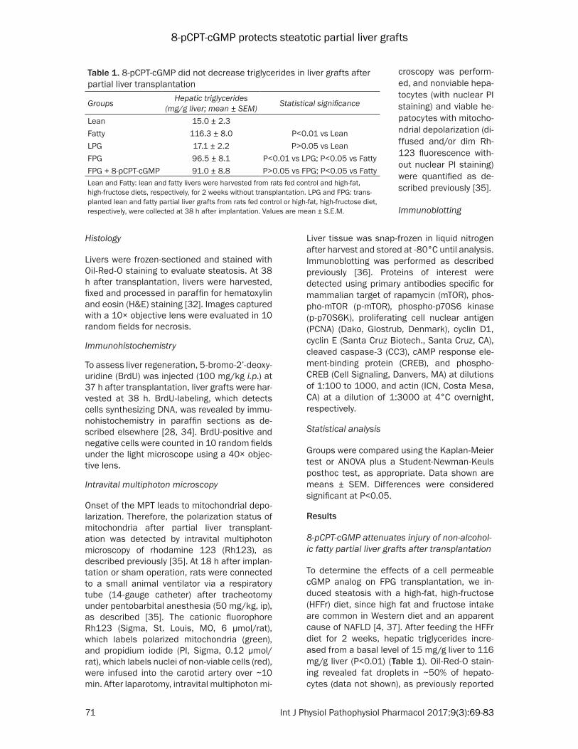
Table 1. 8-pCPT-cGMP did not decrease triglycerides in liver grafts after partial liver transplantation
Groups Hepatic triglycerides (mg/g liver; mean ± SEM) Statistical significance
Lean 15.0 ± 2.3Fatty 116.3 ± 8.0 P<0.01 vs LeanLPG 17.1 ± 2.2 P>0.05 vs LeanFPG 96.5 ± 8.1 P<0.01 vs LPG; P<0.05 vs FattyFPG + 8-pCPT-cGMP 91.0 ± 8.8 P>0.05 vs FPG; P<0.05 vs FattyLean and Fatty: lean and fatty livers were harvested from rats fed control and high-fat, high-fructose diets, respectively, for 2 weeks without transplantation. LPG and FPG: trans-planted lean and fatty partial liver grafts from rats fed control or high-fat, high-fructose diet, respectively, were collected at 38 h after implantation. Values are mean ± S.E.M.
croscopy was perform- ed, and nonviable hepa-tocytes (with nuclear PI staining) and viable he- patocytes with mitocho- ndrial depolarization (di- ffused and/or dim Rh- 123 fluorescence with-out nuclear PI staining) were quantified as de- scribed previously [35].
Immunoblotting

8-pCPT-cGMP protects steatotic partial liver grafts
72 Int J Physiol Pathophysiol Pharmacol 2017;9(3):69-83
[32]. Fat droplets were primarily microvesicular with a small number of macrovesicular drop-lets. Livers from rats fed the control diet sho- wed markedly less Oil-Red-O staining that was confined to small fat droplets [32]. After FPG transplantation with and without 8-pCPT-cGMP treatment hepatic triglycerides decreased slig- htly (18-22%), but differences with and without 8-pCPT-treatment were not statistically signifi-cant (Table 1).
Histology was normal in sham-operated lean livers (Figure 1A). After LPG transplantati- on, scattered focal hepatic necrosis occurred (Figure 1A). By contrast, substantially greater necrosis occurred after FPG transplantation, primarily in periportal and midzonal regions of
the liver lobules (Figure 1A). Some eosinop- hilic inclusion bodies were also observed. How these eosinophilic inclusion bodies formed remains unclear. After FPG transplantation with 8-pCPT-cGMP treatment, necrosis and eosino-philic inclusion bodies were markedly decre- ased (Figure 1A).
Serum ALT, a marker of hepatocellular injury, was 36-40 U/L after sham operation in rats fed control and HFFr diets (Figure 1B). Previously, we showed that serum ALT peaks at ~18 h after partial liver transplantation and then decre- ases gradually afterwards [33]. Therefore, we compared ALT release at 18 h after LPG and FPG transplantation. After LPG transplantation, ALT increased to 655 U/L, which increased to 1540 U/L after FPG transplantation. After transplantation of FPG preserved in UW solu-tion containing 8-pCPT-cGMP, ALT was 675 U/L, nearly identical to that of the LPG group (Figure 1B).
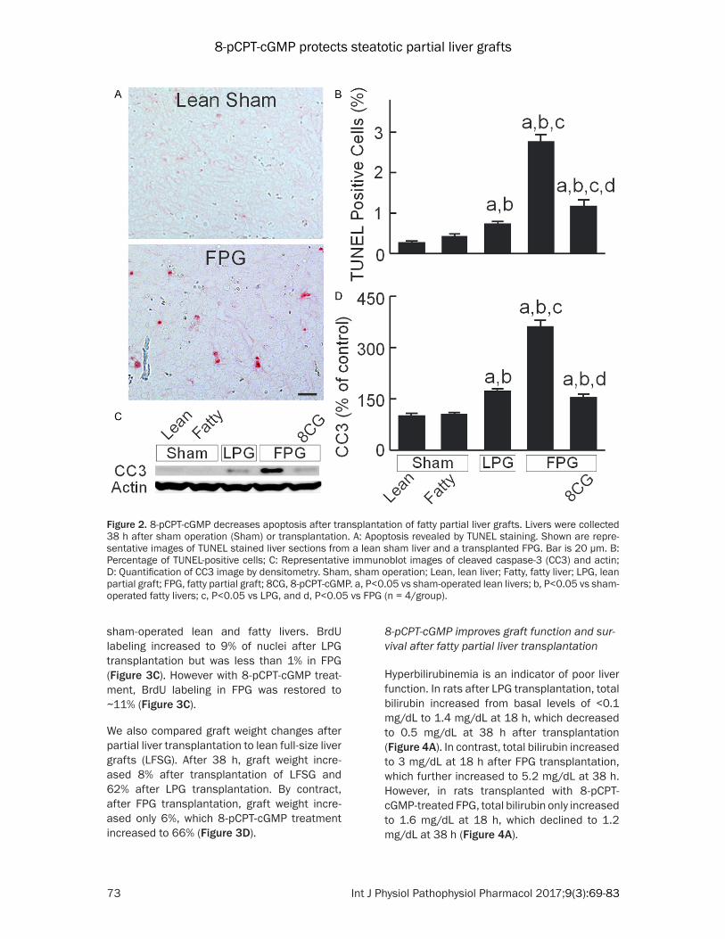
Apoptosis was assessed by TUNEL staining. TUNEL-positive cells were 0.3% and 0.4% in lean and fatty livers after sham-operation, res- pectively (Figure 2A, 2B). At 38 h after trans-plantation, TUNEL-positive cells increased to 0.7% in LPG. After FPG transplantation, apopto-sis further increased to 2.8%, which storage with 8-pCPT-cGMP decreased to 1.2% (Figure 2A, 2B). Activation of caspase-3 mediates ap- optosis. Cleaved caspase-3 was not significant-ly different between lean and fatty livers after sham-operation (Figure 2C, 2D). Cleaved cas-pase-3 increased to 172% after LPG transplan-tation, which further increased to 362% after FPG transplantation. After 8-pCPT-cGMP treat-ment of FPG, cleaved caspase-3 increased only 154% (Figure 2C, 2D).
8-pCPT-cGMP improves liver regeneration after fatty partial liver transplantation
Liver regeneration was evaluated at 38 h after transplantation (Figure 3). PCNA is a marker of cell proliferation [38]. PCNA was barely detect-able after sham operation in either lean or fatty livers but increased ~22 fold after LPG transplantation (Figure 3A, 3B). However af- ter FPG transplantation, PCNA increased only 8-fold in the absence of 8-pCPT-cGMP treat-ment, but with treatment PCNA increased ~16-fold (Figure 3A, 3B). BrdU nuclear labeling, an indicator of synthesis of DNA in the S-phase of the cell cycle, was also barely detectable in
Figure 1. 8-pCPT-cGMP attenuates injury of fatty par-tial liver grafts after transplantation. Rats were fed a low-fat, low-fructose control diet or a high-fat, high-fructose diet for 2 weeks. A: Livers were collected 38 h after sham operation or transplantation. Shown are representative images of H+E-stained liver sec-tions. Arrows identify necrotic areas. Bar is 100 µm. B: Blood was collected at 18 h after sham operation or transplantation for measurement of alanine ami-notransferase (ALT) in sera. Sham, sham operation; Lean, lean liver; Fatty, fatty liver; LPG, lean partial graft; FPG, fatty partial graft; 8CG, 8-pCPT-cGMP. a, P<0.05 vs sham-operated lean livers; b, P<0.05 vs sham-operated fatty livers; c, P<0.05 vs LPG, and d, P<0.05 vs FPG (n = 4/group).
8-pCPT-cGMP protects steatotic partial liver grafts
73 Int J Physiol Pathophysiol Pharmacol 2017;9(3):69-83
sham-operated lean and fatty livers. BrdU labeling increased to 9% of nuclei after LPG transplantation but was less than 1% in FPG (Figure 3C). However with 8-pCPT-cGMP treat-ment, BrdU labeling in FPG was restored to ~11% (Figure 3C).
We also compared graft weight changes after partial liver transplantation to lean full-size liver grafts (LFSG). After 38 h, graft weight incre- ased 8% after transplantation of LFSG and 62% after LPG transplantation. By contract, after FPG transplantation, graft weight incre- ased only 6%, which 8-pCPT-cGMP treatment increased to 66% (Figure 3D).
8-pCPT-cGMP improves graft function and sur-vival after fatty partial liver transplantation
Hyperbilirubinemia is an indicator of poor liver function. In rats after LPG transplantation, total bilirubin increased from basal levels of <0.1 mg/dL to 1.4 mg/dL at 18 h, which decreased to 0.5 mg/dL at 38 h after transplantation (Figure 4A). In contrast, total bilirubin increased to 3 mg/dL at 18 h after FPG transplantation, which further increased to 5.2 mg/dL at 38 h. However, in rats transplanted with 8-pCPT-cGMP-treated FPG, total bilirubin only increased to 1.6 mg/dL at 18 h, which declined to 1.2 mg/dL at 38 h (Figure 4A).
Figure 2. 8-pCPT-cGMP decreases apoptosis after transplantation of fatty partial liver grafts. Livers were collected 38 h after sham operation (Sham) or transplantation. A: Apoptosis revealed by TUNEL staining. Shown are repre-sentative images of TUNEL stained liver sections from a lean sham liver and a transplanted FPG. Bar is 20 µm. B: Percentage of TUNEL-positive cells; C: Representative immunoblot images of cleaved caspase-3 (CC3) and actin; D: Quantification of CC3 image by densitometry. Sham, sham operation; Lean, lean liver; Fatty, fatty liver; LPG, lean partial graft; FPG, fatty partial graft; 8CG, 8-pCPT-cGMP. a, P<0.05 vs sham-operated lean livers; b, P<0.05 vs sham-operated fatty livers; c, P<0.05 vs LPG, and d, P<0.05 vs FPG (n = 4/group).

8-pCPT-cGMP protects steatotic partial liver grafts
74 Int J Physiol Pathophysiol Pharmacol 2017;9(3):69-83
All rats survived after sham operation (data not shown). Our previous study showed that all rats survived after transplantation of LPG cold stored for 2 h [32]. By contrast, survival decre- ased to 0% by 3 days after transplantation of FPG (Figure 4B). 8-pCPT-cGMP increased the survival of FPG to 77% (Figure 4B). Therefore, 8-pCPT-cGMP effectively prevented failure of FPG after transplantation.
8-pCPT-cGMP blunts mitochondrial depolariza-tion in hepatocytes of transplanted fatty partial liver grafts
Energy supply is essential for maintaining cell function and supporting cell proliferation. On-
set of the MPT plays an essential role in liver graft failure from preservation injury [26]. We therefore examined mitochondrial polarization status in transplanted liver grafts by intravital multiphoton microscopy using the fluorescence of Rh123, a membrane-permeant cationic fluo-rophore that labels polarized mitochondria. After sham operation, mitochondria were polar-ized in virtually every hepatocyte, as indicated by punctate Rh123 green fluorescence and in confirmation of previous studies (Figure 5A) [26, 28, 35]. In implanted LPG at 18 h after transplantation, mitochondria in some hepato-cytes (~20%) became depolarized, as indicated by dim and diffuse instead of punctate Rh123 fluorescence (Figure 5A). After FPG transplan-
Figure 3. 8-pCPT-cGMP improves regeneration of FPG after transplantation. Livers were collected 38 h after sham operation or transplantation. Proliferating cell nuclear antigen (PCNA) was detected by immunoblotting (A) and quantified by densitometry (B). 5-Bromo-2’-deoxyuridine (BrdU) incorporation was detected immunohistochemically. BrdU-positive cells (C) were counted in 10 random fields per slide in a blinded manner. Partial grafts were weighed before implantation and at 38 h after transplantation to calculate graft weight increases (D). Sham, sham opera-tion; Lean, lean liver; Fatty, fatty liver; LFSG, lean full-size graft; LPG, lean partial graft; FPG, fatty partial graft; 8CG, 8-pCPT-cGMP. a, P<0.05 vs sham-operated lean livers; b, P<0.05 vs sham-operated fatty livers or LFSG; c, P<0.05 vs LPG, and d, P<0.05 vs FPG (n = 4/group).

8-pCPT-cGMP protects steatotic partial liver grafts
75 Int J Physiol Pathophysiol Pharmacol 2017;9(3):69-83
tation, mitochondrial depolarization increased to 69% of hepatocytes, an about 3.5-fold in- crease compared to LPG. 8-pCPT-cGMP treat-ment of FPG during storage decreased mito-chondrial depolarization after transplantation to 21% (Figure 5B). At this early stage, nonvia-ble cells (with red PI fluorescence in nuclei) were rare, indicating mitochondrial depolariza-tion occurred before cell death.
8-pCPT-cGMP increases mTOR signaling in transplanted fatty partial liver grafts
The mTOR pathway modulates cell proliferation and is highly sensitive to nutrient/energy alter-ations [39, 40]. At 38 h after transplantation, total mTOR protein expression was not signifi-cantly different between LPG, FPG and sham
(data not shown). Weak phospho-mTOR bands were detected in sham-operated lean and fatty livers, which increased 4.4-fold after LPG trans-plantation (Figure 6A, 6B). In contrast, phos-pho-mTOR increased only 1.8-fold after FPG transplantation, indicating lower mTOR activa-tion compared to LPG. 8-pCPT-cGMP treatment of FPG, however, increased phospho-mTOR 3.2-fold after transplantation compared to sham (Figure 6A, 6B). Phosphorylation of p70S6K, a kinase downstream of mTOR, also increased 5.6-fold after LPG transplantation (Figure 6A, 6C). After FPG transplantation, p70S6K phosphorylation increased only 1.6-fold, which 8-pCPT-cGMP treatment increased to 4.9-fold (Figure 6A, 6C).
Figure 4. 8-pCPT-cGMP improves function and in-creases survival of transplanted fatty partial liver grafts. Blood was collected at 18 and 38 h after implantation for measurement of total bilirubin (A). LPG, lean partial graft; FPG, fatty partial graft; 8CG, 8-pCPT-cGMP. a, P<0.05 vs LPG, and b, P<0.05 vs FPG (n = 4/group). Rats were observed 7 days for survival (B). b, P<0.05 vs FPG by the Kaplan-Meier test (n = 9-11/group).
Figure 5. 8-pCPT-cGMP blocks mitochondrial depo-larization in transplanted fatty partial liver grafts. Intravital multiphoton microscopy of Rh123 and PI was performed at 18 h after sham operation or transplantation, as described in “METHODS”. A: Rep-resentative multiphoton images. Bar is 10 µm. Arrow identifies a PI-positive non-viable hepatocyte. B: He-patocytes with depolarized mitochondria were count-ed in 10 random fields per rat as the percentage of the total. a, P<0.05 vs sham-operated lean livers; b, P<0.05 vs LPG and c, P<0.05 vs FPG (n = 4/group).

8-pCPT-cGMP protects steatotic partial liver grafts
76 Int J Physiol Pathophysiol Pharmacol 2017;9(3):69-83
8-pCPT-cGMP increases CREB activation in transplanted fatty partial liver grafts
In hepatocytes, 8-pCPT-cGMP activates PKA, an event that suppresses onset of the MPT [31]. PKA activation also stimulates cell prolif-eration/growth through the activation of CR- EB [41, 42]. We therefore explored whether 8-pCPT-cGMP alters CREB activation. Total CR- EB protein expression was not different be- tween lean and fatty livers and did not change after partial liver transplantation (Figure 7A).
After LPG transplantation, phospho-CREB in- creased 1.5-fold compared to sham operation but decreased 53% after FPG transplantation. After 8-pCPT-cGMP treatment, phospho-CREB instead increased 1.4-fold after FPG transplan-tation (Figure 7A, 7B).
8-pCPT-cGMP increases cyclin expression in transplanted fatty partial liver grafts
Cyclins are important governors of cell cycle progression [43]. We explored the effects of 8-pCPT-cGMP on cyclin D1 and E expression in FPG (Figure 8). Weak bands of cyclin D1 and E were observed in sham-operated lean livers, which were not significantly different from sham-operated fatty livers (Figure 8A). Cyclin D1 increased 7.5-fold in transplanted LPG but increased only 1.9-fold in transplanted FPG (Figure 8A, 8B). In transplanted FPG treated with 8-pCPT-cGMP, cyclin D1 increased 6.1-fold (Figure 8A, 8B). Cyclin E changed in a similar
Figure 6. 8-pCPT-cGMP increases mTOR signaling in transplanted fatty partial liver grafts. Livers were col-lected 38 h after sham operation or transplantation. Phospho-mammalian target of rapamycin (p-mTOR), phospho-p70S6 kinase (p-p70S6K), and actin we- re detected by immunoblotting (A, representative blots). (B) Quantification of p-mTOR by densitometry; (C) Quantification of p-p70S6K. Sham, sham opera-tion; Lean, lean liver; Fatty, fatty liver; LPG, lean par-tial graft; FPG, fatty partial graft; 8CG, 8-pCPT-cGMP. a, P<0.05 vs sham-operated lean livers; b, P<0.05 vs sham-operated fatty livers; c, P<0.05 vs LPG, and d, P<0.05 vs FPG (n = 4/group).
Figure 7. 8-pCPT-cGMP increases CREB activation in transplanted fatty partial liver grafts. Livers were col-lected 38 h after sham operation or transplantation. cAMP response element-binding protein (CREB), phospho-CREB (p-CREB), and actin were detected by immunoblotting (A, representative images). (B) Quantification of p-CREB by densitometry; Sham, sham operation; Lean, lean liver; Fatty, fatty liver; LPG, lean partial graft; FPG, fatty partial graft; 8CG, 8-pCPT-cGMP. a, P<0.05 vs sham-operated lean livers; b, P<0.05 vs sham-operated fatty livers; c, P<0.05 vs LPG, and d, P<0.05 vs FPG (n = 4/group).

8-pCPT-cGMP protects steatotic partial liver grafts
77 Int J Physiol Pathophysiol Pharmacol 2017;9(3):69-83
fashion after the various treatments (Figure 8A, 8C).
Discussion
8-pCPT-cGMP attenuates injury and improves regeneration of non-alcoholic fatty partial liver grafts
The continuing shortage of donor livers has increased the number of patients waiting for liver transplantation and markedly increased waiting-list mortality in the last two decades, which approaches like living and split liver
donation might help to alleviate [11, 44, 45]. However, the current epidemic of obesity and metabolic syndrome has increased the inci-dence of NAFLD and NASH, which when severe is a contraindication to organ donation in liver transplantation, especially in the context of living or split liver donation requiring partial liver transplantation. Therefore, development of effective therapies to improve outcomes of steatotic partial liver transplantation might sig-nificantly impact the shortage of usable donor livers and save lives. Steatotic partial grafts face two risk factors: fatty infiltration and small graft mass. Steatosis itself is among the most important factors that negatively impact the outcome of liver transplantation [20, 21, 46]. Steatotic grafts show substantially greater vul-nerability to I/R injury during cold storage/transplantation, resulting in increased cell dea- th, delayed recovery of graft function and high-er primary non-function in clinical and experi-mental liver transplantation [47, 48]. Compared to full size liver transplantation, grafts for par-tial liver transplantation usually experience shorter cold storage times. Clinically, cold pres-ervation time is about 2 to 6 h for living donor transplantation whereas mean cold ischemic time for full-size transplantation is 6-12 h [49-51]. Although liver tissue is well known to have great regeneration capacity, liver regeneration is suppressed when liver graft mass is re- duced to below a critical level even after short cold preservation times [33, 52]. Therefore, strategies for successful FPG transplantation should both prevent liver injury and improve liver regeneration. Here, we demonstrated that the cell-permeable cGMP analog 8-pCPT-cGMP markedly decreased liver injury (less necrotic and apoptotic cell death and transaminase release), improved graft regeneration (increas- ed BrdU incorporation, PCNA expression, and graft weight gain), and enhanced graft function (lower serum total bilirubin) (Figures 1-4). Most importantly, 8-pCPT-cGMP markedly improved survival of FPG (Figure 4). Therefore, 8-pCPT-cGMP is a promising therapy to improve func-tion and decrease failure of steatotic partial grafts.
8-pCPT-cGMP attenuates graft injury by decreasing mitochondrial depolarization after transplantation of fatty partial liver grafts
Onset of the MPT is a penultimate step leading to cell death after I/R injury, causing necrosis from ATP depletion and apoptosis from mito-
Figure 8. 8-pCPT-cGMP increases cyclin D1 and E expression in transplanted fatty partial liver grafts. Livers were collected 38 h after sham operation or transplantation. Cyclin D1 (CyD1), cyclin E (CyE), and actin were detected by immunoblotting (A, represen-tative images). (B) Quantification of cyclin D1 by den-sitometry; (C) Quantification of cyclin E. Sham, sham operation; Lean, lean liver; Fatty, fatty liver; LPG, lean partial graft; FPG, fatty partial graft; 8CG, 8-pCPT-cGMP. a, P<0.05 vs sham-operated lean livers; b, P<0.05 vs sham-operated fatty livers; c, P<0.05 vs LPG, and d, P<0.05 vs FPG (n = 4/group).

8-pCPT-cGMP protects steatotic partial liver grafts
78 Int J Physiol Pathophysiol Pharmacol 2017;9(3):69-83
chondrial release of proapoptotic factors such as cytochrome c [25, 53] (Figure 9). Cell death in turn leads to release of mitochondrial and other cellular damage-associated molecular pattern molecules (e.g., mitochondrial DNA and high mobility group box 1 protein), which are potent inflammatory mediators that attract leu-kocytes with subsequent production of reactive oxygen species, release of proteases and other activities to amplify liver injury [54-56] (Figure 9). Our previous study demonstrated that the MPT was more extensive in steatotic full-size
cally acts via cGMP-dependent protein kinases (PKG) to regulate many physiological processes (e.g., smooth muscular cell relaxation and intra-cellular calcium concentration). Previous stud-ies show in isolated rat liver mitochondria that cGMP analogs block the Ca2+-induced MPT in the presence of hepatic cytosol [29]. NO ex- erts its biological effects through activation of guanylate cyclase, producing cGMP [57], and NO donors and cGMP analogs prevent MPT-dependent necrotic killing of ischemic hepato-cytes in culture after ischemia/reoxygenation
Figure 9. Potential mechanisms by which 8-pCPT-cGMP improves the out-come of fatty partial liver transplantation. Fatty liver transplantation causes onset of mitochondrial permeability transition (MPT), which decreases ATP formation and causes release of cytochrome c, leading to necrosis and apoptosis. Mitochondrial injury and cell death in turn cause release of mitochondrial and other cellular damage-associated molecular pattern molecules (DAMPs), leading to inflammation which further amplifies liver injury. ATP acts both as an energy source supporting liver regeneration and also a modulator of liver regeneration signaling. For example, mammalian target of rapamycin (mTOR) and its downstream p70S6 kinase (p70S6K), the pathway that regulates protein synthesis necessary for cell proliferation and growth, are highly sensitive to perturbations of nutrient/energy supply. Moreover, activation of a number of growth factor receptors and kinases that are involved in cell proliferation (e.g., extracellular signal-regulated ki-nase [ERK], c-Jun-N-terminal kinase [JNK], etc.) also requires high energy molecules. Formation of cyclin/cyclin-dependent kinase (CDK) complexes is a convergent point of multiple upstream signaling to control cell prolifera-tion. Therefore, ATP depletion from the MPT occurring in FPG suppresses liv-er regeneration. Previous studies show that both cGMP and cAMP analogs protect against the MPT in cultured hepatocytes after ischemia/reperfusion through activation of protein kinase A (PKA). 8-pCPT-cGMP, a cell permeable cGMP analog, activates PKA in hepatocytes and possibly PKG in other cell types to block MPT onset, thus attenuating cell death and improving liver regeneration. 8-pCPT-cGMP also enhances cell proliferation through activa-tion of cAMP response element-binding protein (CREB).
grafts than in lean full-size gr- afts after transplantation [35]. However, this effect occurred after long cold storage. We also observed that the MPT occurred after lean partial gr- afts when liver graft size was reduced to 25% in rat (quarter-size grafts) with a longer cold storage (6 h) than current stu- dy (2 h) [36]. In this study we showed that steatosis incre- ases the MPT in partial grafts even after short cold storage (Figure 5), demonstrating that co-existence of two risk fac-tors (steatosis and reduced graft size) further increases the susceptibility to MPT on- set, leading to severe graft injury and failure after trans-plantation. 8-pCPT-cGMP, whi- ch blocks the MPT in cultured hepatocytes after I/R and in isolated mitochondria after ca- lcium uptake [31], also decre- ased mitochondrial depolar-ization in vivo after FPG trans-plantation, attenuated subse- quent graft injury and im- proved graft survival (Figures 1, 2). These results are consis-tent with the conclusion that MPT onset plays an important role in FPG injury and failure and that blockade of MPT onset is an effective strategy to decrease FPG injury and improve survival.
8-pCPT-cGMP is a cell-perme-able cGMP analog, which typi-

8-pCPT-cGMP protects steatotic partial liver grafts
79 Int J Physiol Pathophysiol Pharmacol 2017;9(3):69-83
[29]. Although PKG is typically the target of cGMP, hepatocytes do not express mRNA for either of the two isoforms of PKG [58]. More- over, analogs of both cGMP and cAMP stimu-late cytosolic PKA from hepatocytes, and a PKA peptide inhibitor but not a PKG peptide in- hibitor abolishes cGMP and cAMP-stimulated kinase activity in hepatocyte cytosol [31]. Thus, by activation of cytosolic PKA, cGMP and cAMP inhibit MPT onset and protect hepatocytes against necrotic cell death after ischemia/reox-ygenation [31].
These findings in vitro remain to be confirmed in vivo. In the present study, we showed that the cGMP analog, 8-pCPT-cGMP, also sup-pressed MPT onset in vivo after fatty partial liver transplantation, as visualized by intravital multiphoton microscopy of Rh123 (Figure 5). This protection of 8-pCPT-cGMP against the MPT in hepatocytes of FPG is thus likely medi-ated by PKA. However, since multiple cell types exist in the liver, we cannot rule out completely the possibility that 8-pCPT-cGMP also works through PKG in cells other than hepatocytes. Moreover, although NO and cGMP production are closely related, protection of 8-pCPT-cGMP is unlikely by increasing NO, since iNOS ex- pression and production of reactive nitrogen species already increases dramatically in FPG after transplantation, effects which are associ-ated with increased graft injury [32]. Moreover, 1400 W, a selective iNOS inhibitor, improves the outcome of FPG transplantation [32].
Potential mechanisms by which 8-pCPT-cGMP improves liver regeneration in FPG
In this study, we confirmed that FPG trans- plantation leads to poor liver regeneration (decreased BrdU incorporation, PCNA expres-sion and liver graft weight gain), poor graft function (hyperbilirubinemia), and graft failure. 8-pCPT-cGMP improved all these adverse out-comes, including the enhancement of liver regeneration (Figure 3). Liver regeneration is essential for survival and recovery of graft mass and function after partial liver transplan-tation. Partial grafts face extra metabolic work load due to their reduced mass. Moreover, extra energy is needed to support liver regen-eration. Therefore, mitochondrial dysfunction not only leads to cell death but can also sup-press liver regeneration (Figure 9). In addition, ATP not only acts as an energy supplier for liver
regeneration but also as a regeneration sig- naling modifier. Activation of a number of growth factor receptors and kinases by phos-phorylation (e.g., epidermal growth factor re- ceptor, MAP kinase kinase, extracellular signal-regulated kinase [ERK], c-Jun-N-terminal kinase [JNK], etc.) requires high energy molecules [59] (Figure 9). mTOR is a serine-threonine kinase that regulates the protein synthesis neces- sary for cell growth and proliferation [60-62]. Activation of its downstream p70S6 kinase in turn regulates the 40S ribosomal protein S6 to control protein synthesis and cell proliferation [60] (Figure 9). A previous study shows that inhibition of mTOR decreases DNA synthesis after partial hepatectomy [63]. The mTOR path-way is highly sensitive to perturbations of nutri-ent/energy supply, and decreased ATP can sup-press the mTOR pathway [39, 40]. Indeed, in FPG with severe mitochondrial dysfunction, we observed decreased mTOR and p70S6 kinase activation (Figure 6). 8-pCPT-cGMP, which pre-vented mitochondrial dysfunction, also improv- ed mTOR and p70S6 kinase activation in FPG (Figure 6).
Additionally, we observed a suppression of CREB activation after FPG activation (Figure 7). Previous studies show that CREB activation regulates hepatocyte proliferation after partial hepatectomy [64, 65]. PKA is an important reg-ulator of CREB. Activation of G-protein coupled receptors stimulates the activity of membrane-associated adenylyl cyclase, which converts ATP to cAMP. Binding of cAMP to the two PKA catalytic subunits causes release of active cat-alytic subunits, which migrate into the nucleus to phosphorylate and thereby activate CREB, a transcriptional factor. Activated CREB then interacts with the cAMP response enhancer element of the promoters of cAMP-responsive genes to cause transcription of genes that reg-ulates various physiological processes includ-ing proliferation [41, 42] (Figure 9). Previous studies show that transient increases of cAMP also cause upregulation of genes expressed in the G1 phase of the cell cycle (e.g., c-fos), whereas decreased cAMP suppresses hepatic DNA synthesis after partial hepatectomy [66, 67]. Interestingly, activation of PKG can also increase CREB phosphorylation. A previous study shows that cGMP-PKG signaling plays an important role preventing apoptosis and pro-moting cell proliferation in both normal and cer-tain cancer cells and that PKG knockdown by

8-pCPT-cGMP protects steatotic partial liver grafts
80 Int J Physiol Pathophysiol Pharmacol 2017;9(3):69-83
siRNA decreases CREB phosphorylation [68]. Another study also shows that cGMP-PKG sig-naling pathway acts in parallel with the cAMP-PKA pathway to produce CREB phosphorylation [69]. Therefore, activation of either PKA or PKG can increase cell proliferation and 8-pCPT-cGMP may increase CREB activation via PKA and/or PKG (Figure 9).
Liver regeneration requires cell proliferation. Cyclins are a family of proteins that control cell cycle progression [43]. Cyclins bind corre-sponding cyclin-dependent kinases, forming complexes that phosphorylate other proteins (e.g., retinoblastoma protein). These phosphor-ylated proteins, in turn, regulate specific ev- ents during cell division [43]. Cyclin D1, which drives hepatocytes to enter cell cycle [70], is highly CREB-dependent, whereas cyclin B1 is co-regulated by both CREB-dependent and -independent mechanisms [71]. JNK1/2 also controls cyclin D1 expression, and inhibition of JNK activation suppresses hepatocyte mitosis after partial hepatectomy [72]. Inhibition of ERK activation also suppresses synthesis of cyclins E and A [73]. Inhibitors of mammalian target of rapamycin complex 1 (mTORC1, a pro-tein complex composed of mTOR itself, regula-tory-associated protein of mTOR [Raptor] and other proteins) [74], including rapamycin and siRNA for raptor, inhibit upregulation of cyclin D1 by inorganic polyphosphate [75]. Therefore, cyclins are a convergent point of multiple upstream signaling to control cell proliferation (Figure 9). In this study, we observed suppre- ssed cyclin D1 and E expression in FPG com-pared to LPG transplantation. 8-pCPT-cGMP restored cyclin expression after FPG trans- plantation (Figure 8), which is consistent with improved FPG regeneration by 8-pCPT-cGMP (Figure 3).
Taken together, we observed more severe graft injury and suppression of regeneration after transplantation of FPG. Such increased injury and suppression of regeneration were associ-ated with more severe mitochondrial dysfunc-tion. 8-pCPT-cGMP improved the outcomes of FPG transplantation, most likely by blocking mitochondrial dysfunction and increasing pro-liferative signaling (Figure 9). Other cGMP ana-logs (e.g., 8-BrcGMP) also protect against the MPT onset in hepatocytes after ischemia/reperfusion in vitro [29, 30]. Whether these analogs also improve the outcomes of FPG
transplantation will be examined in future studies.
Acknowledgements
This study was supported, in part, by Grants from the National Institute of Health [DK- 70844, DK037034, DK073336], the Russian Federation [14.Z50.31.0028] and the Chinese National Natural Foundation [Grant 814708- 78]. The Cell & Molecular Imaging Core of the Hollings Cancer Center at the Medical University of South Carolina supported by NIH Grant 1P30 CA138313 and Shared Instrumentation Grant S10OD018113 provided instrumentation for multiphoton microscopy. Animals were housed in the Animal Resources at Medical University of South Carolina supported by NIH Grant C06 RR015455.
Disclosure of conflict of interest
None.
Address correspondence to: Dr. Zhi Zhong, Depart- ment of Drug Discovery & Biomedical Sciences, Medical University of South Carolina, 280 Calhoun Street, MSC140, Charleston 29425, SC, USA. Tel: 843-792-2163; Fax: 843-792-1617; E-mail: [email protected]
References
[1] Yilmaz Y and Younossi ZM. Obesity-associated nonalcoholic fatty liver disease. Clin Liver Dis 2014; 18: 19-31.
[2] Goceri E, Shah ZK, Layman R, Jiang X and Gur-can MN. Quantification of liver fat: a compre-hensive review. Comput Biol Med 2016; 71: 174-189.
[3] Dongiovanni P, Lanti C, Riso P and Valenti L. Nutritional therapy for nonalcoholic fatty liver disease. J Nutr Biochem 2016; 29: 1-11.
[4] Basciano H, Federico L and Adeli K. Fructose, insulin resistanceand metabolic dyslipidemia. Nutr Metab (Lond) 2005; 2: 5.
[5] Charlton M, Krishnan A, Viker K, Sanderson S, Cazanave S, McConico A, Masuoko H and Gores G. Fast food diet mouse: novel small animal model of NASH with ballooning, pro-gressive fibrosis and high physiological fidelity to the human condition. Am J Physiol Gastroin-test Liver Physiol 2011; 301: G825-G834.
[6] Charlton M. Nonalcoholic fatty liver disease: a review of current understanding and future impact. Clin Gastroenterol Hepatol 2004; 2: 1048-1058.

8-pCPT-cGMP protects steatotic partial liver grafts
81 Int J Physiol Pathophysiol Pharmacol 2017;9(3):69-83
[7] Wanless IR and Lentz JS. Fatty liver hepatitis (steatohepatitis) and obesity: an autopsy study with analysis of risk factors. Hepatology 1990; 12: 1106-1110.
[8] Finkenstedt A and Graziadei IW. Steatosis after liver transplantation: is it really benign? Liver Transpl 2016; 22: 585-7.
[9] Angulo P. Nonalcoholic fatty liver disease. N Engl J Med 2002; 346: 1221-1231.
[10] Canbay A, Sowa JP, Syn WK and Treckmann J. NASH cirrhosis-the new burden in liver trans-plantation: how should it be managed? Visc Med 2016; 32: 234-238.
[11] Fayek SA, Quintini C, Chavin KD and Marsh CL. The current state of liver transplantation in the United States: perspective from American society of transplant surgeons (ASTS) scien- tific studies committee and endorsed by ASTS council. Am J Transplant 2016; 16: 3093-3104.
[12] Rinella ME, Alonso E, Rao S, Whitington P, Fry-er J, Abecassis M, Superina R, Flamm SL and Blei AT. Body mass index as a predictor of he-patic steatosis in living liver donors. Liver Transpl 2001; 7: 409-414.
[13] Garcia Urena MA, Colina Ruiz-Delgado F, More-no GE, Jimenez RC, Garcia GI, Loinzaz SC, Gon-zalez P and Gomez SR. Hepatic steatosis in liver transplant donors: common feature of donor population? World J Surg 1998; 22: 837-844.
[14] Behrns KE, Tsiotos GG, DeSouza NF, Krishna MK, Ludwig J and Nagorney DM. Hepatic ste-atosis as a potential risk factor for major he-patic resection. J Gastrointest Surg 1998; 2: 292-298.
[15] Mittler J, Pascher A, Neuhaus P and Pratschke J. The utility of extended criteria donor organs in severely ill liver transplant recipients. Trans-plantation 2008; 86: 895-896.
[16] Nocito A, El-Badry AM and Clavien PA. When is steatosis too much for transplantation? J Hep-atol 2006; 45: 494-499.
[17] Urena MA, Moreno GE, Romero CJ, Ruiz- Delgado FC and Moreno SC. An approach to the rational use of steatotic donor livers in liver transplantation. Hepatogastroenterology 1999; 46: 1164-1173.
[18] Sun Z, Klein AS, Radaeva S, Hong F, El-Assal O, Pan H, Jaruga B, Batkai S, Hoshino S, Tian Z, Kunos G, Diehl AM and Gao B. In vitro interleu-kin-6 treatment prevents mortality associated with fatty liver transplants in rats. Gastroenter-ology 2003; 125: 125-215.
[19] Spitzer AL, Lao OB, Dick AA, Bakthavatsalam R, Halldorson JB, Yeh MM, Upton MP, Reyes JD and Perkins JD. The biopsied donor liver: incor-porating macrosteatosis into high-risk donor assessment. Liver Transpl 2010; 16: 874-884.
[20] Marsman WA, Wiesner RH, Rodrigues L, Batts KP, Porayko MK, Hay JE, Gores GJ and Krom RAF. Use of fatty donor liver is associated with diminished early patient and graft survival. Transplantation 1997; 62: 1246-1251.
[21] Strasberg SM, Howard TK, Molmenti EP and Hertl M. Selecting the donor liver: risk factors for poor function after orthotopic liver trans-plantation. Hepatology 1994; 20: 829-838.
[22] Lemasters JJ. Modulation of mitochondrial membrane permeability in pathogenesis, au-tophagy and control of metabolism. J Gastro-enterol Hepatol 2007; 22 Suppl 1: S31-S37.
[23] Gottlieb RA. Cell death pathways in acute isch-emia/reperfusion injury. J Cardiovasc Pharma-col Ther 2011; 16: 233-238.
[24] Jassem W, Fuggle SV, Rela M, Koo DD and He-aton ND. The role of mitochondria in ischemia/reperfusion injury. Transplantation 2002; 73: 493-499.
[25] Zhong Z, Ramshesh VK, Rehman H, Currin RT, Sridharan V, Theruvath TP, Kim I, Wright GL and Lemasters JJ. Activation of the oxygen-sensing signal cascade prevents mitochondri-al injury after mouse liver ischemia-reperfu-sion. Am J Physiol Gastrointest Liver Physiol 2008; 295: G823-G832.
[26] Theruvath TP, Zhong Z, Currin RT, Pediaditakis P and Lemasters JJ. Minocycline mitigates storage/reperfusion injury after rat liver trans-plantation through suppression of the mito-chondrial permeability transition. Hepatology 2008; 47: 235-246.
[27] Rehman H, Sun J, Shi Y, Ramshesh VK, Liu Q, Currin RT, Lemasters JJ and Zhong Z. NIM811 prevents mitochondrial dysfunction, attenu-ates liver injury and stimulates liver regenera-tion after massive hepatectomy. Transplanta-tion 2011; 91: 406-412.
[28] Zhong Z, Theruvath TP, Currin RT, Waldmeier PC and Lemasters JJ. NIM811, a mitochondrial permeability transition inhibitor, prevents mito-chondrial depolarization in small-for-size rat liver grafts. Am J Transplant 2007; 7: 1103-1111.
[29] Kim JS, Ohshima S, Pediaditakis P and Le- masters JJ. Nitric oxide protects hepatocytes against mitochondrial permeability transition-induced reperfusion injury. Hepatology 2004; 39: 1533-1543.
[30] Kim JS, Ohshima S, Pediaditakis P and Lemas-ters JJ. Nitric oxide: a signaling molecule against mitochondrial permeability transition- and pH-dependent cell death after reperfu-sion. Free Radic Biol Med 2004; 37: 1943-1950.
[31] Pediaditakis P, Kim JS, He L, Zhang X, Graves LM and Lemasters JJ. Inhibition of the mito-chondrial permeability transition by protein ki-

8-pCPT-cGMP protects steatotic partial liver grafts
82 Int J Physiol Pathophysiol Pharmacol 2017;9(3):69-83
nase A in rat liver mitochondria and hepato-cytes. Biochem J 2010; 431: 411-421.
[32] He S, Rehman H, Wright GL and Zhong Z. Inhi-bition of inducible nitric oxide synthase pre-vents mitochondrial damage and improves survival of steatotic partial liver grafts. Trans-plantation 2010; 89: 291-298.
[33] Zhong Z, Connor HD, Froh M, Bunzendahl H, Lind H, Lehnert M, Mason RP, Thurman RG and Lemasters JJ. Free radical-dependent dys-function of small-for-size rat liver grafts: pre-vention by plant polyphenols. Gastroenterology 2005; 129: 652-664.
[34] Zhong Z, Schwabe RF, Kai Y, He L, Yang L, Bun-zendahl H, Brenner DA and Lemasters JJ. Liver regeneration is suppressed in small-for-size liver grafts after transplantation: involvement of JNK, cyclin D1 and defective energy supply. Transplantation 2006; 82: 241-250.
[35] Liu Q, Rehman H, Krishnasamy Y, Ramshesh VK, Theruvath TP, Chavin KD, Schnellmann RG, Lemasters JJ and Zhong Z. Role of inducible nitric oxide synthase in mitochondrial depolar-ization and graft injury after transplantation of fatty livers. Free Radic Biol Med 2012; 53: 250-259.
[36] Rehman H, Connor HD, Ramshesh VK, Theru-vath TP, Mason RP, Wright GL, Lemasters JJ and Zhong Z. Ischemic preconditioning pre-vents free radical production and mitochon- drial depolarization in small-for-size rat liver grafts. Transplantation 2008; 85: 1322-1331.
[37] Bray GA, Nielsen SJ and Popkin BM. Consump-tion of high-fructose corn syrup in beverages may play a role in the epidemic of obesity. Am J Clin Nutr 2004; 79: 537-543.
[38] Moldovan GL, Pfander B and Jentsch S. PCNA, the maestro of the replication fork. Cell 2007; 129: 665-679.
[39] Riehle KJ, Dan YY, Campbell JS and Fausto N. New concepts in liver regeneration. J Gastro-enterol Hepatol 2011; 26 Suppl 1: 203-212.
[40] Dennis PB, Jaeschke A, Saitoh M, Fowler B, Kozma SC and Thomas G. Mammalian TOR: a homeostatic ATP sensor. Science 2001; 294: 1102-1105.
[41] Servillo G, Penna L, Foulkes NS, Magni MV, Della Fazia MA and Sassone-Corsi P. Cyclic AMP signalling pathway and cellular prolifera-tion: induction of CREM during liver regenera-tion. Oncogene 1997; 14: 1601-1606.
[42] Servillo G, Della Fazia MA and Sassone-Corsi P. Coupling cAMP signaling to transcription in the liver: pivotal role of CREB and CREM. Exp Cell Res 2002; 275: 143-154.
[43] Galderisi U, Jori FP and Giordano A. Cell cycle regulation and neural differentiation. Onco-gene 2003; 22: 5208-5219.
[44] Alqahtani SA. Update in liver transplantation. Curr Opin Gastroenterol 2012; 28: 230-8.
[45] Jones PD, Hayashi PH and Sidney BI. Liver transplantation in 2013: challenges and con-troversies. Minerva Gastroenterol Dietol 2013; 59: 117-131.
[46] Selzner M and Clavien PA. Fatty liver in liver transplantation and surgery. Semin Liver Dis 2001; 21: 105-113.
[47] Verran D, Kusyk T, Painter D, Fisher J, Koorey D, Strasser S, Stewart G and McCaughan G. Clinical experience gained from the use of 120 steatotic donor livers for orthotopic liver trans-plantation. Liver Transpl 2003; 9: 500-505.
[48] Zhong Z, Connor H, Mason RP, Qu W, Stachle-witz RF, Gao W, Lemasters JJ and Thurman RG. Destruction of Kupffer cells increases survival and reduces graft injury after transplantation of fatty livers from ethanol-treated rats. Liver Transplant Surg 1996; 2: 383-387.
[49] Troisi R, Cammu G, Militerno G, De Baerde-maeker L, Decruyenaere J, Hoste E, Smeets P, Colle I, Van Vlierberghe H, Petrovic M, Voet D, Mortier E, Hesse UJ and de Hemptinne B. Mod-ulation of portal graft inflow: a necessity in adult living-donor liver transplantation? Ann Surg 2003; 237: 429-436.
[50] O’Callaghan JM, Morgan RD, Knight SR and Morris PJ. The effect of preservation solutions for storage of liver allografts on transplant out-comes: a systematic review and meta-analysis. Ann Surg 2014; 260: 46-55.
[51] Bejaoui M, Pantazi E, Folch-Puy E, Baptista PM, Garcia-Gil A, Adam R and Rosello-Catafau J. Emerging concepts in liver graft preserva-tion. World J Gastroenterol 2015; 21: 396-407.
[52] Liu Q, Rehman H, Krishnasamy Y, Haque K, Schnellmann RG, Lemasters JJ and Zhong Z. Amphiregulin stimulates liver regeneration af-ter small-for-size mouse liver transplantation. Am J Transplant 2012; 12: 2052-61.
[53] Theruvath TP, Zhong Z, Pediaditakis P, Ramshesh VK, Currin RT, Tikunov A, Holmu-hamedov E and Lemasters JJ. Minocycline and N-methyl-4-isoleucine cyclosporin (NIM811) mitigate storage/reperfusion injury after rat liver transplantation through suppression of the mitochondrial permeability transition. Hep-atology 2008; 47: 236-246.
[54] Zhai Y, Busuttil RW and Kupiec-Weglinski JW. Liver ischemia and reperfusion injury: new in-sights into mechanisms of innate-adaptive im-mune-mediated tissue inflammation. Am J Transplant 2011; 11: 1563-1569.
[55] Boros P and Bromberg JS. New cellular and molecular immune pathways in ischemia/re-perfusion injury. Am J Transplant 2006; 6: 652-658.
[56] Zhang Q, Itagaki K and Hauser CJ. Mitochon-drial DNA is released by shock and activates neutrophils via p38 map kinase. Shock 2010; 34: 55-59.

8-pCPT-cGMP protects steatotic partial liver grafts
83 Int J Physiol Pathophysiol Pharmacol 2017;9(3):69-83
[57] Derbyshire ER and Marletta MA. Structure and regulation of soluble guanylate cyclase. Annu Rev Biochem 2012; 81: 533-559.
[58] Kulhanek-Heinze S, Gerbes AL, Gerwig T, Voll-mar AM and Kiemer AK. Protein kinase a dependent signalling mediates anti-apoptotic effects of the atrial natriuretic peptide in isch-emic livers. J Hepatol 2004; 41: 414-420.
[59] Gabai VL, Meriin AB, Yaglom JA, Wei JY, Mosser DD and Sherman MY. Suppression of stress kinase JNK is involved in HSP72-mediated pro-tection of myogenic cells from transient energy deprivation. HSP72 alleviates the stewss-in-duced inhibition of JNK dephosphorylation. J Biol Chem 2000; 275: 38088-38094.
[60] Laplante M and Sabatini DM. mTOR signaling at a glance. J Cell Sci 2009; 122: 3589-3594.
[61] Gerasimovskaya EV, Tucker DA, Weiser-Evans M, Wenzlau JM, Klemm DJ, Banks M and Sten-mark KR. Extracellular ATP-induced prolifera-tion of adventitial fibroblasts requires phos-phoinositide 3-kinase, Akt, mammalian target of rapamycin and p70 S6 kinase signaling pathways. J Biol Chem 2005; 280: 1838-1848.
[62] Jaeschke A, Dennis PB and Thomas G. mTOR: a mediator of intracellular homeostasis. Curr Top Microbiol Immunol 2004; 279: 283-298.
[63] Francavilla A, Starzl TE, Scotti C, Carrieri G, Az-zarone A, Zeng QH, Porter KA and Schreiber SL. Inhibition of liver, kidney, and intestine regeneration by rapamycin. Transplantation 1992; 53: 496-498.
[64] Boshart M, Weih F, Schmidt A, Fournier RE and Schutz G. A cyclic AMP response element me-diates repression of tyrosine aminotransferase gene transcription by the tissue-specific extin-guisher locus Tse-1. Cell 1990; 61: 905-916.
[65] Zhao G, Wakabayashi R, Shimoda S, Fukunaga Y, Kumagai M, Tanaka M and Nakano K. Im-paired activities of cyclic adenosine mono-phosphate-responsive element binding pro-tein, protein kinase a and calcium-independent phospholipase A2 are involved in deteriorated regeneration of cirrhotic liver after partial hep-atectomy in rats. Hepatol Res 2011; 41: 1110-1119.
[66] MacManus JP, Franks DJ, Youdale T and Brace-land BM. Increases in rat liver cyclic AMP concentrations prior to the initiation of DNA synthesis following partial hepatectomy or hor-mone infusion. Biochem Biophys Res Commun 1972; 49: 1201-1207.
[67] Kruijer W, Skelly H, Botteri F, van der Putten H, Barber JR, Verma IM and Leffert HL. Proto-on-cogene expression in regenerating liver is sim-ulated in cultures of primary adult rat hepato-cytes. J Biol Chem 1986; 261: 7929-7933.
[68] Wong JC, Bathina M and Fiscus RR. Cyclic GMP/protein kinase G type-Ialpha (PKG-Ial-pha) signaling pathway promotes CREB phos-phorylation and maintains higher c-IAP1, livin, survivin, and Mcl-1 expression and the inhibi-tion of PKG-Ialpha kinase activity synergizes with cisplatin in non-small cell lung cancer cells. J Cell Biochem 2012; 113: 3587-3598.
[69] Lu YF and Hawkins RD. Ryanodine receptors contribute to cGMP-induced late-phase LTP and CREB phosphorylation in the hippocam-pus. J Neurophysiol 2002; 88: 1270-1278.
[70] Nelsen CJ, Rickheim DG, Timchenko NA, Stan-ley MW and Albrecht JH. Transient expression of cyclin D1 is sufficient to promote hepatocyte replication and liver growth in vivo. Cancer Res 2001; 61: 8564-8568.
[71] Daniel P, Filiz G, Brown DV, Hollande F, Gonza-les M, D’Abaco G, Papalexis N, Phillips WA, Ma-laterre J, Ramsay RG and Mantamadiotis T. Selective CREB-dependent cyclin expression mediated by the PI3K and MAPK pathways supports glioma cell proliferation. Oncogene-sis 2014; 3: e108.
[72] Schwabe RF, Bradham CA, Uehara T, Hatano E, Bennett BL, Schoonhoven R and Brenner DA. c-Jun-N-terminal kinase drives cyclin D1 ex-pression and proliferation during liver regen-eration. Hepatology 2003; 37: 824-832.
[73] Bokemeyer D, Panek D, Kitahara M, Trzaskos JM, Muller CE, Hockemeyer J, Kunter U, Boor P, Floege J, Kramer HJ and Ostendorf T. The map kinase ERK regulates renal activity of cyclin-dependent kinase 2 in experimental glomeru-lonephritis. Nephrol Dial Transplant 2007; 22: 3431-3441.
[74] Kim DH, Sarbassov DD, Ali SM, King JE, Latek RR, Erdjument-Bromage H, Tempst P and Sa-batini DM. mTOR interacts with raptor to form a nutrient-sensitive complex that signals to the cell growth machinery. Cell 2002; 110: 163-175.
[75] Hassanian SM, Ardeshirylajimi A, Dinarvand P and Rezaie AR. Inorganic polyphosphate pro-motes cyclin D1 synthesis through activation of mTOR/Wnt/beta-catenin signaling in endo-thelial cells. J Thromb Haemost 2016; 14: 2261-2273.