Embed Size (px)
Citation preview
Age-Dependent Decline in �-Cell Proliferation Restrictsthe Capacity of �-Cell Regeneration in MiceShuen-Ing Tschen,
1Sangeeta Dhawan,
1Tatyana Gurlo,
1and Anil Bhushan
1,2
OBJECTIVE—The aim of this study was to elucidate whetherage plays a role in the expansion or regeneration of �-cell mass.
RESEARCH DESIGN AND METHODS—We analyzed the ca-pacity of �-cell expansion in 1.5- and 8-month-old mice inresponse to a high-fat diet, after short-term treatment with theglucagon-like peptide 1 (GLP-1) analog exendin-4, or after strep-tozotocin (STZ) administration.
RESULTS—Young mice responded to high-fat diet by increasing�-cell mass and �-cell proliferation and maintaining normoglyce-mia. Old mice, by contrast, did not display any increases in �-cellmass or �-cell proliferation in response to high-fat diet andbecame diabetic. To further assess the plasticity of �-cell masswith respect to age, young and old mice were injected with asingle dose of STZ, and �-cell proliferation was analyzed toassess the regeneration of �-cells. We observed a fourfoldincrease in �-cell proliferation in young mice after STZ adminis-tration, whereas no changes in �-cell proliferation were observedin older mice. The capacity to expand �-cell mass in response toshort-term treatment with the GLP-1 analog exendin-4 alsodeclined with age. The ability of �-cell mass to expand wascorrelated with higher levels of Bmi1, a polycomb group proteinthat is known to regulate the Ink4a locus, and decreased levelsof p16Ink4a expression in the �-cells. Young Bmi1�/� mice thatprematurely upregulate p16Ink4a failed to expand �-cell mass inresponse to exendin-4, indicating that p16Ink4a levels are a criticaldeterminant of �-cell mass expansion.
CONCLUSIONS—�-Cell proliferation and the capacity of�-cells to regenerate declines with age and is regulated by theBmi1/p16Ink4a pathway. Diabetes 58:1312–1320, 2009
Hyperglycemia in type 1 and 2 diabetes is, bydefinition, caused by insufficient insulin secre-tion to meet insulin demand. Defective insulinsecretion in both forms of diabetes is caused in
part by loss of �-cell mass (1–4). Diabetes can be reversedin type 1 and 2 diabetes by replacement of �-cell mass, asdemonstrated by pancreas and islet transplantation (5,6).However, given the shortage of organ donors and the needfor chronic immunosuppression, pancreas transplantationhas limited applicability in the treatment of diabetes.
Regeneration of �-cell mass is one promising approachto replace the deficit in �-cell mass in diabetic patients.Regeneration occurs in rodents after injury or geneticablation of �-cells (7). Lineage tracing experiments showthat new �-cells can arise from proliferation of preexisting�-cells (8). However, both the capacity of regeneration andthe mechanism involved can differ significantly dependingon the experimental model. An alternative source of�-cells has recently been proposed showing that faculta-tive progenitors can be found in regenerating pancreaticducts (9). Several studies have shown that the endocrinepancreas has endogenous renewal capacity in response tometabolic demands such as pregnancy and insulin resis-tance (10). Changes in insulin demand caused by physio-logical states such as insulin resistance have been shownto lead to adaptive changes in �-cell mass. The cell sourceand mechanism leading to the endogenous renewal is notclear, although proliferation of �-cells appears to play animportant role (11). Elucidating mechanisms of regenera-tion and endogenous renewal in response to metabolicdemands may provide novel insights into approaches torestore functional �-cell mass in diabetes.
Most of the studies exploring the capacity of endoge-nous renewal have been carried out on rodents at rela-tively young ages, and several studies suggest that thecapacity to expand or regenerate �-cell mass may declinewith age. For example, a threefold increase of insulincontent was measured in the residual pancreas after 90%pancreatectomy in 1-month-old rats; however, a compara-ble increase was not observed in rats that were 5 or 15months old (12). Moreover, consistent with the adaptiveincrease in pancreatic insulin content in the 1-month-oldbut not older animals after a 90% pancreatectomy, bloodglucose values in the 1-month-old rats declined 2 weeksafter surgery, whereas no such decline was observed in 5-and 15-month-old rats (12). The rate of �-cell proliferationgradually declines with aging in rats to a steady state by 7months of age (13). Furthermore, long-term bromodeoxyuri-dine labeling in 1-year-old mice also suggests that �-cellreplication rates decline with age (14). The decline in �-cellproliferation with age correlates with increased expressionof the cell cycle regulator p16Ink4a in islet cells (15). p16Ink4a
inhibits the cyclin-dependent kinase 4 (CDK4)-cyclin D2complex and can inhibit cell cycle progression and regener-ation of islet cells. Transgenic mice that overexpressedp16Ink4a showed reduced islet cell proliferation and a reduc-tion in the regenerative capacity of islets after toxin-mediateddestruction. However, the mechanisms that regulate theincrease in p16Ink4a with aging are not known.
Establishing the basis of aging in affecting the capacityof adaptive changes in �-cell mass in adult humans versusyoung rodents has important clinical implications. If it is aspecies difference, then caution will need to be exercisedextrapolating findings in rodents to the potential for �-cellregeneration in humans. For example, partial pancreatec-
From the 1Larry L. Hillblom Islet Research Center, Department of Medicine,University of California, Los Angeles, Los Angeles, California; and the2Molecular Biology Institute, University of California, Los Angeles, LosAngeles, California.
Corresponding author: Anil Bhushan, [email protected] 2 September 2008 and accepted 8 February 2009.Published ahead of print at http://diabetes.diabetesjournals.org on 19 Febru-
ary 2009. DOI: 10.2337/db08-1651.© 2009 by the American Diabetes Association. Readers may use this article as
long as the work is properly cited, the use is educational and not for profit,and the work is not altered. See http://creativecommons.org/licenses/by-nc-nd/3.0/ for details.
The costs of publication of this article were defrayed in part by the payment of page
charges. This article must therefore be hereby marked “advertisement” in accordance
with 18 U.S.C. Section 1734 solely to indicate this fact.
See accompanying original article, p. 1365.
ORIGINAL ARTICLE
1312 DIABETES, VOL. 58, JUNE 2009
tomy in young mice is followed by extensive regenerationof �-cells through �-cell replication. In contrast, partialpancreatecomy in adult humans does not lead to �-cellregeneration (16). To date, it is not clear whether thisdifferent outcome is a species difference or a consequenceof partial pancreatectomy at different ages. Also of inter-est, genetically obese mice have a several-fold increase in�-cell mass, whereas obese adult humans have only amuch more modest 0.5-fold increase in �-cell mass (1).Again, it is not known whether this is a species differenceor the consequence of a different response to obesity-induced insulin resistance during aging.
Studies to date that have reported an increase in �-cellmass with glucagon-like peptide 1 (GLP-1)-1 based thera-pies were undertaken in young rodents (17–21). On thestrength of those observations, it has been proposed thatthese therapies might serve to foster �-cell regeneration inhumans with either type 1 or type 2 diabetes (22–25).However, it is plausible that GLP-1–induced expansion of�-cell mass may only be achievable in young subjects.Similarly, the rapid regeneration of �-cell mass in youngmice after a single dose of the �-cell toxin streptozotocin(STZ) has been widely used as a model for �-cell regener-ation in type 1 diabetes, but it is not yet known whetherthere is comparable recovery of �-cell mass in mice duringthe adult phase of �-cell turnover, a circumstance moreclinically relevant to most humans.
In the current studies, the capacity of endogenousrenewal of �-cells in response to either a high-fat diet orthe GLP-1 analog exendin-4 as well as the capacity toregenerate after toxin administration was examined inyoung and old mice. �-Cell mass and metabolic measure-ments were measured after high-fat diet or exendin-4treatment. �-Cell proliferation was measured to probe themechanism by which age could affect the capacity for�-cell renewal. Furthermore, the levels of p16ink4a werelinked to the capacity of �-cell proliferation. Becauseseveral studies have established a role for the polycombgroup protein Bmi1 in the regulation of p16Ink4a (26–28),we analyzed the levels of Bmi1. We further showed thatloss of Bmi1 results in premature increase in the expres-sion of p16ink4a. Because aging can lead to many changesin the �-cell other than p16Ink4a upregulation, we usedBmi1 knockout mice as a model to explore the endoge-nous capacity to renew in young mice in which p16ink4a isprematurely upregulated. This result indicates that levelsof p16ink4a are the primary determinant of the capacity toexpand �-cell mass. Our data suggest that the older mice,unlike younger mice, have a limited capacity to expand�-cell mass because of age-related accumulation ofp16Ink4a.
RESEARCH DESIGN AND METHODS
We obtained 6-week-old (young) and 7- to 8-month-old (old) male C57BL/6mice from The Jackson Laboratory. For the high-fat diet experiment, 12 youngor 12 old mice were fed with either normal diet (4.4% of total calories derivedfrom fat, 3.9 kcal/g; Harlan Teklad) or high-fat diet (55% of total caloriesderived from fat, 4.8 kcal/g; Harlan Teklad) for 8 weeks. For exendin-4experiments, six young and six old mice were injected intraperitoneally witheither exendin-4 (10 nmol/kg; Sigma) or PBS for 7 days. For the STZ(Sigma-Aldrich) experiment, six young and six old mice were injected with asingle dose of 90 mg/kg freshly prepared STZ in citrate buffer (pH 4.5) andkilled at 7 days. Bmi1�/� mice were obtained from Maarten van Lohuizen ofthe Netherlands Cancer Institute. For Bmi1�/� mice, targeted disruption ofthe Bmi1 allele has been described before (29). The animals were maintainedby mating Bmi1�/� males and females on a C57BL/6J background. Mice werefed ad libitum on standard diet and keep under a 12-h light/dark circle. All
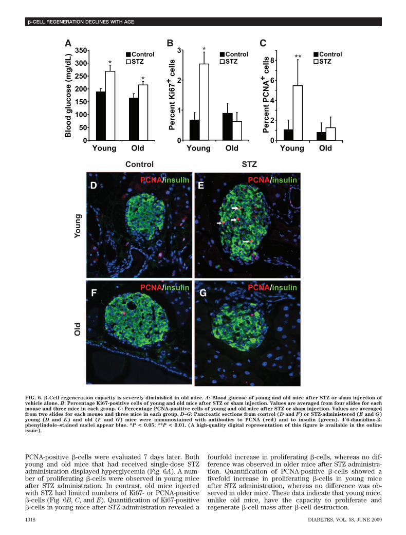
animal protocols were approved by the chancellor’s animal research commit-tee at the University of California, Los Angeles.Metabolic analysis. Fasting blood glucose was measured after overnightfasting. An insulin tolerance test was performed after a 6-h fast. Blood glucosewas measured before intraperitoneal injection of insulin (0.75 mU/g body wt)and then 20, 40, and 60 min after injection. No anesthesia was used during theexperiment. Glucose tolerance testing was performed after overnight fasting.Blood glucose levels (mg/dl) were measured before intraperitoneal injectionof glucose (2 mg dextrose/g body wt) and then 15, 30, 60, and 120 min afterinjection.Immunohistochemistry. Pancreatic tissue was processed as previouslydescribed (11). In brief, the pancreas was dissected and fixed in 4% formal-dehyde before being embedded in paraffin. Then, 5-�m sections were depar-affinized and rehydrated, followed by antigen retrieval using antigenunmasking buffer (Vector Labs), and then permeabilized in 0.4% TritonX-100/Tris-buffered saline. Slides were blocked with 3% IgG-free BSA (JacksonImmunoResearch Laboratories) and treated with anti-mouse insulin (Dako),anti-ki67 (BD Pharmingen), anti–proliferating cell nuclear antigen (PCNA; LabVision), anti-Bmi1 (Millipore), anti-p16Ink4a (Santa Cruz), anti–cyclin D2(Santa Cruz), or anti-p27 (Santa Cruz) antibody followed by fluoresceinisothiocyanate– or Cy3-conjugated secondary antibodies (The Jackson Labo-ratory). Terminal deoxynucleotidyl transferase dUTP nick-end labeling(TUNEL) assay was detected using an in situ cell death detection kit (Roche)according to manufacturer’s instructions. Slides were mounted with Vecta-shield with 4�6-diamidino-2-phenylindole (Vector Labs), and images wereobtained with a Leica DM6000 microscope using Openlab software (Improvi-sion). The immunofluorescence data presented are representatives of at leastfive animals per group in each case.�-Cell mass. �-Cell mass was measured as previously described (11). In brief,five to eight sections from each pancreas were stained with anti-mouse insulinantibody (Dako) and scanned by a Leica DM6000 microscope. Montage imageswere made by ImageJ software. The cross-sectional areas of pancreas and�-cells were determined by ImagePro software. �-Cell mass per pancreas wasestimated as the product of the relative cross-sectional area of �-cells per totaltissue and the weight of the pancreas and was calculated by examiningpancreata from at least three animals for each genotype.Islet isolation and immunoblotting. Liberase propidium iodide–purifiedenzyme blend for rodent islet isolation (Roche) was infused at 3.5 mg/ml intothe pancreas via the bile duct. Inflated pancreata were then removed andincubated in Liberase propidium iodide for 18 min at 37°C. Islets weredissociated from the exocrine tissues by shaking vigorously several times,followed by Histopaque (Sigma-Aldrich) gradient. Islets were handpickedunder a dissecting microscope and lysed by tissue extraction buffer (Invitro-gen). Lysates with equal amounts of protein were resolved by SDS-PAGE,followed by transferring to polyvinylidene fluoride membrane for immuno-blotting. The membranes were probed with specific antibodies against p16Ink4a
(Santa Cruz), Bmi1 (Millipore), and �-tubulin (Sigma-Aldrich). The datapresented are representative of at least three experiments.Chromatin immunoprecipitation. We performed chromatin immunopre-cipitation (ChIP) analysis using a Millipore ChIP kit (no. 17-295) according tothe manufacturer’s instructions, with minor modifications. The islets (150–200islets per group) were treated with 2% paraformaldehyde at room temperaturefor 20 min to cross-link the DNA with bound proteins. After washing, the isletswere resuspended in SDS lysis buffer with protease inhibitors and sonicatedto shear the chromatin. The chromatin was then precleared and incubatedwith 2–5 mg of anti–acetyl histone H3 lysine 9 (H3K9; no. 07-532; Millipore), ornormal mouse IgG as a control, overnight at 4°C with agitation. Afterimmunoprecipitation, the chromatin was harvested, the cross-links werereversed, and the DNA was purified and precipitated. The resulting DNA wasquantified and served as a template for the real-time PCR, performed using aLightCycler FastStartPLUS DNA SYBR Master kit (Roche) and Light CyclerPCR equipment (Roche). The DNA enrichment after ChIP was estimated asthe percentage bound-to-input ratio, determined by real-time PCR. Theprimers used to amplify the Ink4a/Arf locus are as follows: primer set 1:forward 5�GAGTACAGCAGCGGGAGCAT-3�, reverse 5�-GAACTTCACCAAGAAAACCCTCTCT-3�; primer set 2: forward 5�-GTCCGATCCTTTAGCGCTGTT-3�, reverse 5�-AGCCCGGACTACAGAAGAGATG-3�; primer set 3:5�CCGGAGCCACCCATTAAACTA-3�, reverse 5�-CAAGACTTCTCAAAAATAAGACACTGAAA-3�; primer set 4: forward 5�-CCCAACACCCACTTGAGGAA-3�,reverse 5�-CAGAGGTCACAGGCATCGAA-3�; and primer set 5: (negative con-trol, HoxC13 exon 2) forward 5�-CATTTTTCACTGATTTCCTAAGCA-3�, re-verse 5�-CAATGATGTCACCCCTCCTC-3�.Cell culture and transfection. Min6 cells were maintained in Dulbecco’smodified Eagle’s medium with 10% fetal bovine serum and transfected usingLipofectamine 2000 (Invitrogen) following the manufacturer’s instructionswith 1 mg of Bmi1 construct (in pcDNA3 myc-HisA, resulting in expression ofNH2-terminal myc-tagged Bmi1) and the control enhanced green fluorescent
S.-I. TSCHEN AND ASSOCIATES
DIABETES, VOL. 58, JUNE 2009 1313
protein construct (in pcDNA3), allowing expression from the cytomegaloviruspromoter in either case.Statistical analyses. All data were expressed as the means � SE. Mean andSE values were calculated from at least triplicates of a representativeexperiment. The statistical significance of differences was measured byunpaired Student’s t test and confirmed by one-way ANOVA for repeatmeasures. P � 0.05 indicated statistical significance. The P values indicated inthe graphs are from Student’s t test.
RESULTS
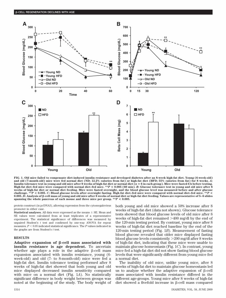
Adaptive expansion of �-cell mass associated withinsulin resistance is age dependent. To ascertainwhether age plays a role in the adaptive �-cell massexpansion associated with insulin resistance, young (6-week-old) and old (7- to 8-month-old) mice were fed ahigh-fat diet. Insulin tolerance testing performed after 8weeks of high-fat diet showed that both young and oldmice displayed decreased insulin sensitivity comparedwith mice on a normal diet (Fig. 1A). No statisticallysignificant difference in body weight between groups wasnoted at the beginning of the study. The body weight of
both young and old mice showed a 50% increase after 8weeks of high-fat diet (data not shown). Glucose tolerancetests showed that blood glucose levels of old mice after 8weeks of high-fat diet remained �400 mg/dl by the end ofthe 120-min testing period. By contrast, young mice after 8weeks of high-fat diet reached baseline by the end of the120-min testing period (Fig. 1B). Measurement of fastingblood glucose revealed that older mice displayed fastingblood glucose levels consistently �200 mg/dl after 8 weeksof high-fat diet, indicating that these mice were unable tomaintain glucose homeostasis (Fig. 1C). In contrast, youngmice fed a high-fat diet did not show fasting blood glucoselevels that were significantly different from young mice feda normal diet.
The inability of old mice, unlike young mice, after 8weeks of high-fat diet to maintain glucose homeostasis ledus to analyze whether the adaptive expansion of �-cellmass associated with insulin resistance differed in thedifferent age-groups. Young mice after 8 weeks of high-fatdiet showed a fivefold increase in �-cell mass compared
0
50
100
150
200
250
300
0 20 40 60
Blo
od G
luco
se (m
g/dL
)
Blo
od G
luco
se (m
g/dL
)
Blo
od G
luco
se (m
g/dL
)
Time
Young NDYoung HFDOld NDOld HFD
A B
C D
00 15 30 120
100
200
300
400
500
600
700
Young NDYoung HFDOld NDOld HFD
0
50
100
150
200
250
300
Young Old
NDHFD
0
1
2
3
4
5
6
7
Young Old
Bet
a C
ell M
ass
(mg)
NDHFD
**
**
**
** **
FIG. 1. Old mice failed to compensate diet-induced insulin resistance and developed diabetes after an 8-week high-fat diet. Young (6-week-old)and old (7-month-old) mice were fed normal diet (ND; 12.2% calories from fat) or high-fat diet (HFD; 55% calories from fat) for 8 weeks. A:Insulin tolerance test in young and old mice after 8 weeks of high-fat diet or normal diet (n � 6 in each group). Mice were fasted 6 h before testing.High-fat diet–fed mice were compared with normal diet–fed mice. **P < 0.005 (60 min). B: Glucose tolerance test in young and old mice after 8weeks of high-fat diet or normal diet feeding. Mice were fasted overnight, and the blood glucose level was measured before and after glucosechallenge. **P < 0.005. C: Blood glucose levels after overnight fasting. High-fat diet–fed mice were compared with normal diet–fed mice. **P <0.005. D: Analysis of �-cell mass of young and old mice after 8 weeks of normal diet or high-fat diet feeding. Values are representative of 5–6 slidesspanning the whole pancreas of each mouse and three mice per group. **P < 0.005.
�-CELL REGENERATION DECLINES WITH AGE
1314 DIABETES, VOL. 58, JUNE 2009
with the normal-diet group. In contrast, old mice fedhigh-fat diet did not show any changes in �-cell masscompared with control old mice (Fig. 1D). Moreover,pancreas weight did not show any significant increaseafter 8 weeks of high-fat diet in both young and old mice(data not shown). Thus, young mice, unlike old mice, havethe capacity to expand �-cell mass to adapt to insulinresistance.Expansion of �-cell mass is correlated with p16Ink4a
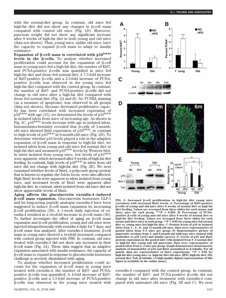
levels in the �-cells. To analyze whether increasedproliferation could account for the expansion of �-cellmass in young mice fed a high-fat diet, the number of Ki67-and PCNA-positive �-cells was quantified in mice fedhigh-fat diet and those fed normal diet. A 7.5-fold increaseof Ki67-positive �-cells and a 2.3-fold increase of PCNA-positive �-cells was observed in the young mice fedhigh-fat diet compared with the control group. In contrast,the number of Ki67- and PCNA-positive �-cells did notchange in old mice after a high-fat diet compared withthose fed normal diet (Fig. 2A and B). No TUNEL staining(as a measure of apoptosis) was observed in all groups(data not shown). Because decreased proliferative capac-ity has been correlated with increased expression ofp16Ink4a with age (15), we determined the levels of p16Ink4a
in isolated islets from mice of increasing age. As shown inFig. 2C, p16Ink4a levels increase with age in isolated islets.Immunohistochemistry revealed that �-cells of 1-month-old mice showed little expression of p16Ink4a, in contrastto high levels of p16Ink4a in 6-month-old mice (Fig. 2D). Todetermine whether p16 levels played a role in the adaptiveexpansion of �-cell mass in response to high-fat diet, weisolated islets from young and old mice fed normal diet orhigh-fat diet and measured p16ink4a levels by Western blot.In islets isolated from young mice, low levels of p16ink4a
were apparent, which decreased after 8 weeks of high-fat dietfeeding. In contrast, high levels of p16Ink4a in islets from oldmice did not change with high-fat diet (Fig. 2E). We nextexamined whether levels of Bmi1, a polycomb group proteinthat is known to regulate the Ink4a locus, were also affected.High Bmi1 levels were apparent in islets isolated from youngmice, and increased levels of Bmi1 were apparent afterhigh-fat diet. In contrast, islets isolated from old mice did notshow appreciable levels of Bmi1.Aging affects the glucoincretin exendin-4–induced�-cell mass expansion. Glucoincretin hormones GLP-1and its long-acting peptide analogue exendin-4 have beensuggested to induce �-cell mass expansion by increasing�-cell proliferation (30). A 1-week daily injection of ex-endin-4 resulted in a twofold increase in �-cell mass (30).To further investigate the effect of aging on �-cell massexpansion and �-cell proliferation, young and old mice wereinjected intraperitoneally with exendin-4 daily for 7 days, and�-cell mass was analyzed. After exendin-4 treatment, �-cellmass in young mice showed a twofold increment comparedwith control group injected with PBS. In contrast, old micetreated with exendin-4 did not show any increases in their�-cell mass (Fig. 3A). These data suggest that as adaptiveexpansion associated with insulin resistance, the capacity of�-cell mass to expand in response to glucoincretin hormoneschallenge is severely diminished with aging.
To analyze whether increased proliferation could ac-count for the expansion of �-cell mass in young micetreated with exendin-4, the number of Ki67- and PCNA-positive �-cells was quantified. A 4-fold increase of Ki67-positive �-cells and a 5.5-fold increase of PCNA-positive�-cells was observed in the young mice treated with
exendin-4 compared with the control group. In contrast,the number of Ki67- and PCNA-positive �-cells did notchange in old mice after treatment with exendin-4 com-pared with untreated old mice (Fig. 3B and C). We next
0
1
2
3
Perc
ent K
i67+
Perc
ent P
CN
A+
4
5
6
0
1
2
3
4
5
6
Young Old Y oung Old
NDHFD
NDHFD
p16
p16
p16
1mC
A B
D
E
4m 6m 11m
β-actin
Bmi1
Bmi-1
Tub
20
15
10
Rel
ativ
e Va
lue
5
0NDYoung Old
HFD HFDND
ND
Young Old
HFD HFDND
****
*** ***
FIG. 2. Increased �-cell proliferation in high-fat diet young micecorrelates with increased Bmi1 levels. A: Percentage of Ki67-positive�-cells of young and old mice after 8 weeks of normal diet or high-fatdiet feeding. Values are averaged from three slides for each mouse andthree mice for each group. ***P < 0.005. B: Percentage of PCNA-positive �-cells of young and old mice after 8 weeks of normal diet orhigh-fat diet feeding. Values are averaged from three slides for eachmouse and three mice in each group. **P < 0.05 for young mice fed normaldiet vs. young mice fed high-fat diet. C: Protein levels of p16 in isolatedislets from 1-, 4-, 6-, and 11-month-old mice. Data were representative ofpooled islets from 3–5 mice per group. D: Representative picture ofpancreatic sections from 1- and 6-month-old wild-type mice stained withantibodies to p16 (red) and insulin (green). E: Levels of p16 and Bmi1protein expression in the pooled islet cells isolated from 3–4 normal dietor high-fat diet young and old pancreata. Data were representative ofpooled islets from 3–4 mice per group. Graph demonstrates densitometricanalysis of immunoblot of p16 and Bmi1 normalized to �-tubulin. For allpanels, data are representative of three experiments. **P < 0.05 forhigh-fat diet young mice vs. high-fat diet old mice. HFD, high-fat diet; ND,normal diet; Tub, �-tubulin. (A high-quality digital representation of thisfigure is available in the online issue).
S.-I. TSCHEN AND ASSOCIATES
DIABETES, VOL. 58, JUNE 2009 1315
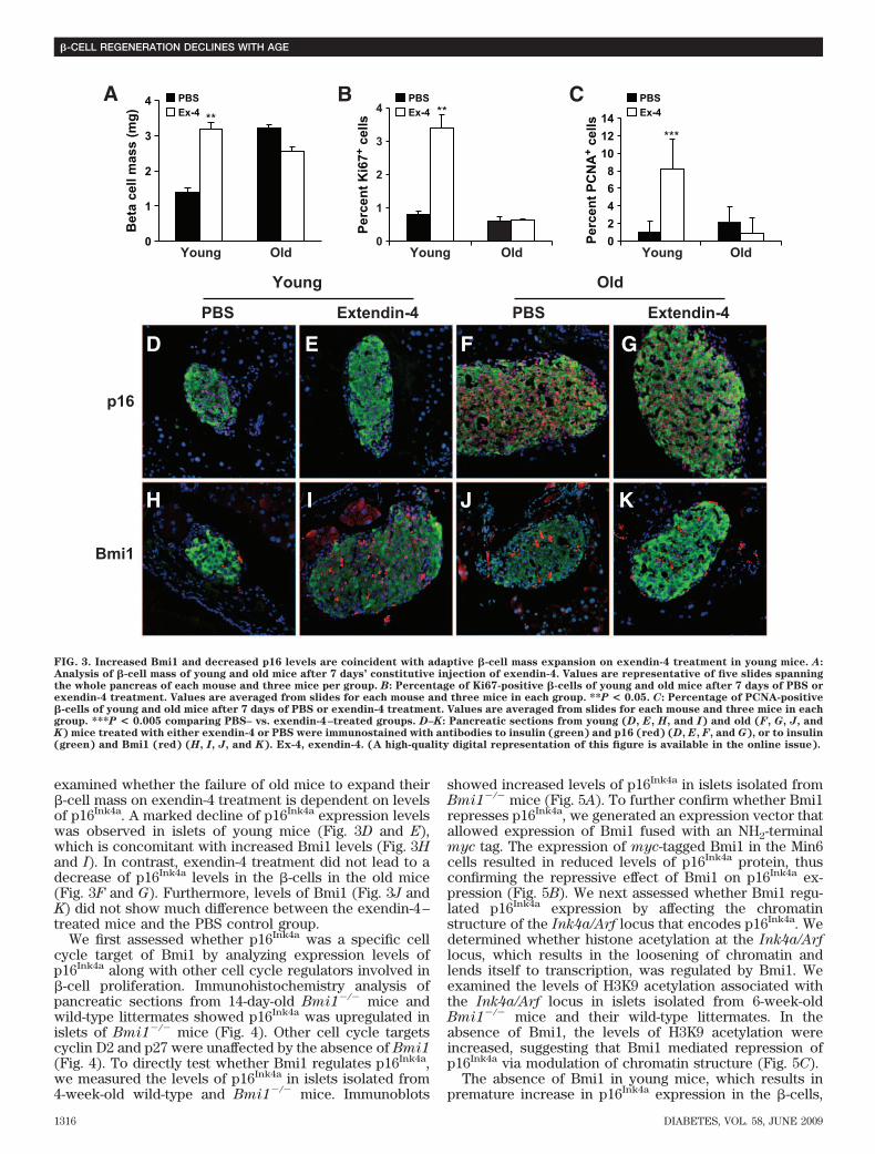
examined whether the failure of old mice to expand their�-cell mass on exendin-4 treatment is dependent on levelsof p16Ink4a. A marked decline of p16Ink4a expression levelswas observed in islets of young mice (Fig. 3D and E),which is concomitant with increased Bmi1 levels (Fig. 3Hand I). In contrast, exendin-4 treatment did not lead to adecrease of p16Ink4a levels in the �-cells in the old mice(Fig. 3F and G). Furthermore, levels of Bmi1 (Fig. 3J andK) did not show much difference between the exendin-4–treated mice and the PBS control group.
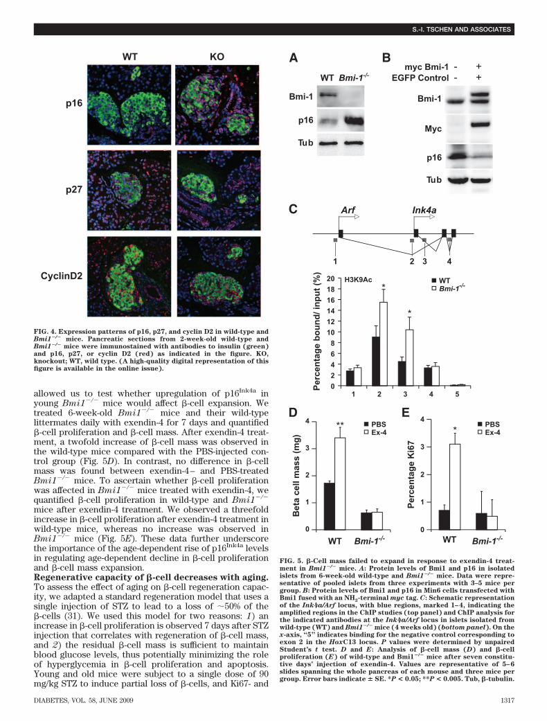
We first assessed whether p16Ink4a was a specific cellcycle target of Bmi1 by analyzing expression levels ofp16Ink4a along with other cell cycle regulators involved in�-cell proliferation. Immunohistochemistry analysis ofpancreatic sections from 14-day-old Bmi1�/� mice andwild-type littermates showed p16Ink4a was upregulated inislets of Bmi1�/� mice (Fig. 4). Other cell cycle targetscyclin D2 and p27 were unaffected by the absence of Bmi1(Fig. 4). To directly test whether Bmi1 regulates p16Ink4a,we measured the levels of p16Ink4a in islets isolated from4-week-old wild-type and Bmi1�/� mice. Immunoblots
showed increased levels of p16Ink4a in islets isolated fromBmi1�/� mice (Fig. 5A). To further confirm whether Bmi1represses p16Ink4a, we generated an expression vector thatallowed expression of Bmi1 fused with an NH2-terminalmyc tag. The expression of myc-tagged Bmi1 in the Min6cells resulted in reduced levels of p16Ink4a protein, thusconfirming the repressive effect of Bmi1 on p16Ink4a ex-pression (Fig. 5B). We next assessed whether Bmi1 regu-lated p16Ink4a expression by affecting the chromatinstructure of the Ink4a/Arf locus that encodes p16Ink4a. Wedetermined whether histone acetylation at the Ink4a/Arflocus, which results in the loosening of chromatin andlends itself to transcription, was regulated by Bmi1. Weexamined the levels of H3K9 acetylation associated withthe Ink4a/Arf locus in islets isolated from 6-week-oldBmi1�/� mice and their wild-type littermates. In theabsence of Bmi1, the levels of H3K9 acetylation wereincreased, suggesting that Bmi1 mediated repression ofp16Ink4a via modulation of chromatin structure (Fig. 5C).
The absence of Bmi1 in young mice, which results inpremature increase in p16Ink4a expression in the �-cells,
0
1
2
Bet
a ce
ll m
ass
(mg)
Perc
ent K
i67+
cel
ls
Perc
ent P
CN
A+
cells
3
4** **
PBSEx-4
PBSEx-4
PBSEx-4
02468
101214
0
1
2
3
4
Young OldYoung Old Young Old
A
PBS
p16
Bmi1
Extendin-4 PBS Extendin-4
Young Old
D E F G
H I J K
B C
***
FIG. 3. Increased Bmi1 and decreased p16 levels are coincident with adaptive �-cell mass expansion on exendin-4 treatment in young mice. A:Analysis of �-cell mass of young and old mice after 7 days’ constitutive injection of exendin-4. Values are representative of five slides spanningthe whole pancreas of each mouse and three mice per group. B: Percentage of Ki67-positive �-cells of young and old mice after 7 days of PBS orexendin-4 treatment. Values are averaged from slides for each mouse and three mice in each group. **P < 0.05. C: Percentage of PCNA-positive�-cells of young and old mice after 7 days of PBS or exendin-4 treatment. Values are averaged from slides for each mouse and three mice in eachgroup. ***P < 0.005 comparing PBS– vs. exendin-4–treated groups. D–K: Pancreatic sections from young (D, E, H, and I) and old (F, G, J, andK) mice treated with either exendin-4 or PBS were immunostained with antibodies to insulin (green) and p16 (red) (D, E, F, and G), or to insulin(green) and Bmi1 (red) (H, I, J, and K). Ex-4, exendin-4. (A high-quality digital representation of this figure is available in the online issue).
�-CELL REGENERATION DECLINES WITH AGE
1316 DIABETES, VOL. 58, JUNE 2009
allowed us to test whether upregulation of p16Ink4a inyoung Bmi1�/� mice would affect �-cell expansion. Wetreated 6-week-old Bmi1�/� mice and their wild-typelittermates daily with exendin-4 for 7 days and quantified�-cell proliferation and �-cell mass. After exendin-4 treat-ment, a twofold increase of �-cell mass was observed inthe wild-type mice compared with the PBS-injected con-trol group (Fig. 5D). In contrast, no difference in �-cellmass was found between exendin-4– and PBS-treatedBmi1�/� mice. To ascertain whether �-cell proliferationwas affected in Bmi1�/� mice treated with exendin-4, wequantified �-cell proliferation in wild-type and Bmi1�/�
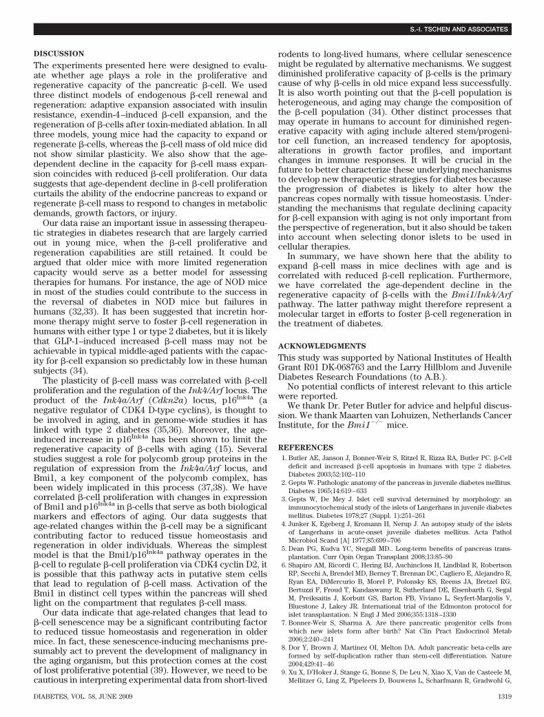
mice after exendin-4 treatment. We observed a threefoldincrease in �-cell proliferation after exendin-4 treatment inwild-type mice, whereas no increase was observed inBmi1�/� mice (Fig. 5E). These data further underscorethe importance of the age-dependent rise of p16Ink4a levelsin regulating age-dependent decline in �-cell proliferationand �-cell mass expansion.Regenerative capacity of �-cell decreases with aging.To assess the effect of aging on �-cell regeneration capac-ity, we adapted a standard regeneration model that uses asingle injection of STZ to lead to a loss of 50% of the�-cells (31). We used this model for two reasons: 1) anincrease in �-cell proliferation is observed 7 days after STZinjection that correlates with regeneration of �-cell mass,and 2) the residual �-cell mass is sufficient to maintainblood glucose levels, thus potentially minimizing the roleof hyperglycemia in �-cell proliferation and apoptosis.Young and old mice were subject to a single dose of 90mg/kg STZ to induce partial loss of �-cells, and Ki67- and
1
Arf Ink4a
2 3 4
WTmyc Bmi-1 - +
EGFP Control
Bmi-1Bmi-1
Myc
p16
p16
Tub
Tub
- +Bmi-1-/-
A B
C
D E
02468
10
Perc
enta
ge b
ound
/ inp
ut (%
)1214161820 H3K9Ac WT
Bmi-1-/-
1 2 3 4 5
0
1
2
Bet
a ce
ll m
ass
(mg)
Perc
enta
ge K
i67
3
4
WT Bmi-1-/- Bmi-1-/-0
1
2
3
4
WT
PBSEx-4
PBSEx-4
*
*
***
FIG. 5. �-Cell mass failed to expand in response to exendin-4 treat-ment in Bmi1�/� mice. A: Protein levels of Bmi1 and p16 in isolatedislets from 6-week-old wild-type and Bmi1�/� mice. Data were repre-sentative of pooled islets from three experiments with 3–5 mice pergroup. B: Protein levels of Bmi1 and p16 in Min6 cells transfected withBmi1 fused with an NH2-terminal myc tag. C: Schematic representationof the Ink4a/Arf locus, with blue regions, marked 1–4, indicating theamplified regions in the ChIP studies (top panel) and ChIP analysis forthe indicated antibodies at the Ink4a/Arf locus in islets isolated fromwild-type (WT) and Bmi1�/� mice (4 weeks old) (bottom panel). On thex-axis, “5” indicates binding for the negative control corresponding toexon 2 in the HoxC13 locus. P values were determined by unpairedStudent’s t test. D and E: Analysis of �-cell mass (D) and �-cellproliferation (E) of wild-type and Bmi1�/� mice after seven constitu-tive days’ injection of exendin-4. Values are representative of 5–6slides spanning the whole pancreas of each mouse and three mice pergroup. Error bars indicate � SE. *P < 0.05; **P < 0.005. Tub, �-tubulin.
WT
p16
p27
CyclinD2
KO
FIG. 4. Expression patterns of p16, p27, and cyclin D2 in wild-type andBmi1�/� mice. Pancreatic sections from 2-week-old wild-type andBmi1�/� mice were immunostained with antibodies to insulin (green)and p16, p27, or cyclin D2 (red) as indicated in the figure. KO,knockout; WT, wild type. (A high-quality digital representation of thisfigure is available in the online issue).
S.-I. TSCHEN AND ASSOCIATES
DIABETES, VOL. 58, JUNE 2009 1317
PCNA-positive �-cells were evaluated 7 days later. Bothyoung and old mice that had received single-dose STZadministration displayed hyperglycemia (Fig. 6A). A num-ber of proliferating �-cells were observed in young miceafter STZ administration. In contrast, old mice injectedwith STZ had limited numbers of Ki67- or PCNA-positive�-cells (Fig. 6B, C, and E). Quantification of Ki67-positive�-cells in young mice after STZ administration revealed a
fourfold increase in proliferating �-cells, whereas no dif-ference was observed in older mice after STZ administra-tion. Quantification of PCNA-positive �-cells showed afivefold increase in proliferating �-cells in young miceafter STZ administration, whereas no difference was ob-served in older mice. These data indicate that young mice,unlike old mice, have the capacity to proliferate andregenerate �-cell mass after �-cell destruction.
0
50
100
150
200
Blo
od g
luco
se (m
g/dL
)
Perc
ent K
i67+
cel
ls
Perc
ent P
CN
A+
cells
250
300
350
Young Old
PCNA/insulin PCNA/insulin
PCNA/insulin PCNA/insulin
Young Old Young Old0
1
2
3ControlSTZ
ControlSTZ
ControlSTZ
0
2
4
6
8
A B C
D
Control
Old
Youn
g
STZ
E
F G
*
*
***
FIG. 6. �-Cell regeneration capacity is severely diminished in old mice. A: Blood glucose of young and old mice after STZ or sham injection ofvehicle alone. B: Percentage Ki67-positive cells of young and old mice after STZ or sham injection. Values are averaged from four slides for eachmouse and three mice in each group. C: Percentage PCNA-positive cells of young and old mice after STZ or sham injection. Values are averagedfrom two slides for each mouse and three mice in each group. D–G: Pancreatic sections from control (D and F) or STZ-administered (E and G)young (D and E) and old (F and G) mice were immunostained with antibodies to PCNA (red) and to insulin (green). 4�6-diamidino-2-phenylindole–stained nuclei appear blue. *P < 0.05; **P < 0.01. (A high-quality digital representation of this figure is available in the onlineissue).
�-CELL REGENERATION DECLINES WITH AGE
1318 DIABETES, VOL. 58, JUNE 2009
DISCUSSION
The experiments presented here were designed to evalu-ate whether age plays a role in the proliferative andregenerative capacity of the pancreatic �-cell. We usedthree distinct models of endogenous �-cell renewal andregeneration: adaptive expansion associated with insulinresistance, exendin-4–induced �-cell expansion, and theregeneration of �-cells after toxin-mediated ablation. In allthree models, young mice had the capacity to expand orregenerate �-cells, whereas the �-cell mass of old mice didnot show similar plasticity. We also show that the age-dependent decline in the capacity for �-cell mass expan-sion coincides with reduced �-cell proliferation. Our datasuggests that age-dependent decline in �-cell proliferationcurtails the ability of the endocrine pancreas to expand orregenerate �-cell mass to respond to changes in metabolicdemands, growth factors, or injury.
Our data raise an important issue in assessing therapeu-tic strategies in diabetes research that are largely carriedout in young mice, when the �-cell proliferative andregeneration capabilities are still retained. It could beargued that older mice with more limited regenerationcapacity would serve as a better model for assessingtherapies for humans. For instance, the age of NOD micein most of the studies could contribute to the success inthe reversal of diabetes in NOD mice but failures inhumans (32,33). It has been suggested that incretin hor-mone therapy might serve to foster �-cell regeneration inhumans with either type 1 or type 2 diabetes, but it is likelythat GLP-1–induced increased �-cell mass may not beachievable in typical middle-aged patients with the capac-ity for �-cell expansion so predictably low in these humansubjects (34).
The plasticity of �-cell mass was correlated with �-cellproliferation and the regulation of the Ink4/Arf locus. Theproduct of the Ink4a/Arf (Cdkn2a) locus, p16Ink4a (anegative regulator of CDK4 D-type cyclins), is thought tobe involved in aging, and in genome-wide studies it haslinked with type 2 diabetes (35,36). Moreover, the age-induced increase in p16Ink4a has been shown to limit theregenerative capacity of �-cells with aging (15). Severalstudies suggest a role for polycomb group proteins in theregulation of expression from the Ink4a/Arf locus, andBmi1, a key component of the polycomb complex, hasbeen widely implicated in this process (37,38). We havecorrelated �-cell proliferation with changes in expressionof Bmi1 and p16Ink4a in �-cells that serve as both biologicalmarkers and effectors of aging. Our data suggests thatage-related changes within the �-cell may be a significantcontributing factor to reduced tissue homeostasis andregeneration in older individuals. Whereas the simplestmodel is that the Bmi1/p16Ink4a pathway operates in the�-cell to regulate �-cell proliferation via CDK4 cyclin D2, itis possible that this pathway acts in putative stem cellsthat lead to regulation of �-cell mass. Activation of theBmi1 in distinct cell types within the pancreas will shedlight on the compartment that regulates �-cell mass.
Our data indicate that age-related changes that lead to�-cell senescence may be a significant contributing factorto reduced tissue homeostasis and regeneration in oldermice. In fact, these senescence-inducing mechanisms pre-sumably act to prevent the development of malignancy inthe aging organism, but this protection comes at the costof lost proliferative potential (39). However, we need to becautious in interpreting experimental data from short-lived
rodents to long-lived humans, where cellular senescencemight be regulated by alternative mechanisms. We suggestdiminished proliferative capacity of �-cells is the primarycause of why �-cells in old mice expand less successfully.It is also worth pointing out that the �-cell population isheterogeneous, and aging may change the composition ofthe �-cell population (34). Other distinct processes thatmay operate in humans to account for diminished regen-erative capacity with aging include altered stem/progeni-tor cell function, an increased tendency for apoptosis,alterations in growth factor profiles, and importantchanges in immune responses. It will be crucial in thefuture to better characterize these underlying mechanismsto develop new therapeutic strategies for diabetes becausethe progression of diabetes is likely to alter how thepancreas copes normally with tissue homeostasis. Under-standing the mechanisms that regulate declining capacityfor �-cell expansion with aging is not only important fromthe perspective of regeneration, but it also should be takeninto account when selecting donor islets to be used incellular therapies.
In summary, we have shown here that the ability toexpand �-cell mass in mice declines with age and iscorrelated with reduced �-cell replication. Furthermore,we have correlated the age-dependent decline in theregenerative capacity of �-cells with the Bmi1/Ink4/Arfpathway. The latter pathway might therefore represent amolecular target in efforts to foster �-cell regeneration inthe treatment of diabetes.
ACKNOWLEDGMENTS
This study was supported by National Institutes of HealthGrant R01 DK-068763 and the Larry Hillblom and JuvenileDiabetes Research Foundations (to A.B.).
No potential conflicts of interest relevant to this articlewere reported.
We thank Dr. Peter Butler for advice and helpful discus-sion. We thank Maarten van Lohuizen, Netherlands CancerInstitute, for the Bmi1�/� mice.
REFERENCES
1. Butler AE, Janson J, Bonner-Weir S, Ritzel R, Rizza RA, Butler PC. �-Celldeficit and increased �-cell apoptosis in humans with type 2 diabetes.Diabetes 2003;52:102–110
2. Gepts W. Pathologic anatomy of the pancreas in juvenile diabetes mellitus.Diabetes 1965;14:619–633
3. Gepts W, De Mey J. Islet cell survival determined by morphology: animmunocytochemical study of the islets of Langerhans in juvenile diabetesmellitus. Diabetes 1978;27 (Suppl. 1):251–261
4. Junker K, Egeberg J, Kromann H, Nerup J. An autopsy study of the isletsof Langerhans in acute-onset juvenile diabetes mellitus. Acta PatholMicrobiol Scand [A] 1977;85:699–706
5. Dean PG, Kudva YC, Stegall MD.. Long-term benefits of pancreas trans-plantation. Curr Opin Organ Transplant 2008;13:85–90
6. Shapiro AM, Ricordi C, Hering BJ, Auchincloss H, Lindblad R, RobertsonRP, Secchi A, Brendel MD, Berney T, Brennan DC, Cagliero E, Alejandro R,Ryan EA, DiMercurio B, Morel P, Polonsky KS, Reems JA, Bretzel RG,Bertuzzi F, Froud T, Kandaswamy R, Sutherland DE, Eisenbarth G, SegalM, Preiksaitis J, Korbutt GS, Barton FB, Viviano L, Seyfert-Margolis V,Bluestone J, Lakey JR. International trial of the Edmonton protocol forislet transplantation. N Engl J Med 2006;355:1318–1330
7. Bonner-Weir S, Sharma A. Are there pancreatic progenitor cells fromwhich new islets form after birth? Nat Clin Pract Endocrinol Metab2006;2:240–241
8. Dor Y, Brown J, Martinez OI, Melton DA. Adult pancreatic beta-cells areformed by self-duplication rather than stem-cell differentiation. Nature2004;429:41–46
9. Xu X, D’Hoker J, Stange G, Bonne S, De Leu N, Xiao X, Van de Casteele M,Mellitzer G, Ling Z, Pipeleers D, Bouwens L, Scharfmann R, Gradwohl G,
S.-I. TSCHEN AND ASSOCIATES
DIABETES, VOL. 58, JUNE 2009 1319
Heimberg H. Beta cells can be generated from endogenous progenitors ininjured adult mouse pancreas. Cell 2008;132:197–207
10. Bouwens L, Rooman I. Regulation of pancreatic beta-cell mass. PhysiolRev 2005;85:1255–1270
11. Zhong L, Georgia S, Tschen SI, Nakayama K, Nakayama K, Bhushan A.Essential role of Skp2-mediated p27 degradation in growth and adaptiveexpansion of pancreatic beta cells. J Clin Invest 2007;117:2869–2876
12. Tanigawa K, Nakamura S, Kawaguchi M, Xu G, Kin S, Tamura K. Effect ofaging on B-cell function and replication in rat pancreas after 90% pancre-atectomy. Pancreas 1997;15:53–59
13. Montanya E, Nacher V, Biarnes M, Soler J. Linear correlation between�-cell mass and body weight throughout the lifespan in Lewis rats: role of�-cell hyperplasia and hypertrophy. Diabetes 2000;49:1341–1346
14. Teta M, Long SY, Wartschow LM, Rankin MM, Kushner JA. Very slowturnover of �-cells in aged adult mice. Diabetes 2005;54:2557–2567
15. Krishnamurthy J, Ramsey MR, Ligon KL, Torrice C, Koh A, Bonner-Weir S,Sharpless NE. p16INK4a induces an age-dependent decline in islet regen-erative potential. Nature 2006;443:453–457
16. Menge BA, Tannapfel A, Belyaev O, Drescher R, Muller C, Uhl W, SchmidtWE, Meier JJ. Partial pancreatectomy in adult humans does not provoke�-cell regeneration. Diabetes 2008;57:142–149
17. Farilla L, Hui H, Bertolotto C, Kang E, Bulotta A, Di Mario U, Perfetti R.Glucagon-like peptide-1 promotes islet cell growth and inhibits apoptosisin Zucker diabetic rats. Endocrinology 2002;143:4397–4408
18. Hadjiyanni I, Baggio LL, Poussier P, Drucker DJ. Exendin-4 modulatesdiabetes onset in nonobese diabetic mice. Endocrinology 2008;149:1338–1349
19. Li Y, Hansotia T, Yusta B, Ris F, Halban PA, Drucker DJ. Glucagon-likepeptide-1 receptor signaling modulates beta cell apoptosis. J Biol Chem2003;278:471–478
20. Sherry NA, Chen W, Kushner JA, Glandt M, Tang Q, Tsai S, Santamaria P,Bluestone JA, Brillantes AM, Herold KC. Exendin-4 improves reversal ofdiabetes in NOD mice treated with anti-CD3 monoclonal antibody byenhancing recovery of beta-cells. Endocrinology 2007;148:5136–5144
21. Xu G, Stoffers DA, Habener JF, Bonner-Weir S. Exendin-4 stimulates both�-cell replication and neogenesis, resulting in increased �-cell mass andimproved glucose tolerance in diabetic rats. Diabetes 1999;48:2270–2276
22. Creutzfeldt WO, Kleine N, Willms B, Orskov C, Holst JJ, Nauck MA.Glucagonostatic actions and reduction of fasting hyperglycemia by exog-enous glucagon-like peptide I(7–36) amide in type I diabetic patients.Diabetes Care 1996;19:580–586
23. Gutniak M, Orskov C, Holst JJ, Ahren B, Efendic S. Antidiabetogenic effectof glucagon-like peptide-1 (7–36)amide in normal subjects and patientswith diabetes mellitus. N Engl J Med 1992;326:1316–1322
24. Kjems LL, Holst JJ, Volund A, Madsbad S. The influence of GLP-1 onglucose-stimulated insulin secretion: effects on �-cell sensitivity in type 2and nondiabetic subjects. Diabetes 2003;52:380–386
25. Meneilly GS, McIntosh CH, Pederson RA, Habener JF, Ehlers MR, Egan JM,Elahi D. Effect of glucagon-like peptide 1 (7–36 amide) on insulin-mediatedglucose uptake in patients with type 1 diabetes. Diabetes Care 2003;26:837–842
26. Bruggeman SW, Valk-Lingbeek ME, van der Stoop PP, Jacobs JJ, KieboomK, Tanger E, Hulsman D, Leung C, Arsenijevic Y, Marino S, van LohuizenM. Ink4a and Arf differentially affect cell proliferation and neural stem cellself-renewal in Bmi1-deficient mice. Genes Dev 2005;19:1438–1443
27. Jacobs JJ, Kieboom K, Marino S, DePinho RA, van Lohuizen M. Theoncogene and Polycomb-group gene bmi-1 regulates cell proliferation andsenescence through the ink4a locus. Nature 1999;397:164–168
28. Molofsky AV, Slutsky SG, Joseph NM, He S, Pardal R, Krishnamurthy J,Sharpless NE, Morrison SJ.. Increasing p16INK4a expression decreasesforebrain progenitors and neurogenesis during ageing. Nature 2006;443:448–452
29. Park IK, Qian D, Kiel M, Becker MW, Pihalja M, Weissman IL, Morrison SJ,Clarke MF. Bmi-1 is required for maintenance of adult self-renewinghaematopoietic stem cells. Nature 2003;423:302–305
30. Buteau J, Spatz ML, Accili D. Transcription factor FoxO1 mediatesglucagon-like peptide-1 effects on pancreatic �-cell mass. Diabetes 2006;55:1190–1196
31. Bonner-Weir S, Trent DF, Honey RN, Weir GC. Responses of neonatal ratislets to streptozotocin: limited B-cell regeneration and hyperglycemia.Diabetes 1981;30:64–69
32. Kodama S, Kuhtreiber W, Fujimura S, Dale EA, Faustman DL. Isletregeneration during the reversal of autoimmune diabetes in NOD mice.Science 2003;302:1223–1227
33. Roep BO, Atkinson M. Animal models have little to teach us about type 1diabetes. 1. In support of this proposal. Diabetologia 2004;47:1650–1656
34. Schuit FC, In’t Veld PA, Pipeleers DG. Glucose stimulates proinsulinbiosynthesis by a dose-dependent recruitment of pancreatic beta cells.Proc Natl Acad Sci U S A 1988;85:3865–3869
35. Zeggini E, Weedon MN, Lindgren CM, Frayling TM, Elliott KS, Lango H,Timpson NJ, Perry JR, Rayner NW, Freathy RM, Barrett JC, Shields B,Morris AP, Ellard S, Groves CJ, Harries LW, Marchini JL, Owen KR, KnightB, Cardon LR, Walker M, Hitman GA, Morris AD, Doney AS, McCarthy MI,Hattersley AT. Replication of genome-wide association signals in UKsamples reveals risk loci for type 2 diabetes. Science 2007;316:1336–1341
36. Scott LJ, Mohlke KL, Bonnycastle LL, Willer CJ, Li Y, Duren WL, Erdos MR,Stringham HM, Chines PS, Jackson AU, Prokunina-Olsson L, Ding CJ, SwiftAJ, Narisu N, Hu T, Pruim R, Xiao R, Li XY, Conneely KN, Riebow NL,Sprau AG, Tong M, White PP, Hetrick KN, Barnhart MW, Bark CW,Goldstein JL, Watkins L, Xiang F, Saramies J, Buchanan TA, Watanabe RM,Valle TT, Kinnunen L, Abecasis GR, Pugh EW, Doheny KF, Bergman RN,Tuomilehto J, Collins FS, Boehnke M. A genome-wide association study oftype 2 diabetes in Finns detects multiple susceptibility variants. Science2007;316:1341–1345
37. Park IK, Morrison SJ, Clarke MF. Bmi1, stem cells, and senescenceregulation. J Clin Invest 2004;113:175–179
38. Valk-Lingbeek ME, Bruggeman SW, van Lohuizen M. Stem cells andcancer; the polycomb connection. Cell 2004;118:409–418
39. Kim WY, Sharpless NE. The regulation of INK4/ARF in cancer and aging.Cell 2006;127:265–275
�-CELL REGENERATION DECLINES WITH AGE
1320 DIABETES, VOL. 58, JUNE 2009