Embed Size (px)
Citation preview
268
Copyright © 2020 by Korean Society of Otorhinolaryngology-Head and Neck Surgery.This is an open-access article distributed under the terms of the Creative Commons Attribution Non-Commercial License (https://creativecommons.org/licenses/by-nc/4.0) which permits unrestricted non-commercial use, distribution, and reproduction in any medium, provided the original work is properly cited.
Clinical and Experimental Otorhinolaryngology Vol. 13, No. 3: 268-273, August 2020 https://doi.org/10.21053/ceo.2019.01599
Original Article
INTRODUCTION
Rhinoplasty is considered to be a popular and effective form of cosmetic surgery [1]. Nevertheless, its postoperative complica-tions may include significant eyelid edema and ecchymosis [2]. Periorbital edema may cause difficulties in visual acuity in the early postoperative period, and ecchymosis may increase perior-
bital pigmentation and cause disruptions in the patient’s social activities. The main cause of eyelid edema and ecchymosis after rhinoplasty is soft tissue damage at the osteotomy site [2]. Care-ful and delicate surgical manipulation can help to reduce such damage, but it cannot be completely prevented.
External percutaneous osteotomy and the internal endonasal technique are the two most common approaches used in rhino-plasty. Considering the risk of periosteal injury and the manipu-lation of the surrounding soft tissue during osteotomy, lateral osteotomy can be done with or without periosteal elevation (subperiosteal tunneling) [3]. However, there is no consensus re-garding which osteotomy method is more effective for reducing morbidity in rhinoplasty procedures. Since rhinoplasty remains a challenging procedure to this day, it is important for clinicians to follow the best practices for reducing postoperative complica-
• Received October 8, 2019 Revised December 20, 2019 Accepted January 2, 2020
• Corresponding author: Se Hwan Hwang Department of Otolaryngology-Head and Neck Surgery, Bucheon St. Mary’s Hospital, College of Medicine, The Catholic University of Korea, 327 Sosa-ro, Wonmi-gu, Bucheon 14647, Korea Tel: +82-32-340-7044, Fax: +82-32-340-2674 E-mail: [email protected]
pISSN 1976-8710 eISSN 2005-0720
Effects of Periosteal Elevation Before Lateral Osteotomy in Rhinoplasty: A Meta-Analysis of
Randomized Controlled Trials
Ji-Sun Kim ·Sun Hong Kim ·Hyesook Lee ·Byung Guk Kim ·Se Hwan Hwang
Department of Otolaryngology-Head and Neck Surgery, College of Medicine, The Catholic University of Korea, Seoul, Korea
Objectives. Although osteotomy is an important part of rhinoplasty, it is known to be closely related to postoperative eye-lid edema and ecchymosis. We aimed to evaluate the effects of periosteal elevation prior to osteotomy on eyelid ede-ma and ecchymosis.
Methods. Two authors reviewed studies in the Medline, Scopus, and Cochrane databases published through May 2019. Randomized controlled trials comparing periosteal elevation (subperiosteal tunneling) with periosteal preservation that reported eyelid edema or ecchymosis or subconjunctival hemorrhage as outcomes of interest were included. From each study, the baseline characteristics of the study subjects, the quality of the study, the number of patients in the treatment and control groups, and outcomes were extracted.
Results. Data for meta-analysis were identified in six studies with a total of 208 patients. Eyelid ecchymosis and edema within 3 days postoperatively were significantly more common in the periosteal elevation group than in the preserva-tion group, although such an association was not found for edema on postoperative day 7 (standardized mean differ-ence [SMD], 0.21; 95% confidence interval [CI], –0.09 to 0.50; I2=0%). There was no significant difference in sub-conjunctival hemorrhage on day 1 (SMD, 0.31; 95% CI, –0.09 to 0.72; I2=0%).
Conclusion. Periosteal preservation during lateral osteotomy may reduce eyelid edema and ecchymosis compared to peri-osteal elevation. Further studies with rigorous research methods should be carried out to determine the effectiveness of different techniques in lateral osteotomy.
Keywords. Osteotomy; Periosteum; Edema; Ecchymosis; Systematic Review; Meta-analysis
Kim JS et al. Subperiosteal Tunneling in Rhinoplasty 269
tions. This review aimed to evaluate the efficacy of periosteal el-evation. To assess the evidence for periosteal elevation prior to lateral osteotomy, we conducted a review of the literature to identify studies investigating eyelid edema, ecchymosis, and subconjunctival hemorrhage in patients undergoing lateral oste-otomy with or without periosteal elevation.
MATERIALS AND METHODS
Search strategy and selection of studies Studies published in English until May 2019 from Medline, Sco-pus, and Cochrane Register of Controlled Trials were identified. Search terms were “rhinoplasty,” “osteotomy,” “periosteum,” ‘edema,” and “ecchymosis.” We also checked the reference lists of identified studies to ensure that relevant studies were not missed.
Two reviewers (JSK, SHH) independently screened all ab-stracts and titles for candidate studies, excluding those that were not associated with lateral osteotomy of rhinoplasty. Random-ized controlled trials that studied patients undergoing rhinoplas-ty and periosteal elevation or periosteal preservation were in-cluded. Studies were excluded from analysis if patients under-went additional surgery (such as blepharoplasty or orthognathic surgery) or if reports were duplicated or if the results were not clearly reported as quantifiable data, or if appropriate data could not be extracted and calculated from published results. Fig. 1 shows the search strategy for meta-analysis.
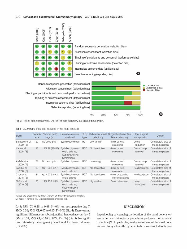
Data extraction and risk of bias assessmentData from included studies were extracted using standardized forms. Primary outcomes were degree of eyelid edema [3-6], ec-chymosis [3-8], and subconjunctival hemorrhage [3,4] compar-ing different osteotomy methods with another methods during the postoperative period (within 3 days or the seventh postop-erative day). Eyelid edema and ecchymosis were assessed indi-vidually using visual grading scales. On the grading scale, the smallest number indicated no edema or ecchymosis, and the largest number indicated severe edema enough to close the eye-lid or severe ecchymosis spreading to the lateral canthus.
In studies of the effect of subperiosteal tunneling on mea-sured outcomes, data on the number of patients, grading scale, and P-value were extracted. Quality assessment of the included randomized controlled studies was performed using the Co-chrane risk of bias tool. We used a “risk of bias” table including random sequence generation, allocation concealment, blinding, incomplete outcome data, and free of selective reporting (Fig. 2).
Statistical analysisA meta-analysis of the selected studies was conducted by R sta-tistical software (R Foundation, Vienna, Austria) with continuous measurements comparing the mean and standard deviation be-tween the control and experimental groups. The effect sizes of edema, ecchymosis, and subconjunctival hemorrhage were ex-pressed as the standardized mean difference (SMD). Heteroge-neity was confirmed by Cochran Q statistic test and I2 test. In this analysis, outcomes that did not show heterogeneity (I2<50) were analyzed using fixed-effects model.
RESULTS
Six studies with a total of 208 patients were included in this meta-analysis. Due to the incomplete reporting of patient vari-ables in these studies, a clear comparison of patients’ overall characteristics could not be made. The characteristics of the in-cluded studies are shown in Table 1.
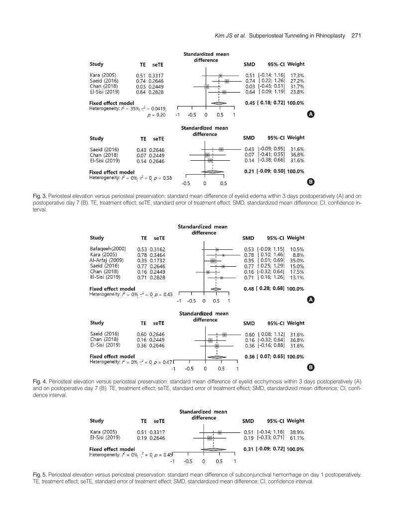
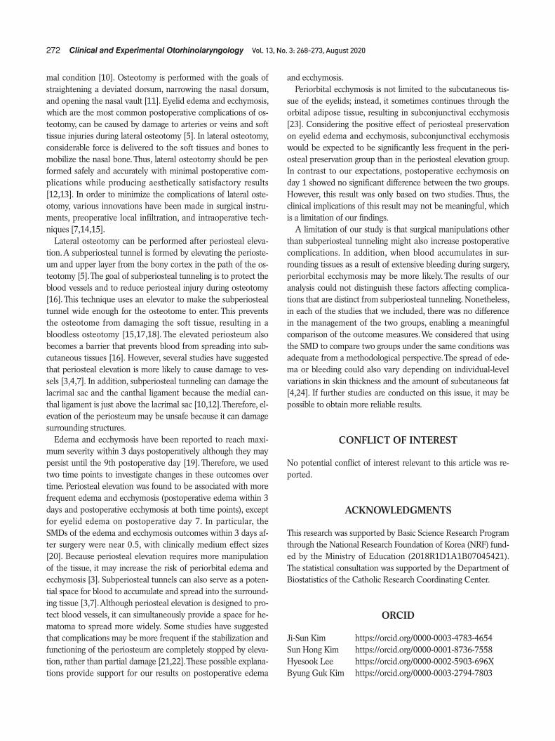
Eyelid edema (SMD, 0.45; 95% confidence interval [CI], 0.18 to 0.72; I2=35%) within 3 days postoperatively was significantly more common in the periosteal elevation group than in the pres-ervation group. However, edema on postoperative day 7 (SMD, 0.21; 95% CI, –0.09 to 0.50; I2=0%) showed no significant dif-ference between the two groups (Fig. 3). Eyelid ecchymosis was significantly more common in the periosteal elevation group than in the preservation group (within 3 days postoperatively: SMD,
Eyelid edema within 3 days after rhinoplasty was significantly more common in the periosteal elevation group than in the periosteal preservation group.
Eyelid ecchymosis was significantly more common in the peri-osteal elevation group than in the periosteal preservation group, both within 3 days and at 7 days after rhinoplasty.
Periosteal preservation during osteotomy may reduce eyelid edema and ecchymosis compared to periosteal elevation.
H LI IG GH H T S
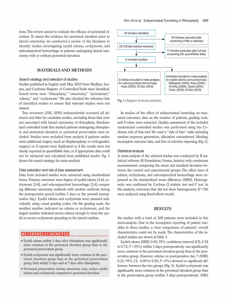
Fig. 1. Diagram of study selection.
2 Articles included in meta-analysis for subconjunctival hemorrhage : Kara (2005), El-Sisi (2019)
78 Studies identified
23 Full-text articles reviewed
6 Included studies
55 Articles excluded after screening of title or abstract
17 Articles excluded after full text screening (No quantifiable data)
6 Articles included in meta-analysis for eyelid edema and ecchymosis : Bafaqeeh (2000), Kara (2005),
Al-Arfaj (2009), Saeid (2016), Chan (2018), El-Sisi (2019)
270 Clinical and Experimental Otorhinolaryngology Vol. 13, No. 3: 268-273, August 2020
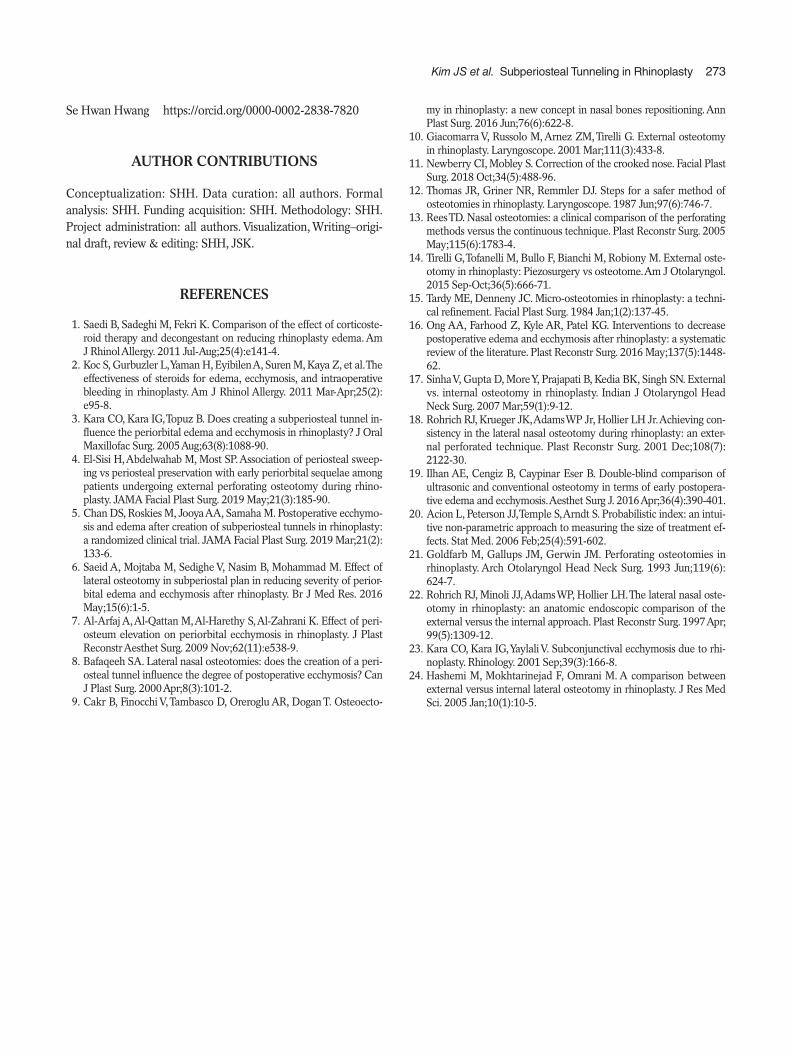
0.48; 95% CI, 0.28 to 0.68; I2=0%; on postoperative day 7: SMD, 0.36; 95% CI, 0.07 to 0.65; I2=0%) (Fig. 4). There was no significant difference in subconjunctival hemorrhage on day 1 (SMD, 0.31; 95% CI, –0.09 to 0.72; I2=0%) (Fig. 5). No signifi-cant inter-study heterogeneity was found for these outcomes (I2<50%).
DISCUSSION
Repositioning or changing the location of the nasal bone is es-sential in most rhinoplasty procedures performed for external correction [9]. In particular, medial movement of the nasal bone via osteotomy allows the pyramid to be reconstructed to its nor-
Table 1. Summary of studies included in the meta-analysis
Study Sample
sizeNumber (M/F);
age (yr)Outcome measure
analyzedStudy type
Pathway of lateral osteotomy
Surgical instruments of lateral osteotomy
Other surgical manipulation
Control
Bafaqeeh et al. (2000) [8]
20 No description Eyelid ecchymosis RCT Low-to-high 4-mm curved osteotome
Dorsal reduction
Contralateral side of the same patient
Kara et al. (2005) [3]
18 15/3; 26 (18–53) Eyelid ecchymosis, eyelid edema, Subconjunctival hemorrhage
RCT No description 4-mm curved osteotome
Dorsal hump removal
Contralateral side of the same patient
Al-Arfaj et al. (2009) [7]
78 No description Eyelid ecchymosis RCT Low-to-high 4-mm curved osteotome
Dorsal hump removal
Contralateral side of the same patient
Saeid et al. (2016) [6]
30 9/21; 20.4±2.7 Eyelid ecchymosis, eyelid edema
RCT No description 4-mm curved osteotome
No description Contralateral side of the same patient
Chan et al. (2018) [5]
34 6/28; 27.9±9.2 Eyelid ecchymosis, eyelid edema
RCT No description 6-mm unguarded cottle osteotome
No description Contralateral side of the same patient
El-Sisi et al. (2019) [4]
28 19/9; 23.7±3.9 Eyelid ecchymosis, eyelid edema, subconjunctival hemorrhage
RCT High-to-low 2-mm osteotome Hump resection
Contralateral side of the same patient
Values are presented as mean (range) or mean±standard deviation.M, male; F, female; RCT, randomized controlled trial.
Fig. 2. Risk of bias assessment. (A) Risk of bias summary. (B) Risk of bias graph.
Random sequence generation (selection bias)
Allocation concealment (selection bias)
Blinding of participants and personnel (performance bias)
Blinding of outcome assessment (detection bias)
Incomplete outcome data (attrition bias)
Selective reporting (reporting bias)
Sae
id (2
016)
Kar
a (2
005)
El-S
isi (
2019
)
Cha
n (2
018)
Baf
aqee
h (2
000)
Al-A
rfaj (
2009
)A
B0% 25% 50% 75% 100%
Random sequence generation (selection bias)
Allocation concealment (selection bias)
Blinding of participants and personnel (performance bias)
Blinding of outcome assessment (detection bias)
Incomplete outcome data (attrition bias)
Selective reporting (reporting bias)
Low risk of biasUnclear risk of biasHigh risk of bias
Kim JS et al. Subperiosteal Tunneling in Rhinoplasty 271
A
Fig. 3. Periosteal elevation versus periosteal preservation: standard mean difference of eyelid edema within 3 days postoperatively (A) and on postoperative day 7 (B). TE, treatment effect; seTE, standard error of treatment effect; SMD, standardized mean difference; CI, confidence in-terval.
B
A
B
Fig. 4. Periosteal elevation versus periosteal preservation: standard mean difference of eyelid ecchymosis within 3 days postoperatively (A) and on postoperative day 7 (B). TE, treatment effect; seTE, standard error of treatment effect; SMD, standardized mean difference; CI, confi-dence interval.
Fig. 5. Periosteal elevation versus periosteal preservation: standard mean difference of subconjunctival hemorrhage on day 1 postoperatively. TE, treatment effect; seTE, standard error of treatment effect; SMD, standardized mean difference; CI, confidence interval.
272 Clinical and Experimental Otorhinolaryngology Vol. 13, No. 3: 268-273, August 2020
mal condition [10]. Osteotomy is performed with the goals of straightening a deviated dorsum, narrowing the nasal dorsum, and opening the nasal vault [11]. Eyelid edema and ecchymosis, which are the most common postoperative complications of os-teotomy, can be caused by damage to arteries or veins and soft tissue injuries during lateral osteotomy [5]. In lateral osteotomy, considerable force is delivered to the soft tissues and bones to mobilize the nasal bone. Thus, lateral osteotomy should be per-formed safely and accurately with minimal postoperative com-plications while producing aesthetically satisfactory results [12,13]. In order to minimize the complications of lateral oste-otomy, various innovations have been made in surgical instru-ments, preoperative local infiltration, and intraoperative tech-niques [7,14,15].
Lateral osteotomy can be performed after periosteal eleva-tion. A subperiosteal tunnel is formed by elevating the perioste-um and upper layer from the bony cortex in the path of the os-teotomy [5]. The goal of subperiosteal tunneling is to protect the blood vessels and to reduce periosteal injury during osteotomy [16]. This technique uses an elevator to make the subperiosteal tunnel wide enough for the osteotome to enter. This prevents the osteotome from damaging the soft tissue, resulting in a bloodless osteotomy [15,17,18]. The elevated periosteum also becomes a barrier that prevents blood from spreading into sub-cutaneous tissues [16]. However, several studies have suggested that periosteal elevation is more likely to cause damage to ves-sels [3,4,7]. In addition, subperiosteal tunneling can damage the lacrimal sac and the canthal ligament because the medial can-thal ligament is just above the lacrimal sac [10,12]. Therefore, el-evation of the periosteum may be unsafe because it can damage surrounding structures.
Edema and ecchymosis have been reported to reach maxi-mum severity within 3 days postoperatively although they may persist until the 9th postoperative day [19]. Therefore, we used two time points to investigate changes in these outcomes over time. Periosteal elevation was found to be associated with more frequent edema and ecchymosis (postoperative edema within 3 days and postoperative ecchymosis at both time points), except for eyelid edema on postoperative day 7. In particular, the SMDs of the edema and ecchymosis outcomes within 3 days af-ter surgery were near 0.5, with clinically medium effect sizes [20]. Because periosteal elevation requires more manipulation of the tissue, it may increase the risk of periorbital edema and ecchymosis [3]. Subperiosteal tunnels can also serve as a poten-tial space for blood to accumulate and spread into the surround-ing tissue [3,7]. Although periosteal elevation is designed to pro-tect blood vessels, it can simultaneously provide a space for he-matoma to spread more widely. Some studies have suggested that complications may be more frequent if the stabilization and functioning of the periosteum are completely stopped by eleva-tion, rather than partial damage [21,22]. These possible explana-tions provide support for our results on postoperative edema
and ecchymosis.Periorbital ecchymosis is not limited to the subcutaneous tis-
sue of the eyelids; instead, it sometimes continues through the orbital adipose tissue, resulting in subconjunctival ecchymosis [23]. Considering the positive effect of periosteal preservation on eyelid edema and ecchymosis, subconjunctival ecchymosis would be expected to be significantly less frequent in the peri-osteal preservation group than in the periosteal elevation group. In contrast to our expectations, postoperative ecchymosis on day 1 showed no significant difference between the two groups. However, this result was only based on two studies. Thus, the clinical implications of this result may not be meaningful, which is a limitation of our findings.
A limitation of our study is that surgical manipulations other than subperiosteal tunneling might also increase postoperative complications. In addition, when blood accumulates in sur-rounding tissues as a result of extensive bleeding during surgery, periorbital ecchymosis may be more likely. The results of our analysis could not distinguish these factors affecting complica-tions that are distinct from subperiosteal tunneling. Nonetheless, in each of the studies that we included, there was no difference in the management of the two groups, enabling a meaningful comparison of the outcome measures. We considered that using the SMD to compare two groups under the same conditions was adequate from a methodological perspective. The spread of ede-ma or bleeding could also vary depending on individual-level variations in skin thickness and the amount of subcutaneous fat [4,24]. If further studies are conducted on this issue, it may be possible to obtain more reliable results.
CONFLICT OF INTEREST
No potential conflict of interest relevant to this article was re-ported.
ACKNOWLEDGMENTS
This research was supported by Basic Science Research Program through the National Research Foundation of Korea (NRF) fund-ed by the Ministry of Education (2018R1D1A1B07045421). The statistical consultation was supported by the Department of Biostatistics of the Catholic Research Coordinating Center.
ORCID
Ji-Sun Kim https://orcid.org/0000-0003-4783-4654Sun Hong Kim https://orcid.org/0000-0001-8736-7558Hyesook Lee https://orcid.org/0000-0002-5903-696X Byung Guk Kim https://orcid.org/0000-0003-2794-7803
Kim JS et al. Subperiosteal Tunneling in Rhinoplasty 273
Se Hwan Hwang https://orcid.org/0000-0002-2838-7820
AUTHOR CONTRIBUTIONS
Conceptualization: SHH. Data curation: all authors. Formal analysis: SHH. Funding acquisition: SHH. Methodology: SHH. Project administration: all authors. Visualization, Writing–origi-nal draft, review & editing: SHH, JSK.
REFERENCES
1. Saedi B, Sadeghi M, Fekri K. Comparison of the effect of corticoste-roid therapy and decongestant on reducing rhinoplasty edema. Am J Rhinol Allergy. 2011 Jul-Aug;25(4):e141-4.
2. Koc S, Gurbuzler L, Yaman H, Eyibilen A, Suren M, Kaya Z, et al. The effectiveness of steroids for edema, ecchymosis, and intraoperative bleeding in rhinoplasty. Am J Rhinol Allergy. 2011 Mar-Apr;25(2): e95-8.
3. Kara CO, Kara IG, Topuz B. Does creating a subperiosteal tunnel in-fluence the periorbital edema and ecchymosis in rhinoplasty? J Oral Maxillofac Surg. 2005 Aug;63(8):1088-90.
4. El-Sisi H, Abdelwahab M, Most SP. Association of periosteal sweep-ing vs periosteal preservation with early periorbital sequelae among patients undergoing external perforating osteotomy during rhino-plasty. JAMA Facial Plast Surg. 2019 May;21(3):185-90.
5. Chan DS, Roskies M, Jooya AA, Samaha M. Postoperative ecchymo-sis and edema after creation of subperiosteal tunnels in rhinoplasty: a randomized clinical trial. JAMA Facial Plast Surg. 2019 Mar;21(2): 133-6.
6. Saeid A, Mojtaba M, Sedighe V, Nasim B, Mohammad M. Effect of lateral osteotomy in subperiostal plan in reducing severity of perior-bital edema and ecchymosis after rhinoplasty. Br J Med Res. 2016 May;15(6):1-5.
7. Al-Arfaj A, Al-Qattan M, Al-Harethy S, Al-Zahrani K. Effect of peri-osteum elevation on periorbital ecchymosis in rhinoplasty. J Plast Reconstr Aesthet Surg. 2009 Nov;62(11):e538-9.
8. Bafaqeeh SA. Lateral nasal osteotomies: does the creation of a peri-osteal tunnel influence the degree of postoperative ecchymosis? Can J Plast Surg. 2000 Apr;8(3):101-2.
9. Cakr B, Finocchi V, Tambasco D, Oreroglu AR, Dogan T. Osteoecto-
my in rhinoplasty: a new concept in nasal bones repositioning. Ann Plast Surg. 2016 Jun;76(6):622-8.
10. Giacomarra V, Russolo M, Arnez ZM, Tirelli G. External osteotomy in rhinoplasty. Laryngoscope. 2001 Mar;111(3):433-8.
11. Newberry CI, Mobley S. Correction of the crooked nose. Facial Plast Surg. 2018 Oct;34(5):488-96.
12. Thomas JR, Griner NR, Remmler DJ. Steps for a safer method of osteotomies in rhinoplasty. Laryngoscope. 1987 Jun;97(6):746-7.
13. Rees TD. Nasal osteotomies: a clinical comparison of the perforating methods versus the continuous technique. Plast Reconstr Surg. 2005 May;115(6):1783-4.
14. Tirelli G, Tofanelli M, Bullo F, Bianchi M, Robiony M. External oste-otomy in rhinoplasty: Piezosurgery vs osteotome. Am J Otolaryngol. 2015 Sep-Oct;36(5):666-71.
15. Tardy ME, Denneny JC. Micro-osteotomies in rhinoplasty: a techni-cal refinement. Facial Plast Surg. 1984 Jan;1(2):137-45.
16. Ong AA, Farhood Z, Kyle AR, Patel KG. Interventions to decrease postoperative edema and ecchymosis after rhinoplasty: a systematic review of the literature. Plast Reconstr Surg. 2016 May;137(5):1448- 62.
17. Sinha V, Gupta D, More Y, Prajapati B, Kedia BK, Singh SN. External vs. internal osteotomy in rhinoplasty. Indian J Otolaryngol Head Neck Surg. 2007 Mar;59(1):9-12.
18. Rohrich RJ, Krueger JK, Adams WP Jr, Hollier LH Jr. Achieving con-sistency in the lateral nasal osteotomy during rhinoplasty: an exter-nal perforated technique. Plast Reconstr Surg. 2001 Dec;108(7): 2122-30.
19. Ilhan AE, Cengiz B, Caypinar Eser B. Double-blind comparison of ultrasonic and conventional osteotomy in terms of early postopera-tive edema and ecchymosis. Aesthet Surg J. 2016 Apr;36(4):390-401.
20. Acion L, Peterson JJ, Temple S, Arndt S. Probabilistic index: an intui-tive non-parametric approach to measuring the size of treatment ef-fects. Stat Med. 2006 Feb;25(4):591-602.
21. Goldfarb M, Gallups JM, Gerwin JM. Perforating osteotomies in rhinoplasty. Arch Otolaryngol Head Neck Surg. 1993 Jun;119(6): 624-7.
22. Rohrich RJ, Minoli JJ, Adams WP, Hollier LH. The lateral nasal oste-otomy in rhinoplasty: an anatomic endoscopic comparison of the external versus the internal approach. Plast Reconstr Surg. 1997 Apr; 99(5):1309-12.
23. Kara CO, Kara IG, Yaylali V. Subconjunctival ecchymosis due to rhi-noplasty. Rhinology. 2001 Sep;39(3):166-8.
24. Hashemi M, Mokhtarinejad F, Omrani M. A comparison between external versus internal lateral osteotomy in rhinoplasty. J Res Med Sci. 2005 Jan;10(1):10-5.