Embed Size (px)
Citation preview

Bulgarian Journal of Veterinary Medicine, 2014, 17, No 3, 199206 ISSN 1311-1477; online at http://tru.uni-sz.bg/bjvm/bjvm.htm
Original article
ETIOLOGY AND CLINICOEPIDEMIOLOGICAL PROFILE OF APIARIES WITH COLONY COLLAPSE DISORDER-LIKE
SYMPTOMS IN BULGARIA
P. PARVANOV & N. RUSENOVA
Department of Veterinary Microbiology, Infectious and Parasitic
Diseases, Faculty of Veterinary Medicine, Stara Zagora, Bulgaria
Summary
Parvanov, P. & N. Rusenova, 2014. Etiology and clinicoepidemiological profile of apiaries with colony collapse disorder-like symptoms in Bulgaria. Bulg. J. Vet. Med., 17, No 3, 199206. Field clinicoepidemiological and laboratory etiological examinations were performed in apiaries with colony collapsе disorder (CCD)-like symptoms in different regions in Bulgaria. The survey included 5,785 bee families in 41 apiaries, which were depopulated by 3,043 (52.6%) families. In 30 apiaries (71.8 %), the depopulation occurred in the spring (March-April) and in 11 apiaries (26.8%) bees left the hives in the autumn (September-October). The depopulation consisted in a dramatic decrease in the number of bees (the bees disappeared) until only queens with 20–30 bees were left and the family perished. In all apiaries except in one (n=40) Nosema ceranae infection was established. The results from questionnaires and clinicoepidemiological studies in affected apiaries allowed rejecting depopu-lation factors such as starvation, inadequate and poor-quality feed stores, varroatosis, pesticide intoxication, genetically modified cultures, high-voltage, electromagnetic and radiofrequency influ-ences. Severe Nosema ceranae infection probably combined with viral infection resulting in disturbed repair of the intestinal epithelium, nutritional and energy deficiency and impossibility of flying bees to return to their hives were outlined as the main causes for the rapid collapse of bee families and depopulation of hives.
Key words: bee family, colony collapsе disorder, cause, death, Nosema ceranae
INTRODUCTION
The increased death rates of bee families during the last years are of major concern to global apiculture. In the USA only, colony collapsе disorder (CCD; empty hive syndrome) was responsible for the disappearance of 31% and 36% of bee families during the winter and spring of
2006/2007 and 2007/2008 respectively (vanEngelsdorp et al., 2007; 2008), whe-reas in the autumn of 2006 some bee-keepers have lost 30–90% of their bee families (Ellis et al., 2010).
Considerable losses were registered also on Europe – Spain, Greece, Turkey,

Etiology and clinicoepidemiological profile of apiaries with colony collapse disorder-like ...
BJVM, 17, No 3 200
France, Switzerland, Poland, and Ger-many (Fries et al., 2006; Higes et al. 2006; Topolska & Kasprzak, 2007; Giray et al., 2007; 2010). Only in northern Greece, 45 to 65% of bee families were lost during the winter and spring of 2007–2008 (Thrasyvoulou, 2009).
Numerous authors associated the weakening of bee families with pesticides used in agricultural practice (Decourtye et al., 2003, 2004), genetically modified cultures (Malone et al., 1999; Malone & Pham-Delegue, 2001; Huang et al., 2004), temperature deviations during brood rearing (Tautz et al., 2003), inbreeding (Spivak & Gilliam, 1998a,b; Palmer & Oldroyd, 2000), starvation, stress, electro-magnetic influences from TV and GSM retranslators.
The involvement of pathogens in CCD etiology is the most plausible hypothesis (Martin-Hernandez et al., 2007). A spe-cial attention is paid on the Israeli acute paralysis virus (IAPV), transmitted by the Varroa destructor mite. Other viruses have been also isolated from bee families with CCD – Chronic paralysis virus, Deforming wing virus, Kashmir bee virus, Black queen cell virus, but they were not associated with syndrome etiology (Higes et al., 2008а).
The theory for the leading role of the microsporidian Nоsema ceranae (N. cera-nae) in CCD etiology and high mortality of bees is supported by the investigations of Higes et al. (2006) and Martin-Her-nandez et al. (2007) carried out in some regions in Spain. It is however suggested that N. ceranae infection without predis-posing factors could not lead to bee family perishing, determining the agent as opportunistic but not obligate pathogen (Invernizzi et al., 2009).
Johnson et al. (2009) have linked CCD to picornaviruses suppressing pro-
tein synthesis in the organism of bees, whereas Bromenshenk et al. (2010) attri-bute the high death rates of bee families to mixed Iridovirus/Nosema infection. Des-pite the multiple studies however, the main cause of the problem has not been identified.
The present investigation was motiva-ted by the spread of bee family weakening and in many instances, disappearing of bees from entire apiaries in different regions of Bulgaria during the last years reported by beekeepers and according to our own observations.
The purpose of the present survey was to perform a more detailed etiological and clinicoepidemiological profile of Bulgari-an apiaries with CCD-like states, which have not been so far performed.
MATERIALS AND METHODS
From April 2008 to May 2010, together with the National Branch Organisation Bulgarian Union of Beekeepers, a survey was performed in the country by filling feedback forms developed by us for detection and investigation of apiaries with CCD-like syndromes.
The questionnaire consisted of 20 questions about the tentative causes for the syndrome and increased death rates according to our and global experience. The aim was to obtain information about the number of perished bee families in affected apiaries, the time and way of depopulation, the strength of bee families in the autumn, the type, amount and quality of winter feed stores, the used anti-varroatosis drugs, presence of GSM re-translation towers and GMO crops in the neighbourhood, chemical pollutions etc.
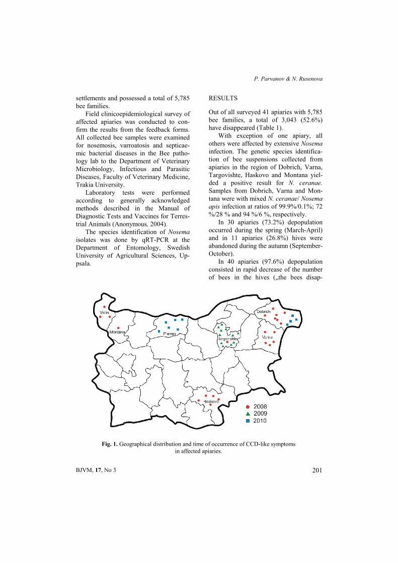
The respondents were owners of apia-ries in different regions in Bulgaria (Fig. 1). Affected apiaries (41) were located in 34
P. Parvanov & N. Rusenova
BJVM, 17, No 3 201
settlements and possessed a total of 5,785 bee families.
Field clinicoepidemiological survey of affected apiaries was conducted to con-firm the results from the feedback forms. All collected bee samples were examined for nosemosis, varroatosis and septicae-mic bacterial diseases in the Bee patho-logy lab to the Department of Veterinary Microbiology, Infectious and Parasitic Diseases, Faculty of Veterinary Medicine, Trakia University.
Laboratory tests were performed according to generally acknowledged methods described in the Manual of Diagnostic Tests and Vaccines for Terres-trial Аnimals (Anonymous, 2004).
The species identification of Nosema isolates was done by qRT-PCR at the Department of Entomology, Swedish University of Agricultural Sciences, Up-psala.
RESULTS
Out of all surveyed 41 apiaries with 5,785 bee families, a total of 3,043 (52.6%) have disappeared (Table 1).
With exception of one apiary, all others were affected by extensive Nosema infection. The genetic species identifica-tion of bee suspensions collected from apiaries in the region of Dobrich, Varna, Targovishte, Haskovo and Montana yiel-ded a positive result for N. ceranae. Samples from Dobrich, Varna and Mon-tana were with mixed N. ceranae/ Nosema apis infection at ratios of 99.9%/0.1%; 72 %/28 % and 94 %/6 %, respectively.
In 30 apiaries (73.2%) depopulation occurred during the spring (March-April) and in 11 apiaries (26.8%) hives were abandoned during the autumn (September-October).
In 40 apiaries (97.6%) depopulation consisted in rapid decrease of the number of bees in the hives („the bees disap-
Fig. 1. Geographical distribution and time of occurrence of CCD-like symptoms in affected apiaries.

Etiology and clinicoepidemiological profile of apiaries with colony collapse disorder-like ...
BJVM, 17, No 3 202
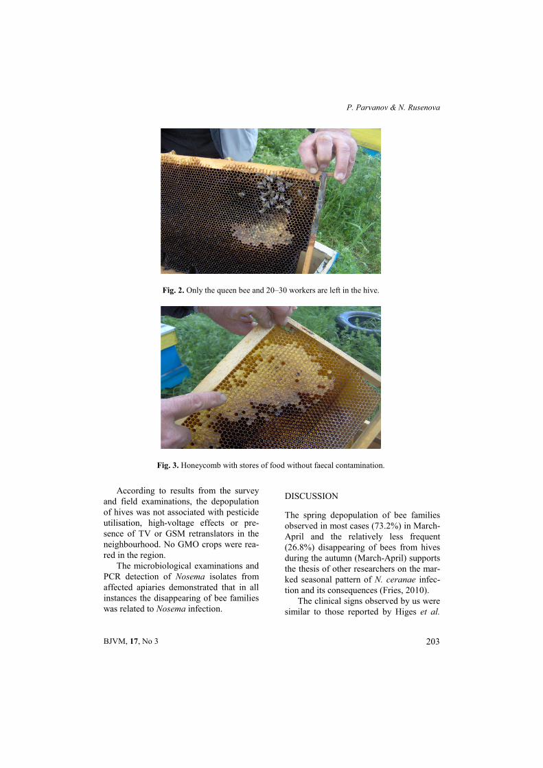
peared”), until only the queen bee and 20–30 workers were left in the hive (Fig. 2) and the family perished. The combs had enough stored food, no dead bees were found lying on the bottom of hives and no diarrhoeic staining was present in most cases (Fig. 3). The apiaries were not affected by infectious diseases during the previous year. At the beginning of the winter, the strength of the bee families was high and moderate, and the food stores – adequate and of high-quality. During both the autumn and the spring, bees were additionally fed honey-sugar paste and sugar syrup. Apiaries were pro-phylactically treated against varroatosis with approved drugs and according to
beekeepers, depopulation was not asso-ciated with either varroatosis or antivar-roatosis treatments.
In one of apiaries in Haskovo region, the depopulation was described by the beekeepers as sudden disappearing of bees together with the bee queen (as a swarm) when the nest was full of food stores and with brood. Out of 52 bee families, 18 have left the hives (34.61%). The brood was defective, with damaged caps and malformed bee bodies inside the cells. This description is compatible with that of severe varroatosis. In this apiary, no antivarroatosis treatments have been performed during the past two years.
Table 1. Surveyed apiaries with CCD-like symptoms
Region and number of settlements
Time of the event
Number of apiaries
Number of bee families
Depopulated bee families
Isolated pathogen
Dobrich region 1 April 2008 3 700 300 N. ceranae
3 April 2008 3 828 528 Nosema spр.
3 May 2010 4 453 216 Nosema spр.
Varna region 1 May 2008 3 800 680 N. ceranae
2 May 2008 2 260 290 Nosema spр.
Haskovo region 1 April 2008 3 146 63 Nosema spр.
1 April 2008 1 32 8 N. ceranae
1 October 2008 1 52 18 V. destructor
Montana region 1 April 2008 1 260 80 N. ceranae
Vidin region 3 April 2008 3 400 80 Nosema spр.
Targovishte region 1 May 2008 1 80 60 N. ceranae
11 October 2009 11 1310 430 Nosema spр.
Pleven region 5 April 2010 5 464 290 Nosema spр.
Total – 34 41 5785 3043
P. Parvanov & N. Rusenova
BJVM, 17, No 3 203
According to results from the survey and field examinations, the depopulation of hives was not associated with pesticide utilisation, high-voltage effects or pre-sence of TV or GSM retranslators in the neighbourhood. No GMO crops were rea-red in the region.
The microbiological examinations and PCR detection of Nosema isolates from affected apiaries demonstrated that in all instances the disappearing of bee families was related to Nosema infection.
DISCUSSION
The spring depopulation of bee families observed in most cases (73.2%) in March-April and the relatively less frequent (26.8%) disappearing of bees from hives during the autumn (March-April) supports the thesis of other researchers on the mar-ked seasonal pattern of N. ceranae infec-tion and its consequences (Fries, 2010).
The clinical signs observed by us were similar to those reported by Higes et al.
Fig. 2. Only the queen bee and 20–30 workers are left in the hive.
Fig. 3. Honeycomb with stores of food without faecal contamination.

Etiology and clinicoepidemiological profile of apiaries with colony collapse disorder-like ...
BJVM, 17, No 3 204
(2008a) in Spain. The depopulation occurred by decrease in the number of bees in the hive until only the bee queen and 20–30 bees were left and finally, the family perished despite the adequate food stores in the nests. On hive bottom, neither dead bees nor diarrhoeic stains were usually found. The lack or few diar-rhoeic stains is considered as characteris-tic feature of N. сeranae infection (Kasp-rzak & Topolska, 2007; Fries, 2010). Furthermore, in such samples, predomi-nance of N. сeranae was established up to a ratio of 99.9%/0.1%. Similar data are also reported by others (Tapaszti et al., 2009; Bourgeois et al., 2010). The diar-rhoeic staining in some hives were pro-bably result of N. сeranae/N. аpis co-in-fection.
The survey, clinicoepidemiological and laboratory examinations of affected apiaries allowed rejecting as the only or major cause for disappearance of bees factors such as starvation, inadequate or low-quality food stores, varroatosis infec-tion, pesticide contamination of nests, GMO, high-voltage or radiofrequency influences.
Considering that N. ceranae infection of bee families is slow and more common-ly, asymptomatic, some of aforementioned predisposing factors could play a role for its exacerbation and hence, rapid depo-pulation of hives (Higes et al., 2008a,b). However, a viral infection could be a more probable cause (Johnson et al., 2009; Bromenshenk et al., 2010), leading to impaired protein synthesis and inhibited repair potential of the intestinal epithe-lium, enhancing and exacerbating N. ceranae infection. These circumstances, in our belief, could explain the rapid wea-kening of bee families and reduction of the number of bees, as severely affected bees leave the hives and due to nutritional
or energy deficiency, could not return back.
ACKNOWLEDGMENTS
Our sincere thanks to Prof. I. Fries for species affiliation and quantitative determination of Nosema spp. in tested spore suspensions and bee samples, as well as to beekeepers who helped us in completing questionnaires and in examination of apiaries. The survey was supported by grant 04/08 from the Faculty of Veterinary Medicine – Stara Zagora.
REFERENCES
Anonymous, 2004. Manual of Diagnostic Tests and Vaccines for Terrestrial Аni-mals. Office International Des Epizooties.
Bourgeois, A. L., T. E. Rinderer, L. D. Beaman & R. G. Danka, 2010. Genetic detection and quantification of Nosema apis and N. ceranae in the honey bee. Journal of Invertebrate Pathology, 103, 53–58.
Bromenshenk, J. J., C. B. Henderson, C. H. Wick, M. F. Stanford, A. W. Zulich, R. E. Jabbour, S. V. Deshpande, P. E. McCub-bin, R. A. Seccomb, P. M. Welch, T. Williams, D. R. Firth, E. Skowronski, M. M. Lehmann, S. L. Bilimoria, J. Gress, K. W. Wanner & R. A. Cramer Jr., 2010. Iridovirus and microsporidian linked to honey bee colony decline. PLoS One, 5, e13181.doi:10.1371/journal.pone. 0013181.
Decourtye, A., E. Lacassie & M. Decourtye & M. H. Pham-Delégue, 2003. Learning performances of honeybees (Apis mellifera L) are differentially affected by imida-cloprid according to the season. Pest Management Science, 59, 269–278.
Decourtye, A., J. Devillers, S. Cluzeau, M. Charreton & M. H. Pham-Delégue, 2004. Effects of imidacloprid and deltamethrin on associative learning in honeybees under semi-field and laboratory conditions. Ecotoxicology and Environmental Safety, 57, 410–419.
P. Parvanov & N. Rusenova
BJVM, 17, No 3 205
Ellis, J., D. J. D. Evans & J. Pettis. 2010. Colony losses, manager colony population and decline colony collapse disorder in the United States. Journal of Apicultural Re-search, 49, 134–136.
Fries, I., R. Martin-Hernandez, A. Meana, P. Palencia & M. Higes, 2006. Natural infections of Nosema ceranae in European honey bees. Journal of Apicultural Re-search, 45, 230–233.
Fries, I., 2010. Nosema ceranae in European honey bees (Apis mellifera). Journal of Invertebrate Pathology, 103, S73–S79.
Giray, T., I. Cakmak, L. Aydin, I. Kandemir, A. Inci, D. Oskay, M. A. Doke, M. Kence & A. Kence, 2007. Preliminary survey results on 2006-2007 colony losses in Turkey. Uludag Bee Journal, 7, 101–107.
Giray, T., M. Kence, D. Oskay, M. A. Doke & A. Kence, 2010. Scientific note: Colony losses survey in Turkey and causes of bee deaths. Apidologie 41, 451–453.
Higes, M., R. Martin & A. Meana, 2006. Nosema ceranae, a new microsporidian parasite in honeybees in Europe. Journal of Invertebrate Pathology, 92, 81–83.
Higes, M., R. Martin-Hernandez, C. Botias, E. G. Bailon, A. V. Gonzalez-Porto, L. Barrios, M. J Del Nozal, J. L. Bernal, J. J. Jimenez, P. G. Palencia & A. Meana, 2008a. How natural infection by Nosema ceranae causes honeybee colony collapse. Environmental Microbiology, 10, 2659–2669.
Higes, M., R. Martin-Hernandez, B. Garrido, P. Palencia & A. Meana, 2008b. Detection of infective Nosema ceranae (Microspo-ridia) spores in corbicular pollen of forager honeybees. Journal of Invertebrate Patho-logy, 97, 76–78.
Huang, Z. Y, A. V. Hanley, W. L. Pett, M. Langenberger & J. J. Duan, 2004. Field and semifield evaluation of impacts of transgenic canola pollen on survival and development of worker honey bees. Jour-nal of Economic Entomology, 97, 1517–1523.
Invernizzi, C., C. Abud, I. H. Tomasco, J. Harriet, G. Ramallo, J. Campá, H. Katz, G. Gardiol & Y. Mendoza, 2009. Presence of Nosema ceranae in honeybees (Apis mellifera) in Uruguay. Journal of Inver-tebrate Pathology, 101, 150–153.
Jonson, R. M., J. D. Evans, G. E. Robinson & M. R. Berenbaum, 2009. Changes in transcript abundance relating to colony collapse disorder in honey bees (Apis mellifera). Proceedings of the National Academy of Sciences of the United States of America, 106, 14790–14795.
Kasprzak, S. & G. Topolska, 2007. Nosema ceranae (Eukaryota: Fungi: Microsporea) – a new parasite of western honey bee Apis mellifera L. Wiadomości Parazytologicz-ne, 53, 281–284.
Malone, L. A., E. P. J. Burgess & D. Stefanovic, 1999. Effects of a Bacillus thuringiensis toxin, two Bacillus thu-ringiensis biopesticide formulations, and a soybean trypsin inhibitor on honey bee (Apis mellifera L.) survival and food consumption. Apidologie, 30, 465–473.
Malone, L. A. & M. H. Pham-Delégue, 2001. Effects of transgene products on honey bees (Apis mellifera) and bumblebees (Bombus sp.). Apidologie, 32, 287–304.
Martin-Hernandez, R., А. Meana, L. Prieto, A. M. Salvador, E. Garrido-Bailón & M. Higes, 2007. Outcome of colonisation of Apis mellifera by Nosema ceranae. Appli-ed and Environmental Microbiology, 73, 6331–6338.
Palmer, K. A. & B. P. Oldroyd, 2000. Evo-lution of multiple mating in the genus Apis. Apidologie, 31, 235–248.
Spivak, M. & M. Gilliam, 1998a. Hygienic behaviour of honey bees and its application for control of brood diseases and varroa. Part I. Hygienic behaviour and resistance to American foulbrood. Bee World, 79, 124–134.
Spivak, M. & M. Gilliam, 1998b. Hygienic behaviour of honey bees and its application for control of brood diseases and varroa – Part II. Studies on hygienic

Etiology and clinicoepidemiological profile of apiaries with colony collapse disorder-like ...
BJVM, 17, No 3 206
behaviour since the Rothenbuhler era. Bee World, 79, 169–186.
Tautz, J., S. Maier, C. Groh, W. Rӧssler & A. Brockmann, 2003. Behavioral perfor-mance in adult honey bees is influenced by the temperature experienced during their larval development. Proceedings of the National Academy of Science of the United States of America, 100, 7343–7347.
Tapaszti, Z., P. Forgách, C. Kövágó, L. Békési, T. Bakonyi & M. Rusvai, 2009. First detection and dominance of Nosema ceranae in Hungarian honeybee colonies. Acta Veterinaria Hungarica, 57, 383–388.
Thrasyvoulou, A., 2009. Nosema cerenae in Greece. Some attempts to control it. http:// www.apimondia.org/2009/proceedings. htm#Health.
Topolska, G. & S. Kasprzak, 2007. First cases of Nosema ceranae infection in bees in Poland. Medycyna Weterynaryjna, 63, 1504–1506.
VanEngelsdorp, D., R. Underwood, D. Caron, J. J. Hayes, 2007. An estimate of managed colony losses in the winter of 2006-2007: A report commissioned by the Apiary
Inspectors of America. American Bee Journal, 147, 599–603.
VanEngelsdorp, D., R. Underwood, J. J. Hay-es, D. Caron, J. S. Pettis, 2008. A survey of honey bee colony losses in the U.S., fall 2007 to spring 2008. PLoS One, 3, e4071, doi:10.1371/journal.pone. 0004071.
Paper received 02.04.2013; accepted for publication 21.06.2013
Correspondence: Assoc. Prof. Parvan Parvanov Department of Veterinary Microbiology, Infectious and Parasitic Diseases, Faculty of Veterinary Medicine, Stara Zagora, Bulgaria e-mail: [email protected]