Embed Size (px)
Citation preview
Am J Cancer Res 2017;7(6):1252-1269www.ajcr.us /ISSN:2156-6976/ajcr0041068
Original Article Heat shock protein 70-2 (HSP70-2) a novel cancer testis antigen that promotes growth of ovarian cancer
Namita Gupta1, Nirmala Jagadish1, Avadhesha Surolia2, Anil Suri1
1Cancer Microarray, Genes and Proteins Laboratory, National Institute of Immunology, Aruna Asaf Ali Marg, New Delhi-110 067, India; 2Molecular Biophysics Unit, Indian Institute of Science, Bangalore 560012, India
Received October 1, 2016; Accepted October 18, 2016; Epub June 1, 2017; Published June 15, 2017
Abstract: Heat shock protein 70-2 (HSP70-2) is known to be involved in tumor progression. However, its molecular role and mechanism in epithelial ovarian cancer (EOC) remains unknown. In the present investigation, we examined the role of HSP70-2 in cell cycle, apoptosis and epithelial mesenchymal transition pathways in EOC cells in in vitro and in-vivo xenograft mouse model. To investigate the role of HSP70-2 in ovarian cancer, plasmid driven short hair-pin RNA approach was used to examine HSP70-2 gene and protein expression in ovarian cancer cell line A-10 (ori-gin: serous papillary cystadenocarcinoma), Caov-3 (origin: adenocarcinoma) and SKOV3 (origin: adenocarcinoma; derived from metastatic site: ascites) by RT-PCR, quantitative-PCR, immunohistochemistry and Western blotting. Light microscopy, scanning electron microscopy, viability tests, and flow cytometry were used to study the cellular proliferation, onset of senescence, colony forming ability and morphological features of cancer cells. Cell migration and invasion ability was evaluated by wound healing and Boyden chamber assays. Further, we studied the effect of HSP70-2 protein ablation on human ovarian xenograft mice model. At molecular level, various molecules involved in apoptosis, cell cycle and epithelial-mesenchymal-transition were also examined both in in-vitro and in-vivo xeno-graft mouse model. The knockdown of HSP70-2 expression by gene silencing resulted in the onset of apoptosis, senescence, reduced cellular growth and colony forming ability of EOC cells. Interestingly, the migration, invasion and wound healing abilities of cells were also significantly inhibited. In addition, the ablation of HSP70-2 resulted in the upregulation of cytochrome-C, caspase 3, caspase 7, caspase 9, APAF1, BAX, BIM, BAK, BAD, BID, PUMA, NOXA, p16, p21, Rb, E-cadherin, cytokeratin 18, EMA in these cells as well as in the xenograft tumor specimens. However, there was downregulation of PARP1, BCL-2, Bcl-xL, MCL-1, Survivin, XIAP, cIAP2, CDK1, CDK2, CDK4, CDK6, cyclin D1, cyclin E, cyclin A2, cyclin B1, p-Rb, N-cadherin, SNAIL, SLUG, VIMENTIN, SMA, MMP2, MMP3, MMP9 and TWIST in these samples. Furthermore, the xenograft studies showed significant reduction in the tumor growth. Our results suggest that HSP70-2 can promote cellular growth and invasion of EOC cells and therefore may be a potential therapeutic target in EOC.
Keywords: HSP70-2, gene silencing, migration, invasion, tumor regression, ovarian cancer
Introduction
Ovarian cancer is the most lethal gynecological cancer worldwide [1]. Majority of the ovarian cancers are diagnosed at advanced stage because of which the treatment options are limited [2]. Among ovarian cancer, epithelial ovarian cancer (EOC) of serous histotype is the most prevalent and aggressive subtype [3]. Recently, cancer testis (CT) antigens have been the main focus of research for exploring novel therapeutics for cancer treatment [4]. CT anti-gens are unique class of tumor associated anti-gens which are highly immunogenic and are
abundantly expressed in various malignancies but not in somatic tissues except testis [5] and hence may be a potential target for novel therapeutic approach. In this context, a novel CT antigen, heat shock protein 70-2 (HSP70-2) a member of HSP70 family protein [6], has be- en demonstrated to be expressed in various malignancies [6-11] and is being investigated as a target for development of novel therapeu-tics. HSP70-2 has been proposed to be involv- ed in the formation of an active complex of CDC2/Cyclin B during metaphase of the first meiotic division in germ cells during spermato-genesis [12], suggesting that it is a chaperone
HSP70-2 promotes ovarian cancer cellular growth and invasion
1253 Am J Cancer Res 2017;7(6):1252-1269
necessary for the progression of meiosis in the germ cells [13]. Moreover, in HSP70-2 gene knock-out [Hsp70-2(-/-)] mice, it was demon-strated that primary spermatocytes failed to complete meiosis, indicating a link between HSP70-2 and CDC2 kinase activity during this phase of spermatogenesis [12].
Recently, our laboratory has shown that HSP- 70-2 is involved in cellular proliferation, early spread and progression of bladder cancer [7], cervical cancer [8], breast cancer [9] and colorectal [10]. However, the role of HSP70-2 in various molecular pathways contributing towards cellular proliferation, migration and invasion ability in EOC cells remains unclear. Therefore, there is a need to understand the role of HSP70-2 in EOC in order to delineate the underlying mechanisms for developing a new therapeutic target for better cancer mana- gement.
The molecular pathology of EOC is heteroge-neous and involves alterations in various path-ways which contribute to multistep and multi-factorial carcinogenesis. Defects in cell signal-ing and epithelial-mesenchymal transition (EMT) pathways play a vital role in cancer cell growth, survival, invasion and metastasis. Here, we have investigated the effect of knock-down of HSP70-2 on various properties of ovar-ian cancer cells using in in-vitro and in-vivo human ovarian xenograft mouse model and studied its role in various pathways contribut-ing towards ovarian carcinogenesis. Our study has put forth evidence that HSP70-2 promotes cellular growth and multistep motility process since its ablation result in cell cycle arrest, onset of senescence state, apoptosis and inhibits cellular motility. The resulting changes were confirmed both at morphological and at molecular levels. In-vivo studies carried out in immuno-compromised mice model corroborat-ed our cell culture findings. Thus, HSP70-2 may be a potential target for developing as a new treatment modality for ovarian cancer.
Material and methods
Cell lines and culture
Ovarian cancer cell line, A-10 (origin: serous papillary cystadenocarcinoma) is a kind gift from Dr. Kunle Odunsi (Roswell Park Cancer Institute, Buffalo, NY). Caov-3 (origin: ovary,
adenocarcinoma) and SKOV3 (origin: ovary; adenocarcinoma; derived from metastatic site: ascites) were procured from American Type Culture Collection (ATCC, Manassas, USA). A-10 and Caov-3 cell were cultured in Dulbecco’s Modified Eagle Media (DMEM) with 10% Fetal Bovine Sera (FBS) and SKOV3 in McCoy’s 5A media with 15% FBS and maintained at 37°C with 5% CO2 incubator. The cell lines were used within a month of procurement and mycoplas-ma contamination was checked by mycoplas-ma PCR detection kit (Applied Biological Materials Inc., Richmond, Canada).
HSP70-2 mRNA expression by reverse tran-scription-polymerase chain reaction (RT-PCR)
HSP70-2 mRNA expression was checked by RT-PCR in all three ovarian cancer cells as described earlier [14]. RT-PCR was carried using HSP70-2 specific primers as mentioned in Supplementary Table 1. β-actin was used as a loading control. The PCR product was elec-trophoresed on 2% agarose gel and sub-cloned into TOPO vector to confirm the nucleotide sequence.
HSP70-2 protein expression validation by Western blotting, indirect immunofluorescence (IIF) and flow cytometry
In order to investigate the HSP70-2 protein expression in ovarian cancer, Western blotting was carried out as described earlier [14] using rabbit anti-HSP70-2 antibody as primary anti-body and goat anti-rabbit IgG Horseradish Peroxidase [HRP, (Jackson Immuno-Research Laboratories, Inc., Baltimore, USA)] as second-ary antibody. The flow cytometric analysis was performed as described earlier [14] by incubat-ing fixed cells with rabbit anti-HSP70-2 anti-body and subsequently with donkey anti-rabbit IgG FITC. IIF was carried out to investigate HSP70-2 localization in various organelles of cancer cells as described earlier [14]. The imag-es were acquired using Carl Zeiss LSM 510 meta confocal microscope (Germany).
Plasmid-based shRNA gene silencing, quantita-tive-PCR and Western blotting
To study the various malignant properties of ovarian cancer cells, gene silencing approach was employed using plasmid driven small inter-fering RNA. Four HSP70-2 shRNA plasmids and
HSP70-2 promotes ovarian cancer cellular growth and invasion
1254 Am J Cancer Res 2017;7(6):1252-1269
the negative control (NC) shRNA plasmid were procured from SureSilencing shRNA plasmids (SuperArray, Frederick, MD, USA) as detailed earlier [7]. Post 48 hours of transfections, total RNA was isolated from transfected cells using RNeasy mini kit (Qiagen, Germany). Quantita- tive-PCR (qPCR) was performed as describ- ed earlier [14] using HSP70-2 primers. β-actin was included as an internal control. Similarly, qPCR analysis was carried out for various molecules involved in apoptosis, cell cycle and EMT pathway using primers as listed in Supplementary Tables 1 and 2. In addition, Western blotting was carried out as described earlier [14] to check the HSP70-2 protein expression post transfection with four HSP70-2 shRNA targets as compared to NC shRNA trans-fected cells. Western blot analysis was also done similarly for various molecules involved in apoptosis, cell cycle and EMT using specific antibodies as mentioned in Supplementary Information. The two shRNA targets that showed maximum ablation of HSP70-2 gene and protein expression were selected for all subsequent experiments.
Cellular proliferation, cell viability and colony forming ability
Effect of ablation of HSP70-2 protein was assessed as described earlier [14]. Cellular growth of cells transfected with NC shRNA, HSP70-2 shRNA3 and shRNA4 was assessed as described earlier [14]. For checking cell via-bility, cells transfected with NC shRNA, HSP70-2 shRNA3 and shRNA4 were seeded in a 96-well plate. Subsequently MTT assay was performed as described earlier [14]. Further, colony forming ability was carried out as described earlier [14]. The experiments have been performed three independent times and in triplicates.
Scanning electron microscopy
Cells transfected with NC shRNA, HSP70-2 shRNA3 and shRNA4 were fixed and processed at different time intervals as described earlier [14]. The images were captured using electron microscope (EVO LSM10 Zeiss, Germany) at 20 kV using SmartSEM software. As controls, ovar-ian cancer cells were also treated with pacli-taxel (2.5 µM) and DMSO (1.5%).
Annexin V staining
To study the effect of HSP70-2 shRNA3 and shRNA4 on apoptosis as compared to NC
shRNA, transfected cells were stained with annexin V using annexinV-PerCP-Cy5-5-A stain-ing kit (Biovision, Milpitas, CA, USA) and assay carried out as described earlier [14]. The exper-iments have been performed three indepen-dent times and in triplicates.
TUNEL assay
TUNEL assay was carried out to assess the DNA damage in HSP70-2 shRNA3 and shRNA4 treated cells using Apo-BrdU-Red in-situ DNA fragmentation assay kit (Biovision, Milpitas, CA, USA) as described earlier [14]. The experi-ments have been performed three indepen-dent times and in triplicates.
M30 assay
M30 assay was carried out to determine apo- ptotic events in cells and to detect the epitope of cytokeratin 18 presented on the surface of cells after cleavage of caspases. The assay was carried out as described earlier [14]. The experiments have been performed three inde-pendent times and in triplicates.
Cell cycle analysis by Propidium Iodide (PI) staining using flow cytometry
Cells transfected with NC shRNA, HSP70-2 shRNA3 and shRNA4 were fixed in 70% etha- nol and treated with PI (25 µg/ml) and RNA- ase (10 µg/ml) solution for 30 min. The ac- quisition-analysis was done using BD-FACS CALIBUR (BD Biosciences, California, USA). The experiments have been performed three inde-pendent times and in triplicates.
Cellular senescence assay
Cells were transfected with NC shRNA, HSP70-2 shRNA3 and shRNA4 and β-galactosidase activity was assessed using Senescence kit (Sigma-Aldrich, St. Louis, MO, USA) as described earlier [14]. The experiments have been per-formed three independent times and in triplicates.
Cell migration, invasion and wound healing assay
Cellular motility was analyzed by carrying out cell migration and invasion assay as described earlier [14]. In addition, wound healing assay was also performed. Cells transfected with NC shRNA, HSP70-2 shRNA3 and shRNA4 were
HSP70-2 promotes ovarian cancer cellular growth and invasion
1255 Am J Cancer Res 2017;7(6):1252-1269
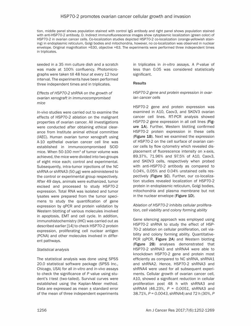
Figure 1. HSP70-2 gene and protein expression in ovarian cancer cell lines. A. RT-PCR analysis depicts HSP70-2 mRNA expression in A10, Caov3 and SKOV3 cells. B. Western blotting demonstrates the presence of 70 KDa HSP70-2 protein in ovarian cancer cells. β-actin was used as a loading control. C. Flow cytometric analyses show surface expression of HSP70-2 protein showing displacement in fluorescence intensity on x-axis, in ovarian cancer cells. Left panel shows unstained popula-
HSP70-2 promotes ovarian cancer cellular growth and invasion
1256 Am J Cancer Res 2017;7(6):1252-1269
seeded in a 35 mm culture dish and a scratch was made at 100% confluency. Photomicro- graphs were taken till 48 hour at every 12 hour interval. The experiments have been performed three independent times and in triplicates.
Effects of HSP70-2 shRNA on the growth of ovarian xenograft in immunocompromised mice
In-vivo studies were carried out to examine the effects of HSP70-2 ablation on the malignant properties of ovarian cancer. All investigations were conducted after obtaining ethical clear-ance from Institute animal ethical committee (IAEC). Human ovarian tumor xenograft using A-10 epithelial ovarian cancer cell line was established in immunocompromised SCID mice. When 50-100 mm3 of tumor volume was achieved, the mice were divided into two groups of eight mice each; control and experimental. Subsequently, intra-tumor injections of the NC shRNA or shRNA3 (50 µg) were administered to the control or experimental group respectively. After 49 days, animals were euthanized, tumor excised and processed to study HSP70-2 expression. Total RNA was isolated and tumor lysates were prepared from the tumor speci-mens to study the quantification of gene expression by qPCR and protein validation by Western blotting of various molecules involved in apoptosis, EMT and cell cycle. In addition, immunohistochemistry (IHC) was carried out as described earlier [14] to check HSP70-2 protein expression, proliferating cell nuclear antigen (PCNA) and other molecules involved in differ-ent pathways.
Statistical analysis
The statistical analysis was done using SPSS 20.0 statistical software package (SPSS Inc., Chicago, USA) for all in-vitro and in-vivo assays to check the significance of P value using stu-dent’s t-test (two-tailed). Survival curves were established using the Kaplan-Meier method. Data are expressed as mean ± standard error of the mean of three independent experiments
in triplicates in in-vitro assays. A P-value of less than 0.05 was considered statistically significant.
Results
HSP70-2 gene and protein expression in ovar-ian cancer cells
HSP70-2 gene and protein expression was examined in A10, Caov3, and SKOV3 ovarian cancer cell lines. RT-PCR analysis showed HSP70-2 gene expression in all cell lines (Fig- ure 1A). Further, Western blotting confirmed HSP70-2 protein expression in these cells (Figure 1B). Next we examined the expression of HSP70-2 on the cell surface of ovarian can-cer cells by flow cytometry which revealed dis-placement of fluorescence intensity on x-axis, 89.37%, 71.96% and 97.5% of A10, Caov3, and SKOV3 cells, respectively when probed with anti-HSP70-2 antibody as compared to 0.04%, 0.05% and 0.04% unstained cells res- pectively (Figure 1C). Further, our co-localiza-tion studies revealed localization of HSP70-2 protein in endoplasmic reticulum, Golgi bodies, mitochondria and plasma membrane but not in the nuclear envelope (Figure 1D).
Ablation of HSP70-2 inhibits cellular prolifera-tion, cell viability and colony forming ability
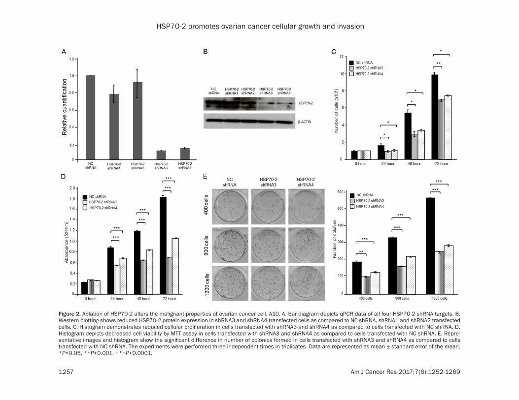
Gene silencing approach was employed using HSP70-2 shRNA to study the effect of HSP- 70-2 ablation on cellular proliferation, cell via-bility and colony forming ability. Quantitative-PCR (qPCR, Figure 2A) and Western blotting (Figure 2B) analyses demonstrated that HSP70-2 shRNA3 and shRNA4 were able to knockdown HSP70-2 gene and protein most efficiently as compared to NC shRNA, shRNA1 and shRNA2. Hence, HSP70-2 shRNA3 and shRNA4 were used for all subsequent experi-ments. Cellular growth of ovarian cancer cell, A10, showed a significant reduction in cellular proliferation post 48 h with shRNA3 and shRNA4 (46.23%, P = 0.0051, shRNA3 and 38.71%, P = 0.0043, shRNA4) and 72 h (30%, P
tion, middle panel shows population stained with control IgG antibody and right panel shows population stained with anti-HSP70-2 antibody. D. Indirect immunofluorescence images show cytoplasmic localization (green color) of HSP70-2 in ovarian cancer cells. Co-localization studies depicted HSP70-2 co-localization (orange-yellowish stain-ing) in endoplasmic reticulum, Golgi bodies and mitochondria, however, no co-localization was observed in nuclear envelope. Original magnification ×630, objective ×63. The experiments were performed three independent times in triplicates.
HSP70-2 promotes ovarian cancer cellular growth and invasion
1257 Am J Cancer Res 2017;7(6):1252-1269
Figure 2. Ablation of HSP70-2 alters the malignant properties of ovarian cancer cell, A10. A. Bar diagram depicts qPCR data of all four HSP70-2 shRNA targets. B. Western blotting shows reduced HSP70-2 protein expression in shRNA3 and shRNA4 transfected cells as compared to NC shRNA, shRNA1 and shRNA2 transfected cells. C. Histogram demonstrates reduced cellular proliferation in cells transfected with shRNA3 and shRNA4 as compared to cells transfected with NC shRNA. D. Histogram depicts decreased cell viability by MTT assay in cells transfected with shRNA3 and shRNA4 as compared to cells transfected with NC shRNA. E. Repre-sentative images and histogram show the significant difference in number of colonies formed in cells transfected with shRNA3 and shRNA4 as compared to cells transfected with NC shRNA. The experiments were performed three independent times in triplicates. Data are represented as mean ± standard error of the mean. *P<0.05, **P<0.001, ***P<0.0001.
HSP70-2 promotes ovarian cancer cellular growth and invasion
1258 Am J Cancer Res 2017;7(6):1252-1269
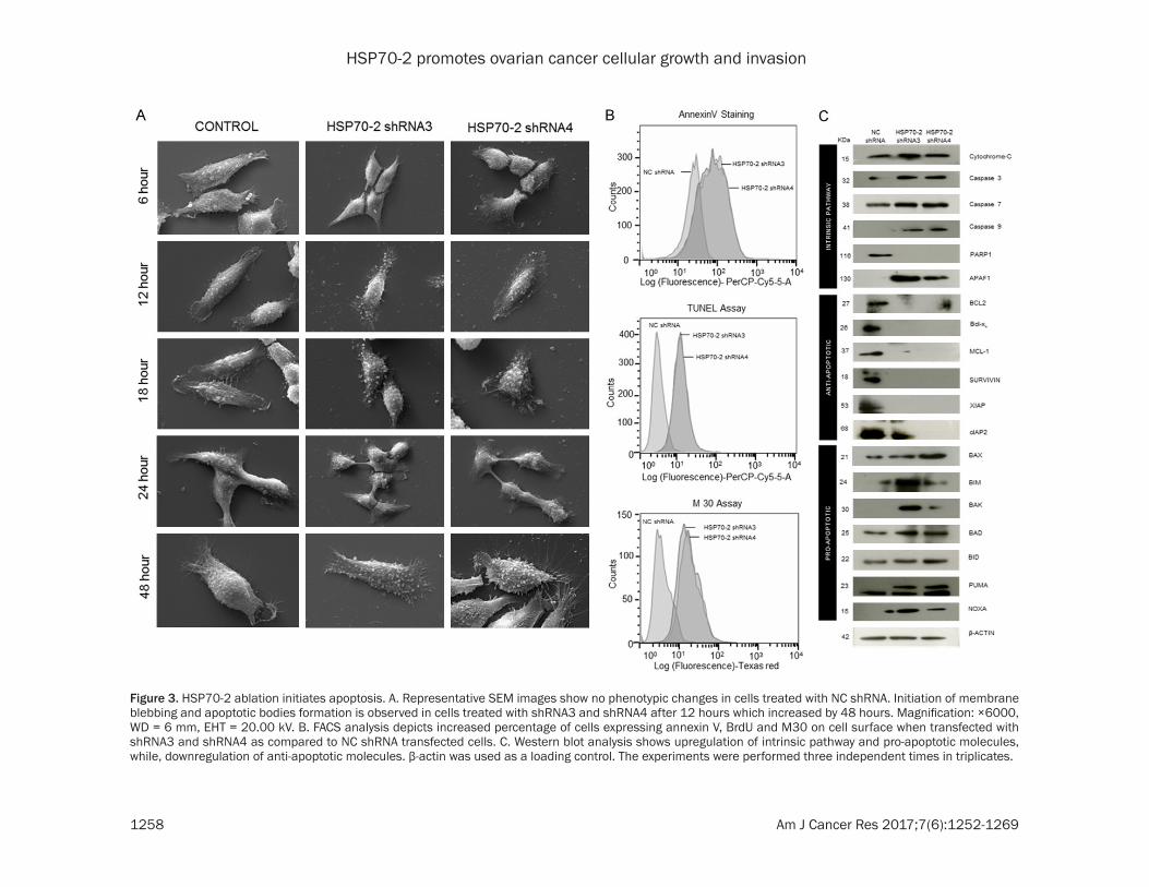
Figure 3. HSP70-2 ablation initiates apoptosis. A. Representative SEM images show no phenotypic changes in cells treated with NC shRNA. Initiation of membrane blebbing and apoptotic bodies formation is observed in cells treated with shRNA3 and shRNA4 after 12 hours which increased by 48 hours. Magnification: ×6000, WD = 6 mm, EHT = 20.00 kV. B. FACS analysis depicts increased percentage of cells expressing annexin V, BrdU and M30 on cell surface when transfected with shRNA3 and shRNA4 as compared to NC shRNA transfected cells. C. Western blot analysis shows upregulation of intrinsic pathway and pro-apoptotic molecules, while, downregulation of anti-apoptotic molecules. β-actin was used as a loading control. The experiments were performed three independent times in triplicates.
HSP70-2 promotes ovarian cancer cellular growth and invasion
1259 Am J Cancer Res 2017;7(6):1252-1269
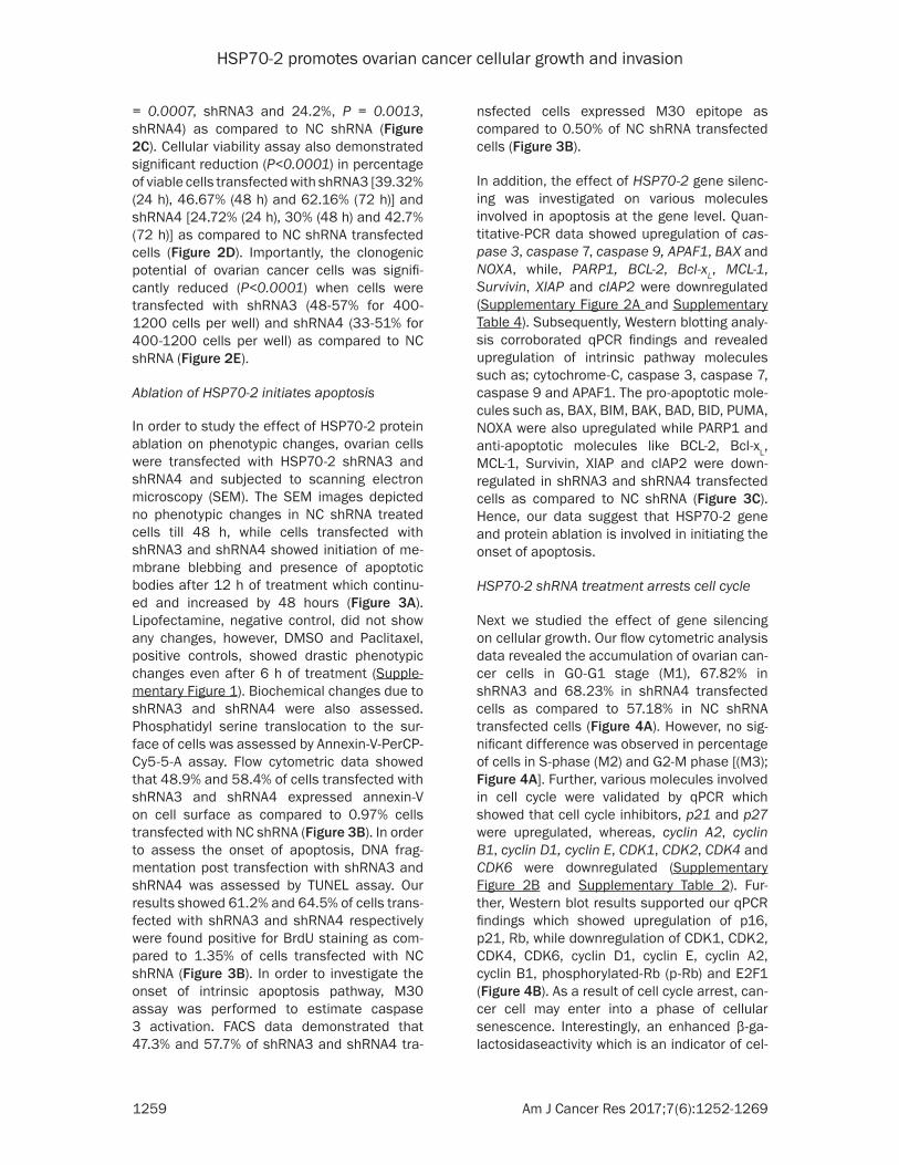
= 0.0007, shRNA3 and 24.2%, P = 0.0013, shRNA4) as compared to NC shRNA (Figure 2C). Cellular viability assay also demonstrated significant reduction (P<0.0001) in percentage of viable cells transfected with shRNA3 [39.32% (24 h), 46.67% (48 h) and 62.16% (72 h)] and shRNA4 [24.72% (24 h), 30% (48 h) and 42.7% (72 h)] as compared to NC shRNA transfected cells (Figure 2D). Importantly, the clonogenic potential of ovarian cancer cells was signifi-cantly reduced (P<0.0001) when cells were transfected with shRNA3 (48-57% for 400-1200 cells per well) and shRNA4 (33-51% for 400-1200 cells per well) as compared to NC shRNA (Figure 2E).
Ablation of HSP70-2 initiates apoptosis
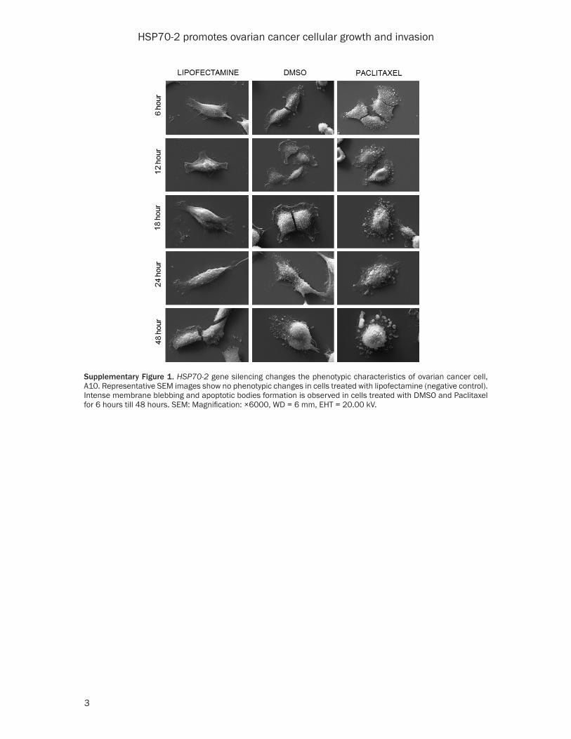
In order to study the effect of HSP70-2 protein ablation on phenotypic changes, ovarian cells were transfected with HSP70-2 shRNA3 and shRNA4 and subjected to scanning electron microscopy (SEM). The SEM images depicted no phenotypic changes in NC shRNA treated cells till 48 h, while cells transfected with shRNA3 and shRNA4 showed initiation of me- mbrane blebbing and presence of apoptotic bodies after 12 h of treatment which continu- ed and increased by 48 hours (Figure 3A). Lipofectamine, negative control, did not show any changes, however, DMSO and Paclitaxel, positive controls, showed drastic phenotypic changes even after 6 h of treatment (Supple- mentary Figure 1). Biochemical changes due to shRNA3 and shRNA4 were also assessed. Phosphatidyl serine translocation to the sur-face of cells was assessed by Annexin-V-PerCP-Cy5-5-A assay. Flow cytometric data showed that 48.9% and 58.4% of cells transfected with shRNA3 and shRNA4 expressed annexin-V on cell surface as compared to 0.97% cells transfected with NC shRNA (Figure 3B). In order to assess the onset of apoptosis, DNA frag-mentation post transfection with shRNA3 and shRNA4 was assessed by TUNEL assay. Our results showed 61.2% and 64.5% of cells trans-fected with shRNA3 and shRNA4 respectively were found positive for BrdU staining as com-pared to 1.35% of cells transfected with NC shRNA (Figure 3B). In order to investigate the onset of intrinsic apoptosis pathway, M30 assay was performed to estimate caspase 3 activation. FACS data demonstrated that 47.3% and 57.7% of shRNA3 and shRNA4 tra-
nsfected cells expressed M30 epitope as compared to 0.50% of NC shRNA transfected cells (Figure 3B).
In addition, the effect of HSP70-2 gene silenc-ing was investigated on various molecules involved in apoptosis at the gene level. Quan- titative-PCR data showed upregulation of cas-pase 3, caspase 7, caspase 9, APAF1, BAX and NOXA, while, PARP1, BCL-2, Bcl-xL, MCL-1, Survivin, XIAP and cIAP2 were downregulated (Supplementary Figure 2A and Supplementary Table 4). Subsequently, Western blotting analy-sis corroborated qPCR findings and revealed upregulation of intrinsic pathway molecules such as; cytochrome-C, caspase 3, caspase 7, caspase 9 and APAF1. The pro-apoptotic mole-cules such as, BAX, BIM, BAK, BAD, BID, PUMA, NOXA were also upregulated while PARP1 and anti-apoptotic molecules like BCL-2, Bcl-xL, MCL-1, Survivin, XIAP and cIAP2 were down- regulated in shRNA3 and shRNA4 transfected cells as compared to NC shRNA (Figure 3C). Hence, our data suggest that HSP70-2 gene and protein ablation is involved in initiating the onset of apoptosis.
HSP70-2 shRNA treatment arrests cell cycle
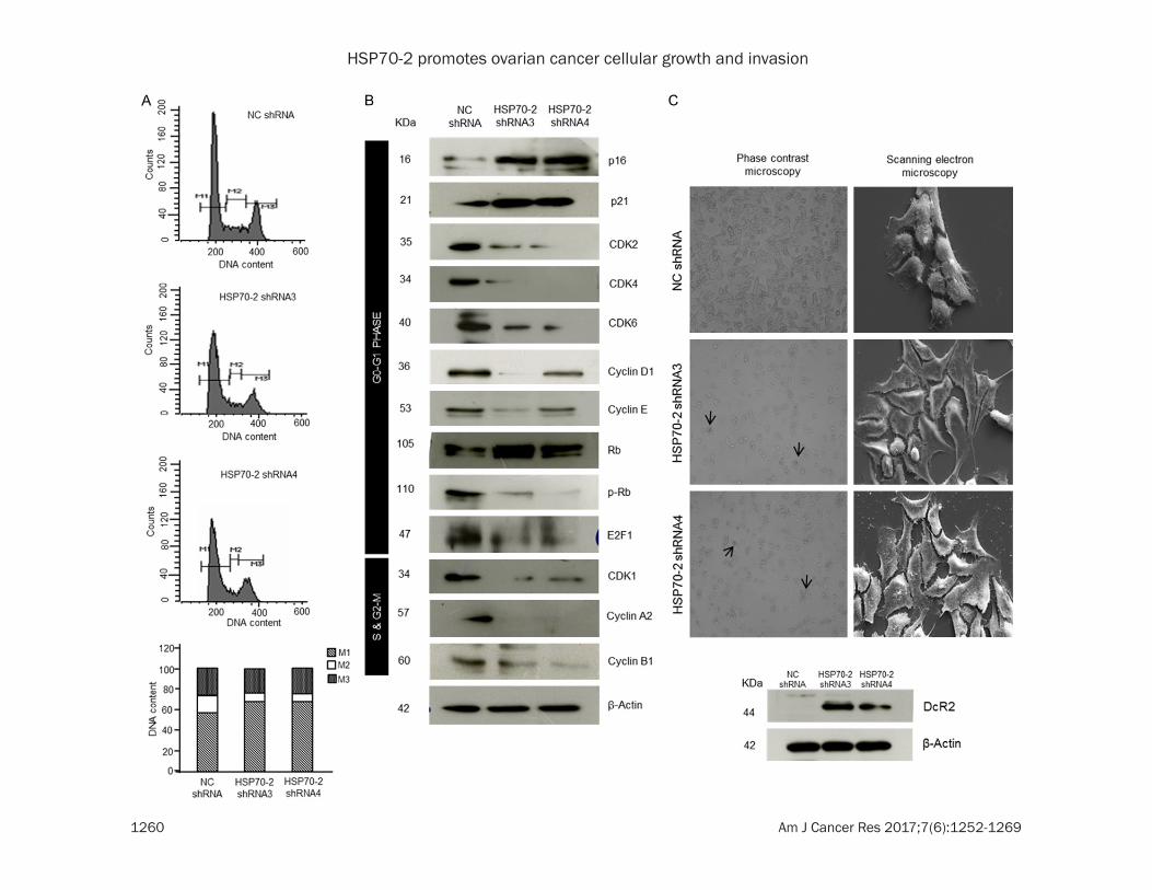
Next we studied the effect of gene silencing on cellular growth. Our flow cytometric analysis data revealed the accumulation of ovarian can-cer cells in G0-G1 stage (M1), 67.82% in shRNA3 and 68.23% in shRNA4 transfected cells as compared to 57.18% in NC shRNA transfected cells (Figure 4A). However, no sig-nificant difference was observed in percentage of cells in S-phase (M2) and G2-M phase [(M3); Figure 4A]. Further, various molecules involved in cell cycle were validated by qPCR which showed that cell cycle inhibitors, p21 and p27 were upregulated, whereas, cyclin A2, cyclin B1, cyclin D1, cyclin E, CDK1, CDK2, CDK4 and CDK6 were downregulated (Supplementary Figure 2B and Supplementary Table 2). Fur- ther, Western blot results supported our qPCR findings which showed upregulation of p16, p21, Rb, while downregulation of CDK1, CDK2, CDK4, CDK6, cyclin D1, cyclin E, cyclin A2, cyclin B1, phosphorylated-Rb (p-Rb) and E2F1 (Figure 4B). As a result of cell cycle arrest, can-cer cell may enter into a phase of cellular senescence. Interestingly, an enhanced β-ga- lactosidaseactivity which is an indicator of cel-
HSP70-2 promotes ovarian cancer cellular growth and invasion
1260 Am J Cancer Res 2017;7(6):1252-1269
HSP70-2 promotes ovarian cancer cellular growth and invasion
1261 Am J Cancer Res 2017;7(6):1252-1269
lular senescence was observed in the cells transfected with shRNA3 and shRNA4 as com-pared to NC shRNA transfected cells (Figure 4C). In addition, SEM images were captured which further validated our data revealing flat-tening of cells in shRNA3 and shRNA4 trans-fected cells as compared to NC shRNA trans-fected cells (Figure 4C) suggesting cellular senescence state. Further, Western blot data also supported the phenotypic characteristics of cellular senescence wherein Decoy receptor 2 (DcR2), a marker for senescence onset was found to be upregulated in shRNA3 and shRNA4 transfected cells as compared to NC shRNA transfected cells (Figure 4C). Collectively, our data suggest that HSP70-2 shRNA treatment arrests the cell growth in G0-G1 stage and induces cellular senescence.
Knockdown of HSP70-2 inhibits cellular motil-ity
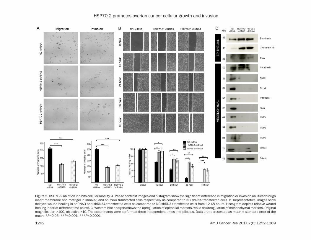
Metastasis is a multistep process and was assessed by studying migration, invasion and cellular motility of ovarian cancer cells. Our cell migration assay revealed a significant reduc-tion in cells migrating through insert membrane in shRNA3 (45.5%, P<0.0001) and shRNA4 (37.64%, P<0.001) transfected cells as com-pared to NC shRNA transfected cells (Figure 5A). Similarly, our invasion assay showed re- duced number of shRNA3 (55.21%, P<0.0001) and shRNA4 (48.56%, P<0.0001) transfected cells invading through matrigel as compared to NC shRNA transfected cells (Figure 5A). Cellular motility was also assessed by wound healing assay which demonstrated the inhibition of cel-lular motility. HSP70-2 shRNA3 and shRNA4 transfected cells showed delayed wound heal-ing ability as compared to NC shRNA transf- ected cells (Figure 5B). Relative wound healing index was found to be significantly higher for shRNA3 and shRNA4 transfected cells (P<
0.001) as compared to NC shRNA transfected cells after 24 hours of transfection (Figure 5B).
Next we studied the status of various mole-cules associated with migration, invasion and cellular motility. Various genes under investiga-tion showed (Supplementary Figure 2C and Supplementary Table 2) the relative quantifica-tion of various mesenchymal marker genes such as; N-cadherin, SNAIL, SLUG, VIMENTIN, SMA, MMP2, MMP3, MMP9 and TWIST in cells transfected with shRNA3 and shRNA4. In order to validate gene expression data at the pro- tein level, Western blotting was carried out. Epithelial markers, such as E-cadherin, cyto-keratin 18 and EMA were upregulated while, mesenchymal markers, N-cadherin, SNAIL, SLUG, VIMENTIN, SMA, MMP2, MMP3, MMP9 and TWIST were downregulated (Figure 5C). Our study clearly indicates that HSP70-2 abla-tion inhibits migratory, invasive and cellular motility ability of ovarian cancer cells by alter-ing the key molecules involved in various meta-static processes.
HSP70-2 knockdown retards growth of the human ovarian xenografts in in-vivo mouse model
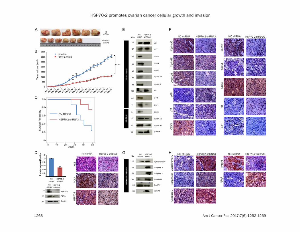
For in-vivo studies, the ovarian human xeno-graft was established in immunocompromised SCID mice treated with HSP70-2 shRNA3. There was a significant reduction (P = 0.0015) in tumor size and tumor volume of shRNA3 treated mice as compared to those treated with NC shRNA (Figure 6A, 6B). Moreover, signifi-cant survival (P = 0.037) was observed in mice treated with HSP70-2 shRNA3 as compared to NC shRNA (Figure 6C). Further, qPCR and Western blot analysis showed a significant reduction (P = 0.003) in HSP70-2 mRNA (50%) (Figure 6D) and protein expression (Figure 6D) in tumor specimens treated with shRNA3 as compared to NC shRNA. In addition, immuno-
Figure 4. HSP70-2 shRNA treatment arrests cell growth. A. Flow cytometric analysis shows cell cycle analysis of NC shRNA, shRNA3 and shRNA4 transfected cells stained with PI (M1: G0-G1, M2: S and M3: G2-M phase). Bar dia-gram depicts cumulative percentage accumulation of cells in M1 (57.18%: NC shRNA, 67.82%: shRNA3, 68.23%: shRNA4), M2 (16.81%: NC shRNA, 8.49%: shRNA3, 6.75%: shRNA4) and M3 (25.65%: NC shRNA, 23.33%: shRNA3, 25.33%: shRNA4). B. Western blotting depicts the upregulation of p16, p21, Rb, while downregulation of cyclins, cyclin dependent kinases, p-Rb and E2F1. β-actin was used as a loading control. C. Representative phase contrast images show enhanced β-galactosidase activity (arrow) in shRNA3 and shRNA4 transfected cells. Representative SEM images show initiation of senescence in shRNA3 and shRNA4 transfected cells revealing flattened phenotypic characteristics. Western blotting shows increased DcR2 expression in shRNA3 and shRNA4 transfected cells. Phase contrast microscopy: Original magnification ×100, objective ×10. SEM: Magnification: ×6000, WD = 6 mm, EHT = 20.00 kV. The experiments were performed three independent times in triplicates.
HSP70-2 promotes ovarian cancer cellular growth and invasion
1262 Am J Cancer Res 2017;7(6):1252-1269
Figure 5. HSP70-2 ablation inhibits cellular motility. A. Phase contrast images and histogram show the significant difference in migration or invasion abilities through insert membrane and matrigel in shRNA3 and shRNA4 transfected cells respectively as compared to NC shRNA transfected cells. B. Representative images show delayed wound healing in shRNA3 and shRNA4 transfected cells as compared to NC shRNA transfected cells from 12-48 hours. Histogram depicts relative wound healing index at different time points. C. Western blot analysis shows the upregulation of epithelial markers, while downregulation of mesenchymal markers. Original magnification ×100, objective ×10. The experiments were performed three independent times in triplicates. Data are represented as mean ± standard error of the mean. *P<0.05, **P<0.001, ***P<0.0001.
HSP70-2 promotes ovarian cancer cellular growth and invasion
1263 Am J Cancer Res 2017;7(6):1252-1269
HSP70-2 promotes ovarian cancer cellular growth and invasion
1264 Am J Cancer Res 2017;7(6):1252-1269
histochemical (IHC) analysis of xenograft tu- mor specimens validated these results and showed reduced expression of HSP70-2 and PCNA (proliferating cell nuclear antigen) in the shRNA3 treated tumors as compared to NC shRNA treated tumors (Figure 6D).
Next, we validated our in-vitro findings of cell cycle regulators in the xenograft model system. As expected there was a marked reduction in the gene expression of cyclin A2, CDK1, CDK2, CDK4, CDK6 and E2F1, whereas, increased ex- pression of p21, p27 and Rb was observed in shRNA3 treated mice as compared to NC shRNA treated mice (Supplementary Figure 3A and Supplementary Table 3). Further, Western blot analysis confirmed these observations and showed upregulation of cell cycle inhibi-tors, p21 and p27 and tumor suppressor, Rb. As expected, cyclin A2, cyclin B1, cyclin D1, cyclin E and cyclin dependent kinases like CDK1, CDK2, CDK4 and CDK6 and p-Rb and E2F1 were downregulated (Figure 6E). The par-affin embedded tumor serial sections were subjected to IHC to validate the gene and pro-tein expression data of tumor lysates. IHC images showed reduced immuno-reactivity of cyclin A2, cyclin B1, cyclin D1, CDK1, CDK2, CDK4, CDK6 and E2F1, while, increased immu-no-reactivity of p16, p27 and Rb in shRNA3 treated tumor as compared to NC shRNA treat-ed tumor (Figure 6F).
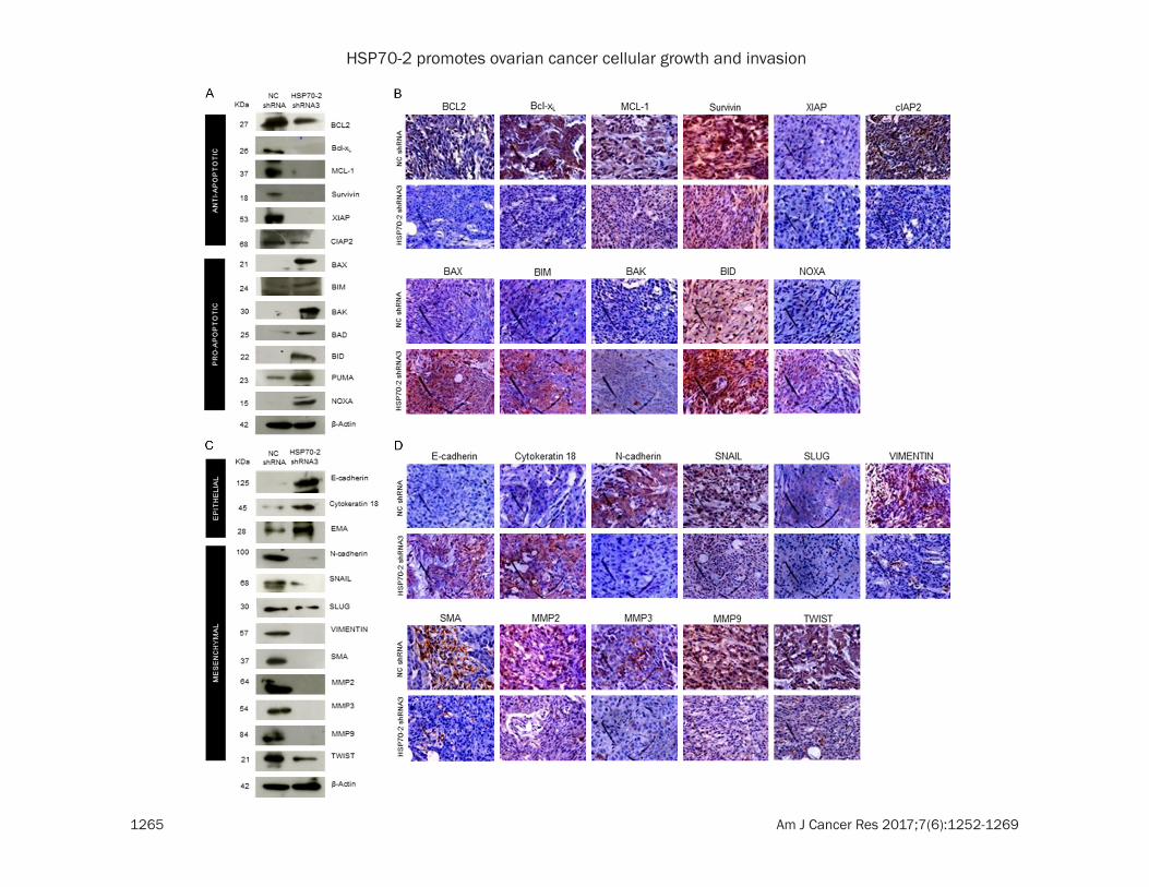
The effect of HSP70-2 shRNA3 treatment was also studied on various molecules of apoptotic pathway in these xenograft tumor specimens. Quantitative-PCR analysis of tumor specimens depicted upregulation of cytochrome-C, cas-
pase 3, caspase 7, caspase 9, APAF1, BAX and NOXA, while, downregulation of BCL-2, Bcl-xL, MCL-1 and XIAP in shRNA3 treated tumor as compared to NC shRNA treated tumor (Supplementary Figure 3B and Supplementary Table 3). Western blot analysis on tumor speci-men lysates confirmed the upregulation of intrinsic pathway molecules, such as cyto-chrome-C, caspase 3, caspase 7, caspase 9, APAF1 and pro-apoptotic molecules like BAX, BIM, BAK, BAD, BID, PUMA and NOXA while, PARP1 and anti-apoptotic molecules like BCL-2, Bcl-xL, MCL-1, Survivin, XIAP and cIAP2 were downregulated (Figures 6G, 7A) in shRNA3 treated animals. Our IHC studies on tumor seri-al sections validated the qPCR and protein expression results of tumor lysate. These results showed increased immuno-reactivity of cytochrome-C, caspase 3, caspase 7, APAF1 and pro-apoptotic molecules like BAX, BIM, BAK, BID and NOXA were upregulated while, PARP1 and anti-apoptotic molecules like BCL-2, Bcl-xL, MCL-1, Survivin, XIAP and cIAP2 showed reduced immuno-reactivity (Figures 6H, 7B).
Various key molecules involved in the EMT were further analyzed. Our qPCR data revealed the upregulation of epithelial markers, E-cadherin, cytokeratin 18 and EMA in the shRNA3 treated tumors as compared to NC shRNA treated tumors. Further there was downregulation of mesenchymal markers, N-cadherin, SNAIL, SLUG, VIMENTIN, SMA, MMP2, MMP3, MMP9 and TWIST in the shRNA3 treated tumors as compared to NC shRNA treated tumors (Supplementary Figure 3C and Supplementary
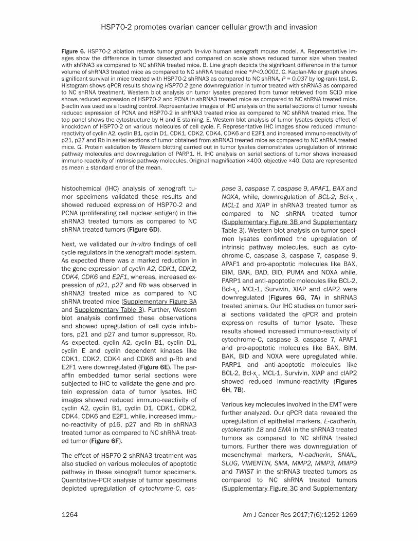
Figure 6. HSP70-2 ablation retards tumor growth in-vivo human xenograft mouse model. A. Representative im-ages show the difference in tumor dissected and compared on scale shows reduced tumor size when treated with shRNA3 as compared to NC shRNA treated mice. B. Line graph depicts the significant difference in the tumor volume of shRNA3 treated mice as compared to NC shRNA treated mice *P<0.0001. C. Kaplan-Meier graph shows significant survival in mice treated with HSP70-2 shRNA3 as compared to NC shRNA, P = 0.037 by log-rank test. D. Histogram shows qPCR results showing HSP70-2 gene downregulation in tumor treated with shRNA3 as compared to NC shRNA treatment. Western blot analysis on tumor lysates prepared from tumor retrieved from SCID mice shows reduced expression of HSP70-2 and PCNA in shRNA3 treated mice as compared to NC shRNA treated mice. β-actin was used as a loading control. Representative images of IHC analysis on the serial sections of tumor reveals reduced expression of PCNA and HSP70-2 in shRNA3 treated mice as compared to NC shRNA treated mice. The top panel shows the cytostructure by H and E staining. E. Western blot analysis of tumor lysates depicts effect of knockdown of HSP70-2 on various molecules of cell cycle. F. Representative IHC images show reduced immuno-reactivity of cyclin A2, cyclin B1, cyclin D1, CDK1, CDK2, CDK4, CDK6 and E2F1 and increased immuno-reactivity of p21, p27 and Rb in serial sections of tumor obtained from shRNA3 treated mice as compared to NC shRNA treated mice. G. Protein validation by Western blotting carried out in tumor lysates demonstrates upregulation of intrinsic pathway molecules and downregulation of PARP1. H. IHC analysis on serial sections of tumor shows increased immuno-reactivity of intrinsic pathway molecules. Original magnification ×400, objective ×40. Data are represented as mean ± standard error of the mean.
HSP70-2 promotes ovarian cancer cellular growth and invasion
1265 Am J Cancer Res 2017;7(6):1252-1269
HSP70-2 promotes ovarian cancer cellular growth and invasion
1266 Am J Cancer Res 2017;7(6):1252-1269
Table 3). Further, the Western blot analysis of tumor xenograft lysate treated with shRNA3 confirmed the qPCR findings and revealed that E-cadherin, cytokeratin 18 and EMA were up- regulated whereas downregulation of N-cad- herin, SNAIL, SLUG, VIMENTIN, SMA, MMP2, MMP3, MMP9 and TWIST was observed (Fig- ure 7C). Validation for qPCR and Western blot-ting results were further confirmed by IHC whi- ch showed that immuno-reactivity of E-cadh- erin and cytokeratin 18 was increased, while decreased immuno-reactivity of N-cadherin, SNAIL, SLUG, VIMENTIN, SMA, MMP2, MMP3, MMP9 and TWIST was observed in shRNA3 treated tumor as compared to NC shRNA treat-ed tumor (Figure 7D).
Discussion
Ovarian cancer is one of the most common can-cer amongst gynecological cancers [1]. Especially serous ovarian carcinoma is the most prevalent subtype of epithelial ovarian cancer (EOC) and accounts for about 80-90% of all ovarian cancers [3]. Early-stage ovarian cancers are normally asymptomatic and hence are diagnosed at an advanced disease stage, when treatment modalities are limited [15]. Till date few cancer testis (CT) antigens expres-sion have been shown to be associated with ovarian cancer [16-19] however, none of these CT antigens are in clinical practice yet. In this study, we investigated the role of HSP70-2 in EOC and found its expression in all the EOC cell lines. Small interfering RNA-mediated (shRNA) knockdown of HSP70-2 expression in EOC cells resulted in reduced cellular proliferation, cell viability, colony forming ability, cellular motility, invasion ability and retarded tumor growth in in-vivo ovarian cancer xenograft mouse model. Our recent studies also demonstrated that ectopic expression HSP70-2 does promote cel-lular proliferation, migration and invasion in in vitro and tumor growth in in vivo in bladder can-cer [7], cervix cancer [8], breast cancer [9] and
colorectal cancer [10]. Recently, it has been reported [20] that employing shRNA approach to silence genes involved in disease progres-sion may be a novel approach for development of a new class of therapeutics.
In an attempt for developing a novel therapeu-tic target for ovarian cancer, our study demon-strated that HSP70-2 depleted cells showed downregulation of anti-apoptotic proteins and increased expression of pro-apoptotic genes. This in turn further led to upregulation of cyto-chrome-C, caspase 3, caspase 7 and caspase 9. Also, Apoptotic protease activating factor 1 (APAF1) was found to be upregulated resulting in reduced cellular growth and hence the cell death. Similarly, recent studies also showed that anti-apoptotic gene BCL-2 along with other members, Bcl-xL and MCL-1 suppress the activ-ity of pro-apoptotic proteins BAX and BAK [21]. In the absence of anti-apoptotic genes, BAX and BAK disrupt the integrity of the outer mito-chondrial membrane, causing the release of pro-apoptotic signaling protein, cytochrome-C, which in turn activates the cascade of caspas-es leading to multiple cellular changes associ-ated with apoptosis [21]. Further in support of our data, yet another study has shown that gene silencing of c-myc (proto-oncogene) led to upregulation of apoptotic-related molecules caspase 3, caspase 9 and PARP1 in ovarian cancer cells [22]. Our investigation distinctly show that HSP70-2 promotes cellular growth and thus, depletion of HSP70-2 seems to pro-mote apoptosis in ovarian cancer cells.
Cell cycle dysregulation is a common molecular finding with upregulation of cyclin D1 or E1, E2F1 or cyclin dependent kinases (CDK2), and downregulation of CDK inhibitors (p16, p21 and p27) have been observed in ovarian cancer [23]. Interestingly, our findings revealed that HSP70-2 depleted cells had increased expres-sion of cell cycle inhibitors (p16, p21) which in turn downregulated the expression of cyclin D,
Figure 7. HSP70-2 shRNA treatment initiates the onset of apoptosis and inhibits EMT in tumor cells. A. Western blot data show upregulation of pro-apoptotic molecules and downregulation of anti-apoptotic molecules. B. Represen-tative images of IHC of serial sections exhibit increased immuno-reactivity of pro-apoptotic molecules in shRNA3 treated mice as compared to NC shRNA treated mice. However, decreased immuno-reactivity is observed in case of anti-apoptotic molecules. C. Western blotting in tumor lysates shows upregulation of epithelial markers and downregulation of mesenchymal markers in tumor cells treated with shRNA3 as compared to NC shRNA. D. Rep-resentative micrographs of IHC analysis on serial tumor sections shows increased immuno-reactivity of epithelial markers and reduced immuno-reactivity of mesenchymal markers in shRNA3 treated mice as compared to NC shRNA treated mice. Original magnification ×400, objective ×40.
HSP70-2 promotes ovarian cancer cellular growth and invasion
1267 Am J Cancer Res 2017;7(6):1252-1269
cyclin E thus resulting in cell growth arrest (Figure 4). As a result, downregulation of CDK4/6-cyclin D or CDK2-cyclin E, decreased phosphorylation of Rb was found which further lead to increased expression of E2F1 and hence, arrest of cancer cells at G0-G1 phase of cell cycle. These findings were similar to earlier studies carried out on salt-inducible kinase 3 [24] and COX-1 [25] in ovarian cancer cells. Hence, ablation of HSP70-2 arrests the cell cycle and inhibited cellular growth of ovarian cancer cells.
EOC cells display an aggressive characteristic feature which has the ability to migrate and invade into the peritoneal cavity and metasta-size to local organs [26]. Our study revealed that HSP70-2 depleted cells had reduced migratory and invasive ability indicating that HSP70-2 plays an important role in cellular motility. E-cadherin helps to assemble epitheli-al cells and maintain structural integrity and loss of E-cadherin is considered to be the main alteration in the cancer cells. Moreover, reduc-tion of E-cadherin expression has been shown to be associated with invasion and metasta- sis [23]. Contrary to this, adhesion molecule, N-cadherin is often upregulated and is ass- ociated with the cell migration and invasion in cancer cells [21]. Transcriptional factors, including SNAIL, SLUG and TWIST involved in EMT have been found to be expressed in vari-ous malignancies [23]. These studies suggest-ed that expression of transcription factors lead to loss of adherent junctions, associated con-version from epithelial to spindle morphology, expression of matrix degrading enzymes (MMP’s) and increased motility [21]. Inter- estingly, our data also showed similar finding at molecular level, wherein, HSP70-2 ablated cells had increased expression of epithelial markers including, E-cadherin, while downregu-lation of mesenchymal markers, N-cadherin, VIMENTIN, SMA, MMP2, MMP3 and MMP9. Transcription factors such as SNAIL, SLUG and TWIST which promotes EMT were also down-regulated (Figure 5). Other studies have also shown in ovarian cancer cells that COX-1 depleted cells show reduced expression of genes promoting cell invasion or migration [23]. We are the first to report that CT antigen, HSP70-2 depleted ovarian cancer cells have reduced cellular motility as a result of low expression of EMT molecules.
Encouraged by our observations in cell culture, we confirmed these findings in in-vivo mouse model. Animals treated with HSP70-2 shRNA exhibited retarded tumor growth which was consistent with our previous findings in bladder and cervix cancer xenograft mouse model [7, 8]. Our data further showed that HSP70-2 shRNA treatment altered various genes of cell cycle in G0-G1 stage which resulted in cell cycle arrest and caused senescence. Other studies on COX-2 selective inhibitor [27] and metfor- min (antidiabetic drug [28]) supported our data and showed that these inhibitors led to down-regulation of cyclin D1 as assessed by IHC in in-vivo ovarian cancer system. So far no CT anti-gen has been explored for its involvement at molecular level in various pathways contribut-ing towards tumor growth and cellular motility in ovarian cancer cells. Apparently, this seems to be the first report showing role of HSP70-2 in cellular growth in the ovarian cancer cells. Apoptosis is one such physiological process by which tumor growth can be retarded. We showed the effect of HSP70-2 knockdown on tumor regression in in-vivo mouse model and found that HSP70-2 depleted tumor had in- creased expression of cytochrome-C, caspas-es, pro-apoptotic molecules, while decreased expression of anti-apoptotic molecules at both mRNA as well as protein levels. Some earlier studies have also shown similar trend in tumor regression treated with NCX-4016, a nitro-derivative of aspirin [29] and RY-2f, an isofla-vone analog [30] in human ovarian cancer xenograft model. Thus, HSP70-2 seems to be a key molecule involved in growth and motility of ovarian cancer cells.
HSP70-2 is member of HSP70 family of pro-teins [6]. HSP70 proteins can be thought of as a potent buffering system for cellular stress, either from extrinsic (physiological, viral and environmental) or intrinsic (replicative or onco-genic) stimuli. HSP70-2 protein may have a buffering system in ovarian cancer cells and may utilize this property to stabilize the pro-teins that are required for ovarian cancer cell survival. Therefore, future studies are warrant-ed to target the HSP70-2 as combination thera-py to treat the ovarian cancer patients. Recently it was documented that gene silencing of CDK11 increased the cytotoxic effect of che-motherapeutic agent paclitaxel in ovarian can-cer cells [31]. The antitumor effect of HSP70-2
HSP70-2 promotes ovarian cancer cellular growth and invasion
1268 Am J Cancer Res 2017;7(6):1252-1269
knockdown has paved a way for exploring shRNA based novel cancer treatment. In this context, as monotherapy, recent clinical trials employing siRNAs against VEGF and kinesin spindle protein (KSP) have shown promising results to treat metastatic endometrial and hepatocellular carcinoma [20] as a new thera-peutic treatment modality for cancer.
Collectively, our study shows that HSP70-2 plays an important role in ovarian cancer. Gene silencing studies clearly indicate that HSP70-2 promotes cellular growth and multistep pro-cess including migration and invasion of ovari-an cancer cells. Hence, our study suggests that HSP70-2 may be a putative therapeutic target in combination with other chemotherapeutic agents against ovarian cancer for future treat-ment strategies and warrants future studies.
Acknowledgements
We acknowledge Dr V. Kumar, Senior Staff Scientist, International Centre for Genetic Engineering and Biotechnology, New Delhi, India for critical reading and editing of this manuscript. We also thank technical support by Mrs. Rekha Rani, National Institute of Immunology, New Delhi, India for SEM imaging. This work is supported by grants from Indo-UK Cancer Research Program (Grant No. BT/IN/UK/NII/2006), Centre for Molecular Medicine (Grant No.BT/PR/14549/MED/14/1291), NII-core funding, Department of Biotechnology, Government of India.
Disclosure of conflict of interest
None.
Address correspondence to: Anil Suri, Cancer Re- search Program, Cancer Microarray, Genes and Proteins Laboratory, National Institute of Immuno- logy, ArunaAsaf Ali Marg, New Delhi 110067, In- dia. Tel: 91-11-26703-700; Fax: 91-11-26-1621-25; 91-11-26742125; E-mail: [email protected]
References
[1] Siegel RL, Miller KD, Jemal A. Cancer statistics. CA Cancer J Clin 2015; 65: 5-29.
[2] Rauh-Hain JA, Krivak TC, DelCarmen MG, Olawaiye AB. Ovarian cancer screening and early detection in the general population. Rev Obstet Gynecol 2011; 4: 15-21.
[3] Nolen B, Marrangoni A, Velikokhatnaya L, Prosser D, Winans M, Gorelik E, Lokshin A. A
serum based analysis of ovarian epithelial tu-mourigenesis. Gynecol Oncol 2009; 112: 47-54.
[4] Gjerstorff MF, Andersen MH, Ditzel HJ. Oncogenic cancer/testis antigens: prime can-didates for immunotherapy. Oncotarget 2015; 6: 15772-15787.
[5] Suri A, Saini S, Sinha A, Agarwal S, Verma A, Parashar D, Singh S, Gupta N, Jagadish N. Cancer testis antigens: a new paradigm for cancer therapy. Oncoimmunology 2012; 1: 1194-6.
[6] Rohde M, Daugaard M, Jensen MH, Helin K, Nylandsted J, Jäättelä M. Members of the heat-shock protein 70 family promote cancer cell growth by distinct mechanisms. Genes Dev 2005; 19: 570-582.
[7] Garg M, Kanojia D, Seth A, Kumar R, Gupta A, Surolia A, Suri A. Heat-shock protein 70-2 (HSP70-2) expression in bladder urothelial carcinoma is associated with tumor progres-sion and promotes migration and invasion. Eur J Cancer 2010; 46: 207-215.
[8] Garg M, Kanojia D, Saini S, Suri S, Gupta A, Surolia A, Suri A. Germ cell-specific heat shock protein 70-2 is expressed in cervical carcino-ma and is involved in the growth, migration, and invasion of cervical cells. Cancer 2010; 116: 3785-3796.
[9] Jagadish N, Agarwal S, Gupta N, Fatima R, Devi S, Kumar V, Suri V, Kumar R, Suri V, Sadasukhi TC, Gupta A, Ansari AS, Lohiya NK, Suri A. Heat shock protein 70-2 (HSP70-2) overexpression in breast cancer. J Exp Clin Cancer Res 2016; 35: 150.
[10] Jagadish N, Parashar D, Gupta N, Agarwal S, Suri V, Kumar R, Suri V, Sadasukhi TC, Gupta A, Ansari AS, Lohiya NK, Suri A. Heat shock protein 70-2 (HSP70-2) is a novel therapeutic target for colorectal cancer and is associated with tumor growth. BMC Cancer 2016; 16: 561.
[11] Singh S, Suri A. Targeting the testis-specific heat-shock protein 70-2 (HSP70-2) reduces cellular growth, migration, and invasion in re-nal cell carcinoma cells. Tumor Biol 2014; 35: 12695-12706.
[12] Zhu D, Dix DJ, Eddy EM. HSP70-2 is requ- ired for CDC2 kinase activity in meiosis I of mouse spermatocytes. Development 1997; 124: 3007-3014.
[13] Eddy EM. Role of heat shock protein HSP70-2 in spermatogenesis. Rev Reprod 1999; 4: 23-30.
[14] Jagadish N, Parashar D, Gupta N, Agarwal S, Purohit S, Kumar V, Sharma A, Fatima R, Topno AP, Shaha C, Suri A. A-kinase anchor protein 4 (AKAP4) a promising therapeutic target of
HSP70-2 promotes ovarian cancer cellular growth and invasion
1269 Am J Cancer Res 2017;7(6):1252-1269
colorectal cancer. J Exp Clin Cancer Res 2015; 34: 142.
[15] Romero I, Bast RC Jr. Minireview: Human Ovarian Cancer: Biology, Current Management, and Paths to Personalizing Therapy. Endo- crinology 2012; 153: 1593-602.
[16] Agarwal S, Saini S, Parashar D, Verma A, Sinha A, Jagadish N, Batra A, Suri S, Gupta A, Ansari AS, Lohiya NK, Suri A. The novel can-cer-testis antigen A-kinase anchor protein 4 (AKAP4) is a potential target for immunother-apy of ovarian serous carcinoma. Oncoim- munology 2013; 2: e24270.
[17] Odunsi K, Jungbluth AA, Stockert E, Qian F, Gnjatic S, Tammela J, Intengan M, Beck A, Keitz B, Santiago D, Williamson B, Scanlan MJ, Ritter G, Chen YT, Driscoll D, Sood A, Lele S, Old LJ. NY-ESO-1 and LAGE-1 cancer-testis antigens are potential targets for immunother-apy in epithelial ovarian cancer. Cancer Res 2003; 63: 6076-6083.
[18] Tammela J, Uenaka A, Ono T, Noguchi Y, Jungbluth AA, Mhawech-Fauceglia P, Qian F, Schneider S, Sharma S, Driscoll D, Lele S, Old LJ, Nakayama E, Odunsi K. OY-TES-1 ex-pression and serum immune-reactivity in epi-thelial ovarian cancer. Int J Oncol 2006; 29: 903-910.
[19] Garg M, Chaurasiya D, Rana R, Jagadish N, Kanojia D, Dudha N, Kamran N, Salhan S, Bhatnagar A, Suri S, Gupta A, Suri A. Sperm-associated antigen 9, a novel cancer testis an-tigen, is a potential target for immunotherapy in epithelial ovarian cancer. Clin Cancer Res 2007; 13: 1421-1428.
[20] Tabernero J, Shapiro GI, LoRusso PM, Cervantes A, Schwartz GK, Weiss GJ, Paz-Ares L, Cho DC, Infante JR, Alsina M, Gounder MM, Falzone R, Harrop J, White AC, Toudjarska I, Bumcrot D, Meyers RE, Hinkle G, Svrzikapa N, Hutabarat RM, Clausen VA, Cehelsky J, Nochur SV, Gamba-Vitalo C, Vaishnaw AK, Sah DW, Gollob JA, Burris HA 3rd. First-in humans trial of an RNA interference therapeutic targeting VEGF and KSP in cancer patients with liver in-volvement. Cancer Discov 2013; 3: 406-417.
[21] Hanahan D, Weinberg RA. Review hallmarks of cancer: The next generation. Cell 2011; 144: 646-674.
[22] Reyes-Gonzalez JM, Armaiz-Pena GN, Mangala LS, Valiyeva F, Ivan C, Pradeep S, Echevarría-Vargas IM, Rivera-Reyes A, Sood AK, Vivas-Mejía PE. Targeting c-MYC in platinum-resis-tant ovarian cancer. Mol Cancer Ther 2015; 14: 2260-2269.
[23] Konecny GE, Winterhoff B, Kolarova T, Qi J, Manivong K, Dering J, Yang G, Chalukya M, Wang HJ, Anderson L, Kalli KR, Finn RS, Ginther C, Jones S, Velculescu VE, Riehle D, Cliby WA, Randolph S, Koehler M, Hartmann LC, Slamon DJ. Expression of p16 and retino-blastoma determines response to CDK4/6 in-hibition in ovarian cancer. Clin Cancer Res 2011; 17: 1591-1602.
[24] Charoenfuprasert S, Yang YY, Lee YC, Chao KC, Chu PY, Lai CR, Hsu KF, Chang KC, Chen YC, Chen LT, Chang JY, Leu SJ, Shih NY. Identification of salt-inducible kinase 3 as a novel tumor antigen associated with tumori-genesis of ovarian cancer. Oncogene 2011; 30: 3570-3584.
[25] Wilson AJ, Fadare O, Beeghly-Fadiel A, Son DS, Liu Q, Zhao S, Saskowski J, Uddin MJ, Daniel C, Crews B, Lehmann BD, Pietenpol JA, Crispens MA, Marnett LJ, Khabele D. Aberrant over-expression of COX-1 intersects multiple pro-tumorigenic pathways in high-grade serous ovarian cancer. Oncotarget 2015; 6: 21353-21368.
[26] Lokman NA, Elder ASF, Ween MP, Pyragius CE, Hoffmann P, Oehler MK, Ricciardelli C. Annexin A2 is regulated by ovarian cancer-peritoneal cell interactions and promotes metastasis. Oncotarget 2013; 4: 1199-1211.
[27] Li W, Cai JH, Zhang J, Tang YX, Wan L. Effects of cyclooxygenase inhibitors in combination with taxol on expression of cyclin D1 and Ki-67 in a xenograft model of ovarian carcinoma. Int J MolSci 2012; 13: 9741-9753.
[28] Rattan R, Graham RP, Maguire JL, Giri S, Shridhar V. Metformin suppresses ovarian can-cer growth and metastasis with enhancement of cisplatin cytotoxicity in vivo. Neoplasia 2011; 13: 483-491.
[29] Selvendiran K, Bratasz A, Tong L, Ignarro LJ, Kuppusamy P. NCX-4016, a nitro-derivative of aspirin, inhibits EGFR and STAT3 signaling and modulates Bcl-2 proteins in cisplatin-resistant human ovarian cancer cells and xenografts. Cell Cycle 2008; 7: 81-88.
[30] Liu M, Qi Z, Liu B, Ren Y, Li H, Yang G, Zhang Q. RY-2f, an isoflavone analog, overcomes cis-platin resistance to inhibit ovarian tumorigen-esis via targeting the PI3K/AKT/mTOR signal-ing pathway. Oncotarget 2015; 6: 25281-25294.
[31] Liu X, Gao Y, Shen J, Yang W, Choy E, Mankin H, Hornicek FJ, Duan Z. Cyclin-dependent kinase 11 (CDK11) is required for ovarian cancer cell growth in vitro and in vivo, and its inhibition causes apoptosis and sensitizes cells to pacli-taxel. Mol Cancer Ther 2016; 15: 1691-1701.
HSP70-2 promotes ovarian cancer cellular growth and invasion
1
Supplementary Information
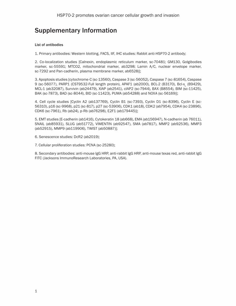
List of antibodies
1. Primary antibodies: Western blotting, FACS, IIF, IHC studies: Rabbit anti-HSP70-2 antibody;
2. Co-localization studies [Calnexin, endoplasmic reticulum marker, sc-70481; GM130, Golgibodies marker, sc-55591; MTCO2, mitochondrial marker, ab3298; Lamin A/C, nuclear envelope marker, sc-7292 and Pan-cadherin, plasma membrane marker, ab6528)];
3. Apoptosis studies [cytochrome-C (sc-13560), Caspase 3 (sc-56052), Caspase 7 (sc-81654), Caspase 9 (sc-56077), PARP1 (CST9532-Full length protein), APAF1 (ab2000), BCL-2 (B3170), Bcl-xL (B9429), MCL-1 (ab32087), Survivin (ab24479), XIAP (ab2541), cIAP2 (sc-7944), BAX (B8554), BIM (sc-11425), BAK (sc-7873), BAD (sc-8044), BID (sc-11423), PUMA (ab54288) and NOXA (sc-56169)];
4. Cell cycle studies [Cyclin A2 (ab137769), Cyclin B1 (sc-7393), Cyclin D1 (sc-8396), Cyclin E (sc-56310), p16 (sc-9968), p21 (sc-817), p27 (sc-53906), CDK1 (ab18), CDK2 (ab7954), CDK4 (sc-23896), CDK6 (sc-7961), Rb (ab24), p-Rb (ab76298), E2F1 (ab179445)];
5. EMT studies [E-cadherin (ab1416), Cytokeratin 18 (ab668), EMA (ab156947), N-cadherin (ab 76011), SNAIL (ab85931), SLUG (ab51772), VIMENTIN (ab92547), SMA (ab7817), MMP2 (ab92536), MMP3 (ab52915), MMP9 (ab119906), TWIST (ab50887)];
6. Senescence studies: DcR2 (ab2019);
7. Cellular proliferation studies: PCNA (sc-25280);
8. Secondary antibodies: anti-mouse IgG HRP, anti-rabbit IgG HRP, anti-mouse texas red, anti-rabbit IgG FITC (Jacksons ImmunoResearch Laboratories, PA, USA).
HSP70-2 promotes ovarian cancer cellular growth and invasion
2
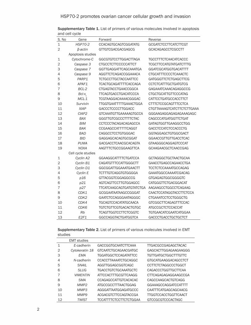
Supplementary Table 1. List of primers of various molecules involved in apoptosis and cell cycleS. No Gene Forward Reverse1 HSP70-2 CCACAGTGCAGTCGGATATG GCGATCTCCTTCATCTTCGT2 β-actin GTTGTCGACGACGAGCG GCACAGAGCCTCGCCTT
Apoptosis studies1 Cytochrome-C GGCGTGTCCTTGGACTTAGA TGCCTTTCTCAACATCACCC2 Caspase 3 CTGCCTCTTCCCCCATTCT TCGCTTCCATGTATGATCTTTG3 Caspase 7 GGTTGAGGATTCAGCAAATGA GGATCGCATGGTGACATTTT4 Caspase 9 AGGTTCTCAGACCGGAAACA CTGCATTTCCCCTCAAACTC5 PARP1 TCTGCCTTGCTACCAATTCC GATGGGTTCTCTGAGCTTCG6 APAF1 TCACTGCAGATTTTCACCAGA CCTCTCATTTGCTGATGTCG7 BCL-2 CTGAGTACCTGAACCGGCA GAGAAATCAAACAGAGGCCG8 Bcl-xL TTCAGTGACCTGACATCCCA CTGCTGCATTGTTCCCATAG9 MCL-1 TCGTAAGGACAAAACGGGAC CATTCCTGATGCCACCTTCT10 Survivin TTGGTGAATTTTTGAAACTGGA CTTTCTCCGCAGTTTCCTCA11 XIAP GACCCTCCCCTTGGACC CTGTTAAAAGTCATCTTCTCTTGAAA12 CIAP2 GTCAAATGTTGAAAAAGTGCCA GGGAAGAGGAGAGAGAAAGAGC13 BAX GGGTTGTCGCCCTTTTCTAC CAGCCCATGATGGTTCTGAT14 BIM CCTCCCTACAGACAGAGCCA GATAGTGGTTGAAGGCCTGG15 BAK CCGAAGCCATTTTTCAGGT GACCTCCATCTCCACCCTG16 BAD CAGGCCTCCTGTGGGAC GGTAGGAGCTGTGGCGACT17 BID GAGGAGCACAGTGCGGAT GGAACCGTTGTTGACCTCAC18 PUMA GACGACCTCAACGCACAGTA GTAAGGGCAGGAGTCCCAT19 NOXA AAGTTTCTGCCGGAAGTTCA GCAAGAACGCTCAACCGAG
Cell cycle studies1 Cyclin A2 GGAAGGCATTTTCTGATCCA GCTAGGGCTGCTAACTGCAA2 Cyclin B1 CAGATGTTTCCATTGGGCTT GAACCTGAGCCAGAACCTGA3 Cyclin D1 GGCGGATTGGAAATGAACTT TCCTCTCCAAAATGCCAGAG4 Cyclin E TCTTTGTCAGGTGTGGGGA GAAATGGCCAAAATCGACAG5 p16 GTTACGGTCGGAGGCCG GTGAGAGTGGCGGGGTC6 p21 AGTCAGTTCCTTGTGGAGCC CATGGGTTCTGACGGACAT7 p27 TTCATCAAGCAGTGATGTATCTGA AAGAAGCCTGGCCTCAGAAG8 CDK1 GCGGAATAATAAGCCGGGAT CAACTCCATAGGTACCTTCTCCA9 CDK2 GAATCTCCAGGGAATAGGGC CTGAAATCCTCCTGGGCTG10 CDK4 TGCAGTCCACATATGCAACA GTCGGCTTCAGAGTTTCCAC11 CDK6 TGTCTGTTCGTGACACTGTGC ATGCCGCTCTCCACCAT12 Rb TCAGTTGGTCCTTCTCGGTC TGTGAACATCGAATCATGGAA13 E2F1 GGCCAGGTACTGATGGTCA GACCCTGACCTGCTGCTCT
Supplementary Table 2. List of primers of various molecules involved in EMT studies
EMT studies1 E-cadherin GACCGGTGCAATCTTCAAA TTGACGCCGAGAGCTACAC2 Cytokeratin 18 GTCAATCTGCAGAACGATGC GAGCACTTGGAGAAGAAGGG3 EMA TGGATGGCTCCAGATATTCC TGTTGATGCTGGCTTTGTTC4 N-cadherin CCACCTTAAAATCTGCAGGC GTGCATGAAGGACAGCCTCT5 SNAIL AGGTTGGAGCGGTCAGC CCTTCTCTAGGCCCTGGCT6 SLUG TGACCTGTCTGCAAATGCTC CAGACCCTGGTTGCTTCAA7 VIMENTIN ATTCCACTTTGCGTTCAAGG CTTCAGAGAGAGGAAGCCGA8 SMA CCAGAGCCATTGTCACACAC CAGCCAAGCACTGTCAGG9 MMP2 ATGCCGCCTTTAACTGGAG GGAAAGCCAGGATCCATTTT10 MMP3 AGGGATTAATGGAGATGCCC CAATTTCATGAGCAGCAACG11 MMP9 ACGACGTCTTCCAGTACCGA TTGGTCCACCTGGTTCAACT12 TWIST TCCATTTTCTCCTTCTCTGGAA GTCCGCGTCCCACTAGC
HSP70-2 promotes ovarian cancer cellular growth and invasion
3
Supplementary Figure 1. HSP70-2 gene silencing changes the phenotypic characteristics of ovarian cancer cell, A10. Representative SEM images show no phenotypic changes in cells treated with lipofectamine (negative control). Intense membrane blebbing and apoptotic bodies formation is observed in cells treated with DMSO and Paclitaxel for 6 hours till 48 hours. SEM: Magnification: ×6000, WD = 6 mm, EHT = 20.00 kV.
HSP70-2 promotes ovarian cancer cellular growth and invasion
4
HSP70-2 promotes ovarian cancer cellular growth and invasion
5
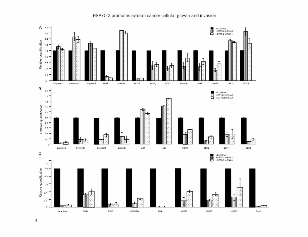
Supplementary Figure 2. Quantitative-PCR analysis of various genes involved in apoptosis, cell cycle and EMT. A. Histogram depicts the upregulation of caspase 3, caspase 7, caspase 9, APAF1, BAX, NOXA while, downregulation of PARP1, BCL-2, Bcl-xL, MCL-1, Survivin, XIAP and cIAP2 in HSP70-2 shRNA3 and shRNA4 transfected cells com-pared to NC shRNA transfected ovarian cancer cells. B. Histogram demonstrates fold changes in expression level of genes of cell cycle, cyclin A2, cyclin B1, cyclin D1, cyclin E, p21, p27, CDK1, CDK2, CDK4 and CDK6 in NC shRNA, HSP70-2 shRNA3 and shRNA4 transfected ovarian cancer cells. C. Histogram depicts downregulation of mesenchy-mal markers, N-cadherin, SNAIL, SLUG, VIMENTIN, SMA, MMP2, MMP3, MMP9 and TWIST in HSP70-2 shRNA3 and shRNA4 transfected cells as compared to NC shRNA transfected cells. The three independent experiments were performed in triplicates. Data is represented as mean ± SEM (standard error of the mean).
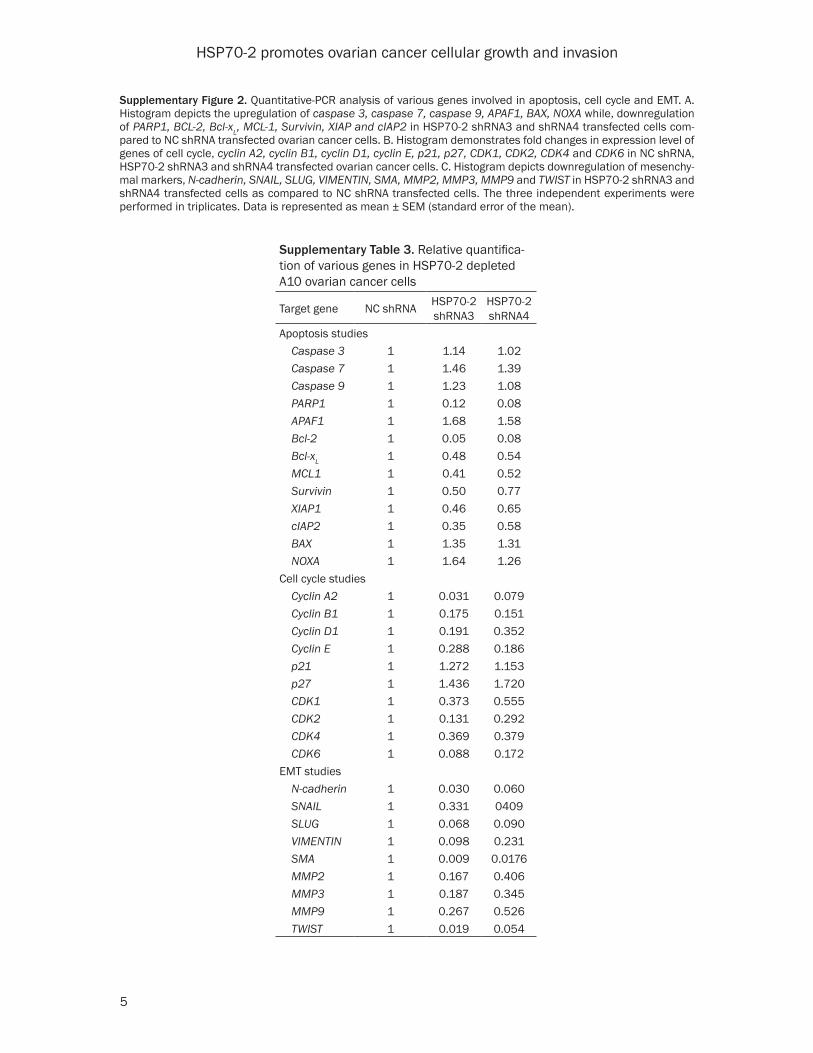
Supplementary Table 3. Relative quantifica-tion of various genes in HSP70-2 depleted A10 ovarian cancer cells
Target gene NC shRNA HSP70-2shRNA3
HSP70-2shRNA4
Apoptosis studies Caspase 3 1 1.14 1.02 Caspase 7 1 1.46 1.39 Caspase 9 1 1.23 1.08 PARP1 1 0.12 0.08 APAF1 1 1.68 1.58 Bcl-2 1 0.05 0.08 Bcl-xL 1 0.48 0.54 MCL1 1 0.41 0.52 Survivin 1 0.50 0.77 XIAP1 1 0.46 0.65 cIAP2 1 0.35 0.58 BAX 1 1.35 1.31 NOXA 1 1.64 1.26Cell cycle studies Cyclin A2 1 0.031 0.079 Cyclin B1 1 0.175 0.151 Cyclin D1 1 0.191 0.352 Cyclin E 1 0.288 0.186 p21 1 1.272 1.153 p27 1 1.436 1.720 CDK1 1 0.373 0.555 CDK2 1 0.131 0.292 CDK4 1 0.369 0.379 CDK6 1 0.088 0.172EMT studies N-cadherin 1 0.030 0.060 SNAIL 1 0.331 0409 SLUG 1 0.068 0.090 VIMENTIN 1 0.098 0.231 SMA 1 0.009 0.0176 MMP2 1 0.167 0.406 MMP3 1 0.187 0.345 MMP9 1 0.267 0.526 TWIST 1 0.019 0.054
HSP70-2 promotes ovarian cancer cellular growth and invasion
6
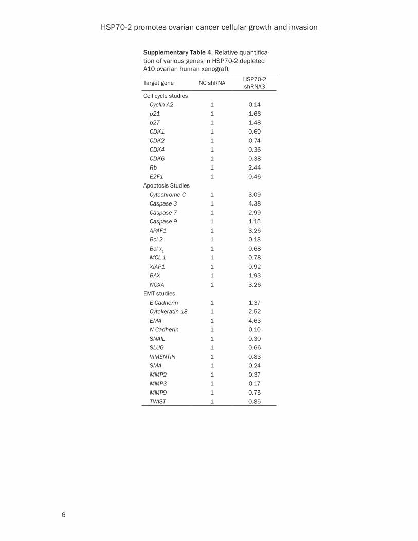
Supplementary Table 4. Relative quantifica-tion of various genes in HSP70-2 depleted A10 ovarian human xenograft
Target gene NC shRNA HSP70-2shRNA3
Cell cycle studies Cyclin A2 1 0.14 p21 1 1.66 p27 1 1.48 CDK1 1 0.69 CDK2 1 0.74 CDK4 1 0.36 CDK6 1 0.38 Rb 1 2.44 E2F1 1 0.46Apoptosis Studies Cytochrome-C 1 3.09 Caspase 3 1 4.38 Caspase 7 1 2.99 Caspase 9 1 1.15 APAF1 1 3.26 Bcl-2 1 0.18 Bcl-xL 1 0.68 MCL-1 1 0.78 XIAP1 1 0.92 BAX 1 1.93 NOXA 1 3.26EMT studies E-Cadherin 1 1.37 Cytokeratin 18 1 2.52 EMA 1 4.63 N-Cadherin 1 0.10 SNAIL 1 0.30 SLUG 1 0.66 VIMENTIN 1 0.83 SMA 1 0.24 MMP2 1 0.37 MMP3 1 0.17 MMP9 1 0.75 TWIST 1 0.85

HSP70-2 promotes ovarian cancer cellular growth and invasion
7
Supplementary Figure 3. Quantitative-PCR analysis of various genes involved in cell cycle, apoptosis and EMT in xenograft tumor cells. A. Histogram demonstrates difference in expression level of genes of cell cycle, cyclin A2, p21, p27, CDK1, CDK2, CDK4, CDK6, Rb and E2F1 in NC shRNA and HSP70-2 shRNA3 treated tumor. B. Histogram depicts upregulation of cytochrome-C, caspase 3, caspase 7, caspase 9, APAF1, BAX and NOXA; and downregula-tion of PARP1, BCL-2, Bcl-xL, MCL-1 and XIAP in HSP70-2 shRNA3 treated tumor as compared to NC shRNA treated tumor. C. Histogram depicts upregulation of epithelial markers like E-cadherin, cytokeratin 18 and EMA and down-regulation of mesenchymal markers, N-cadherin, SNAIL, SLUG, VIMENTIN, SMA, MMP2, MMP3, MMP9 and TWIST in HSP70-2 shRNA3 as compared to NC shRNA treated tumor. The three independent experiments were performed in triplicates. Data is represented as mean ± SEM (standard error of the mean).