Embed Size (px)
Citation preview

Int J Clin Exp Med 2015;8(11):19954-19968www.ijcem.com /ISSN:1940-5901/IJCEM0012052
Original Article Hypoxia/lncRNA-AK123072/EGFR pathway induced metastasis and invasion in gastric cancer
Zhen Yang1*, Ruoming Wang2*, Tengteng Zhang3, Xinhua Dong1
1Department of Gastrointestinal Surgery, 1st Affiliated Hospital of Zhengzhou University, Henan Province, China; 2Department of General Surgery, 1st Renmin Hospital of Shangqiu, Henan Province, China; 3Department of Cancer, 1st Renmin Hospital of Shangqiu, Henan Province, China. *Equal contributors.
Received June 28, 2015; Accepted October 9, 2015; Epub November 15, 2015; Published November 30, 2015
Abstract: Objective: To investigate the role of long noncoding RNAs (lncRNAs) in hypoxia-induced gastric cancer (GC) metastasis and invasion. Methods: We investigated the differentially expressed lncRNAs resulting from hypoxia-in-duced GC and normoxia conditions using microarrays and validated our results through real-time quantitative poly-merase chain reaction. The role of the targeting lncRNA was further detected by in vivo and in vitro assays. Results: We found an lncRNA, AK123072, which was up-regulated by hypoxia. AK123072 was frequently up-regulated in GC samples and promoted GC migration and invasion in vivo and in vitro. Furthermore, AK123072 could mediate the metastasis of hypoxia-induced GC cells. Next, we identified EGFR, which was a metastasis-related gene regulated by AK123072. In addition, we found that the expression of EGFR was positively correlated with that of AK123072 in the clinical GC samples used in our study. Furthermore, we found that the EGFR gene CpG island methylation was significantly increased in GC cells depleted of AK123072. Intriguingly, EGFR expression was also increased by hypoxia, and EGFR up-regulation by AK123072 mediated hypoxia-induced GC cell metastasis. Conclusion: Our results identified hypoxia/lncRNA-AK123072/EGFR pathway in gastric cancer pathogenesis and this might help in the development of new therapeutics in clinics.
Keywords: AK123702, cancer gene therapy, EGFR, gastric cancer (GC), hypoxia, long noncoding RNAs (lncRNAs), metastasis, invasion
Introduction
Gastric cancer (GC) is the second leading cause of cancer mortality in the world, and has a par-ticularly high incidence in Asian countries including China [1]. The high mortality of GC is a consequence of late-stage of diagnosis, the 5-year survival rate for advanced stages is extremely poor and around 5% to 15% [2]. Although diagnosis and treatment of GC have improved, the survival rate has not increased substantially in couple of years. Therefore, an improved understanding of the molecular path-ways involved in the progression of GC will be helpful in improving prevention, diagnosis and therapy of this disease [3]. Hypoxia is an impor-tant microenvironmental factor that induces such metastasis. In fact, hypoxic tumors are often aggressive and more likely to metasta-size [4]. Therefore, investigating the underlying
mechanisms of hypoxia-induced metastasis is critical.
LncRNAs have been identified as a new class of functional RNAs and have kindled our interest. LncRNAs are defined as transcripts of longer than 200 nucleotides without evident protein-coding function [5]. Few studies have implicat-ed lncRNAs in various cancers [6, 7], and sev-eral of the altered lncRNAs can result in the aberrant expression of nearby protein-coding genes, which may contribute to cancer develop-ment [8]. Studies have also demonstrated that lncRNAs play an important role in tumor devel-opment via various mechanisms, such as chro-matin remodeling [9], DNA methylation [10], transcriptional regulation [11], and DNA dam-age repair [13]. All related processes play a piv-otal role in malignant transformation and can-cer treatment. However, the function of most lncRNAs in cancer remains a mystery, and

Hypoxia/lncRNA-AK123072/EGFR in gastric cancer
19955 Int J Clin Exp Med 2015;8(11):19954-19968
lncRNA profiles in hypoxic GC and hypoxia-responsive gene networks remain unknown.
To explore the role of lncRNAs in hypoxic GC, we identified a small number of lncRNAs and mes-senger RNAs (mRNAs) that were aberrantly expressed in GC under hypoxia compared with normoxia using microarrays. We also investi-gated the biological function of the hypoxia-up-regulated AK123072 both in vivo and in vitro. Further analysis demonstrated that EGFR was over-expressed in several types of cancer [14, 15]. In our study, the inhibition of lncRNA-AK123072 and EGFR were found to play an important role in hypoxia-induced metastasis and invasion, suggesting that manipulating new RNA functions can provide a therapeutic opportunity that is worth exploring.
Material and methods
Ethics statement
Investigation has been conducted in accor-dance with the ethical standards and according to the Declaration of Helsinki and according to national and international guidelines and has been approved by the authors’ institutional review board.
Lentivirus infection and construction of stable cell lines with down-regulated lncRNA-AK123072
To observe the effects of AK123072 knock-down on invasion and metastasis in vivo and in vitro, four different small interfering RNAs (siR-NAs) that targeted AK123072 RNA and a scrambled siRNA control were generously pro-vided by GenePharma (Shanghai, China). The four siRNAs were transfected into SGC-7901 and AGS cells using Lipofectamine 2000 (In- vitrogen) following the manufacturer’s protocol. Twenty-four hours after transfection, AK123072 expression levels were measured through reverse transcriptase polymerase chain reac-tion (RT-PCR), and we found that siRNA-AK1 with the sequence 5’-CCACCAGUUACCUGCA- AUATT-3’ (sense) and 5’-UAUUGCAGGUAAC- UGGUGGTT-3’ (antisense) and siRNAAK2 with the sequence 5’-GGAACAAAGAUGGUUUCUA- TT-3’ (sense) and 5’-UAGAAACCAUCUUUGUU- CCTT-3’ (antisense) yielded the highest degree of AK123072 knockdown. Then, we designed and synthesized AK123072-targeting sequen-
ce and inserted this sequence into a Super-silencing Vector (GenePharma, Shanghai, China). An unrelated sequence lentiviral vector was used as a negative control. SGC-7901 and AGS cells were then plated into six-well plates and allowed to adhere for 24 hours. Next, the lentivirus was transfected according to the manufacturer’s instructions. Stably transfected cells were selected with puromycin (Sigma-Aldrich, St. Louis, MO) and confirmed through fluorescence microscopy and RT-PCR.
Cell culture and transfection
Human gastric cancer cell line SGC-7901, AGS and BGC-823 were cultured in RPMI 1640 medium with 10% fetal bovine serum. Cells were transiently transfected by electroporation or LipofectamineTM 2000 (Invitrogen) trans- fection reagent. For luciferase reporter gene assay, SGC-7901 cells (5×10⁵) were seeded onto 6-well plates and then transiently trans-fected. Twenty-four hours after transfection, the transfected cells were infected with adeno-viruses expressing miRNAs or GFP. Twenty-four hours after infection, luciferase activity was measured and then normalized.
MicroRNA real-time PCR
1×10²-1×10⁷ cells were harvested, washed in PBS once, and stored on ice; complete cell lysate was prepared by addition of 600 µl lysis binding buffer and vertex; 60 µl microRNA aomogenete addictive was added to the cell lysate and mixed thoroughly by inverting sever-al times; sample was stored on ice for 10 min, followed by addition of equal volume (600 µl) of phenol: chloroform (1:1) solution; sample was mixed by inverting for 30-60 sec, and then cen-trifuged at 12000 g for 5 min; the supernatant was transferred to a new tube and the volume was estimated; 1/3 volume of 100% ethanol was added and mixed; the mixture was loaded to the column at room temperature and centri-fuged at 10000 g for 15 sec; the flow-though was collected and the volume was then esti-mated; 2/3 volume of 00% ethanol was added and mixed; the mixture was loaded to column at room temperature and centrifuged at 10000 g for 15 sec; the flow-through was discarded; 700 µl microRNA wash solution was added to the column, followed by centrifugation at 10000 for 10 sec; the flow-through was dis-carded; 500 µl microRNA wash solution was
Hypoxia/lncRNA-AK123072/EGFR in gastric cancer
19956 Int J Clin Exp Med 2015;8(11):19954-19968
added to the column, followed by centrifugation at 10000 for 10 sec; the flow-through was dis-carded; the column was transferred to a new tube and 100 µl preheated elution solution (95 degree) was added at room temperature; RNA was collected by centrifugation at 12000 g for 30 sec. The experiments were repeated at least 3 times.
Western blot
Cells were harvested, washed twice in PBS, and lysed in lysis buffer (protease inhibitors were added immediately before use) for 30 min on ice. Lysate was centrifuged at 10000 rmp and the supernatants were collected and stored at -70 in aliquots. All procedures were carried out on ice. Protein concentration was determined using BCA assay kit (Tianlai Biotech). The experiments were repeated at least 3 times.
Cell viability assay
Cells were seeded into 96-well plates (2×10³ cells/well) directly or 24 hours after transfec-tion and allowed to attach overnight. Forty-eight hours later, cell viability was assessed via 3-(4,5-dimethylthiazol-2-yl)-2,5-diphenyl-tetra-zolium bromide (MTT) assay as described previ-ously [14].
Transwell invasion assay
Transwell filters were coated with matrigel (3.9 mg/μL, 60-80 μL) on the upper surface of a polycarbonic membrane (diameter 6.5 mm, pore size 8 mm). After incubating at 37°C for 30 minutes, the matrigel solidified and served as the extracellular matrix for analysis of tumor cell invasion. Harvested cells (1×10⁵) in 100 μL of serum free DMEM were added into the upper compartment of the chamber. A total of 200 μL conditioned medium derived from NIH3T3 cells was used as a source of chemo-attractant, and was placed in the bottom compartment of the chamber. After 24 h incubation at 37°C with 5% CO2, the medium was removed from the upper chamber. The non-invaded cells on the upper side of the chamber were scraped off with a cotton swab. The cells that had migrated from the matrigel into the pores of the inserted filter were fixed with 100% methanol, stained with Hematoxylin, and mounted and dried at 80°C for 30 minutes. The number of cells invading
through the matrigel was counted in three ran-domly selected visual fields from the central and peripheral portion of the filter using an inverted microscope (200× magnification). Each assay was repeated three times.
Plasmid construction and luciferase reporter assay
Wild-type 3’untranslated region (3’UTR) of EGFR containing predicted lncRNA-AK123072 target sites were amplified by PCR from SGC-7901 cell genomic DNA. Primers used: Forward: GAT CTG CAG GGG TTA GCT TGG GGA CCT GAA C; Reverse: GAT CAT ATG AGA GTG ACA TAC TGA TGC CTA C. Mutant 3’UTRs were generated by overlap-extension PCR method. Both wild-type and mutant 3’UTR fragments were subclon- ed into the pGL3-control vector (Promega, Madison, WI) immediately downstream of the stop codon of the luciferase gene. DNA frag-ment coding EGFR protein was amplified by PCR from SGC-7901 cell cDNA, and cloned in- to pCMV-Myc expression vector (Clonetech, Mountain View, CA). Primers used: Forward: GCT GAA TTC ATG CCG GTG GAC CTC AGC AAG T; Reverse: CTG CTC GAG CTA CTT CCC AGA CAG CTG CTC G. For luciferase assay, the reporter plasmid was co-transfected with a control Renilla luciferase vector into SGC-7901 cells in the presence of either miR-15b or NC. After 48 h, cells were harvested, and the luciferase ac- tivity was measured using the Dual-Luciferase Reporter Assay System (Promega, Madison, WI, USA).
Animal studies
60 five-week-old female BALB/c nude mice were purchased from the Animal Center of Zhejiang University (Hangzhou, China) and they were divided into 2 groups evenly. For in vivo metastasis assays, SGC-7901 cells were sub-cutaneously inoculated into nude mice (six per group, 1×10⁶ cells for each mouse). Tumor growth was examined every other day, and tumor volumes were calculated using the equa-tion V=A×B2/2 (mm3), where A is the largest diameter and B is the perpendicular diameter. After 2 weeks, all mice were sacrificed. Trans- planted tumors were excised, and tumor tis-sues were used to perform hematoxylin & eosin (H&E) staining. All research involving animal complied with protocols approved by the
Hypoxia/lncRNA-AK123072/EGFR in gastric cancer
19957 Int J Clin Exp Med 2015;8(11):19954-19968
Zhejiang medical experimental animal care commission.
Data analysis
Image data were processed using SpotData Pro software (Capitalbio). Differentially express- ed genes were identified using SAM package (Significance Analysis of Microarrays, version 2.1).
Results
lncRNA expression profile in hypoxia-induced gastric cancer cells
To examine the overall impact of lncRNAs on hypoxic GC, we analyzed the expression profiles
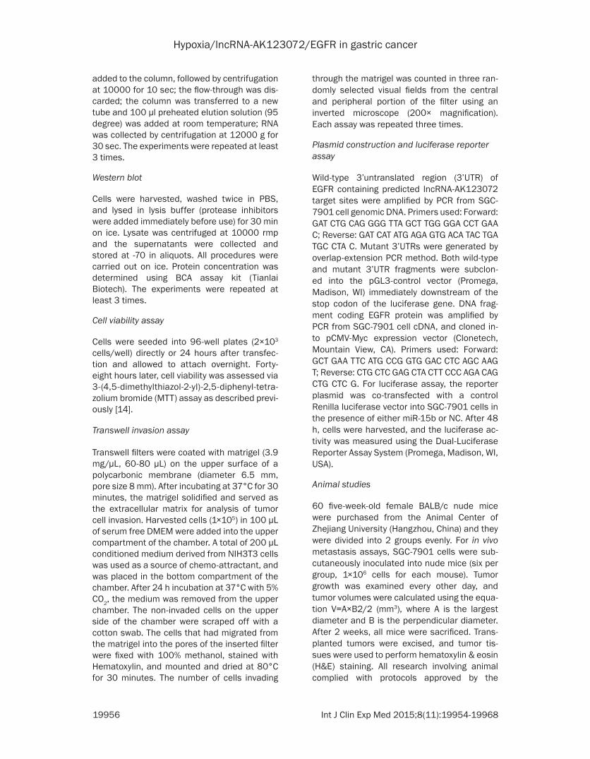
of lncRNAs and protein-coding RNAs in nor- moxia-induced and hypoxia-induced GC cells using microarray analysis. Hierarchical cluster-ing showed the differential lncRNA and protein coding RNA expression profiles between nor-moxia-induced and hypoxia-induced GC cells (Figure 1A and 1B). We set a threshold of a fold change >1.5, P<0.05, and found that 84 lncRNAs were up-regulated and 70 were down-regulated in all hypoxia-induced GC cells com-pared with normoxia-induced GC cells (Figure 1C and 1D). This finding indicated that the lncRNA expression profiles differed between the two groups.
To validate the microarray findings, we random-ly selected six lncRNAs from the differentially expressed lncRNAs with a fold change >3 and
Figure 1. Differentially expressed lncRNAs and mRNAs were analyzed using hierarchical clustering. Hierarchical clustering analysis arranges samples into groups based on expression levels, which allows us to hypothesize the relationships between samples. The dendrogram shows the relationships between the lncRNA (A) and mRNA (B) expression patterns in the samples, that is, which samples are more similar in the expressing relationships. For every gene in each sample, the “Red” indicates high relative expression, and “Blue” indicates low relative expres-sion. Actually, there is no inevitable relation between the lncRNA shown in (A) and mRNA presented in (B) expression patterns in the samples because they were analyzed independently. Schemas of the up-regulated (C) and down-regulated (D) lncRNAs, identified by microarray in the gastric cancer (GC) cells SGC-7901, AGS and BGC-823 under hypoxia. The overlapping areas represent the common lncRNAs in all three GC cell lines under hypoxia.

Hypoxia/lncRNA-AK123072/EGFR in gastric cancer
19958 Int J Clin Exp Med 2015;8(11):19954-19968
analyzed their expression through real-time PCR with hypoxia-induced GC cells (after 24 hours in 1% O2 for the SGC-7901, AGS, and BGC-823 gastric cancer cells) relative to nor-moxia induced GC cells.
Newly identified AK123072 frequently up-regu-lated in gc and induced by hypoxia in gc cells
Among the differentially expressed lncRNAs among hypoxia induced GC cells and normoxia-induced GC cells, we were particularly interest-ed in lncRNA-AK123072 because its expres-sion increased approximately 6.20±1.65-fold
upon hypoxia treatment in all three cell lines. Thus, we studied the role of AK123072, which is an intronic antisense lncRNA. Given that AK123072 is induced by hypoxia in GC cells, we next sought to determine whether AK- 123072 could be induced by hypoxia at differ-ent exposure times (after 4, 8, 16, 24, and 48 hours in 1% O2) in GC cells. We found that AK123072 was induced under hypoxia, with the most robust induction observed after 16 hours in 1% O2 for SGC-7901 cells, 24 hours in 1% O2 for AGS cells, and 48 hours in 1% O2 for BGC-823 cells (Figure 2A-C). The results sug-
Figure 2. AK123072 is often up-regulated in gastric can-cer and is induced by hypoxia in gastric cancer cells. (A-C) AK123072 was induced under hypoxia, with the most robust induction observed after 16 hours in 1% O2 in (A) SGC-7901 cells, 24 hours in 1% O2 in (B) AGS cells, and 48 hours in 1% O2 in (C) BGC-823 cells. (D) RNA was extracted with Trizol reagent from 95 pairs of human gastric cancer and adjacent tissues. AK123072 expression was assessed by real-time PCR. Actin was used as an internal control. The significant differences between samples were analyzed using the Wil-coxon signed-rank test (P=0.002, n=95). (E) Patients with high levels of EGFR expression showed reduced overall sur-vival times compared with patients with low levels of EGFR expression (P=0.0083, log-rank test). *P<0.05; **P<0.01.
Hypoxia/lncRNA-AK123072/EGFR in gastric cancer
19959 Int J Clin Exp Med 2015;8(11):19954-19968
gested that AK123072 could indeed be regu-lated by hypoxia in GC cells; however, no signifi-cant difference was observed in expression after 4 or 8 hours in 1% O2.
Next, we assessed AK123072 expression in 95 pairs of human primary GC tissues and adja-
cent gastric tissues using quantitative RT-PCR to determine AK123072 expression in GC tis-sues. AK123072 expression was remarkably up-regulated in GC tissues compared with non-cancerous gastric tissues (Figure 2D), indicat-ing that AK123072 up-regulation is common in GC.
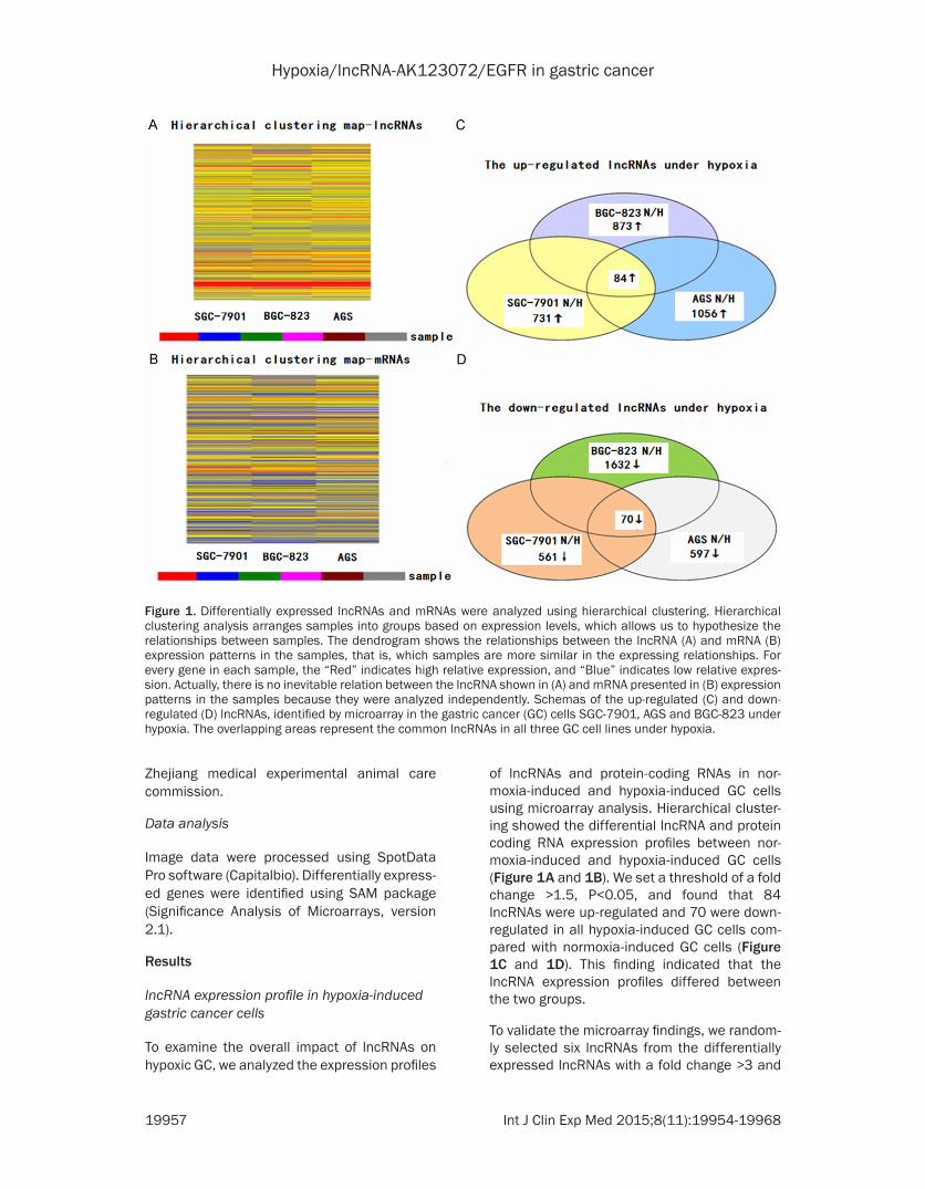
Figure 3. AK123072 promotes gastric cancer cell migration and invasion. A. AK123072 expression following knock-down by four different siRNA (si-AK1, si-AK2, si-AK3, and si-AK4) in the GC cell lines SGC-7901. B. Observation of the infection efficiency of AK123072 (si-AK1 and si-AK2) and scrambled siRNA lentivirus (si-Scr) by fluorescence microscopy. C. After transfection, the cells were collected, and cell mobility assay was done to test the cell inva-sion ability. D. Transwell migration and invasion assays of SGC-7901 cells were performed after transfection with AK123072 si-AK1 and si-AK2 or a scrambled siRNA control (si-Scr). E. Cell mobility was examined with a Cellomics ArrayScan VTI 1700 Plus. The relative distance traveled because of SGC-7901 cell mobility was calculated with this instrument. In all panels, the results are representative of at least three independent experiments.
Hypoxia/lncRNA-AK123072/EGFR in gastric cancer
19960 Int J Clin Exp Med 2015;8(11):19954-19968
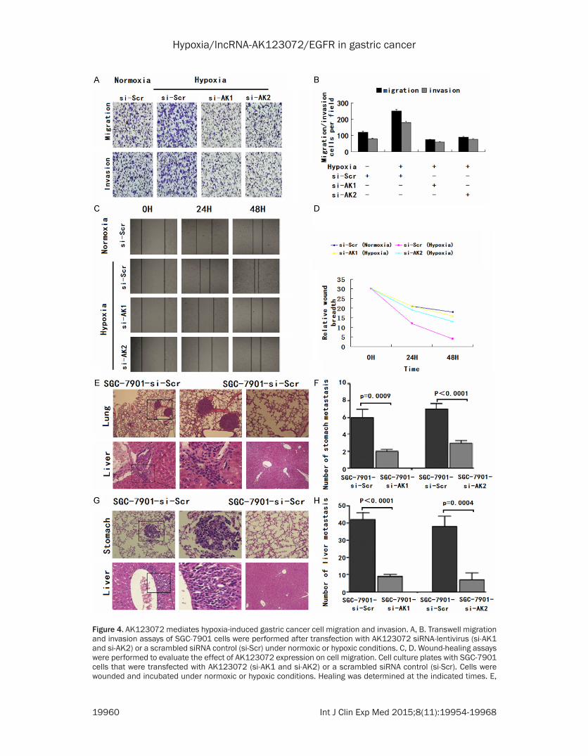
Figure 4. AK123072 mediates hypoxia-induced gastric cancer cell migration and invasion. A, B. Transwell migration and invasion assays of SGC-7901 cells were performed after transfection with AK123072 siRNA-lentivirus (si-AK1 and si-AK2) or a scrambled siRNA control (si-Scr) under normoxic or hypoxic conditions. C, D. Wound-healing assays were performed to evaluate the effect of AK123072 expression on cell migration. Cell culture plates with SGC-7901 cells that were transfected with AK123072 (si-AK1 and si-AK2) or a scrambled siRNA control (si-Scr). Cells were wounded and incubated under normoxic or hypoxic conditions. Healing was determined at the indicated times. E,

Hypoxia/lncRNA-AK123072/EGFR in gastric cancer
19961 Int J Clin Exp Med 2015;8(11):19954-19968
We further determined whether the expression level of EGFR correlated with the clinical out-come of gastric cancer patients. Kaplan-Meier survival analysis and log-rank tests using patient postoperative survival were conducted to further evaluate the correlation between EGFR and prognosis of patients with gastric cancer. According to the median ratio of rela-tive EGFR expression (5.44) in tumor tissues, the gastric cancer patients were classified into two groups: High-EGFR group: EGFR expression ratio ≥ median ratio; and Low-EGFR group: EGFR expression ratio ≤ median ratio. Kaplan-Meier survival analysis showed that high EGFR expression in gastric carcinoma tissues is sig-nificantly associated with worse overall survival (P=0.0083, log-rank test) (Figure 2E). These
results suggest that EGFR may play an impor-tant role in the progression of gastric cancer.
Effect of AK123072 on GC cell migration and invasion and hypoxia-induced migration and invasion
The frequent AK123072 up-regulation in hypox-ic GC cells implies that AK123072 may play a role in hypoxia-induced GC. To test this hypoth-esis, the effects of reduced AK123072 expres-sion on cell proliferation, migration, and inva-sion were investigated in two GC cell lines. Four different siRNA molecules were tested for their knockdown efficiencies, and the two most effi-cient of these molecules (siRNA-AK1 and siR-NA-AK2) were selected for subsequent studies
F. The incidence of metastasis in mice and the mean number of visible tumor nodules in the liver and lung due to SGC-7901-transfected cells. SGC-7901 cells were transfected with the AK123072 siRNA-lentivirus (si-AK1 and si-AK2) or a scrambled siRNA control (si-Scr). The cells were then injected into nude mice via the tail vein for an in vivo metastasis assay, and the animals were sacrificed 6 weeks after injection. G, H. Representative hematoxylin and eosin staining of stomach and liver isolated from mice injected with SGC-7901-si-Scr or SGC-7901-si-AK cells. The arrows indicate tumor foci in the lungs and livers. Magnification, ×100 (left) and ×200 (right).
Figure 5. AK123072 up-regulates EGFR in gastric cancer cells. (A, B) The mRNA (A) and protein (B) levels of EGFR were assessed through RT-PCR and Western blotting, respectively, SGC-7901 cells were transfected with an AK123072 siRNA-lentivirus (si-AK1 and si-AK2) or a scrambled siRNA control (si-Scr) in a dose-dependent manner. (C) Correlation between the relative mRNA levels of AK123072 and EGFR in 95 gastric cancer tissue specimens (r=0.5262, P<0.001, Pearson’s correlation). (D) Methylation mapping of 15 CpG island in EGFR exon 1 region obtained from bisulfite sequencing in scrambled siRNA control or SGC-7901 cells, or SGC-7901 cells infected with AK123072 siRNA. CpG positions are indicated relative to the translation start codon, and each circle in the figure represents a single CpG site. For each cell line, the percentage methylation at a single CpG site is calculated from the sequencing results of 10 independent clones. Black circles, 100% methylated; white circles, 0% methylation.
Hypoxia/lncRNA-AK123072/EGFR in gastric cancer
19962 Int J Clin Exp Med 2015;8(11):19954-19968

Hypoxia/lncRNA-AK123072/EGFR in gastric cancer
19963 Int J Clin Exp Med 2015;8(11):19954-19968
(Figure 3A). We first established SGC-7901 cell lines that stably repressed AK123072 expres-sion by using anAK123072 siRNA-lentivirus (si-AK) vector, as verified by fluorescence micros-copy (Figure 3B) and RT-PCR (Figure 3D). To investigate whether AK123072 might have a role in hypoxia induced metastasis, we first determined whether AK123072 affected nor-moxic GC cell migration and invasion. In tran-swell assays with or without Matrigel, SGC-7901 cells with stable AK123072 knockdown showed significantly decreased migration and invasion compared with control cells (Figure 3E). Moreover, we examined cell motility in dif-ferent groups under normoxia using high-con-tent screening, which indicated that cell motility was reduced in AK123072 siRNA-lentivirus-treated cells compared with scrambled siRNA-treated cells (Figure 3C). Taken together, these results indicate that although AK123072 did not affect GC cell growth, the lncRNA promoted migration and invasion.
Given that AK123072 can promote normoxic GC cell migration and invasion, we hypothe-sized that AK123072 might play a role in hypox-ia-induced migration and invasion. To test this hypothesis, we first measured hypoxic GC cell migration and invasion. Hypoxia significantly increased the migration and invasion potential of SGC-7901 cells (Figure 4A, 4B), which is con-sistent with previous reports [4, 5]. After trans-fection with a siRNA-lentivirus to decrease en- dogenous AK123072 expression, the hypoxia-induced migration and invasion of SGC-7901 cells were dramatically diminished (Figure 4A, 4B). In addition, wound-healing assays showed that, under hypoxia, the cells more rapidly closed wounds (Figure 4C, 4D). However, after AK123072 knockdown in GC cells, wound heal-ing was not significantly promoted under hypox-ia compared with the control cells (Figure 4H).
To further explore the role of AK123072 in tumor invasion and metastasis in vivo, SGC-
7901 cells with stable AK123072 repression by an AK123072 siRNA-lentivirus or a scram-bled siRNA vector-transfected control cells were delivered into nude mice via tail vein injec-tion. We found that the number and size of lung and liver metastatic nodules dramatica- lly decreased in mice administered cells with low AK123072 expression compared with the scrambled siRNA controls (Figure 4G, 4H). Taken together, these observations suggest that AK123072 is a positive metastatic regula-tor of GC.
EGFR an AK123072-regulated gene in GC
To explore the mechanism by which AK123072 promotes GC cell migration and invasion, we attempted to identify the lncRNA’s potential target genes. Recent studies have reported that lncRNAs may function by positively or neg-atively regulating the expression of their neigh-boring protein-coding genes [14, 16, 26-28]. Thus, we retrieved the genomic locus informa-tion from the UCSC genome browser (http://genome.ucsc.edu/) and found that the tumor oncogene EGFR is located 8.6 kb downstream of AK123072, whereas another tumor suppres-sor gene, BMPR1A, is found 33.3 kb upstream of AK123072. To validate the association between the lncRNA AK123072 and EGFR, we performed RT-PCR and western blotting using SGC-7901 and AGS cells with AK123072 knockdown. We observed significantly decrea- sed EGFR levels in cells with low AK123072 expression compared with scrambled siRNA control cells (Figure 5A, 5B). Moreover, we showed that AK123072 suppressed EGFR mRNA and protein levels in GC cells and that there was a dose-dependent relationship be- tween the expression of EGFR and AK123072 (Figure 5A, 5B). These results suggest that EGFR is an AK123072-regulated gene in GC. We also found that EGFR was predominantly located in the cytoplasm of GC cells. Moreover, we found that EGFR expression was heteroge-
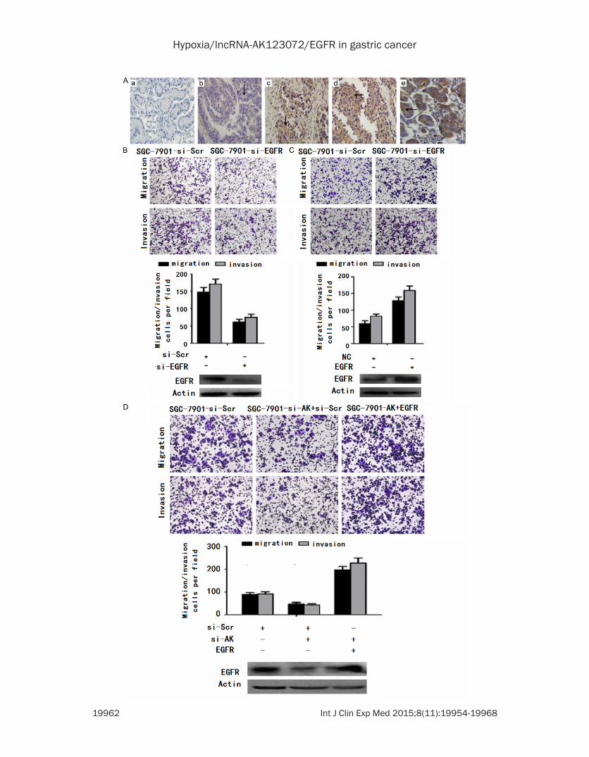
Figure 6. EGFR functions as a metastasis-related gene in gastric cancer and can promote AK123072-induced gas-tric cancer cell migration and invasion. A. Immunohistochemical analysis of EGFR in metastatic and nonmetastatic GC: (a) noncancerous region of GC, (b) primary site of nonmetastatic GC, (c, d) primary site of metastatic GC, and (e) breast cancer as positive control. B. Transwell migration and invasion assays of SGC-7901 cells were performed after transfection with siRNA directed against EGFR or a scrambled siRNA control (si-Scr). EGFR protein levels were detected by Western blot analysis as well. C. Transwell migration and invasion assays of SGC-7901 cells were performed after transduction with EGFR or a negative control. EGFR protein levels were detected by Western blot analysis as well. D. Transwell migration and invasion assays of SGC-7901-si-Scr or SGC-7901-si-AK cells were per-formed after transduction with a scrambled siRNA control (si-Scr) or EGFR. EGFR protein was detected by Western blot analysis as well. In all panels, the results are representative of at least three independent experiments.

Hypoxia/lncRNA-AK123072/EGFR in gastric cancer
19964 Int J Clin Exp Med 2015;8(11):19954-19968

Hypoxia/lncRNA-AK123072/EGFR in gastric cancer
19965 Int J Clin Exp Med 2015;8(11):19954-19968
neous in tumor tissue; it was predominantly located in the center of the tumor but had almost negative expression in the stroma of GC tissues.
Next, we analyzed the correlation between EGFR expression and the clinicopathologic parameters of GC patients. EGFR expression in GC patients did not correlate with age, sex, or cell differentiation. We further investigated the correlation between AK123072 and EGFR ex- pression in 95 clinical GC tissues and found that AK123072 and EGFR expression levels were positively correlated (Figure 5C). These observations indicate that EGFR is frequently up-regulated in GC samples, possibly because of AK123072 over-expression. Next, to inve- stigate the potential mechanism by which AK123072 contributes to malignant behaviors of GC cells, we focused on DNA methylation of EGFR because lncRNAs are known to be involved in epigenetic regulation and DNA hypo-methylation of EGFR has been confirmed in laryngeal cancer, including GC. Then, we per-formed bisulfite sequencing of cloned alleles over the region of -169 to 81 of the EGFR exon1 CpG islands, which has been demonstrated to be involved in GC. The results showed that EGFR CpG islands were densely hypomethyl-ated in control SGC-7901-si-Scr and AGS-si-Scr cells, but GC cells transduced with AK123072 siRNA showed more methylated CpG dinucleo-tides (Figure 5D). Taken together, these results indicate that knockdown of AK123072 can down-regulate the expression of EGFR in GC cells by regulating EGFR DNA methylation, which illustrates that EGFR may act as a down-stream target of AK123072 in GC cells.
EGFR mediates AK123072-induced migration and invasion of GC cells
Additional analysis revealed that EGFR showed strong staining at primary sites from patients with metastatic GC compared with samples
from patients with non-metastatic GC (Figure 6A). To further explore the biological function of EGFR in GC, siRNAs targeting EGFR were designed and transfected into SGC-7901 cells. These siRNAs significantly decreased EGFR expression (Figure 6C). The migration and inva-sion of the two cell lines were significantly decreased by EGFR siRNA but not by the scram-bled siRNA control (Figure 6B and 6C). To sup-port these findings, we over-expressed EGFR in SGC-7901 cells, which significantly increased cell migration and invasion (Figure 6D).
EGFR regulation by AK123072 mediates hypoxia-induced GC metastasis
To determine whether EGFR is involved in AK123072-induced hypoxic GC cell metasta-sis, we first detected EGFR expression under hypoxia. The results showed that EGFR ex- pression in GC cells increased under hypoxia compared with normoxia (Figure 7A). When AK123072 expression was down-regulated in SGC-7901 cells, the hypoxia-induced EGFR in- crease was abrogated (Figure 7A), indicating that EGFR over-expression was mediated by AK123072 up-regulation under hypoxia. More- over, transwell assays indicated that inhibiting EGFR expression promoted hypoxia-induced migration and invasion (Figure 7B). To deter-mine the function of EGFR in AK123072-induced GC metastasis under hypoxia, an EGFR expression vector and a scramble control were co-transfected into SGC-7901 cells with stable AK123072 suppression. In transwell experi-ments, the impairment of the effects of siRNA-mediated AK123072 on hypoxia induced GC cell migration and invasion was partially re- lieved by EGFR but not by the negative control (Figure 7C). Taken together, these results indi-cate that EGFR expression is increased by hypoxia and that EGFR up-regulation by AK- 123072 mediates hypoxia-induced GC me- tastasis.
Figure 7. Restoration of EGFR significantly increased the gastric cancer cell migration and invasiveness inhibited by AK123072 knockdown under hypoxia. A. EGFR protein levels in SGC-7901 cells. The cells were transfected with an AK123072 siRNA-lentivirus (si-AK) or a scrambled siRNA control (si-Scr) and then exposed to hypoxia after trans-fection. The cells were then collected and subjected to Western blot analysis at specific time points, as indicated. β-Actin served as an internal control. B. Transwell migration and invasion assays of SGC-7901-si-Scr and SGC-7901-si-EGFR cells were performed under normoxic or hypoxic conditions. EGFR protein levels were determined by Western blot assays as well. C. Transwell migration and invasion assays of SGC-7901 cells were performed after transfection with an AK123072 siRNAlentivirus (si-AK), EGFR expression vector, or a scrambled siRNA control (si-Scr) under normoxic or hypoxic conditions. EGFR protein levels were determined through Western blot assays as well. In all panels, the results are representative of at least three independent experiments.
Hypoxia/lncRNA-AK123072/EGFR in gastric cancer
19966 Int J Clin Exp Med 2015;8(11):19954-19968
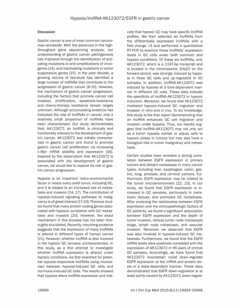
Discussion
Gastric cancer is one of most common carcino-mas worldwide. With the advances in the high-throughput gene sequencing analysis, our understanding of gastric cancer pathogenesis has improved through the identification of acti-vating mutations in and amplifications of onco-genes [14], and inactivating mutations in tumor suppressive genes [15]. In the past decade, a growing volume of literature has identified a large number of miRNAs that contribute to the progression of gastric cancer [8-10]. However, the mechanism of gastric cancer progression, including the factors that promote cancer cell invasion, proliferation, apoptosis-resistance and chemo-therapy resistance remain largely unknown. Although accumulating evidence has indicated the role of lncRNAs in cancer, only a relatively small proportion of lncRNAs have been characterized. Our study demonstrates that, AK123072, an lncRNA, is clinically and functionally relevant to the development of gas-tric cancer. AK123072 was initially character-ized in gastric cancer and found to promote gastric cancer cell proliferation via increasing c-Myc mRNA stability and expression [16]. Inspired by the observation that AK123072 is associated with the development of gastric cancer, we would like to explore its role in gas-tric cancer progression.
Hypoxia is an important micro-environmental factor in nearly every solid tumor, including GC, and it is related to an increased risk of metas-tasis and invasion [14, 17]. The contribution of hypoxia-induced signaling pathways to malig-nancy is of great interest [17-19]. Previous stud-ies found that many protein-coding genes asso-ciated with hypoxia correlation with GC metas-tasis and invasion [20]. However, the exact mechanism in this process has not been thor-oughly elucidated. Recently, mounting evidence suggests that the expression of many lncRNAs is altered in different types of human cancer [21]. However, whether lncRNA is also involved in the hypoxic GC remains uncharacterized. In this study, as a first attempt to investigate whether lncRNA expression is altered under hypoxic conditions, we first searched for poten-tial hypoxia-responsive lncRNAs using microar-rays between hypoxia-induced GC cells and normoxia-induced GC cells. The results showed that hypoxia alters lncRNA expression and indi-
cate that hypoxic GC may have specific lncRNA profiles. We then selected six lncRNAs from the differentially expressed lncRNAs with a fold change >3 and performed a quantitative RT-PCR to examine these lncRNAs’ expression levels in GC cells under both normoxic and hypoxic conditions. Of these six lncRNAs, only AK123072, which is a 1197-bp transcript and is located in the chromosome 10q22 on the forward strand, was strongly induced by hypox-ia in three GC cells and up-regulated in GC samples. In addition, lncRNA-AK123072 was induced by hypoxia at a time-dependent man-ner in different GC cells. These data indicate the specificity of lncRNA-AK123072 in hypoxic induction. Moreover, we found that AK123072 mediated hypoxia-induced GC migration and invasion in vitro and in vivo. To our knowledge, this study is the first report demonstrating that an lncRNA enhances GC cell migration and invasion under hypoxia. Thus, our results sug-gest that lncRNA-AK123072 may not only act as a tumor hypoxia marker or adjust cells to hypoxic stress in tumors but may also have a biological role in tumor malignancy and metas- tasis.
Certain studies have revealed a strong corre- lation between EGFR expression in primary tumors and distant metastasis in many cancer types, including liver, esophageal, colon, gas-tric, lung, prostate, and cervical cancers. Fur- thermore, EGFR expression may be related to the tumor microenvironment [22, 23]. In our study, we found that EGFR expression is in- creased in GC samples, particularly in meta-static tissues, and promotes GC metastasis. After analyzing the relationship between EGFR expression and the clinicopathologic factors of GC patients, we found a significant association between EGFR expression and the depth of tumor invasion, clinical tumor node metastasis stage, lymph node metastasis, and vascular invasion. Moreover, we observed that EGFR was also involved in hypoxia-induced GC me- tastasis. Furthermore, we found that the EGFR mRNA levels were positively correlated with the expression of AK123072 in 95 pairs of clinical GC samples. Accordingly, we have found that AK123072 knockdown could down-regulate EGFR expression at the mRNA and protein lev-els in a dose-dependent manner. These data demonstrated that EGFR down-regulation is at least partly caused by AK123072 down-regula-

Hypoxia/lncRNA-AK123072/EGFR in gastric cancer
19967 Int J Clin Exp Med 2015;8(11):19954-19968
tion in GC. However, the mechanism of AK123072 regulation of EGFR is still unknown. Recent reports demonstrated that mature microRNAs repress protein expression primari-ly through base pairing of a seed region with the 3’-UTR of EGFR [24]. Furthermore, several studies reported that EGFR expression is acti-vated by demethylation of the EGFR CpG island in the development of cancer, including GC [25-27]. Recently, increasing evidence confirmed that lncRNA can regulate DNA methylation of protein-coding genes during the development of disease [28-30].
In conclusion, our results indicated that lncRNA-AK123072 expression is frequently increased in hypoxic GC and promotes hypoxia-induced GC metastasis and invasion. EGFR, a metasta-sis associated gene in cancer that is involved in hypoxia-induced GC metastasis, was identified as a functional gene that was regulated by AK123072 through DNA demethylation. EGFR up-regulation by AK123072 mediates hypoxia-induced GC cell migration and invasion. Further study will explore the exact mechanism of EGFR DNA demethylation that is induced by AK123072. In summary, this finding suggests that the hypoxia/AK123072/EGFR pathway may contribute to the development of new anti-cancer therapeutics directed against hypoxic tumor targets.
Disclosure of conflict of interest
None.
Address correspondence to: Zhen Yang, Department of Gastrointestinal Surgery, 1st Affiliated Hospital of Zhengzhou University, China. E-mail: [email protected]
References
[1] Xie X, Liu HT, Mei J, Ding FB, Xiao HB, Hu FQ, Hu R, Wang MS. LncRNA HMlincRNA717 is down-regulated in non-small cell lung cancer and associated with poor prognosis. Int J Clin Exp Pathol 2014; 7: 8881-6.
[2] Zhang Y, Ma M, Liu W, Ding W, Yu H. Enhanced expression of long noncoding RNA CARLo-5 is associated with the development of gastric cancer. Int J Clin Exp Pathol 2014; 7: 8471-9.
[3] Zeng X, Shi H, Wang J, Cui S, Tang H, Zhang X. Long noncoding RNA aberrant expression pro-files after cytoreductive surgery and hyperther-mic intraperitoneal chemotherapy of AGC as-
certained by microarray analysis. Tumour Biol 2015; 36: 5021-94.
[4] Pan W, Liu L, Wei J, Ge Y, Zhang J, Chen H, Zhou L, Yuan Q, Zhou C, Yang M. A functional lncRNA HOTAIR genetic variant contributes to gastric cancersusceptibility. Mol Carcinog 2015; [Epub ahead of print].
[5] Zhang ZZ, Shen ZY, Shen YY, Xu J, Zhao EH, Wang M, Wang CJ, Cao H. HOTAIR long non- coding RNA promotes gastric cancer metasta-sis through suppression of Poly r(C) Binding Protein (PCBP) 1. Mol Cancer Ther 2015; 14: 1162-70.
[6] Li LJ, Zhu JL, Bao WS, Chen DK, Huang WW, Weng ZL. Long noncoding RNA GHET1 pro-motes the development of bladder cancer. Int J Clin Exp Pathol 2014; 7: 7196-205.
[7] Li Q, Shao Y, Zhang X, Zheng T, Miao M, Qin L, Wang B, Ye G, Xiao B, Guo J. Plasma long non-coding RNA protected by exosomes as a poten-tial stable biomarker for gastric cancer. Tumour Biol 2015; 36: 2007-12.
[8] Ma G, Gu D, Lv C, Chu H, Xu Z, Tong N, Wang M, Tang C, Xu Y, Zhang Z, Wang B, Chen J. Genetic variant in 8q24 is associated with prognosis for gastric cancer in a Chinese population. J Gastroenterol Hepatol 2015; 30: 689-95.
[9] Okugawa Y, Toiyama Y, Hur K, Toden S, Saigusa S, Tanaka K, Inoue Y, Mohri Y, Kusunoki M, Boland CR, Goel A. Metastasis-associated long non-coding RNA drives gastric cancer develop-ment and promotes peritoneal metastasis. Carcinogenesis 2014; 35: 2731-9.
[10] Hu Y, Wang J, Qian J, Kong X, Tang J, Wang Y, Chen H, Hong J, Zou W, Chen Y, Xu J, Fang JY. Long noncoding RNA GAPLINC regulates CD44-dependent cell invasiveness and associ-ates with poor prognosis of gastric cancer. Cancer Res 2014; 74: 6890-902.
[11] Chen X, Sun J, Song Y, Gao P, Zhao J, Huang X, Liu B, Xu H, Wang Z. The novel long noncoding RNA AC138128.1 may be a predictive bio-marker ingastric cancer. Med Oncol 2014; 31: 262.
[12] Ding J, Li D, Gong M, Wang J, Huang X, Wu T, Wang C. Expression and clinical significance of the long non-coding RNA PVT1 in humangas-tric cancer. Onco Targets Ther 2014; 7: 1625-30.
[13] He X, Tan X, Wang X, Jin H, Liu L, Ma L, Yu H, Fan Z. C-Myc-activated long noncoding RNA CCAT1 promotes colon cancer cell proliferation and invasion. Tumour Biol 2014; 35: 12181-8.
[14] Xu TP, Huang MD, Xia R, Liu XX, Sun M, Yin L, Chen WM, Han L, Zhang EB, Kong R, De W, Shu YQ. Decreased expression of the long non-cod-ing RNA FENDRR is associated with poor prog-nosis in gastric cancer and FENDRR regulates gastric cancer cell metastasis by affecting fi-

Hypoxia/lncRNA-AK123072/EGFR in gastric cancer
19968 Int J Clin Exp Med 2015;8(11):19954-19968
bronectin1 expression. J Hematol Oncol 2014; 7: 63.
[15] Zhang S, Chen S, Yang G, Gu F, Li M, Zhong B, Hu J, Hoffman A, Chen M. Long noncoding RNA HOTAIR as an independent prognostic marker in cancer: a meta-analysis. PLoS One 2014; 9: e105538.
[16] Xia T, Liao Q, Jiang X, Shao Y, Xiao B, Xi Y, Guo J. Long noncoding RNA associated-competing endogenous RNAs in gastric cancer. Sci Rep 2014; 4: 6088.
[17] Chen S, Li P, Xiao B, Guo J. Long noncoding RNA HMlincRNA717 and AC130710 have been officially named as gastric cancer associated transcript 2 (GACAT2) and GACAT3, respective-ly. Tumour Biol 2014; 35: 8351-2.
[18] Ma MZ, Chu BF, Zhang Y, Weng MZ, Qin YY, Gong W, Quan ZW. Long non-coding RNA CCAT1 promotes gallbladder cancer develop-ment via negative modulation of miRNA-218-5p. Cell Death Dis 2015; 6: e1583.
[19] Colon Cancer Associated Transcript-1 (CCAT1) Expression in Adenocarcinoma of the Stomach. Mizrahi I, Mazeh H, Grinbaum R, Beglaibter N, Wilschanski M, Pavlov V, Adileh M, Stojadinovic A, Avital I, Gure AO, Halle D, Nissan A. J Cancer 2015; 6: 105-10.
[20] Xu MD, Qi P, Weng WW, Shen XH, Ni SJ, Dong L, Huang D, Tan C, Sheng WQ, Zhou XY, Du X. Long non-coding RNA LSINCT5 predicts nega-tive prognosis and exhibits oncogenic activity in gastric cancer. Medicine (Baltimore) 2014; 93: e303.
[21] Nagatsuma AK, Aizawa M, Kuwata T, Doi T, Ohtsu A, Fujii H, Ochiai A. Expression profiles of HER2, EGFR, MET and FGFR2 in a large co- hort of patients with gastric adenocarcinoma. Gastric Cancer 2015; 18: 227-38.
[22] Zhang J, Cao J, Li J, Zhang Y, Chen Z, Peng W, Sun S, Zhao N, Wang J, Zhong D, Zhang X, Zhang J. A phase I study of AST1306, a novel irreversible EGFR and HER2 kinase inhibitor, in patients with advanced solid tumors. J Hematol Oncol 2014; 7: 22.
[23] Ye B, Jiang LL, Xu HT, Zhou DW, Li ZS. Expression of PI3K/AKT pathway in gastric cancer and its blockade suppresses tumor growth and metastasis. Int J Immunopathol Pharmacol 2012; 25: 627-36.
[24] Xie J, Chen M, Zhou J, Mo MS, Zhu LH, Liu YP, Gui QJ, Zhang L, Li GQ. miR-7 inhibits the inva-sion and metastasis of gastric cancer cells by suppressing epidermal growth factor receptor expression. Oncol Rep 2014; 31: 1715-22.
[25] Jiang L, Chen Y, Li Y, Lan T, Wu M, Wang Y, Qian H. Type II cGMP-dependent protein kinase in-hibits ligand-induced activation of EGFR ingas-tric cancer cells. Mol Med Rep 2014; 9: 1405-9.
[26] Chaturvedi R, Asim M, Piazuelo MB, Yan F, Barry DP, Sierra JC, Delgado AG, Hill S, Casero RA Jr, Bravo LE, Dominguez RL, Correa P, Polk DB, Washington MK, Rose KL, Schey KL, Morgan DR, Peek RM Jr, Wilson KT. Activation of EGFR and ERBB2 by Helicobacter pylori re-sults in survival of gastricepithelial cells with DNA damage. Gastroenterology 2014; 146: 1739-51.e14.
[27] Gala K, Chandarlapaty S. Molecular pathways: HER3 targeted therapy. Clin Cancer Res 2014; 20: 1410-6.
[28] Agaimy A, Rau TT, Hartmann A, Stoehr R. SMARCB1 (INI1)-negative rhabdoid carcino-mas of the gastrointestinal tract: clinicopatho-logic and molecular study of a highly aggres-sive variant with literature review. Am J Surg Pathol 2014; 38: 910-20.
[29] Tajiri R, Ooi A, Fujimura T, Dobashi Y, Oyama T, Nakamura R, Ikeda H. Intratumoral heteroge-neous amplification of ERBB2 and subclonal genetic diversity in gastric cancers revealed by multiple ligation-dependent probe amplifica-tion and fluorescence in situ hybridization. Hum Pathol 2014; 45: 725-34.
[30] Shi M, Shi H, Ji J, Cai Q, Chen X, Yu Y, Liu B, Zhu Z, Zhang J. Cetuximab Inhibits Gastric Cancer Growth in vivo, Independent of KRAS Status. Curr Cancer Drug Targets 2014; 14: 217-24.


![lncRNA Mediated Hijacking of T-cell Hypoxia Response Pathway … · processing and presentation pathway which includes phagosome-lysosome fusion [3]. Once the soluble antigens are](https://img.pdfslide.net/doc/110x75/5f0d9d1a7e708231d43b3948/lncrna-mediated-hijacking-of-t-cell-hypoxia-response-pathway-processing-and-presentation.jpg)