Embed Size (px)
Citation preview
41http://www.ecevr.org/
CLINICAL EXPERIMENTALVACCINERESEARCH
Original article
Introduction
Dengue viruses are belonging to genus Flavivirus of the family Flaviviridae. They are
transmitted via mosquito Aedes aegypti. The dengue virus group comprises an inde-
pendent serogroup of four closely related but antigenically distinct serotypes 1, 2, 3,
and 4 [1]. It is estimated that more than 2.5 billion people are at the risk of infection by
dengue virus. Dengue fever, dengue hemorrhagic fever (DHF), and dengue shock syn-
drome are important viral diseases caused by any of the four serotypes [2]. There is still
no approved vaccine or drug for this virus. One of the challenges of dengue infection is
that, although antibodies produced against the infecting serotype confer lifelong im-
munity against the infecting serotype, re-infection by another serotype has been asso-
ciated with sever disease. Individuals with secondary dengue infection produce cross-
reactive and non-neutralizing antibodies and also memory T cells, which can enhance
© Korean Vaccine Society.This is an Open Access article distributed under the terms of the Creative Commons Attribution Non-Com-mercial License (http://creativecommons.org/licenses/by-nc/3.0) which permits unrestricted non-commercial use, distribution, and reproduction in any medium, pro-vided the original work is properly cited.
K O R E A N V A C C I N E S O C I E T Y
K O R E A N V A C C I N E S O C I E T Y
K O R E A N A C C I N E O C I E T Y
VS
Clin Exp Vaccine Res 2016;5:41-49http://dx.doi.org/10.7774/cevr.2016.5.1.41pISSN 2287-3651 • eISSN 2287-366X
Hossein Fahimi1, Majid Sadeghizadeh2, Mahshid Mohammadipour3
1Department of Molecular and Cellular Sciences, Faculty of Advanced Sciences and Technology, Pharmaceutical Sciences Branch, Islamic Azad University (IAUPS), Tehran; 2Department of Molecular Genetics, Faculty of Biological Sciences, Tarbiat Modares University, Tehran; 3Blood Transfusion Research Center, High Institute for Research and Education in Transfusion Medicine, Tehran, Iran
Received: July 31, 2015Revised: September 10, 2015Accepted: October 2, 2015
Corresponding author: Majid Sadeghizadeh, PhDDepartment of Molecular Genetics, Faculty of Biological Sciences, Tarbiat Modares University, P.O. Box 14115-154, Tehran, IranTel: +98-21-8288-4409, Fax: +98-21-8288-4484E-mail: [email protected]
No potential conflict of interest relevant to this article was reported.
Purpose: Dengue virus infection is now a global problem. Currently, there is no licensed vac-cine or proven antiviral treatment against this virus. All four serotypes (1-4) of dengue virus can infect human. An effective dengue vaccine should be tetravalent to induce protective immune responses against all four serotypes. Most of dengue vaccine candidates are monovalent, or in the form of physically mixed multivalent formulations. Recently envelope protein domain III of virus is considered as a vaccine candidate, which plays critical roles in the most important viral activities. Development of a tetravalent protein subunit vaccine is very important for equal induction of immune system and prevention of unbalanced immunity. Here, we have presented and used a rational approach to design a tetravalent dengue vaccine candidate.Materials and Methods: We designed a multi domain antigen by fusing four consensus do-main III sequences together with appropriate hydrophobic linkers and used several types of bioinformatics software and neural networks to predict structural and immunological proper-ties of the designed tetravalent antigen.Results: We designed a tetravalent protein (EDIIIF) based on domain III of dengue virus enve-lope protein. According to the results of the bioinformatics analysis, the constructed models for EDIIIF protein were structurally stable and potentially immunogenic.Conclusion: The designed tetravalent protein can be considered as a potential dengue vac-cine candidate. The presented approach can be used for rational design and in silico evalua-tion of chimeric dengue vaccine candidates.
Keywords: In silico, Dengue virus, Vaccine
In silico analysis of an envelope domain III-based multivalent fusion protein as a potential dengue vaccine candidate
Hossein Fahimi et al • An in silico dengue vaccine
42 http://www.ecevr.org/ http://dx.doi.org/10.7774/cevr.2016.5.1.41
virus replication and over production of cytokines, that in
turn are risk factors for DHF [3]. The leading hypothesis to ex-
plain pathogenesis of dengue virus is the antibody-dependent
enhancement of infection; which suggests cross reactive sub-
neutralizing antibodies may enhance the severity of infection
[2]. Therefore, an effective vaccine against dengue virus must
induce a simultaneous and strong immune response against
all four serotypes.
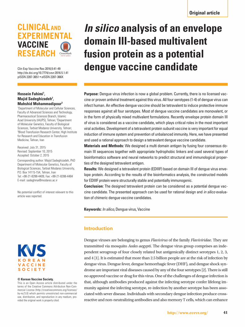
Dengue virus genome comprised of an 11-kb plus-sensed
RNA, surrounded by an icosahedral scaffold and covered by
a lipid envelope. The genome contains one open reading
frame encoding a poly-protein, which after cleavage by host
and viral enzymes, produces three structural and seven non-
structural proteins (Fig. 1) [4]. The envelope protein (E pro-
tein) is the major component of the virus, which contains
specific sites for host cell attachment and fusion. This protein
is a dominant antigen, which is potent for induction of im-
mune response in infected hosts and eliciting virus-neutral-
izing antibodies [5,6].
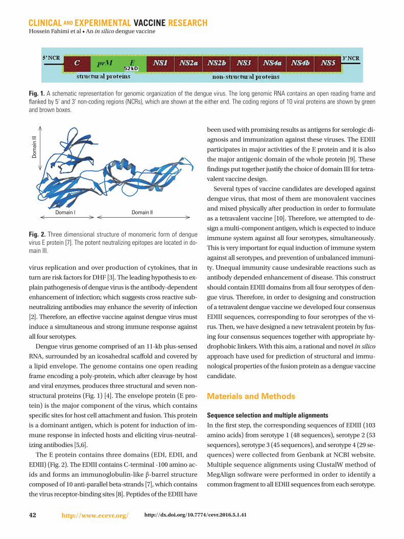
The E protein contains three domains (EDI, EDII, and
EDIII) (Fig. 2). The EDIII contains C-terminal -100 amino ac-
ids and forms an immunoglobulin-like β-barrel structure
composed of 10 anti-parallel beta-strands [7], which contains
the virus receptor-binding sites [8]. Peptides of the EDIII have
been used with promising results as antigens for serologic di-
agnosis and immunization against these viruses. The EDIII
participates in major activities of the E protein and it is also
the major antigenic domain of the whole protein [9]. These
findings put together justify the choice of domain III for tetra-
valent vaccine design.
Several types of vaccine candidates are developed against
dengue virus, that most of them are monovalent vaccines
and mixed physically after production in order to formulate
as a tetravalent vaccine [10]. Therefore, we attempted to de-
sign a multi-component antigen, which is expected to induce
immune system against all four serotypes, simultaneously.
This is very important for equal induction of immune system
against all serotypes, and prevention of unbalanced immuni-
ty. Unequal immunity cause undesirable reactions such as
antibody depended enhancement of disease. This construct
should contain EDIII domains from all four serotypes of den-
gue virus. Therefore, in order to designing and construction
of a tetravalent dengue vaccine we developed four consensus
EDIII sequences, corresponding to four serotypes of the vi-
rus. Then, we have designed a new tetravalent protein by fus-
ing four consensus sequences together with appropriate hy-
drophobic linkers. With this aim, a rational and novel in silico
approach have used for prediction of structural and immu-
nological properties of the fusion protein as a dengue vaccine
candidate.
Materials and Methods
Sequence selection and multiple alignmentsIn the first step, the corresponding sequences of EDIII (103
amino acids) from serotype 1 (48 sequences), serotype 2 (53
sequences), serotype 3 (45 sequences), and serotype 4 (29 se-
quences) were collected from Genbank at NCBI website.
Multiple sequence alignments using ClustalW method of
MegAlign software were performed in order to identify a
common fragment to all EDIII sequences from each serotype.
Fig. 1. A schematic representation for genomic organization of the dengue virus. The long genomic RNA contains an open reading frame and flanked by 5' and 3' non-coding regions (NCRs), which are shown at the either end. The coding regions of 10 viral proteins are shown by green and brown boxes.
Fig. 2. Three dimensional structure of monomeric form of dengue virus E protein [7]. The potent neutralizing epitopes are located in do-main III.
Dom
ain
III
Domain I Domain II
Hossein Fahimi et al • An in silico dengue vaccine
43http://www.ecevr.org/http://dx.doi.org/10.7774/cevr.2016.5.1.41
Construct designNet servers and several types of bioinformatics software are
applied in our study [11-22]. The in silico gene analysis and
optimization of the chimeric gene was performed using
GeneRunner, EditSeq and online software such as Optimizer
[11] and NEB cutter [12]. The mRNA secondary structure of
the chimeric gene was predicted using Mfold program [13].
Bioinformatics analysis of recombinant fusion proteinFor prediction of primary properties of fusion protein, EditSeq
and online ProtParam software were used (http://web.expasy.
org/protparam/). Secondary-structure prediction for fusion
protein was performed by using the neural-network-based al-
gorithm program PSIPRED (http://bioinf.cs.ucl.ac.uk/
psipred/) and GOR4 method [14]. The Chou and Fasman
method used for further analysis of secondary structure of
protein (http://web.expasy.org/protscale/). Tertiary structure
prediction was performed by three methods including Mod-
eller (http://salilab.org/modeller/), TASSER (http://zhanglab.
ccmb.med.umich.edu/I-TASSER/), and FOLDpro (http://
mine10.ics.uci.edu/). The stability of produced structural
models was analyzed by Pymol and swiss-pdbviewer. Evalua-
tion of model stability was performed based on energy mini-
mization and Ramachandran plot. The Ramachandran plots
were created by Procheck based at the biotech validation site
(http://nihserver.mbi.ucla.edu/SAVS1/). Relative solvent ac-
cessibility of different residues was evaluated by online
SCRATCH server (http://scratch.proteomics.ics.uci.edu/).
The DiNNNA online software was used for disulfide bound
prediction (http://clavius.bc.edu/~clotelab/DiANNA/).
Prediction of B-cell epitopesWeb-based algorithm servers, Bcepred (http://www.imtech.
res.in/raghava/bcepred/) and Discotope (http://www.cbs.dtu.
dk/services/discotop/), were used for prediction of continuous
and discontinue B-cell epitopes, respectively. MHC super type
A1 by NetCTL 1.2 Server (http://www.cbs.dtu.dk/services/
NetCTL/). Finally, we used the VaxiJen server to predict the im-
munogenicity of the whole tetravalent fusion protein (http://
www.ddg-pharmfac.net/vaxijen/VaxiJen/VaxiJen.html).
Results
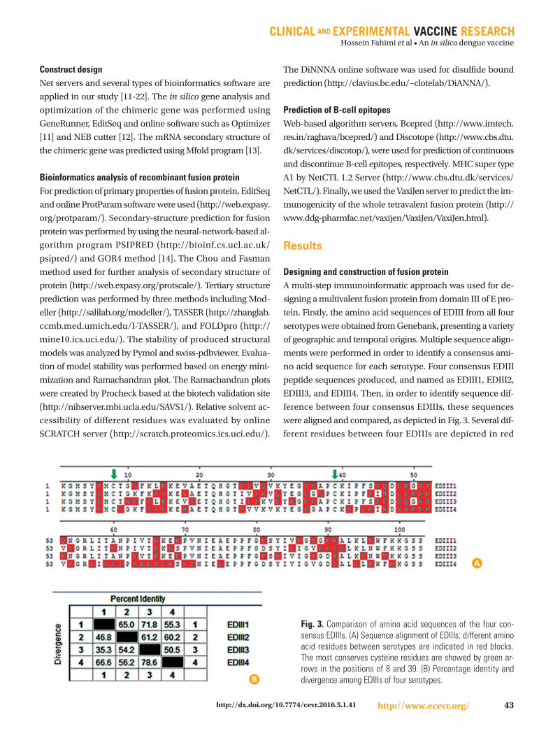
Designing and construction of fusion proteinA multi-step immunoinformatic approach was used for de-
signing a multivalent fusion protein from domain III of E pro-
tein. Firstly, the amino acid sequences of EDIII from all four
serotypes were obtained from Genebank, presenting a variety
of geographic and temporal origins. Multiple sequence align-
ments were performed in order to identify a consensus ami-
no acid sequence for each serotype. Four consensus EDIII
peptide sequences produced, and named as EDIII1, EDIII2,
EDIII3, and EDIII4. Then, in order to identify sequence dif-
ference between four consensus EDIIIs, these sequences
were aligned and compared, as depicted in Fig. 3. Several dif-
ferent residues between four EDIIIs are depicted in red
Fig. 3. Comparison of amino acid sequences of the four con-sensus EDIIIs. (A) Sequence alignment of EDIIIs; different amino acid residues between serotypes are indicated in red blocks. The most conserves cysteine residues are showed by green ar-rows in the positions of 8 and 39. (B) Percentage identity and divergence among EDIIIs of four serotypes.B
A

Hossein Fahimi et al • An in silico dengue vaccine
44 http://www.ecevr.org/ http://dx.doi.org/10.7774/cevr.2016.5.1.41
Fig. 4. Schematic representation of the EDIIIF construct.
Table 1. The properties of EDIIIF protein, predicted by temp-prot considering the antigenicity and the number and position of disulfide bonds
Antigen Total No. of cysteine Cysteine 1-position Cysteine 2-position Predicted No. of bonds Predicted probability of antigenicity
EDIII1 2 8 39 1 0.83EDIII2 2 8 39 1 0.88EDIII3 2 8 39 1 0.86EDIII4 2 8 39 1 0.79EDIIIF 8 8 39 4 0.94
131 162254 285377 408
blocks. As depicted in Fig. 3B, EDIII4 sequence shows maxi-
mum divergence with other EDIIIs, whereas the EDIII1 and
EDIII3 are the most identical sequences (with 71.8% identity
and 35.5% divergence).
Consensus EDIII sequences were used for designing a tet-
ravalent fusion protein, which named as EDIIIF (GenBank
accession No. JN985899). In order to efficient separation of
the EDIIIs in fusion protein, linkers containing four repeated
EAAAK sequences were inserted between EDIII sequences. It
has been shown that the salt bridge Glu--Lys+ between re-
peated Ala residues can stabilize helix formation [15], which
expected to provide independent folding of four EDIII do-
mains. In order to, efficient purification of EDIIIF protein, a
6×-His tag sequence was added to carboxyl terminus of de-
signed protein. The arrangement of fragments junctions and
linker sites are shown in Fig. 4.
Prediction of primary and secondary structure of EDIIIF proteinOn the basis of data obtained from prediction of EDIIIF prop-
erties by EditSeq software and online software ProtParam (in
ExPASy tools), it is an approximately 52 kD protein (478 ami-
no acid) with pH isoelectric 7.30. The instability index for this
protein was computed to be 31.87, which classifies the pro-
tein as a stable protein in Escherichia coli. A similar analysis
carried out using DiNNNA online software for disulfide
bound prediction (Table 1). As depicted in the Table 1, pre-
diction for numbers and positions of disulfide bonds in
EDIIIF protein, showed that all of eight cysteine residues par-
ticipate in disulfide bound formation. Clearly, there is one in-
tra-domain disulfide bridge in each domain and no any inter-
domain disulfide bond was predicted. These properties are
essential for accurate conformation of EDIIIF protein, which
predicted by temp-prot server analysis as well. Furthermore,
predicted probability of antigenicity according the analysis
by this server was 94%, which showed the EDIIIF has enough
antigenicity as a vaccine candidate. The predicted antigenici-
ty for the chimeric protein was higher than the predicted an-
tigenicity for each EDIII proteins in separate form.
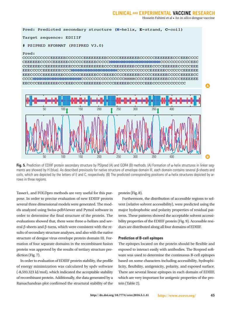
The secondary structure of the EDIIIF protein was predict-
ed by online software. Several prediction methods were used
and the results were compared for evaluation of the structure
of this protein. These results indicated that helix structures lie
in the regions of amino acid 104 to 123, 227 to 246, and 350 to
369, which are related to the hydrophobic amino acids en-
tered between EDIII domains (Fig. 5).
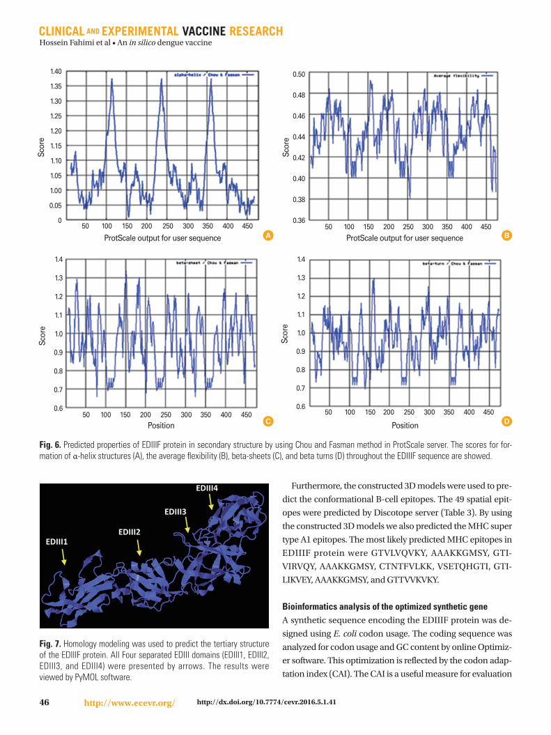
Furthermore, the accuracy of this secondary structure pre-
diction confirmed using ProtScale server. These properties
include the scores for α-helix structures, the average flexibili-
ty, the beta-sheets, and the beta turns (Fig. 6).
Prediction of tertiary structure of the EDIIIF proteinComparative and homology modeling of the EDIIIF se-
quence was exploited to produce three dimensional models
of the fusion protein. We used three methods for construc-
tion of three dimensional models of protein. The Modeller,
Hossein Fahimi et al • An in silico dengue vaccine
45http://www.ecevr.org/http://dx.doi.org/10.7774/cevr.2016.5.1.41
Tasser1, and FOLDpro methods are very useful for this pur-
pose. In order to precise evaluation of new EDIIIF protein
several three dimensional models were generated. The mod-
els analyzed using Swiss-pdbViewer and Pymol software in
order to determine the final structure of the protein. The
evaluations showed that, there were three α-helixes and sev-
eral β-sheets and β-turns, which were consistent with the re-
sults of secondary structure analyses, and also with the native
structure of dengue virus envelope protein domain III. For-
mation of four separate domains in the recombinant fusion
protein was approved by the results of tertiary structure pre-
diction (Fig. 7).
In order to evaluation of EDIIIF protein stability, the profile
of energy minimization was calculated by spdv software
(-8,593.323 kJ/mol), which indicated the acceptable stability
of recombinant protein. Additionally, the data generated by a
Ramachandran plot confirmed the structural stability of the
protein (Fig. 8).
Furthermore, the distribution of accessible regions to sol-
vent (relative solvent accessibility), were predicted using the
major hydrophobic and polarity properties of residual pat-
terns. These patterns showed the acceptable solvent accessi-
bility properties of the EDIIIF protein (Fig. 9). Accessible resi-
dues are distributed along all four domains of EDIIIF.
Prediction of B-cell epitopesThe epitopes located on the protein should be flexible and
exposed to interact easily with antibodies. The Bcepred soft-
ware was used to determine the continuous B-cell epitopes
based on some characters including accessibility, hydrophi-
licity, flexibility, antigenicity, polarity, and exposed surface.
There are several linear epitopes in each domain of EDIIIF,
which are very important for antigenic properties of the pro-
tein (Table 2).
Fig. 5. Prediction of EDIIIF protein secondary structure by PSIpred (A) and GOR4 (B) methods. (A) Formation of α-helix structures in linker seg-ments are showed by H (blue). As described previously for native structure of envelope domain III, each domain contains several β-sheets and coils, which are depicted by the letters of E and C, respectively. (B) The predicted corresponding positions of α-helix structures depicted by ar-rows in three regions.
50 100 150 200 250 300 350 400
50 100 150 200 250 300 350 400 B
A
Hossein Fahimi et al • An in silico dengue vaccine
46 http://www.ecevr.org/ http://dx.doi.org/10.7774/cevr.2016.5.1.41
Fig. 7. Homology modeling was used to predict the tertiary structure of the EDIIIF protein. All Four separated EDIII domains (EDIII1, EDIII2, EDIII3, and EDIII4) were presented by arrows. The results were viewed by PyMOL software.
Furthermore, the constructed 3D models were used to pre-
dict the conformational B-cell epitopes. The 49 spatial epit-
opes were predicted by Discotope server (Table 3). By using
the constructed 3D models we also predicted the MHC super
type A1 epitopes. The most likely predicted MHC epitopes in
EDIIIF protein were GTVLVQVKY, AAAKKGMSY, GTI-
VIRVQY, AAAKKGMSY, CTNTFVLKK, VSETQHGTI, GTI-
LIKVEY, AAAKKGMSY, and GTTVVKVKY.
Bioinformatics analysis of the optimized synthetic geneA synthetic sequence encoding the EDIIIF protein was de-
signed using E. coli codon usage. The coding sequence was
analyzed for codon usage and GC content by online Optimiz-
er software. This optimization is reflected by the codon adap-
tation index (CAI). The CAI is a useful measure for evaluation
Fig. 6. Predicted properties of EDIIIF protein in secondary structure by using Chou and Fasman method in ProtScale server. The scores for for-mation of α-helix structures (A), the average flexibility (B), beta-sheets (C), and beta turns (D) throughout the EDIIIF sequence are showed.
1.40
1.35
1.30
1.25
1.20
1.15
1.10
1.05
1.00
0.05
0
Scor
e
50 100 150 200 250 300 350 400 450
ProtScale output for user sequence
1.4
1.3
1.2
1.1
1.0
0.9
0.8
0.7
0.6
Scor
e
50 100 150 200 250 300 350 400 450Position
A
1.4
1.3
1.2
1.1
1.0
0.9
0.8
0.7
0.6
Scor
e
Position
50 100 150 200 250 300 350 400 450DC
0.50
0.48
0.46
0.44
0.42
0.40
0.38
0.36
Scor
e
ProtScale output for user sequence 50 100 150 200 250 300 350 400 450
B

Hossein Fahimi et al • An in silico dengue vaccine
47http://www.ecevr.org/http://dx.doi.org/10.7774/cevr.2016.5.1.41
Table 2. The linear epitopes predicted in EDIIIF protein based on different parameters by using Bcepred softwarea)
Prediction parameter Epitope peptide sequence
Flexibility IPFSTQDE, PIVTDKE, LSWFKKGSS, VQYEGDG, PIVTEKD, LNWFKKGSS, FVLKKEV, IPFSTEDGQ, PVVTKKE, INWYKKGSS, EIRDVNK, LHWFRKGSS
Accessibility S FKLEKEVAETQ, QVKYEGTDAP, PFSTQDEKGVTQN, IVTDKEKPVNIEAEPP, KLSWFKKGSSEA, KFKVVKEIAETQ, RVQYEGDGS, MDLEKRHV, VTEKDSPVNIEAEPP, KLNWFKKGSSEA, KEAAAKEAAAKEAAAKKGMSYA, LKKEVSETQHGT, KVEYKGEDAP, TEDGQGKAHNGR, NPVVTKKEEPVNIEAEPP
Antigenic propensity Q HGTVLVQVKYE, PCKIPFS, GESYIVVG, FKVVKEI, GTIVIRVQYE, SPCKIPFEI, LEKRHVLGRLITVNPIV, SYIIIGVEP, CTNTFVLKKEVS, GTILIKVEYK, PCKIPFS, GTTVVKVKYE, PCKVPIEI, VNKEKVVGRIIS, SYIVIGVGD
Exposed surface SFKLEKE, VTDKEKPVNI, KFKVVKE, KLNWFKK, LKKEVSET, KVEYKGE, VVTKKEEPVN, KINWYKKGSSE, KEAAAKK, RDVNKEKV
Hydrophilicity E KEVAETQHGT, KYEGTDAPCK, VTDKEKPVN, FSTQDEKGVTQNG, KKGSSEAA, AETQHGT, QYEGDGSPCK, VTEKDSPVN, KKEVSETQHGT, VEYKGEDAPCK, FSTEDGQGKAHNGR, KKEEPVN, YKKGSSEAAAKEAAAKEAAAKE, DKEMAETQHGTT, RDVNKEK, AENTNSVTN, RKGSSHH, KINWYKKGSSEA, KFSIDKEMAETQHGTT, KVKYEGA, EIRDVNKEKVVGR, PFAENTNSVTN, FRKGSSHH
Polarity S FKLEKEVAETQH, IVTDKEKPVNIE, KLSWFKKGSSE, KFKVVKEIAETQH, EIMDLEKRHVLGRL, KLNWFKKGSSE, KEAAAKEAAAKEAAAKK, LKKEVSETQH, KVEYKGEDA, GKAHNGRL, PVVTKKEEPVNIE, KINWYKKGSSE, KFSIDKEMAETQH, KVPIEIRDVNKEKVVGR, TLHWFRKGSSHHHHHH
a)A linear epitope, AENTNSVT, was also predicted in a turn.
Table 3. The identified 49 B-cell epitope residues out of 478 total residues as conformational B-cell epitopes of EDIIIF; which were predicted by Discotope server
Amino acid position and sequence
6 (Lys) 143-145 (Glu-Thr-Gln) 285-286 (Cys-Lys) 416 (Asp)20-22 (Glu-Thr- Gln) 263 (Glu) 292-299 (Glu-Asp-Gly-Gln-Gly-Lys-Ala-His) 418 (Asn)83 (Tyr) 268 (Gln) 304-305 (Ile-Thr) 420-421 (Glu-Lys)101 (Gly) 279 (Lys) 391 (Gln) 447-450 (Pro-Phe-Gly_asp)126-127 (Met-Ser) 281-283 (Glu-Asp-Ala) 393 (Gly) 467-475 (Phe-Arg-Lys-Gly-Ser-Ser-His-His-His)
180
135
90
45
0
-45
-90
-135
Psi (
degr
ees)
Ramachandran plot file
-180 -135 -90 -45 0 45 90 135 180
Fig. 8. Evaluation of model stability by using the Ramachandran plot. According to the plot statistics, more than 82% of amino acid residues are in the most favored regions (A, B, L), and 13% are in additional allowed regions (a, b, l, p); whereas only 2.7% are in gen-erously allowed (-a, -b, -l, -p) and 1.7% are in disallowed regions. Accordingly, the constructed model has a good quality.
Fig. 9. Prediction of relative solvent accessibility by using Scratch server.
of the relative adaptiveness of desired sequences with the se-
quence of highly expressed genes in the same host. The opti-
mized gene (edIIIf) showed a CAI of 1.0, compared to the pri-
mary deduced gene, which was only 0.7. The GC content of
this sequence optimized on about 52%, which is appropriate
for efficient expression in E. coli host. Furthermore, the nec-
essary restriction enzyme sequence sites (NdeI and XhoI)
were introduced at the ends of the coding sequence for clon-
ing purposes.
Finally, secondary structure of corresponding mRNA for
edIIIf gene was predicted. As the folding pattern of mRNA is
very important factor affecting translation efficiency, we used
the mFold web based software, in order to determine the po-
tential folding pattern of mRNA. The minimum free energy
for secondary structure formed by mRNA molecule was also
predicted (dG=-17.12). The results of this prediction showed
Hossein Fahimi et al • An in silico dengue vaccine
48 http://www.ecevr.org/ http://dx.doi.org/10.7774/cevr.2016.5.1.41
enough stability of mRNA for efficient translation in bacterial
host.
Discussion
In silico analysis of vaccine candidates is a useful approach
for rational design of chimeric vaccines [16]. Bioinformatics
tools for predicting antigenic properties and candidate vac-
cines analysis are now a standard approach. Rational design
of vaccines and in silico analysis, combined with in vitro and
in vivo verification, accelerate the vaccine discovery process
by approximately 10-20 fold. Several bioinformatics software
and servers are available that can help in the process for de-
signing of chimeric vaccine design [17].
The minimalistic approach for dengue vaccine comes from
the observations that EDIII based vaccines could induce vi-
rus neutralizing antibodies which were preventive for virus
infection [9]. In the diagnostic context, it has been proposed
that recombinant EDIII peptides containing virus-specific
epitopes could be used for specific serological diagnosis of
infections [18]. Recently, we reported immunogenicity of a
recombinant EDIII protein from dengue virus type-3 in mice
[19]. On the base of these studies, we conclude that the de-
signed recombinant fusion protein also can be used for sero-
logic diagnosis of dengue viruses from other flaviviruses.
Bioinformatics approach used for rational design of re-
combinant EDIIIF protein as a multivalent vaccine candi-
date, and the corresponding synthetic gene was optimized
for appropriate protein expression, considering the codon
usage and GC content of the expression host genome. Since
the efficiency of heterologous protein production can be di-
minished by biased codon usage [20], the synthetic DNA
fragment encoding the EDIIIF protein was constructed based
on the codon usage table of E. coli. This index was increased
from 0.75 in the primary sequence to 0.98 in designed syn-
thetic gene. Furthermore, the percentage and distribution of
GC in the coding gene were adjusted on 52%.
In the protein structure prediction, the EDIIIF protein
models formed four domains that are separated by three
main α-helix structures. In the 3D models of protein the three
α-helix structures are related to the repeated EAAAK residues
(104-123, 227-246, and 350-369), which are inserted between
EDIII domains. The α-helix structures provide independent
conformation possibility for all four domains without any in-
teraction and by minimal steric hindrance between domains
[21]. With these results we could expect that these α-helical
parts could support the stable structure of protein. B-cell epi-
topes for the fusion protein could be predicted on the basis of
the structural predictions and solvent accessibility. Several
methods based on accessibility, hydrophilicity, flexibility, an-
tigenicity, and secondary structure of proteins have been de-
veloped [22]. The integrated results based on different pa-
rameters showed that the most likely B-cell epitopes of this
fusion protein were located in four distinct domains selected
as the EDIII1, EDIII2, EDIII3, and EDIII4.
In conclusion, our results from bioinformatics analysis of
the designed tetravalent EDIIIF, as a model, showed the fea-
sibility of this approach in rational design and in silico evalu-
ation of chimeric dengue vaccines.
ORCID
Hossein Fahimi https://orcid.org/0000-0002-1744-8675
Majid Sadeghizadeh https://orcid.org/0000-0002-2497-3152
Ma hshid Mohammadipour https://orcid.org/0000-0002-
9813-9953
References
1. Kuno G, Chang GJ, Tsuchiya KR, Karabatsos N, Cropp CB.
Phylogeny of the genus Flavivirus. J Virol 1998;72:73-83.
2. Halstead SB. Dengue. Lancet 2007;370:1644-52.
3. Rothman AL. Dengue: defining protective versus patholog-
ic immunity. J Clin Invest 2004;113:946-51.
4. Fields BN, Knipe DM, Howley PM. Fields’ virology. 5th ed.
Philadelphia: Wolters Kluwer Health/Lippincott Williams
& Wilkins; 2007.
5. Crill WD, Chang GJ. Localization and characterization of
flavivirus envelope glycoprotein cross-reactive epitopes. J
Virol 2004;78:13975-86.
6. Roehrig JT, Bolin RA, Kelly RG. Monoclonal antibody map-
ping of the envelope glycoprotein of the dengue 2 virus, Ja-
maica. Virology 1998;246:317-28.
7. Modis Y, Ogata S, Clements D, Harrison SC. Structure of
the dengue virus envelope protein after membrane fusion.
Nature 2004;427:313-9.
8. Crill WD, Roehrig JT. Monoclonal antibodies that bind to
domain III of dengue virus E glycoprotein are the most effi-
cient blockers of virus adsorption to Vero cells. J Virol 2001;
75:7769-73.
9. Chavez JH, Silva JR, Amarilla AA, Moraes Figueiredo LT.
Domain III peptides from flavivirus envelope protein are
Hossein Fahimi et al • An in silico dengue vaccine
49http://www.ecevr.org/http://dx.doi.org/10.7774/cevr.2016.5.1.41
useful antigens for serologic diagnosis and targets for im-
munization. Biologicals 2010;38:613-8.
10. Swaminathan S, Batra G, Khanna N. Dengue vaccines:
state of the art. Expert Opin Ther Pat 2010;20:819-35.
11. Puigbo P, Guzman E, Romeu A, Garcia-Vallve S. OPTIMIZ-
ER: a web server for optimizing the codon usage of DNA
sequences. Nucleic Acids Res 2007;35:W126-31.
12. Vincze T, Posfai J, Roberts RJ. NEBcutter: a program to
cleave DNA with restriction enzymes. Nucleic Acids Res
2003;31:3688-91.
13. Zuker M. Mfold web server for nucleic acid folding and
hybridization prediction. Nucleic Acids Res 2003;31:3406-
15.
14. Garnier J, Gibrat JF, Robson B. GOR method for predicting
protein secondary structure from amino acid sequence.
Methods Enzymol 1996;266:540-53.
15. Arai R, Ueda H, Kitayama A, Kamiya N, Nagamune T. De-
sign of the linkers which effectively separate domains of a
bifunctional fusion protein. Protein Eng 2001;14:529-32.
16. Amani J, Mousavi SL, Rafati S, Salmanian AH. In silico
analysis of chimeric espA, eae and tir fragments of Esche-
richia coli O157:H7 for oral immunogenic applications.
Theor Biol Med Model 2009;6:28.
17. Koraka P, Martina BE, Osterhaus AD. Bioinformatics in
new generation flavivirus vaccines. J Biomed Biotechnol
2010;2010:864029.
18. Holbrook MR, Shope RE, Barrett AD. Use of recombinant
E protein domain III-based enzyme-linked immunosor-
bent assays for differentiation of tick-borne encephalitis
serocomplex flaviviruses from mosquito-borne flavivirus-
es. J Clin Microbiol 2004;42:4101-10.
19. Fahimi H, Allahyari H, Hassan ZM, Sadeghizadeh M. Den-
gue virus type-3 envelope protein domain III; expression
and immunogenicity. Iran J Basic Med Sci 2014;17:836-43.
20. Burgess-Brown NA, Sharma S, Sobott F, Loenarz C, Op-
permann U, Gileadi O. Codon optimization can improve
expression of human genes in Escherichia coli: a multi-
gene study. Protein Expr Purif 2008;59:94-102.
21. Tong JC, Tan TW, Ranganathan S. Methods and protocols
for prediction of immunogenic epitopes. Brief Bioinform
2007;8:96-108.
22. Kolaskar AS, Tongaonkar PC. A semi-empirical method
for prediction of antigenic determinants on protein anti-
gens. FEBS Lett 1990;276:172-4.
![Neisseria meningitis serogroup X outbreak in Burkina Faso ... · recent meningitis except in an epidemic reported in 1979 in Uper Volta, now Burkina Faso [3,4,6], that serogroup was](https://img.pdfslide.net/doc/110x75/5f0ebc637e708231d440af89/neisseria-meningitis-serogroup-x-outbreak-in-burkina-faso-recent-meningitis.jpg)