Embed Size (px)
Citation preview

69
Original article
In silico structural and functional analysis of copalyl diphosphate synthaseenzyme in Andrographis paniculata (Burm. f.) Wall. ex Nees:
A plant of immense pharmaceutical valueAayeti Shailaja, Byreddi Bhavani Venkata Bindu, Mote Srinath and Charu Chandra Giri
Centre for Plant Molecular Biology (CPMB), Osmania University, Hyderabad-500007, Telangana State, India
Received April 13, 2018: Revised May 30, 2018: Accepted June 5, 2018: Published online June 30, 2018
Abstract
Andrographis paniculata (Burm. f.) Wall. ex Nees (Acanthaceae) with immense medicinalimportance lacks information on its biosynthetic pathway genes and their regulatory role in theproduction of pharmaceutically important andrographolide. Copalyl diphosphate synthase (CPS)is involved in the production of copalyl diphosphate, a precursor for many bioactive compoundswith particular reference to diterpene lactone. In this study, we elucidated the structural andfunctional aspects of A. paniculata CPS (ApCPS). Composition of amino acids and hydrophobicnature of ApCPS were analysed and identified as non trans-membrane protein. A chloroplasttransit peptide and mitochondrial targeting peptide in ApCPS were identified. Protein secondarystructure prediction has given insight on the distribution of helix (52.52%), loop (45.91%) andstrands (1.56%) in ApCPS. The homology modelling of ApCPS was carried out with SWISS MODEL.The validation of 3D model using PROCHECK revealed that 91.74% of the residues have averaged3D-1D score >= 0.2 which is structurally reliable. In Ramachandran plot, 90.9% amino acidresidues were found in most favoured region. Phylogenetic tree was constructed using MEGA 7.0by taking eudicots, monocots, gymnosperms and fungal species. Among them, ApCPS was clusteredwithin eudicots and closely related to Sesumum indicum in Laminales. Protein-protein interactionstudy using STRING10 revealed that CPS interacts with gibberilic acid and terpene synthase relatedproteins. In Arabidopsis thaliana, CPS coexpression was seen with gibberelic acid related proteins.The present in silico analysis will be useful in understanding the structural, functional andevolutionary diversification of ApCPS.
Key words: Andrographis paniculata (Burm. f.) Wall. ex Nees, ApCPS protein, motifs anddomains, domain linkers, 3D modelling, phylogenetic analysis
Copyright @ 2018 Ukaaz Publications. All rights reserved.Email: [email protected]; Website: www.ukaazpublications.com
1. Introduction
Andrographis paniculata (Burm. f.) Wall. ex Nees (Acanthaceae) isan important medicinal herb and a valuable source for importantditerpene lactone, andrographolide and its derivatives. It hasimmense effect on various diseases and considered to be a valuablesource in medicine. It has pharmacological effects such asantimicrobial (Singha et al., 2003), anti-inflammatory, anti-cancerousand immuno-stimulatory (Kumar et al., 2004; Subramanian et al.,2012; Islam et al., 2018), immuno-modulatory and anti-atherosclerotic (Chao and Lin, 2010). This plant has shown effecton suppression of esophageal cancer and metastasis (Li et al., 2018).The demand for such valuable compound diterpene lactones of thisplant is very high. However, the detailed mechanisms and thebiosynthetic pathway genes are not yet elucidated clearly (Singh etal., 2018).
All the secondary metabolites (specifically diterpenes) of the plantshave a common origin from IPP and DMAPP (Figure 1). These can
Annals of Phytomedicine 7(1): 69-77, 2018DOI: 10.21276/ap.2018.7.1.8; Print ISSN : 2278-9839 and Online ISSN : 2393-9885 7(1):69-77 (2018)
Ann. Phytomed.,
Author for correspondence: Dr. Charu Chandra GiriProfessor, Centre for Plant Molecular Biology (CPMB) , OsmaniaUniversity, Hyderabad-500007, Telangana State, IndiaE-mail: [email protected].: +91-040-27098087
be derived either from MEP or MVA pathway which are interlinkedand have connection between them (Chen et al., 2011; Vranová etal., 2013).The enzyme copalyl diphosphate synthase (CPS)catalyzes conversion of geranyl geranyl diphosphate, to copalyldiphosphate (CPP) which serves as intermediate for all diterpenoidreactions (Beale, 1990; Su et al., 2016).This CPS belongs toisomerase super family which involves in the synthesis ofterpenoids/isoprenoids. CPP is the direct precursor of gibberellicacid synthesis, other phytoalexins and labdane-related diterpenoidsin plants (Prisic et al., 2004; Harris et al., 2005).
Figure 1: Biosynthetic MEP and MVA pathway showing CPS enzymefor the biosynthesis of diterpenoids.

70
The genomic and metabolite studies when combined with variousbioinformatic analysis brings out the unknown diterpenoid scaffoldsand the enzyme information (Andersen-Ranberg et al., 2016). Inour laboratory, we have a comprehensive research programme onA.paniculata distribution, enhanced production of bioactivecompound andrographolide and study of key enzymes/proteins(Neeraja et al., 2015; Parlapally et al., 2015; Zaheer and Giri, 2015;Zaheer et al., 2017a; Zaheer et al., 2017b; ; Bindu et al., 2017;Srinath et al., 2017). In the present communication, in silico studiessuch as structural, functional features and the evolutionaryrelationship were elucidated using various bioinformatics tools.
2. Materials and Methods2.1 Analysis of biochemical properties in ApCPS protein
Amino acid sequence of A. paniculata ent-copalyl diphosphatesynthase (ApCPS) was obtained from NCBI with accession numberAEM00024.1 . Expasy Compute pI/Mw tool is used for theestimation of pI (isoelectric point) and Mw (molecular weight) forthe given protein sequence of ApCPS (Kyte and Doolittle, 1982).For the visualization of hydrophobicity for a peptide sequence thehydropathy plots were developed using Kyte and Doolittle (1982)method for each amino acid. The amino acid composition was shown(in %) using ProtParam tool in Expasy online server (Walker, 2005).
2.2 Prediction of signal peptide sequence
ChloroP was used for finding the choloplast transit peptide (cTP)and TargetP (Emmanuelsson et al., 2000), iPSORT (Bannai et al.,2001, 2002) used to find out the subcellular location and signalpeptide sequences in ApCPS.
2.3 Elucidation of secondary structure of ApCPS and predictionof trans-membrane helices
The secondary structure of ApCPS representing the families ofrelated proteins was characterised using PredictProtein tool. Solventaccessibility and trans-membrane helix prediction was done by thistool. The analysis of trans-membrane helices in ApCPS was doneusing HMMTOP (Hidden and Markov Model Topology of Proteins)as per Tusnady and Simon (2001) and Tied Mixture Hidden MarkovModel (TMHMM). The secondary structure prediction of ApCPSwas carried out using CFFSP prediction server (Chou and Fasman1974; Dor et al., 2006).
2.4 Domain and motif analysis of ApCPS
The prediction of domain in ApCPS was carried out using Interproand NCBI-CD search also used for identifying the super family ofApCPS (Marchler-Bauer et al., 2016).
The Motif analysis was done using multiple Em for motif elicitation(MEME: version 4.10.2). Motif Alignment and Search Tool wasused for computation of pairwise correlation between each pair ofmotifs. Motifs with correlations below 0.60 have little effect onthe accuracy of the E-values computed by MAST. DLP-SVM Domainprediction tool was used for identifying the Domain linkers (Ebinaet al., 2009).
2.5 Tertiary structure prediction / 3D modelling and validationof ApCPS
The tertiary structure (3D) modelling of ApCPS enzyme structurewas predicted using SWISS MODEL server (Kiefer et al., 2008) andPhyre2 tool (Kelley et al., 2015). PROCHECK analysis (Laskowskiet al., 1996) was carried out for the validation and for analysing thestereochemical reliability of the 3D model using Ramachandran plot.
The ligand binding site in the 3D model was predicted using 3D Ligand Site tool (Wass et al., 2010). The 3D model generated by Phyre2was used for generating ligand binding site.
2.6 Phylogenetic analysis, protein interactions and expressionstudies
MEGA 7 software was used for phylogenetic tree construction andevolutionary relationship analysis of ApCPS with other CPSenzymes (dicots-33, monocots-4, gymnosprerms-2 and fungi-2)as per Kumar et al. (2016). The evolutionary history was inferredusing UPGMA method. The optimal tree with the sum of branchlength = 6.99757378 is shown next to the branches. The evolutionarydistances were computed using the Poisson correction method andare in the units of the number of amino acid substitutions per site.All positions containing gaps and missing data were eliminated.There were a total of 623 positions in the final dataset, which wasemployed for construction of phylogenetic tree.
2.6.1 Protein protein interaction and co-expression analysis
STRING10 tool was used for analysing the retrieval of interactingproteins or genes (Szklarczyk et al., 2015). The model plantArabidopsis thaliana CPS was taken as reference protein sequencefor the co expression study of CPS.
3. Results and Discussion3.1 Biochemical property analysis of ApCPS protein
Amino acid sequence analysis in A. paniculata using Compute pI/Mw tool revealed that ApCPS contained 832 amino acid residueswith approximately 2.5 kb size (2496 bp). The theoretical pI valuewas shown as 7.07 and molecular weight was 95370.83 daltons,i.e., 95 Kda. The similar result obtained earlier in ApCPS strengthenedour finding in the present study (Garg et al., 2015).
Prot Scale helps in computing and representing the profile producedby any amino acid scale on a selected protein. The hydrophobicityplots for ApCPS with a window size of 9 and 21 shown in Figure2A and 2B, where the relative weight of the window edges comparedto the window center is 100%. The linear weight variation model isused for development of hydropathy plots without normalization.The more positive value of amino acids indicated that isoleucine,valine and leucine are highly hydrophobic at that region(Figure 2C).
(A)

71
(B)
(C)
Figure 2: Hydropathy plot of ApCPS protein showing peaks for eachamino acid at window size 9 (A); at window size 21 (B);The individual values for 20 amino acids using the scaleHydrophob./Kyte and Doolittle (C).
The amino acid composition and distribution showed that a highestof 11.1% residues of leucine are present in protein which is highlyhydrophobic. After leucine, serine (7.5%) and glutamic acid (7.1%)were having major part of the composition (Figure 3A and 3B). Thetotal number of 103 negatively charged residues of (Asp+Glu) and102 positively charged residues of (Arg + Lys) were present.
(A)
(B)
Figure 3: The amino acid composition of ApCPS using ProtParamtool of ExPASy server (3A); diagrammatic representationof amino acid composition (3B) Lys (K); Ser (S); Thr (T); Ile (I);Glu (E); Pro (P); Arg (R); Gln (Q); Phe (F); Tyr (Y) His (H); Asp (N);Met (M); Cys (C); Val (V); Ala (A); Gly (G); Leu (L); Asp (D); Trp (W).
3.2 Prediction of signal peptide sequence
A chloroplast transit peptide (cTP) sequence containing 27 aminoacids was identified in ApCPS. iPSORT prediction indicated that theprotein sequence has no N-terminal sorting signal but having amitochondrial targeting peptides in it (Figure 4). In an earlier studythe presence of chloroplast transit peptide and mitochondrialtargeting peptide in CPS was also observed in Salvia miltirriza (Suet al., 2016).
Figure 4: Prediction of mitochondrial targeting peptide using iPSORT.
3.3 ApCPS secondary structure elucidation and prediction oftrans-membrane helices
Secondary structure elements and solvent accessibility waspredicted using evolutionary information from multiple sequencealignments and a multi-level system (Rost and Sander, 1993). Threestates of secondary structure were predicted such as helix (H;includes alpha-, pi- and 3_10-helix), (beta-) strand (E = extendedstrand in beta-sheet conformation of at least two residues length)
72
and loop (L). Secondary structure is predicted by a system ofneural networks with an expected average accuracy of more than72% (Rost and Sander, 1994).
PROFsec method in PredictProtein tool was helpful in finding thepercentage of amino acid residues covered by helix, loops and strandsin the protein. Secondary structure analysis revealed that 611 aminoacids are possible for -helix, 369 residues for extended sheets and110 residues for turns (Figure 5). About 52.52% of amino acidsequence was covered with helix, 45.91% with loop and 1.56%region was covered with strand (Figure 6). This shows that -helices and extended sheets were abundant structures in ApCPS.The coils and turns were found to be distributed intermittently inthe protein. There were no disulphide bridges and trans-membranehelices found in the ApCPS amino acid sequence.
Figure 5: Secondary structure of ApCPS showing helix, sheet, turnand coil.
Figure 6: Graphical representation of secondary structure of ApCPS.
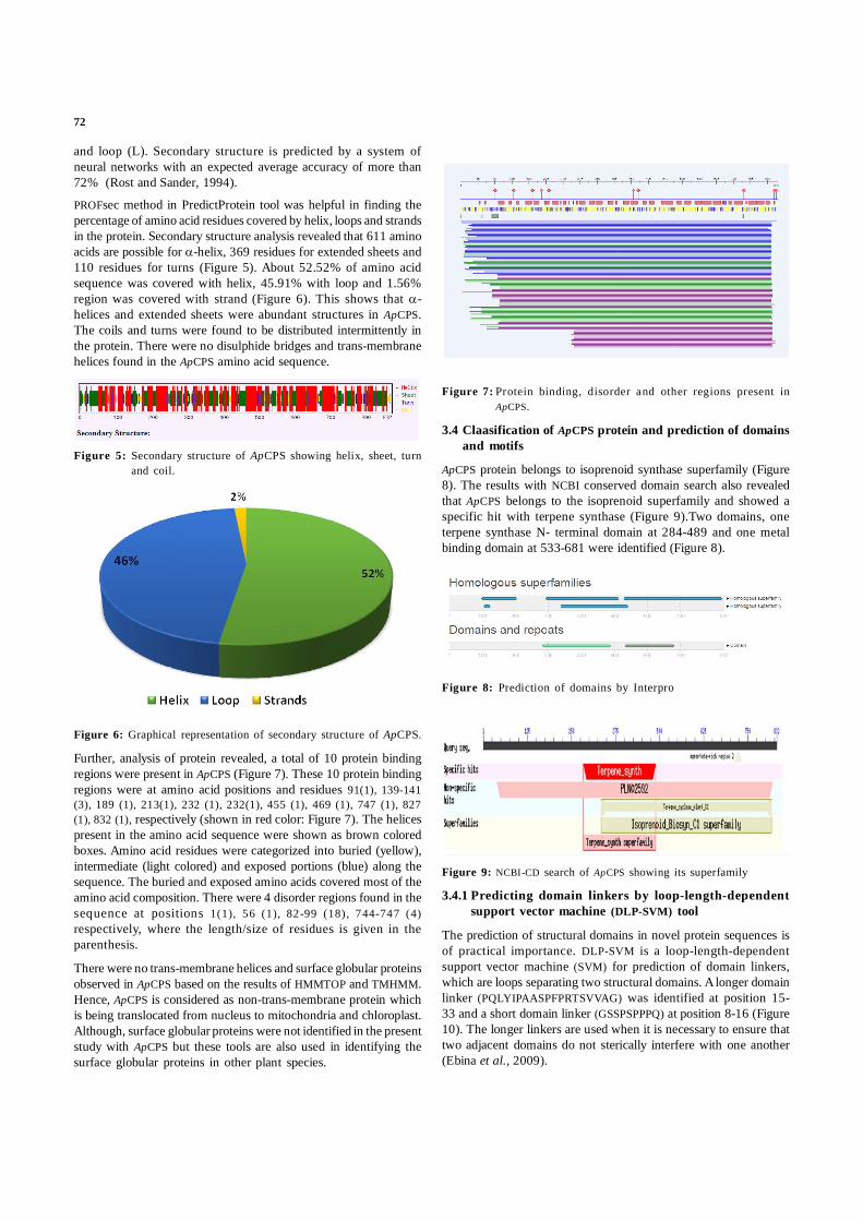
Further, analysis of protein revealed, a total of 10 protein bindingregions were present in ApCPS (Figure 7). These 10 protein bindingregions were at amino acid positions and residues 91(1), 139-141(3), 189 (1), 213(1), 232 (1), 232(1), 455 (1), 469 (1), 747 (1), 827(1), 832 (1), respectively (shown in red color: Figure 7). The helicespresent in the amino acid sequence were shown as brown coloredboxes. Amino acid residues were categorized into buried (yellow),intermediate (light colored) and exposed portions (blue) along thesequence. The buried and exposed amino acids covered most of theamino acid composition. There were 4 disorder regions found in thesequence at positions 1(1), 56 (1), 82-99 (18), 744-747 (4)respectively, where the length/size of residues is given in theparenthesis.
There were no trans-membrane helices and surface globular proteinsobserved in ApCPS based on the results of HMMTOP and TMHMM.Hence, ApCPS is considered as non-trans-membrane protein whichis being translocated from nucleus to mitochondria and chloroplast.Although, surface globular proteins were not identified in the presentstudy with ApCPS but these tools are also used in identifying thesurface globular proteins in other plant species.
Figure 7: Protein binding, disorder and other regions present inApCPS.
3.4 Claasification of ApCPS protein and prediction of domainsand motifs
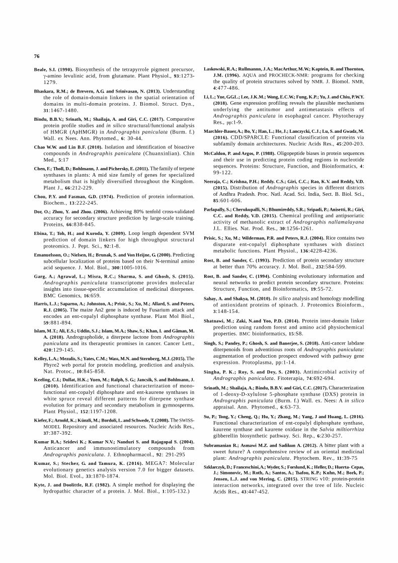
ApCPS protein belongs to isoprenoid synthase superfamily (Figure8). The results with NCBI conserved domain search also revealedthat ApCPS belongs to the isoprenoid superfamily and showed aspecific hit with terpene synthase (Figure 9).Two domains, oneterpene synthase N- terminal domain at 284-489 and one metalbinding domain at 533-681 were identified (Figure 8).
Figure 8: Prediction of domains by Interpro
Figure 9: NCBI-CD search of ApCPS showing its superfamily
3.4.1 Predicting domain linkers by loop-length-dependentsupport vector machine (DLP-SVM) tool
The prediction of structural domains in novel protein sequences isof practical importance. DLP-SVM is a loop-length-dependentsupport vector machine (SVM) for prediction of domain linkers,which are loops separating two structural domains. A longer domainlinker (PQLYIPAASPFPRTSVVAG) was identified at position 15-33 and a short domain linker (GSSPSPPPQ) at position 8-16 (Figure10). The longer linkers are used when it is necessary to ensure thattwo adjacent domains do not sterically interfere with one another(Ebina et al., 2009).

73
The domain linkers were found to be rich in proline residues whichcontributes for structural confirmation, and flexibility of twodomains. They also acted in the prevention of unfavourablecommunication between the domains. Further, depending on lengthof the linkers the interaction between the domains varied (Bhaskaraet al., 2013). This study is also helpful in protein targeted drugdevelopment and other proteomic studies (Shatnawi et al., 2014).
Figure 10: Domain linkers (short and long) identified in ApCPS.
3.4.2 Motif analysis of ApCPS
There were 3 motifs found using MEME with Mast Alignment andSearch Tool (Figure 11). The detail analysis of three motifs wascharacterized and the repetition in the ApCPS sequence detectedFigure 12. Each of the sequence has an e value less than 10. Themotif YIPAASPF occurred twice at positions 18-25 and 674-689 inthe sequence. Motif MHRDWTDKGICW also repeated twice atpositions 189-200 and 369-380. The third motif WQKWLRSWoccurred at positions 554-561and 674-689 along the sequence.
Figure 11: MEME results showing motifs in the ApCPS.
Figure 12: Different motifs and their positions on the sequence ofApCPS (The boxes on the line representing the motifs).
3.5 Elucidation of tertiary structure of ApCPS by 3D homologymodelling, validation and ligand binding site prediction
The 3D structure was predicted using Swiss Model which showed713 amino acid coverage and with 56.81% identity (Figure 13). Theelucidated 3D model was structurally validated with SAVESPROCHECK where 91.74% of the residues have averaged 3D-1Dscore >= 0.2 (Figure 14). This 3D model has shown a overall qualityfactor (OQF) of 92.1986 (Figure 15).
Figure 13: Elucidated 3D structure of ApCPS using Swiss Model.
Figure 14: Validation of 3D structure.
Figure 15: ERRAT result showing the Quality factor for ApCPS.
In Ramachnadran plot, 90.9% amino acid residues were observedin favoured region with 8.4% additional allowed regions (Figure16). Only 0.2% disallowed regions were found in the plot. Thisvalidation studies showing that the predicted 3D model of ApCPSenzyme was structurally reliable. The distribution of main chainbond angles and length were found to be within the limits. Homologymodelling and validation of antioxidant proteins were also shownin similar way in Spinach (Sahay and Shakya, 2010). A single ligandbinding site was identified in validated 3D structure of ApCPS (Figure17). It consists of GLY, ASP, VAL residues at (510-528).

74
Figure 16: Ramachandran Plot of ApCPS 3D structure. The mostfavoured regions are represented in red color; additionalallowed regions in yellow color. Figure 17: Predicted ligand binding site showed in blue color.
Figure 18: The phylogenetic tree showing the evolutionary relation of ApCPS with other SQS from diverse organisms constructed by neighbour-joining method.

75
3.6 Phylogenetic analysis of ApCPS
The phylogenetic tree mainly divided into eudicots, monocots,gymnosperms and fungi. Among the 41 sequences taken for thephylogenetic construction, the ApCPS was clustered within theeudicots (Figure 18). ApCPS was shown close relationship withSesamum indicum and Olea europaea plants belongs to the sameorder Lamiales. After Lamiale plants, it was closer to the orderSolanales. In an earlier study, comparable result was found withthis CPS enzyme by Garg et al. (2015).
The evolutionary divergence study of this plant showed closerelation to Laminales, followed by Solaneles in ApHMGR and ApDXSenzymes related to the diterpenoid pathways (Bindu et al., 2017;Srinath et al., 2017). After the relation with eudicots, ApCPS wasclosely linked to monocots particularly Zea mays and Triticumaestivum and extended to gymnosperms and fungal species.
3.7 Protein interaction study
Arabidopsis thaliana CPS also called as GA1 was used for the proteininteraction study (Figure 19). The protein interaction study shownthat CPS mainly involved in gibberellic acid mediated signallingpathway. Further, it also involves proteins response to oxygen-containing compound which includes the enzymes such as GA1,RGL1, RGL-2, and GA20OX1, etc. The molecular functions such astranscription factor activity and binding included interaction withRGA1, RGL1, RGL2, RGL3 enzymes, iron ion binding includedinteraction with CYP88A3, KA02. Terpene synthase activity mainlyinvolved the interaction within GA1, GA2, TPS04 enzymes. Mostof the interacted proteins shown to be involved in inter cellular andintra cellular membrane bounded organelles. The CPS also shownthe relation to proteins involved in diterpene biosynthesis pathwaywhich includes GA1, TPS04, CYP 82G1, GA2, GA3OX1, GA3,GA20OX1, CYP88A3, KAO2 enzymes. These were also involvedin plant hormone signal transduction and biosynthesis of secondarymetabolites (Figure 20).
Figure 19: Protein-protein interactions using Arabidopsis CPS asreference sequence using STRING 10.
Gene expression and co-expression studies will provide outlineabout CPS protein where the exchange of molecules and theirinteraction is high or low (Figure 20). CPS found to be involved inbiosynthesis of gibberellin and diterpenoids in various studies(Keeling et al., 2010; Yamamura et al., 2018). This finding strengthensour result which showed the CPS co-expression mainly with
gibberellin and terpenoid synthase biosynthesis genes such asGibberellin 3-oxidase 1, Gibberellin 20 oxidase 1 and Terpenesynthase 04, etc.
Figure 20: Co-expression analysis of different proteins with referenceplant CPS (Arabidopsis thaliana CPS). GA1-Gibberillic acidrequiring1; GA3 - GA requiring 3; GA3OX1- Gibberellin 3-oxidase 1; GAI- DELLA protein GAI; GA2- GA REQUIRING2; GA20OX1- Gibberellin 20 oxidase 1; RGL2- RGA-like 2;RGL1- RGA-like 1; SPY- SPINDLY; TPS04- Terpene synthase04; SLY1- SLEEPY1.
4. Conclusion
The present in silico analysis has given structural, functional andphysicochemical aspects of protein ApCPS. The secondary structureprediction showed the alpha helices and beta sheets distribution ofApCPS protein folding. The reliable 3D modelling of this enzymegives information about the protein folding and can be exploited forvarious docking and drug targeting studies. The protein- proteininteraction study will be helpful for understanding the protein atmolecular level in various biosynthetic mechanisms. Evolutionaryrelation will give a scope for molecular biology and gene isolationstudies where the primer designing for genes would be easy.
Acknowledgements
We would like to acknowledge the financial support from OU-UGC-CPEPA programme sponsored by University GrantsCommission (UGC), New Delhi. Authors also thank OU-UGC-CPEPAprogramme and UGC-BSR-RFSMS for research fellowships to AS,BBVB, MS.
Conflict of interest
We declare that we have no conflict of interest
ReferencesAndersen Ranberg, J.; Kongstad, K.T.; Nielsen, M.T.; Jensen, N.B.; Pateraki, I.;
Bach, S.S. and Møller, B.L. (2016). Expanding the landscape of diterpenestructural diversity through stereochemically controlledcombinatorial biosynthesis. Angew. Chem. Int. Ed., 55:2142-2146.
Bannai, H.; Tamada, Y.; Maruyama, O.; Nakai, K. and Miyano, S. (2001). Views:Fundamental building blocks in the process of knowledge discovery.In: Proceedings of the 14th International FLAIRS Conference,pp:233-238.
Bannai, H.; Tamada, Y.; Maruyama, O.; Nakai, K. and Miyano, S. (2002).Extensive feature detection of N-terminal protein sorting signals.Bioinformatics, 18:298-305.
76
Beale, S.I. (1990). Biosynthesis of the tetrapyrrole pigment precursor,-amino levulinic acid, from glutamate. Plant Physiol., 93:1273-1279.
Bhaskara, R.M.; de Brevern, A.G. and Srinivasan, N. (2013). Understandingthe role of domain-domain linkers in the spatial orientation ofdomains in multi-domain proteins. J . Biomol. Struct. Dyn.,31:1467-1480.
Bindu, B.B.V.; Srinath, M.; Shailaja, A. and Giri, C.C. (2017). Comparativeprotein profile studies and in silico structural/functional analysisof HMGR (ApHMGR) in Andrographis paniculata (Burm. f.)Wall. ex Nees. Ann. Phytomed., 6: 30-44.
Chao W.W. and Lin B.F. (2010). Isolation and identification of bioactivecompounds in Andrographis paniculata (Chuanxinlian). ChinMed., 5:17
Chen, F.; Tholl, D.; Bohlmann, J. and Pichersky, E. (2011). The family of terpenesynthases in plants: A mid size family of genes for specializedmetabolism that is highly diversified throughout the Kingdom.Plant J., 66:212-229.
Chou, P.Y. and Fasman, G.D. (1974). Prediction of protein information.Biochem., 13:222-245.
Dor, O.; Zhou, Y. and Zhou. (2006). Achieving 80% tenfold cross-validatedaccuracy for secondary structure prediction by large-scale training.Proteins, 66:838-845.
Ebina, T.; Toh, H.; and Kuroda, Y. (2009). Loop length dependent SVMprediction of domain linkers for high throughput structuralproteomics. J. Pept. Sci., 92:1-8.
Emanuelsson, O.; Nielsen, H.; Brunak, S. and Von Heijne, G. (2000). Predictingsubcellular localization of proteins based on their N-terminal aminoacid sequence. J. Mol. Biol., 300:1005-1016.
Garg, A.; Agrawal, L.; Misra, R.C.; Sharma, S. and Ghosh, S. (2015).Andrographis paniculata transcriptome provides molecularinsights into tissue-specific accumulation of medicinal diterpenes.BMC Genomics, 16:659.
Harris, L.J.; Saparno, A.; Johnston, A.; Prisic, S.; Xu, M.; Allard, S. and Peters,R.J. (2005). The maize An2 gene is induced by Fusarium attack andencodes an ent-copalyl diphosphate synthase. Plant Mol Biol.,59:881-894.
Islam, M.T.; Ali, E.S.; Uddin, S.J.; Islam, M.A.; Shaw, S.; Khan, I. and Gãman, M.A. (2018). Andrographolide, a diterpene lactone from Andrographispaniculata and its therapeutic promises in cancer. Cancer Lett.,420:129-145.
Kelley, L.A.; Mezulis, S.; Yates, C.M.; Wass, M.N. and Sternberg, M.J. (2015). ThePhyre2 web portal for protein modeling, prediction and analysis.Nat. Protoc., 10:845-858.
Keeling, C.I.; Dullat, H.K.; Yuen, M.; Ralph, S. G.; Jancsik, S. and Bohlmann, J.(2010). Identification and functional characterization of mono-functional ent-copalyl diphosphate and ent-kaurene synthases inwhite spruce reveal different patterns for diterpene synthaseevolution for primary and secondary metabolism in gymnosperms.Plant Physiol., 152:1197-1208.
Kiefer, F.; Arnold, K.; Künzli, M.; Bordoli, L. and Schwede, T. (2008). The SWISS-MODEL Repository and associated resources. Nucleic Acids Res.,37:387-392.
Kumar R.A.; Sridevi K.; Kumar N.V.; Nanduri S. and Rajagopal S. (2004).Anticancer and immunostimulatory compounds fromAndrographis paniculata. J. Ethnopharmacol., 92: 291-295
Kumar, S.; Stecher, G. and Tamura, K. (2016). MEGA7: Molecularevolutionary genetics analysis version 7.0 for bigger datasets.Mol. Biol. Evol., 33:1870-1874.
Kyte, J. and Doolittle, R.F. (1982). A simple method for displaying thehydropathic character of a protein. J. Mol. Biol., 1:105-132.)
Laskowski, R.A.; Rullmannn, J.A.; MacArthur, M.W.; Kaptein, R. and Thornton,J.M. (1996). AQUA and PROCHECK-NMR: programs for checkingthe quality of protein structures solved by NMR. J. Biomol. NMR,4:477-486.
Li, L.; Yue, G.G.L.; Lee, J.K.M.; Wong, E.C.W.; Fung, K.P.; Yu, J. and Chiu, P.W.Y.(2018). Gene expression profiling reveals the plausible mechanismsunderlying the antitumor and antimetastasis effects ofAndrographis paniculata in esophageal cancer. PhytotherapyRes., pp:1-9.
Marchler-Bauer, A.; Bo, Y.; Han, L.; He, J.; Lanczycki, C.J.; Lu, S. and Gwadz, M.(2016). CDD/SPARCLE: Functional classification of proteins viasubfamily domain architectures. Nucleic Acids Res., 45:200-203.
McCaldon, P. and Argos, P. (1988). Oligopeptide biases in protein sequencesand their use in predicting protein coding regions in nucleotidesequences. Proteins: Structure, Function, and Bioinformatics, 4:99-122.
Neeraja, C.; Krishna, P.H.; Reddy. C.S.; Giri, C.C.; Rao, K.V. and Reddy, V.D.(2015). Distribution of Andrographis species in different districtsof Andhra Pradesh. Proc. Natl. Acad. Sci. India, Sect. B. Biol. Sci.,85:601-606.
Parlapally, S.; Cherukupalli, N.; Bhumireddy, S.R.; Sripadi, P.; Anisetti, R.; Giri,C.C. and Reddy, V.D. (2015). Chemical profiling and antipsoriaticactivity of methanolic extract of Andrographis nallamalayanaJ.L. Ellies. Nat. Prod. Res., 30:1256-1261.
Prisic, S.; Xu, M.; Wilderman, P.R. and Peters, R.J. (2004). Rice contains twodisparate ent-copalyl diphosphate synthases with distinctmetabolic functions. Plant Physiol., 136:4228-4236.
Rost, B. and Sander, C. (1993). Prediction of protein secondary structureat better than 70% accuracy. J. Mol. Boil., 232:584-599.
Rost, B. and Sander, C. (1994). Combining evolutionary information andneural networks to predict protein secondary structure. Proteins:Structure, Function, and Bioinformatics, 19:55-72.
Sahay, A. and Shakya, M. (2010). In silico analysis and homology modellingof antioxidant proteins of spinach. J. Proteomics Bioinform.,3:148-154.
Shatnawi, M.; Zaki, N.and Yoo, P.D. (2014). Protein inter-domain linkerprediction using random forest and amino acid physiochemicalproperties. BMC bioinformatics, 15:S8.
Singh, S.; Pandey, P.; Ghosh, S. and Banerjee, S. (2018). Anti-cancer labdanediterpenoids from adventitious roots of Andrographis paniculata:augmentation of production prospect endowed with pathway geneexpression. Protoplasma, pp:1-14.
Singha, P. K.; Roy, S. and Dey, S. (2003). Antimicrobial activity ofAndrographis paniculata. Fitoterapia, 74:692-694.
Srinath, M.; Shailaja, A.; Bindu, B.B.V. and Giri, C.C. (2017). Characterizationof 1-deoxy-D-xylulose 5-phosphate synthase (DXS) protein inAndrographis paniculata (Burm. f.) Wall. ex. Nees: A in silicoappraisal. Ann. Phytomed., 6:63-73.
Su, P.; Tong, Y.; Cheng, Q.; Hu, Y.; Zhang, M.; Yang, J and Huang, L. (2016).Functional characterization of ent-copalyl diphosphate synthase,kaurene synthase and kaurene oxidase in the Salvia miltiorrhizagibberellin biosynthetic pathway. Sci. Rep., 6:230-257.
Subramanian R.; Asmawi M.Z. and Sadikun A. (2012). A bitter plant with asweet future? A comprehensive review of an oriental medicinalplant: Andrographis paniculata . Phytochem. Rev., 11:39-75
Szklarczyk, D.; Franceschini, A.; Wyder, S.; Forslund, K.; Heller, D.; Huerta- Cepas,J.; Simonovic, M.; Roth, A.; Santos, A.; Tsafou, K.P.; Kuhn, M.; Bork, P.;Jensen, L.J. and von Mering, C. (2015). STRING v10: protein-proteininteraction networks, integrated over the tree of life. NucleicAcids Res., 43:447-452.

77
Tusnády, G.E. and Simon, I. (1998). Principles governing amino acidcomposition of integral membrane proteins: Applications totopology prediction. J. Mol. Biol., 283:489-506.
Vranová, E.; Coman, D.; and Gruissem, W. (2013). Network analysis of theMVA and MEP pathways for isoprenoid synthesis. Annu. Rev.Plant Biol., 64:665-700.
Walker, J.M. (Ed.). (2005). The proteomics protocols handbook. HumanaPress.
Wass, M.N.; Kelley, L.A. and Sternberg, M.J. (2010). 3DLigandSite: predictingligand-binding sites using similar structures. Nucleic Acids Res.,38:469-73.
Yamamura, Y.; Taguchi, Y.; Ichitani, K.; Umebara, I.; Ohshita, A.; Kurosaki, F.and Lee, J.B. (2018). Characterization of ent-kaurene synthase andkaurene oxidase involved in gibberellin biosynthesis fromScopariadulcis. J. Nat. Med., pp:1-8.
Zaheer, M. and Giri, C.C. (2015). Multiple shoot induction and jasmonicversus salicylic acid driven elicita tion for enhancedandrographolide production in Andrographis paniculata . PlantCell Tissue Organ Cult., 122:553-563.
Zaheer, M. and Giri, C.C. (2017a). Enhanced diterpene lactone(andrographolide) production from elicited adventitious rootcultures of Andrographis paniculata . Res. Chem. Intermed.,43:2433–2444.
Zaheer, M. and Giri, C.C. (2017b). Influence of cotyledon, hypocotylextracts and authentic andrographolide on selective Agrobac-terium rhizogenes stra ins growth: A deterrent to hairy rootinduction in Andrographis paniculata (Burm.f.) Wall. ex Nees.Ann. Phytomed., 6:51-56.