Embed Size (px)
Citation preview

Induction of Human �-Cell Proliferation andEngraftment Using a Single G1/S Regulatory Molecule,cdk6Nathalie M. Fiaschi-Taesch,
1Fatimah Salim,
1Jeffrey Kleinberger,
1Ronnie Troxell,
1
Irene Cozar-Castellano,2
Karen Selk,1
Edward Cherok,1
Karen K. Takane,1
Donald K. Scott,1
and
Andrew F. Stewart1
OBJECTIVE—Most knowledge on human �-cell cycle controlderives from immunoblots of whole human islets, mixtures of�-cells and non-�-cells. We explored the presence, subcellularlocalization, and function of five early G1/S phase molecules—cyclins D1–3 and cdk 4 and 6—in the adult human �-cell.
RESEARCH DESIGN AND METHODS—Immunocytochemis-try for the five molecules and their relative abilities to drivehuman �-cell replication were examined. Human �-cell replica-tion, cell death, and islet function in vivo were studied in thediabetic NOD-SCID mouse.
RESULTS—Human �-cells contain easily detectable cdks 4 and6 and cyclin D3 but variable cyclin D1. Cyclin D2 was onlymarginally detectable. All five were principally cytoplasmic, notnuclear. Overexpression of the five, alone or in combination, ledto variable increases in human �-cell replication, with thecdk6/cyclin D3 combination being the most robust (15% versus0.3% in control �-cells). A single molecule, cdk6, proved to becapable of driving human �-cell replication in vitro and enhanc-ing human islet engraftment/proliferation in vivo, superior tonormal islets and as effectively as the combination of cdk6 plusa D-cyclin.
CONCLUSIONS—Human �-cells contain abundant cdk4, cdk6,and cyclin D3, but variable amounts of cyclin D1. In contrast torodent �-cells, they contain little or no detectable cyclin D2. Theyare primarily cytoplasmic and likely ineffective in basal �-cellreplication. Unexpectedly, cyclin D3 and cdk6 overexpressiondrives human �-cell replication most effectively. Most impor-tantly, a single molecule, cdk6, supports robust human �-cellproliferation and function in vivo. Diabetes 59:1926–1936,2010
While broadly similar, human pancreatic �-cells differ from their rodent counterparts inmany ways. For example, rodent �-cellscan be induced to replicate using many
strategies, including partial pancreatectomy, induction ofobesity and insulin resistance, infusion of glucose, admin-istration of growth factors, and activation of signaling
pathways downstream of these growth factors and nutri-ents (1–8). In contrast, while evidence suggests thathuman fetal and early neonatal �-cells are able to replicate(9–11), no investigator has been able to demonstrate orinduce robust rates of proliferation in adult human �-cellsusing the same growth factors, nutrients, signaling path-ways, and maneuvers that have been effective in rodents(12–19). For example, unlike events in rodents, obesity ortype 2 diabetes in human adults is not associated withaccelerated �-cell proliferation (19), and even partial pan-createctomy does not induce �-cell replication or regen-eration in humans (17). This inability to induce substantialhuman �-cell replication is problematic, for it is now clearthat �-cell replacement therapy for diabetes will requirelarge numbers of human or xenogeneic �-cells (20).
While much has been learned from mouse geneticmodels regarding the molecular control of �-cell replica-tion (21–35), little is known regarding the molecules thatregulate the G1/S transition in the human adult �-cell. Wehave begun to explore these molecules in adult humanislets, mapping the members of the G1/S proteome that arepresent in human cadaveric islets (12,36). Human islets arebelieved to contain most of the key molecules that governthe G1/S transition in other cell types (12,36,37). Theseinclude the three members of the pocket protein family(pRb, p107, and p130); cyclins D1 and D3; cdks 1, 2, 4, and6; the four members of the INK4 family (p15, p16, p18, andp19); the three members of the KIP/CIP family (p21, p27,and p57); several E2F family members; and p53 and its E3ligase, HDM2 (supplementary Fig 1, available in an onlineappendix at http://diabetes.diabetesjournals.org/cgi/content/full/db09-1776/DC1). Upstream of these G1/S molecules areother regulatory molecules such as skp2, menin, FoxM1,bmi1, and ezh2 (26–29,34). We and others have also shownthat human islets differ from their rodent counterparts inthat they express cdk6, a cdk that is not detectable inmouse islets (12,35). We have also shown that overexpres-sion of cdk6 in association with a D-cyclin, cyclin D1,markedly increases the replication of adult human �-cells(12), and it does so in a way that does not lead todedifferentiation or accelerated �-cell death but insteadleads to enhanced engraftment and function of adulthuman cadaveric islets in vivo (12). We also have shownthat cdk4 (36) and cdk6 (12) in conjunction with cyclin D1can enhance pRb phosphorylation and replication in hu-man �-cells. Despite these advances, it is important tounderscore that, of the 30� molecules that regulate thehuman �-cell G1/S transition (supplementary Fig. 1), thetherapeutic potential has been explored for only three:cdk4, cdk6, and cyclin D1. For example, nothing is known
From the 1The Division of Endocrinology, the University of Pittsburgh Schoolof Medicine, Pittsburgh, Pennsylvania, and the 2Unidad de Investigacion,Hospital Universitario Puerta del Mar, Cadiz, Spain.
Corresponding author: Nathalie M. Fiaschi-Taesch, [email protected] 4 December 2009 and accepted 4 May 2010.DOI: 10.2337/db09-1776© 2010 by the American Diabetes Association. Readers may use this article as
long as the work is properly cited, the use is educational and not for profit,and the work is not altered. See http://creativecommons.org/licenses/by-nc-nd/3.0/ for details.
The costs of publication of this article were defrayed in part by the payment of page
charges. This article must therefore be hereby marked “advertisement” in accordance
with 18 U.S.C. Section 1734 solely to indicate this fact.
ORIGINAL ARTICLE
1926 DIABETES, VOL. 59, AUGUST 2010 diabetes.diabetesjournals.org
about the efficacy of the other two D-cyclins, cyclins D2and D3, in driving proliferation of human �-cells.
Another difference between human and rodent isletsmay involve cyclin D2. In mice, excellent evidence indi-cates that cyclin D2 is indispensible for �-cell growth andislet development and function: animals that lack cyclinD2 develop islet hypoplasia, hypoinsulinemia, and diabe-tes (32,33). In contrast to mouse studies, in humans, cyclinD2 has been reported to be difficult to detect or undetect-able, despite the use of multiple antisera from multiplevendors and appropriate positive controls (12,37).
Here, we asked four questions. 1) Which D-cyclins aredefinitively present in human islets? 2) Which of the threeD-cyclins might be the optimal partner for cdk4 or cdk6 forenhancing adult human �-cell replication? 3) Are cdk4,cdk6, and the three D-cyclins actually present in �-cells [incontrast to immunoblot studies of whole islet extractsdescribed previously (12,37)], and if so, in which subcel-lular compartment? 4) From a therapeutic standpoint, is itessential to deliver both a D-cyclin and a cdk4/6 familymember to enhance human islet engraftment, or can thisbe simplified by using only a single member of thecdk4/6–D-cyclin complex?
We report here that 1) cyclin D1 is variably present, D2is only marginally detectable, and D3 is readily detectablein human islets; 2) cyclin D3 appears to be surprisinglyeffective in inducing human �-cell proliferation and, incombination with cdk6, may be the most effective indriving human �-cell replication; 3) cdks 4 and 6, andcyclins D1 and D3, are located principally in the cytoplasmand not the nucleus; and 4) a single cdk, cdk6, is surpris-ingly as effective in driving human islet engraftment as thepreviously reported combination of cdk6 plus cyclin D1.
RESEARCH DESIGN AND METHODS
All of the methods and procedures have been described previously (5–7,12,18,24,36). Supplementary detailed methods are available in an onlineappendix. The University of Pittsburgh Institutional Review Board approvedin advance both receipt of, and work with, human islets.
RESULTS
Human islets contain easily detectable cyclin D3, butonly variable and marginal quantities of cyclins D1and D2. The cyclin D family includes three D-cyclins,shown schematically in supplementary Fig. 2A. As shownin supplementary Fig. 3 (available in an online appendix),mRNA encoding each of the three D-cyclins is present inhuman islets, as assessed by conventional PCR as well asby quantitative PCR.
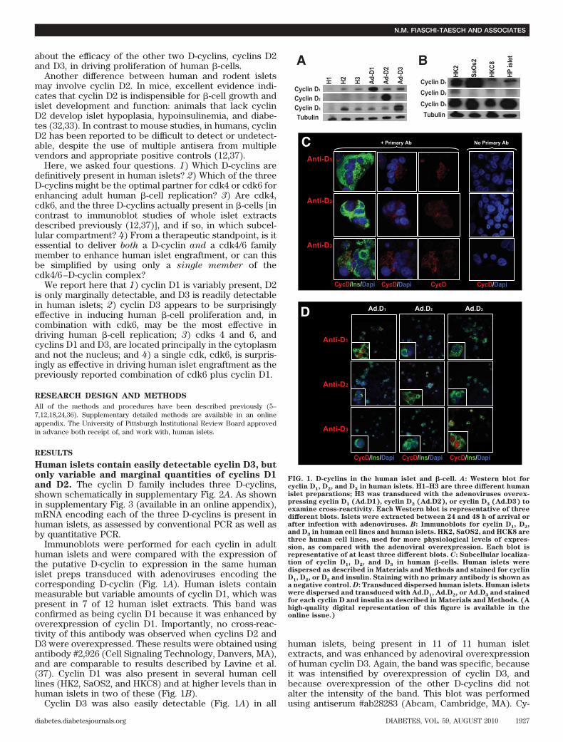
Immunoblots were performed for each cyclin in adulthuman islets and were compared with the expression ofthe putative D-cyclin to expression in the same humanislet preps transduced with adenoviruses encoding thecorresponding D-cyclin (Fig. 1A). Human islets containmeasurable but variable amounts of cyclin D1, which waspresent in 7 of 12 human islet extracts. This band wasconfirmed as being cyclin D1 because it was enhanced byoverexpression of cyclin D1. Importantly, no cross-reac-tivity of this antibody was observed when cyclins D2 andD3 were overexpressed. These results were obtained usingantibody #2,926 (Cell Signaling Technology, Danvers, MA),and are comparable to results described by Lavine et al.(37). Cyclin D1 was also present in several human celllines (HK2, SaOS2, and HKC8) and at higher levels than inhuman islets in two of these (Fig. 1B).
Cyclin D3 was also easily detectable (Fig. 1A) in all
human islets, being present in 11 of 11 human isletextracts, and was enhanced by adenoviral overexpressionof human cyclin D3. Again, the band was specific, becauseit was intensified by overexpression of cyclin D3, andbecause overexpression of the other D-cyclins did notalter the intensity of the band. This blot was performedusing antiserum #ab28283 (Abcam, Cambridge, MA). Cy-
H1 H2 H3 Ad-D
1
Ad-D
2
Ad-D
3
Cyclin D3
Cyclin D2
Cyclin D1
Cyclin D3
Cyclin D2
Cyclin D1
Tubulin
HK2
SaOs
2
HKC8
HP is
let
Tubulin
A
+ Primary Ab No Primary Ab
Anti-D1
Anti-D2
Anti-D3
CycD/Ins/Dapi CycD/Dapi CycD/DapiCycD
C
Ad.D1 Ad.D2 Ad.D3D
Anti-D1
Anti-D2
Anti-D3
CycD/Ins/Dapi CycD/Ins/Dapi CycD/Ins/Dapi
B
FIG. 1. D-cyclins in the human islet and �-cell. A: Western blot forcyclin D1, D2, and D3 in human islets. H1–H3 are three different humanislet preparations; H3 was transduced with the adenoviruses overex-pressing cyclin D1 (Ad.D1), cyclin D2 (Ad.D2), or cyclin D3 (Ad.D3) toexamine cross-reactivity. Each Western blot is representative of threedifferent blots. Islets were extracted between 24 and 48 h of arrival orafter infection with adenoviruses. B: Immunoblots for cyclin D1, D2,and D3 in human cell lines and human islets. HK2, SaOS2, and HCK8 arethree human cell lines, used for more physiological levels of expres-sion, as compared with the adenoviral overexpression. Each blot isrepresentative of at least three different blots. C: Subcellular localiza-tion of cyclin D1, D2, and D3 in human �-cells. Human islets weredispersed as described in Materials and Methods and stained for cyclinD1, D2, or D3 and insulin. Staining with no primary antibody is shown asa negative control. D: Transduced dispersed human islets. Human isletswere dispersed and transduced with Ad.D1, Ad.D2, or Ad.D3 and stainedfor each cyclin D and insulin as described in Materials and Methods. (Ahigh-quality digital representation of this figure is available in theonline issue.)
N.M. FIASCHI-TAESCH AND ASSOCIATES
diabetes.diabetesjournals.org DIABETES, VOL. 59, AUGUST 2010 1927
clin D3 was also easily detectable in each of the threehuman cell lines (Fig. 1B).
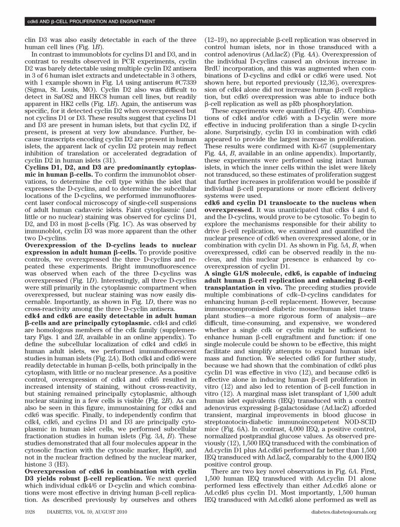
In contrast to immunoblots for cyclins D1 and D3, and incontrast to results observed in PCR experiments, cyclinD2 was barely detectable using multiple cyclin D2 antiserain 3 of 6 human islet extracts and undetectable in 3 others,with 1 example shown in Fig. 1A using antiserum #C7339(Sigma, St. Louis, MO). Cyclin D2 also was difficult todetect in SaOS2 and HKC8 human cell lines, but readilyapparent in HK2 cells (Fig. 1B). Again, the antiserum wasspecific, for it detected cyclin D2 when overexpressed butnot cyclins D1 or D3. These results suggest that cyclins D1and D3 are present in human islets, but that cyclin D2, ifpresent, is present at very low abundance. Further, be-cause transcripts encoding cyclin D2 are present in humanislets, the apparent lack of cyclin D2 protein may reflectinhibition of translation or accelerated degradation ofcyclin D2 in human islets (31).Cyclins D1, D2, and D3 are predominantly cytoplas-mic in human �-cells. To confirm the immunoblot obser-vations, to determine the cell type within the islet thatexpresses the D-cyclins, and to determine the subcellularlocations of the D-cyclins, we performed immunofluores-cent laser confocal microscopy of single-cell suspensionsof adult human cadaveric islets. Faint cytoplasmic (andlittle or no nuclear) staining was observed for cyclins D1,D2, and D3 in most �-cells (Fig. 1C). As was observed byimmunoblot, cyclin D3 was more apparent than the othertwo D-cyclins.Overexpression of the D-cyclins leads to nuclearexpression in adult human �-cells. To provide positivecontrols, we overexpressed the three D-cyclins and re-peated these experiments. Bright immunofluorescencewas observed when each of the three D-cyclins wasoverexpressed (Fig. 1D). Interestingly, all three D-cyclinswere still primarily in the cytoplasmic compartment whenoverexpressed, but nuclear staining was now easily dis-cernable. Importantly, as shown in Fig. 1D, there was nocross-reactivity among the three D-cyclin antisera.cdk4 and cdk6 are easily detectable in adult human�-cells and are principally cytoplasmic. cdk4 and cdk6are homologous members of the cdk family (supplemen-tary Figs. 1 and 2B, available in an online appendix). Todefine the subcellular localization of cdk4 and cdk6 inhuman adult islets, we performed immunofluorescentstudies in human islets (Fig. 2A). Both cdk4 and cdk6 werereadily detectable in human �-cells, both principally in thecytoplasm, with little or no nuclear presence. As a positivecontrol, overexpression of cdk4 and cdk6 resulted inincreased intensity of staining, without cross-reactivity,but staining remained principally cytoplasmic, althoughnuclear staining in a few cells is visible (Fig. 2B). As canalso be seen in this figure, immunostaining for cdk4 andcdk6 was specific. Finally, to independently confirm thatcdk4, cdk6, and cyclins D1 and D3 are principally cyto-plasmic in human islet cells, we performed subcellularfractionation studies in human islets (Fig. 3A, B). Thesestudies demonstrated that all four molecules appear in thecytosolic fraction with the cytosolic marker, Hsp90, andnot in the nuclear fraction defined by the nuclear marker,histone 3 (H3).Overexpression of cdk6 in combination with cyclinD3 yields robust �-cell replication. We next queriedwhich individual cdk4/6 or D-cyclin and which combina-tions were most effective in driving human �-cell replica-tion. As described previously by ourselves and others
(12–19), no appreciable �-cell replication was observed incontrol human islets, nor in those transduced with acontrol adenovirus (Ad.lacZ) (Fig. 4A). Overexpression ofthe individual D-cyclins caused an obvious increase inBrdU incorporation, and this was augmented when com-binations of D-cyclins and cdk4 or cdk6 were used. Notshown here, but reported previously (12,36), overexpres-sion of cdk4 alone did not increase human �-cell replica-tion, but cdk6 overexpression was able to induce both�-cell replication as well as pRb phosphorylation.
These experiments were quantified (Fig. 4B). Combina-tions of cdk4 and/or cdk6 with a D-cyclin were moreeffective in inducing proliferation than a single D-cyclinalone. Surprisingly, cyclin D3 in combination with cdk6appeared to provide the largest increase in proliferation.These results were confirmed with Ki-67 (supplementaryFig. 4A, B, available in an online appendix). Importantly,these experiments were performed using intact humanislets, in which the inner cells within the islet were likelynot transduced, so these estimates of proliferation suggestthat further increases in proliferation would be possible ifindividual �-cell preparations or more efficient deliverysystems were used.cdk6 and cyclin D1 translocate to the nucleus whenoverexpressed. It was unanticipated that cdks 4 and 6,and the D-cyclins, would prove to be cytosolic. To begin toexplore the mechanisms responsible for their ability todrive �-cell replication, we examined and quantified thenuclear presence of cdk6 when overexpressed alone, or incombination with cyclin D1. As shown in Fig. 5A, B, whenoverexpressed, cdk6 can be observed readily in the nu-cleus, and this nuclear presence is enhanced by co-overexpression of cyclin D1.A single G1/S molecule, cdk6, is capable of inducingadult human �-cell replication and enhancing �-celltransplantation in vivo. The preceding studies providemultiple combinations of cdk–D-cyclins candidates forenhancing human �-cell replacement. However, becauseimmunocompromised diabetic mouse/human islet trans-plant studies—a more rigorous form of analysis—aredifficult, time-consuming, and expensive, we wonderedwhether a single cdk or cyclin might be sufficient toenhance human �-cell engraftment and function: if onesingle molecule could be shown to be effective, this mightfacilitate and simplify attempts to expand human isletmass and function. We selected cdk6 for further study,because we had shown that the combination of cdk6 pluscyclin D1 was effective in vivo (12), and because cdk6 iseffective alone in inducing human �-cell proliferation invitro (12) and also led to retention of �-cell function invitro (12). A marginal mass islet transplant of 1,500 adulthuman islet equivalents (IEQ) transduced with a controladenovirus expressing �-galactosidase (Ad.lacZ) affordedtransient, marginal improvements in blood glucose instreptozotocin-diabetic immunoincompetent NOD-SCIDmice (Fig. 6A). In contrast, 4,000 IEQ, a positive control,normalized postprandial glucose values. As observed pre-viously (12), 1,500 IEQ transduced with the combination ofAd.cyclin D1 plus Ad.cdk6 performed far better than 1,500IEQ transduced with Ad.lacZ, comparably to the 4,000 IEQpositive control group.
There are two key novel observations in Fig. 6A. First,1,500 human IEQ transduced with Ad.cyclin D1 aloneperformed less effectively than either Ad.cdk6 alone orAd.cdk6 plus cyclin D1. Most importantly, 1,500 humanIEQ transduced with Ad.cdk6 alone performed as well as
cdk6 AND �-CELL PROLIFERATION AND ENGRAFTMENT
1928 DIABETES, VOL. 59, AUGUST 2010 diabetes.diabetesjournals.org

1,500 IEQ transduced with the combination of Ad.cdk6plus cyclin D1. Thus, a single cdk, cdk6, is as effective asthe combination of cdk6 plus cyclin D1 in enhancinghuman islet graft function.
To more rigorously assess the function of the humanislet grafts, we performed intraperitoneal glucose toler-ance tests three weeks after transplant (Fig. 6B). Micetransplanted with 1,500 IEQ transduced with Ad.cdk6displayed fasting glucose values and glucose tolerancecomparable to normal NOD-SCID mice (not treated withstreptozotocin) and to streptozotocin-diabetic NOD-SCIDmice transplanted with 4,000 IEQ, or 1,500 IEQ transduced
with Ad.cdk6 plus cyclin D1. Glucose tolerance in theAd.cdk6 group was superior to that in the 1,500 IEQAd.lacZ or Ad.cyclin D1 groups.Ad.cdk6 induces sustained human �-cell replicationin vivo, with little cell death. We previously demon-strated that Ad.cdk6 plus D1 induces replication in human�-cells in vivo three days after transplant (12). Here weexplored whether this early proliferation could be sus-tained for longer periods of time. We harvested renalcapsular grafts 28 days after transplant and examinedproliferation in �-cells using combined insulin and Ki-67immunohistochemistry (Fig. 7A). Whereas little Ki-67
+ Primary Ab+ Primary Ab
+ Blocking Peptide
Anti-C4
Anti-C6
Cdk/Ins/Dapi Cdk/Dapi Cdk/DapiCdk
A
Ad.C4 Ad.C4
Ad.C4 Ad.C4
Ad.C6 Ad.C6
Ad.C6 Ad.C6
+ Primary Ab + Primary Ab+ Primary Ab
+ Blocking Peptide+ Primary Ab
+ Blocking PeptideB
Anti-C4
Anti-C6
Cdk/Ins/Dapi Cdk/Ins/Dapi Cdk/Ins/Dapi Cdk/Ins/Dapi
FIG. 2. Subcellular localization of cdk4 and cdk6 in human �-cells. A: Uninfected dispersed human islets. Human islets were dispersed as describedin Materials and Methods and stained for cdk4 or cdk6 and insulin. Staining with primary antibody and blocking peptide is shown as a negativecontrol. B: Dispersed human islet cells transduced with Ad.cdk4 or Ad.cdk6. Human islets were dispersed and transduced with Ad.cdk4 or Ad.cdk6and stained for cdk4 or cdk6 and insulin as described in Materials and Methods. As can be seen, overexpressed cdk4 and cdk6 are easilydetectable, and cdk4 and cdk6 staining is specific. (A high-quality digital representation of this figure is available in the online issue.)
N.M. FIASCHI-TAESCH AND ASSOCIATES
diabetes.diabetesjournals.org DIABETES, VOL. 59, AUGUST 2010 1929

staining is observed in �-cells exposed to Ad.lacZ, mark-edly increased �-cell Ki-67 staining is present in graftsharvested at 28 days from islets transduced with Ad.cdk6alone, Ad.cyclin D1 alone, and Ad.cdk6 plus Ad.cyclin D1in combination. These observations are quantified in Fig.7B, which shows that �-cell replication rates are very lowin control grafts and remain elevated for at least 28 daysafter transplant in human islets transduced with cdk6,cyclin D1, or the combination. We also examined the isletgrafts for cdk6 immunostaining (Fig. 7C). These figuresshow that cdk6 is particularly abundant in human �-cellsin vivo, even 28 days after transplant, and that it isabundant in the nuclear compartment.
�-cell death, as assessed using transferase-mediateddUTP nick-end labeling (TUNEL) staining, was barelydetectable in these grafts at 28 days, although it was easilydetectable in the positive control (Fig. 8A). We had earliershown that �-cell death was not increased at 3 daysposttransplant (12). Because the immediate posttransplantperiod is associated with cell death, and because theengraftment response to cdk6 occurred very early post-transplant, we also examined TUNEL staining in �-cells at24 h posttransplant (Fig. 8B, C). Again, no differences in�-cell apoptosis were observed in cdk6-expressing versuscontrol �-cells.
DISCUSSION
We report several novel observations. First, we find thatmultiple cdk4/6 and D-cyclin combinations can robustlystimulate human �-cell replication in vitro. Surprisingly,among all of the possible cdk4/6–D-cyclin combinations,cyclin D3 appeared to be a particularly effective partnerfor cdk6 and cdk4. Second, we demonstrate that, althoughcyclin D2 is a very effective partner for both cdk4 and cdk6in stimulating human �-cell replication, and despite itsbeing both present and essential for rodent �-cell replica-tion and function, it is only marginally detectable in human�-cells. Third, we observe that the D-cyclins and cdks 4and 6 are principally cytosolic proteins in the human�-cell. Fourth, we report that a single member of thecdk4/6 D-cyclin complex, cdk6, is able to enhance human�-cell transplantation in vivo. Fifth, we demonstrate that
human �-cell replication can be sustained in vivo for atleast four weeks using cdk6.
The rapid proliferation induced by the D3 combinations(Fig. 4) was unanticipated. Of the three D-cyclins, cyclinD2 has repeatedly been demonstrated to be essential formouse islet development and function, because its lossresults in early-onset diabetes (32,33). Combined loss ofboth cyclins D1 and D2 accelerates this �-cell failure anddiabetes, suggesting an additional or complimentary rolefor cyclin D1 in the �-cell (33). In human insulinoma,cyclin D1 overexpression has been implicated etiologically(38). In contrast, loss of cyclin D3 in mice has no apparenteffect on �-cells (33,39), and no prior reports suggest a rolefor cyclin D3 in human �-cell physiology or therapy.Analogously, whereas cdk4 is well known to be essentialin the rodent islet where its loss leads to diabetes and�-cell failure (23), genetic loss of cdk6 has no evidenteffect on the �-cell in mice (40). Indeed, normal mouseislets are essentially cdk6 deleted (12,35). Thus, one mighthave anticipated from mouse studies that cdk4 and cyclinsD1 and D2 might have been most effective in drivinghuman �-cell replication and engraftment. These observa-tions should be interpreted with some caution, however,for no attempt was made to document that the actualconcentrations of the several D-cyclins and cdk4/6 wereidentical when overexpressed, experiments which wouldbe particularly challenging to perform. Thus, while wecannot definitively conclude that cyclin D3 is the optimalD-cyclin partner for cdks 4 and 6 in driving human �-cellreplication, the cyclin D3–cdk6 combination is particu-larly attractive for future transplant studies.
Cyclin D2 is marginally detectable or nondetectable inhuman islets by immunoblot and barely detectable inhuman �-cells by immunofluorescence. These results cor-roborate those by Lavine et al. (37) and were confirmed byusing appropriate controls (cyclin D2 was easily observedby immunoblot and immunohistochemistry in human is-lets transduced with cyclin D2 adenovirus and was alsoeasily observed in at least one human cell line) andmultiple different antisera. This observation is interestingfor at least three reasons. First, the apparent indispens-ability of cyclin D2 in rodent islets, in combination with itsapparent ability to markedly stimulate human �-cell repli-cation, raises the possibility that the failure of adult �-cellsto generate cyclin D2 may be relevant to their inability toreplicate. Second, cyclin D2 is undetectable in humanislets despite their containing easily measurable mRNAencoding cyclin D2. Together, these findings may indicatethat lack of cyclin D2 could play an essential role inrestraining human beta replication and suggest that cyclinD2 and its regulation warrant further investigation inhuman �-cell replication, particularly at the level of pro-tein stability as suggested by Kushner and coworkers (31).For example, it would be interesting to examine whethercyclin D2 is present in �-cells in late embryonic or earlyneonatal human life when �-cells are actively replicating(9–11). Finally, they underscore the point that mouse isletstudies do not predict with complete fidelity events thattranspire in human �-cells. Other well documented exam-ples are the far lower percentage of �-cells in the humanislet as compared with the rodent (41,42), the absence ofan endocrine cell mantle and more heterogeneous distri-bution of cell types in human islet (41,42), the use ofdifferent principal glucose transporters (Glut2 in the ro-dent, Glut1 in the human �-cell) (43–45), the lack ofproliferative responses to obesity or partial pancreatec-
D3
D1
Hsp 90
C N
H3
AC N
cdk6
H3
cdk4
Hsp 90
BW W
FIG. 3. Subcellular fractionation of human islets. Human islets weresubcellularly fractionated into cytoplasmic fractions (C), as markedwith heat shock protein 90 (Hsp90), and nuclear fractions (N) markedwith histone 3, and compared to the initial prep of whole (W) humanislets. The three components were then immunoblotted for cyclins D1or D3 (A) or cdk6, cdk4 (B). As can be seen, all four cdk–cyclins areenriched in the cytosolic compartment and not detected in the nuclearcompartment. These experiments are representative of a minimum ofthree human islet preparations.
cdk6 AND �-CELL PROLIFERATION AND ENGRAFTMENT
1930 DIABETES, VOL. 59, AUGUST 2010 diabetes.diabetesjournals.org

tomy in the human �-cell (17,19), and the refractoriness ofadult human �-cells to proliferative mitogens and growthfactors that readily induce rodent �-cell replication(9,10,12–17,36).
The immunohistochemical subcellular localization stud-ies were important for several reasons. First, they providemethodology for studying cdks 4 and 6 and the D-cyclins inhuman �-cells. Second, they confirm that this family,which had been described in immunoblots of whole isletspreviously, is present in adult human �-cells. Third, theyshow that all five of these molecules are principallylocated in the cytoplasm, a result that was confirmed bysubfractionation studies. This is surprising, because theseare generally thought to be in nuclear kinase complexes
that function by phosphorylating nuclear substrates suchas pRb. Of course, in contrast to human cancers andmouse embryonic fibroblasts, which are rapidly replicat-ing cell types and which inform most studies on this classof molecules, adult human �-cells are typically quiescent,failing to enter the cell cycle. That the cdk–D-cyclin familyis cytoplasmic in human �-cells may provide an explana-tion for this quiescence: their low abundance and inabilityto translocate to the nuclear compartment may be rate-limiting for adult human �-cell replication. Recently, inaccord with these findings, He et al. have reported thatcyclin D2, the key D-cyclin in the mouse �-cell, is alsocytoplasmic (31). It is widely believed that cdk4/6–D-cyclins, which lack nuclear localization signals, require
Uninfected
Ad.LZ
Ad.D1
Ad.D2
Ad.D3 Ad.D3+C4 Ad.D3+C6
Ad.D2+C6
Ad.D1+C6
Ad.D2+C4
Ad.D1+C4
BrdU/insulin
A
4 6 6 6 6 6 5 6 4n =
0
2
4
6
8
10
12
14
16
18
None LZ D1 D1+C4 D2+C4 D3+C4 D1+C6 D2+C6 D3+C6D2 D3
% B
rdU
+ /Insu
lin+
* *
*
*
*
*
**#
#
#
§§
§
§§
§B
5 6
FIG. 4. �-cell proliferation in vitro. A: This panel shows examples of isolated whole human islets, embedded in paraffin, sectioned, and stainedfor insulin (green) and BrdU (red) 72 h after transduction with adenoviruses encoding cdks and D-cyclins. B: Quantification of the BrdU-positive�-cells under each of the conditions. Bars indicate mean � SEM. n refers to the numbers of human pancreatic islet samples examined. None refersto uninfected islets, LZ refers to Ad.lacZ, D1 refers to Ad.cyclin D1, D2 refers to Ad.cyclin D2, D3 refers to Ad.cyclin D3, C4 refers to Ad.cdk4, andC6 refers to cdk6. (A high-quality digital representation of this figure is available in the online issue.)
N.M. FIASCHI-TAESCH AND ASSOCIATES
diabetes.diabetesjournals.org DIABETES, VOL. 59, AUGUST 2010 1931
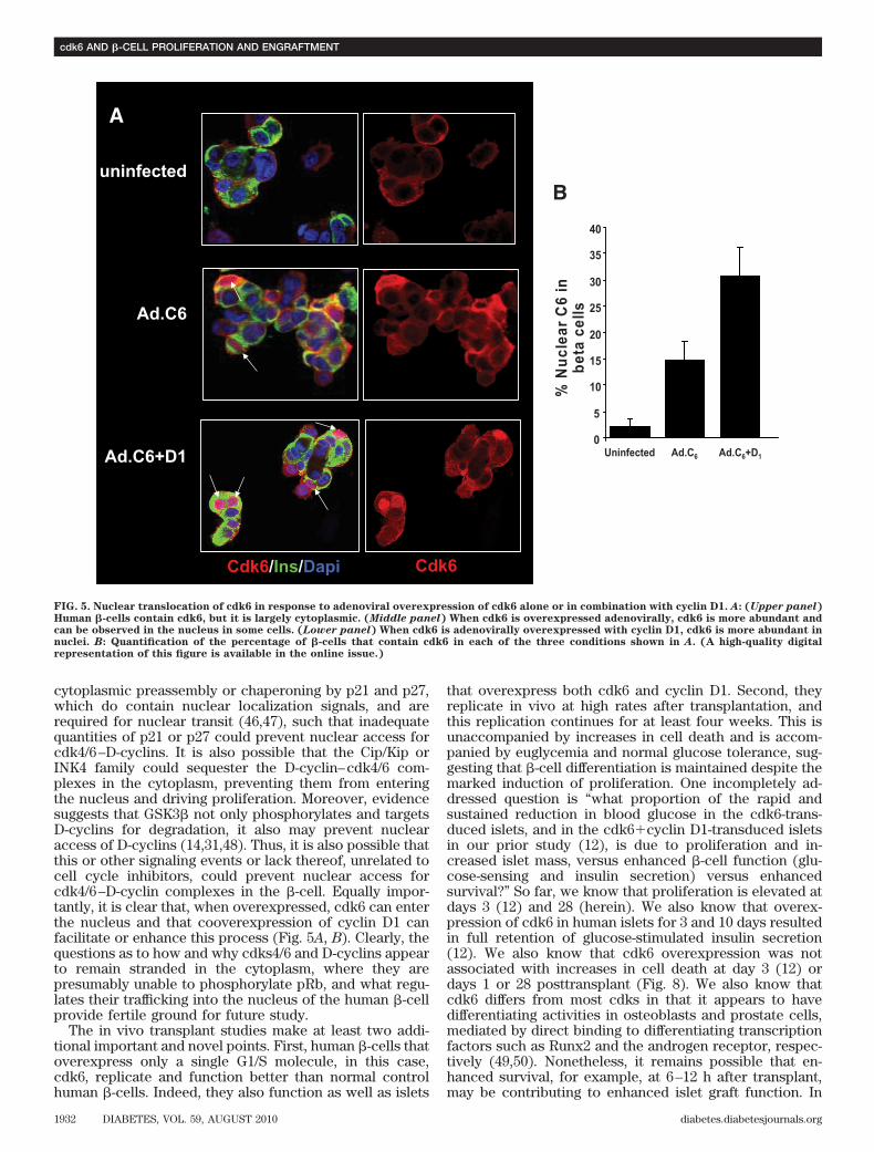
cytoplasmic preassembly or chaperoning by p21 and p27,which do contain nuclear localization signals, and arerequired for nuclear transit (46,47), such that inadequatequantities of p21 or p27 could prevent nuclear access forcdk4/6–D-cyclins. It is also possible that the Cip/Kip orINK4 family could sequester the D-cyclin–cdk4/6 com-plexes in the cytoplasm, preventing them from enteringthe nucleus and driving proliferation. Moreover, evidencesuggests that GSK3� not only phosphorylates and targetsD-cyclins for degradation, it also may prevent nuclearaccess of D-cyclins (14,31,48). Thus, it is also possible thatthis or other signaling events or lack thereof, unrelated tocell cycle inhibitors, could prevent nuclear access forcdk4/6–D-cyclin complexes in the �-cell. Equally impor-tantly, it is clear that, when overexpressed, cdk6 can enterthe nucleus and that cooverexpression of cyclin D1 canfacilitate or enhance this process (Fig. 5A, B). Clearly, thequestions as to how and why cdks4/6 and D-cyclins appearto remain stranded in the cytoplasm, where they arepresumably unable to phosphorylate pRb, and what regu-lates their trafficking into the nucleus of the human �-cellprovide fertile ground for future study.
The in vivo transplant studies make at least two addi-tional important and novel points. First, human �-cells thatoverexpress only a single G1/S molecule, in this case,cdk6, replicate and function better than normal controlhuman �-cells. Indeed, they also function as well as islets
that overexpress both cdk6 and cyclin D1. Second, theyreplicate in vivo at high rates after transplantation, andthis replication continues for at least four weeks. This isunaccompanied by increases in cell death and is accom-panied by euglycemia and normal glucose tolerance, sug-gesting that �-cell differentiation is maintained despite themarked induction of proliferation. One incompletely ad-dressed question is “what proportion of the rapid andsustained reduction in blood glucose in the cdk6-trans-duced islets, and in the cdk6�cyclin D1-transduced isletsin our prior study (12), is due to proliferation and in-creased islet mass, versus enhanced �-cell function (glu-cose-sensing and insulin secretion) versus enhancedsurvival?” So far, we know that proliferation is elevated atdays 3 (12) and 28 (herein). We also know that overex-pression of cdk6 in human islets for 3 and 10 days resultedin full retention of glucose-stimulated insulin secretion(12). We also know that cdk6 overexpression was notassociated with increases in cell death at day 3 (12) ordays 1 or 28 posttransplant (Fig. 8). We also know thatcdk6 differs from most cdks in that it appears to havedifferentiating activities in osteoblasts and prostate cells,mediated by direct binding to differentiating transcriptionfactors such as Runx2 and the androgen receptor, respec-tively (49,50). Nonetheless, it remains possible that en-hanced survival, for example, at 6–12 h after transplant,may be contributing to enhanced islet graft function. In
Ad.C6
uninfected
Ad.C6+D1
Cdk6/Ins/Dapi Cdk6
Uninfected Ad.C6 Ad.C6+D1
% N
ucle
ar C
6 in
be
ta c
ells
0
5
10
15
20
25
30
35
40
A
B
FIG. 5. Nuclear translocation of cdk6 in response to adenoviral overexpression of cdk6 alone or in combination with cyclin D1. A: (Upper panel)Human �-cells contain cdk6, but it is largely cytoplasmic. (Middle panel) When cdk6 is overexpressed adenovirally, cdk6 is more abundant andcan be observed in the nucleus in some cells. (Lower panel) When cdk6 is adenovirally overexpressed with cyclin D1, cdk6 is more abundant innuclei. B: Quantification of the percentage of �-cells that contain cdk6 in each of the three conditions shown in A. (A high-quality digitalrepresentation of this figure is available in the online issue.)
cdk6 AND �-CELL PROLIFERATION AND ENGRAFTMENT
1932 DIABETES, VOL. 59, AUGUST 2010 diabetes.diabetesjournals.org

addition, it is important to point out that no attempt wasmade to quantify �-cell mass in this model. Thus, it isunknown at present precisely how cdk6 alone or incombination enhances human islet engraftment. Theseobservations make cdk6 a particularly attractive target forfuture longer studies in vitro and in vivo aimed at enhanc-ing human �-cell proliferation, mass, and function, as wellas at better understanding the mechanisms through whichit acts.
These studies have limitations and raise additional newquestions for future study. For example, as noted above,they are not quantitative with regard to the absolute levelsof expression of the different D-cyclins and cdks: theyreflect the affinity and specificity of available antisera,which were not directly compared in strict quantitativeterms.
As another example, while we demonstrate that cdk6 isparticularly effective in vivo, we did not examine all of the
other possible cdk4/6–D-cyclin combinations: there aresome 25 potential single, double, and/or triple cdk4/6–D-cyclin combinations that we might have explored in the invivo NOD-SCID diabetes model. Examining all of thesepossibilities would be prohibitive with regard to time,expense, and availability of human islets. Because we
Mean 4,000 (n=9)
1,500 LZ (n=10)
1,500 D1+C6(n=15)
1,500 D1+C6(n=7)
1,500 C6(n=8)
1,500 C6(n=7)
1,500 D 1 (n=6)
Blo
od G
luco
se(m
g/dl
)
Days
0
50
100
150
200
250
300
350
400
450
500
0 7 14 21
AB
lood
Glu
cose
(mg/
dl)
min
1,500 LZ (n=6)1,500 D1(n=4)
4,000 (n=9)
IPGTT at day 21
Non-diabetic (n=8)
050
100150200250300350400450500
0 15 30 60 120
B
FIG. 6. cdk6 alone enhances human islet function in vivo in streptozo-tocin diabetic NOD-SCID mice. Bars indicate mean � SEM. A: Micetransplanted with 1,500 IEQ transduced with Ad.lacZ are shown in theblack lines, as described in the key within the figure. Mice transplantedwith 1,500 IEQ transduced with Ad.cdk6 alone (C6), with Ad.cyclin D1alone (D1), or both Ad.cdk6 and Ad.cyclin D1 (D1�C6) are shown inblue, purple, and green, respectively, and compared with 4,000 normal,nontransduced IEQ. The numbers in the key refer to the number ofexperimental animals in each group. B: Intraperitoneal glucose toler-ance testing (IPGTT) in normal (nondiabetic) NOD-SCID mice, dia-betic NOD-SCID mice transplanted with 4,000 IEQ human islets, 1,500human IEQ transduced with Ad.lacZ, or 1,500 IEQ transduced withAd.cdk6 alone (C6), with Ad.cyclin D1 alone (D1) or both Ad.cdk6 andAd.cyclin D1 (D1�C6). Studies were performed 21 days after transplan-tation. The numbers in parentheses indicate the numbers of animalsstudied. (A high-quality digital representation of this figure is avail-able in the online issue.)
Ad.LZ
Ad.D1
Ad.C6
Ad.D1+C6
Ki67/insulin/DAPI
4000
Intestine
A
05
10152025303540
% K
i-67
posi
tive/
Insu
lin p
ositi
ve
4000 Ad.LZ Ad.C6 Ad.C1 Ad.D1+C6
n=3 n=3 n=4 n=3 n=3
3/269 6/674
162/654
181/1073
80/623
B
C + PrimaryAb + Primary
+ Blocking PeptideAb
Cdk6/Ins/Dapi Cdk6/DapiCdk6
Uninfected Uninfected Uninfected
6 6Ad.C6 Ad.C6 Ad.C6
FIG. 7. Effects of cdk6, cyclin D1, or cdk6 and cyclin D1 on �-cellproliferation in vivo. A: Proliferation in human �-cells in vivo 28 daysafter transplantation. Ki-67 is shown in red, and �-cells are shown ingreen. Human islets grafts were removed at day 28 after transplanta-tion, fixed in 4% paraformaldehyde, embedded in paraffin, and stainedfor Ki-67. Ki-67 staining in samples of intestine cofixed, coembedded,and cosectioned with the islet grafts were performed as a positivecontrol in the lower right panel. B: Quantification of Ki-67-positiveinsulin-positive cells as a function of total insulin-positive cells. Thenumbers shown within the bars indicate the number of Ki-67-positive(red) �-cells and of insulin-positive (green) cells. The numbers belowthe bars indicate the numbers of animals studied, with two sections peranimal. C: cdk6 staining in human �-cells in islet grafts at 28 daysposttransplant. Note that cdk6 is still easily visible 28 days posttrans-plant and that, in many �-cells, it is nuclear as well as cytoplasmic. (Ahigh-quality digital representation of this figure is available in theonline issue.)
N.M. FIASCHI-TAESCH AND ASSOCIATES
diabetes.diabetesjournals.org DIABETES, VOL. 59, AUGUST 2010 1933

were limited with regard to experimental transplant para-digms, we elected to approach the human �-cell prolifer-ation/engraftment issue with an eye toward simplification,selecting a head-to-head comparison of cdk6 versus cyclinD1. To our surprise, cdk6 alone proved to be as effective asthe combination and superior to cyclin D1 alone. Thus,proliferation rates induced in vitro do not necessarilypredict transplant efficacy in vivo: although we foundpreviously that combined overexpression of cdk6 pluscyclin D1 in vitro produces far higher proliferation thaneither alone, we found here that cdk6 alone produced graftfunction that was comparable to the combination. This
observation was unanticipated, for during cell cycle pro-gression, cdks remain constant, awaiting a D-cyclin part-ner to activate their kinase activity. Thus, we had assumedthat cyclin D1 might have been rate-limiting and that cdk6overexpression alone might have had limited effects, butwe observed the opposite. These observations raise anumber of questions, such as “what is the D-cyclin partnerthat binds to cdk6 in �-cells when it alone is overex-pressed and activates proliferation?” Also, as noted above,“is cdk6 regulation a normal checkpoint in human �-cellreplication?” Another question is, “what exact cell in isletis the target of the adenoviral cdk6?” Because cdk6
TUNEL/Insulin/DAPI
Ad.C6Ad.LZ4000
Ad.D1 Ad.D1+C6 Intestine
0
2
4
6
8
10
12
14
% T
UN
EL-IN
S po
sitiv
e
Ad.LZ Ad.C6
49/50678/835
B C
Ad.LZ
Ad.C6
A
TUN
EL/In
sulin
/DA
PI
FIG. 8. Effects of cdk6, cyclin D1, or cdk6 and cyclin D1 on �-cell death in vivo. A: TUNEL assay was performed on each of the 2–6 fields from 3–4human islet grafts shown in Fig. 7, 28 days after transplantation. No TUNEL-positive nuclei were observed. TUNEL-positive nuclei are shown ingreen, and �-cells are shown in red. Examples of TUNEL-positive nuclei are indicated by arrows. TUNEL staining in samples of intestine cofixed,coembedded, and cosectioned with the islet grafts were performed as a positive control in the lower right panel. B: Similar experiments wererepeated, but grafts were removed 24 h after transplantation. As can be seen, �-cell apoptosis is similar in the Ad.cdk6-versus Ad.lacZ-transducedislets. (A high-quality digital representation of this figure is available in the online issue.)
cdk6 AND �-CELL PROLIFERATION AND ENGRAFTMENT
1934 DIABETES, VOL. 59, AUGUST 2010 diabetes.diabetesjournals.org

delivery was driven by the CMV promoter, it is anticipatedthat many cell types in addition to the �-cell would beinduced to proliferate, a prediction that is supported byour previous report (12). These cells do not appear to beendocrine cells (12), but may be endothelial, ductal stro-mal, mesenchymal, progenitor, or other cells. Thus, it willbe important to learn whether cdk6 overexpression usinga �-cell-specific promoter leads to the same enhancementof engraftment and function. From a therapeutic stand-point, the implications are unequivocal: cdk6 is a particu-larly attractive agent, both as a therapeutic molecule forenhancing �-cell replication and also as a “druggable”target for small-molecule therapeutics that might enhancehuman �-cell regulation. As noted, these findings alsosuggest that cdk6/cyclin D3 might also be a particularlyuseful combination therapeutically.
We demonstrate that adenoviral cdk6 can drive human�-cell replication for at least 28 days in vivo. This raisesadditional questions as well. For example, “how longwill/can �-cell proliferation continue in this in vivomodel?” For another example, “is sustained proliferation agood thing, or will it eventually lead to hypoglycemia oroncogenic transformation in �-cells?” Another question is,“how long is cdk6 expression required to ensure sustained�-cell engraftment?” “Will cdk6 be required indefinitely, orcan transient proliferation during the critical period ofengraftment suffice to ensure long-term engraftment?” Arelated question is, “can either regulated or sustaineddelivery of cdk6 produce sustained human �-cell replica-tion in vitro, in a way that will permit expansion of human�-cells ex vivo with retention of differentiation?” These arequestions that should be examined in future studies inwhich cdk6 is delivered using regulatable promoters.
With regard to the oncogenicity question, it is importantto recall that the G1/S family is also likely responsible fornormal human �-cell proliferation in embryonic and neo-natal life (9,10) and that they are not necessarily onco-genes. Having said this, it would seem likely thatunregulated, long-term expression of cdk6 might haveoncogenic consequences. This concern of course appliesnot only to G1/S cell cycle-activating molecules such as thecdks and D-cyclins, but also to most mitogenic signalingpathways, from receptors (e.g., EGF receptor, G-proteinreceptors, etc.) to signaling pathways (PI3 kinase, MAPkinase, Ras/Raf, JAK-STAT pathways, etc.) as well. Thus,one principal goal of these studies would be to use thesestrategies to identify key cellular molecules and pathwaysthat could be targets for small-molecule agonists. Forexample, now that it is clear that cdk6 is effective individ-ually in driving human �-cell expansion, it would bereasonable to screen small-molecule libraries to identifymolecules that activate cdk6 in human �-cells. An addi-tional goal might be to develop tools that would permitregulated expression of G1/S molecules such as cdk6 andD-cyclins such that they can be transiently activated todrive �-cell expansion for a few days or weeks and theninactivated to avoid sustained �-cell proliferation (51).
In conclusion, the G1/S transition is regulated by manyproteins in addition to the five studied here, as describedin the Introduction and supplementary Fig. 1. Each ofthese merits future study as both a normal and a potentialtherapeutic regulator of human �-cell replication. Theefficacy of these, alone or in combination, and whetherthey may prove to be as or more effective than cdk6 as atherapeutic target for ex vivo �-cell expansion or in vivo�-cell engraftment, warrants additional study. Finally,
these studies emphasize that, while broadly similar, hu-man and rodent �-cells display differences in G1/S reper-toires. These studies provide experimental models andmultiple targets exploiting human �-cell replication, re-placement, and engraftment in patients with diabetes.
ACKNOWLEDGMENTS
This work was supported by the Juvenile Diabetes Re-search Foundation (Grants 1-2008-39 and 34-2008-630), theNIH/NIDDK (Grant R-01 DK55023), the NIH/NIDDK BetaCell Biology Consortium (Grant U-01 DK072473), the NIH/NCRR- and NIDDK-supported Islet Cell Resource Consor-tium, its Administrative Bioinformatics CoordinatingCenter, The Spanish Ministry of Science and Innovation(CP08/00094), and the Pam and Scott Kroh and Don andArleen Wagner Family Foundations. No potential conflictsof interest relevant to this article were reported.
Parts of this study were presented in abstract form atthe 70th Scientific Sessions of the American DiabetesAssociation, Orlando, Florida, 25–29 June 2010.
We also thank Drs. Adolfo Garcia-Ocana, Laura C.Alonso, and Rupangi C. Vasavada for helpful discussionduring the development of this project.
REFERENCES
1. Dor Y, Brown J, Martinez OI, Melton DA. Adult pancreatic beta-cells areformed by self-duplication rather than stem-cell differentiation. Nature2004;429:41–46
2. Flier SN, Kulkarni RN, Kahn CR. Evidence for a circulating islet cell growthfactor in insulin-resistant states. Proc Nat Acad Sci 2001;98:7475–7480
3. Alonso LC, Yokoe T, Zhang P, Scott DK, Kim SK, O’Donnell CP, Garcia-Ocana A. Glucose infusion in mice: a new model to induce beta-cellreplication. Diabetes 2007;56:1792–1801
4. Miettinen PJ, Ustinov J, Ormio P, Gao R, Palgi J, Hakonen E, Juntti-Berggren L, Berggren PO, Otonkoski T. Downregulation of EGF receptorsignaling in pancreatic islets causes diabetes due to impaired postnatalbeta-cell growth. Diabetes 2006;55:3299–3308
5. Vasavada RC, Garcia-Ocana A, Zawalich WS, Sorenson RL, Dann P, SyedM, Ogren L, Talamantes F, Stewart AF. Targeted expression of placentallactogen in the beta cells of transgenic mice results in beta cell prolifera-tion, islet mass augmentation, and hypoglycemia. J Biol Chem 2000;275:15399–15406
6. Garcia-Ocana A, Takane KK, Syed MA, Philbrick WM, Vasavada RC,Stewart AF. Hepatocyte growth factor overexpression in the islet oftransgenic mice increases beta cell proliferation, enhances islet mass, andinduces mild hypoglycemia. J Biol Chem 2000;275:1226–1232
7. Fujinaka Y, Sipula D, Garcia-Ocana A, Vasavada RC. Characterization ofmice doubly transgenic for parathyroid hormone-related protein andmurine placental lactogen: a novel role for placental lactogen in pancreaticbeta-cell survival. Diabetes 2004;53:3120–3130
8. Fatrai S, Elghazi L, Balcazar N, Cras-Meneur C, Krits I, Kiyokawa H,Bernal-Mizrachi E. Akt induces beta-cell proliferation by regulating cyclinD1, cyclin D2, and p21 levels and cyclin-dependent kinase-4 activity.Diabetes 2006;55:318–325
9. Meier JJ, Butler AE, Saisho Y, Monchamp T, Galasso R, Bhushan A, RizzaRA, Butler PC. Beta-cell replication is the primary mechanism subservingthe postnatal expansion of beta-cell mass in humans. Diabetes 2008;57:1584–1594
10. Kassem SA, Ariel I, Thornton PS, Scheimberg I, Glaser B. Beta-cellproliferation and apoptosis in the developing normal human pancreas andin hyperinsulinism of infancy. Diabetes 2000;49:1325–1333
11. Hayek A, Beattie GM. Experimental transplantation of human fetal andadult pancreatic islets. J Clin Endocrinol Metab 1997;82:2471–2475
12. Fiaschi-Taesch N, Bigatel TA, Sicari B, Takane KK, Salim F, Velazquez-Garcia S, Harb G, Selk K, Cozar-Castellano I, Stewart AF. Survey of thehuman pancreatic beta-cell G1/S proteome reveals a potential therapeuticrole for cdk-6 and cyclin D1 in enhancing human beta-cell replication andfunction in vivo. Diabetes 2009;58:882–893
13. Parnaud G, Bosco D, Berney T, Pattou F, Kerr-Conte J, Donath MY, BruunC, Mandrup-Poulsen T, Billestrup N, Halban PA. Proliferation of sortedhuman and rat beta cells. Diabetologia 2008;51:91–100
14. Liu H, Remedi MS, Pappan KL, Kwon G, Rohatgi N, Marshall CA, McDaniel
N.M. FIASCHI-TAESCH AND ASSOCIATES
diabetes.diabetesjournals.org DIABETES, VOL. 59, AUGUST 2010 1935
ML. Glycogen synthase kinase-3 and mammalian target of rapamycinpathways contribute to DNA synthesis, cell cycle progression, and prolif-eration in human islets. Diabetes 2009;58:663–672
15. Tyrberg B, Eizirik DL, Hellerstrom C, Pipeleers DG, Andersson A. Humanpancreatic beta-cell deoxyribonucleic acid-synthesis in islet grafts de-creases with increasing organ donor age but increases in response toglucose stimulation in vitro. Endocrinology 1996;137:5694–5699
16. Davalli AM, Ogawa Y, Ricordi C, Scharp DW, Bonner-Weir S, Weir GC. Aselective decrease in the beta cell mass of human islets transplanted intodiabetic nude mice. Transplantation 1995;59:817–820
17. Menge BA, Tannapfel A, Belyaev O, Drescher R, Muller C, Uhl W, SchmidtWE, Meier JJ. Partial pancreatectomy in adult humans does not provokebeta-cell regeneration. Diabetes 2008;57:142–149
18. Rao P, Roccisana J, Takane KK, Bottino R, Zhao A, Trucco M, García-Ocana A. Gene transfer of constitutively active Akt markedly improveshuman islet transplant outcomes in diabetic severe combined immunode-ficient mice. Diabetes 2005;54:1664–1675
19. Butler AE, Janson J, Bonner-Weir S, Ritzel R, Rizza RA, Butler PC. Beta-celldeficit and increased beta-cell apoptosis in humans with type 2 diabetes.Diabetes 2003;52:102–110
20. Shapiro AM, Lakey JR, Ryan EA, Korbutt GS, Toth E, Warnock GL,Kneteman NM, Rajotte RV. Islet transplantation in seven patients with type1 diabetes mellitus using a glucocorticoid-free immunosuppressive regi-men. N Engl J Med 2000;343:230–238
21. Cozar-Castellano I, Fiaschi-Taesch N, Bigatel TA, Takane KK, Garcia-Ocana A, Vasavada R, Stewart AF. Molecular control of cell cycleprogression in the pancreatic beta-cell. Endocr Rev 2006;27:356–370
22. Heit JJ, Karnik SK, Kim SK. Intrinsic regulators of pancreatic beta-cellproliferation. Annu Rev Cell Dev Biol 2006;22:311–338
23. Rane SG, Dubus P, Mettus RV, Galbreath EJ, Boden G, Reddy EP, BarbacidM. Loss of Cdk4 expression causes insulin-deficient diabetes and Cdk4activation results in beta-islet cell hyperplasia. Nat Genet 1999;22:44–52
24. Harb G, Vasavada RC, Cobrinik D, Stewart AF. The retinoblastoma proteinand its homolog p130 regulate the G1/S transition in pancreatic beta-cells.Diabetes 2009;58:1852–1862
25. Uchida T, Nakamura T, Hashimoto N, Matsuda T, Kotani K, Sakaue H, KidoY, Hayashi Y, Nakayama KI, White MF, Kasuga M. Deletion of Cdkn1bameliorates hyperglycemia by maintaining compensatory hyperinsulin-emia in diabetic mice. Nat Med 2005;11:175–182
26. Zhong L, Georgia S, Tschen SI, Nakayama K, Nakayama K, Bhushan A.Essential role of Skp2-mediated p27 degradation in growth and adaptiveexpansion of pancreatic beta cells. J Clin Invest 2007;117:2869–2876
27. Dhawan S, Tschen SI, Bhushan A. Bmi-1 regulates the Ink4a/Arf locus tocontrol pancreatic beta-cell proliferation. Genes and Development 2009;23:906–911
28. Chen H, Gu X, Su IH, Bottino R, Contreras JL, Tarakhovsky A, Kim SK.Polycomb protein Ezh2 regulates pancreatic beta-cell Ink4a/Arf expressionand regeneration in diabetes mellitus. Genes and Development 2009;23:975–985
29. Ackermann Misfeldt A, Costa RH, Gannon M. Beta-cell proliferation, butnot neogenesis, following 60% partial pancreatectomy is impaired in theabsence of FoxM1. Diabetes 2008;57:3069–3077
30. Zhang X, Gaspard JP, Mizukami Y, Li J, Graeme-Cook F, Chung DC.Overexpression of cyclin D1 in pancreatic beta-cells in vivo results in islethyperplasia without hypoglycemia. Diabetes 2005;54:712–719
31. He LM, Sartori DJ, Teta M, Opare-Addo LM, Rankin MM, Long SY, Diehl JA,Kushner JA. Cyclin D2 protein stability is regulated in pancreatic beta-cells. Mol Endocrinol 2009;23:1865–1875
32. Georgia S, Bhushan A. Beta cell replication is the primary mechanism formaintaining postnatal beta cell mass. J Clin Invest 2004;114:963–968
33. Kushner JA, Ciemerych MA, Sicinska E, Wartschow LM, Teta M, Long SY,Sicinski P, White MF. Cyclins D2 and D1 are essential for postnatalpancreatic beta-cell growth. Mol Cell Biol 2005;25:3752–3762
34. Karnik SK, Hughes CM, Gu X, Rozenblatt-Rosen O, McLean GW, Xiong Y,
Meyerson M, Kim SK. Menin regulates pancreatic islet growth by promot-ing histone methylation and expression of genes encoding p27Kip1 andp18INK4c. Proc Natl Acad Sci U S A 2005;102:14659–14664
35. Martín J, Hunt SL, Dubus P, Sotillo R, Nehme-Pelluard F, Magnuson MA,Parlow AF, Malumbres M, Ortega S, Barbacid M. Genetic rescue of Cdk4null mice restores pancreatic beta-cell proliferation but not homeostaticcell number. Oncogene 2003;22:5261–5269
36. Cozar-Castellano I, Takane KK, Bottino R, Balamurugan AN, Stewart AF.Induction of beta-cell proliferation and retinoblastoma protein phosphor-ylation in rat and human islets using adenovirus-mediated transfer ofcyclin-dependent kinase-4 and cyclin D1. Diabetes 2004;53:149–159
37. Lavine JA, Raess PW, Davis DB, Rabaglia ME, Presley BK, Keller MP,Beinfeld MC, Kopin AS, Newgard CB, Attie AD. Contamination withE1A-positive wild-type adenovirus accounts for species-specific stimula-tion of islet cell proliferation: a cautionary note. Mol Endocrinol 2010;24:464–467
38. Chung DC, Brown SB, Graeme-Cook F, Seto M, Warshaw AL, Jensen RT,Arnold A. Overexpression of cyclin D1 occurs frequently in humanpancreatic endocrine tumors. J Clin Endocrinol Metab 2000;85:4373–4378
39. Kozar K, Ciemerych MA, Rebel VI, Shigematsu H, Zagozdzon A, Sicinska E,Geng Y, Yu Q, Bhattacharya S, Bronson RT, Akashi K, Sicinski P. Mousedevelopment and cell proliferation in the absence of D-cyclins. Cell2004;118:477–491
40. Malumbres M, Sotillo R, Santamaría D, Galan J, Cerezo A, Ortega S, DubusP, Barbacid M. Mammalian cells cycle without the D-type cyclin-dependentkinases Cdk4 and Cdk6. Cell 2004;118:493–504
41. Brissova M, Fowler MJ, Nicholson WE, Chu A, Hirshberg B, Harlan DM,Powers AC. Assessment of human pancreatic islet architecture andcomposition by laser scanning confocal microscopy. J Histochem Cyto-chem 2005;53:1087–1097
42. Cabrera O, Berman DM, Kenyon NS, Ricordi C, Berggren PO, Caicedo A.The unique cytoarchitecture of human pancreatic islets has implicationsfor islet cell function. Proc Natl Acad Sci U S A 2006;103:2334–2339
43. De Vos A, Heimberg H, Quartier E, Huypens P, Bouwens L, Pipeleers D,Schuit F. Human and rat beta cells differ in glucose transporter but not inglucokinase gene expression. J Clin Invest 1995;96:2489–2495
44. Ferrer J, Benito C, Gomis R. Pancreatic islet GLUT2 glucose transportermRNA and protein expression in humans with and without NIDDM.Diabetes 1995;44:1369–1374
45. Schuit FC. Is GLUT2 required for glucose sensing? Diabetologia 1997;40:104–111
46. Malumbres M, Barbacid M. Mammalian cyclin-dependent kinases. TrendsBiochem Sci 2005;30:630–641
47. Sherr CJ, Roberts JM. CDK inhibitors: positive and negative regulators ofG1-phase progression. Genes Dev 1999;13:1501–1512
48. Kida A, Kakihana K, Kotani S, Kurosu T, Miura O. Glycogen synthasekinase-3beta and p38 phosphorylate cyclin D2 on Thr280 to trigger itsubiquitin/proteasome-dependent degradation in hematopoietic cells. On-cogene 2007;26:6630–6640
49. Thomas DM, Johnson SA, Sims NA, Trivett MK, Slavin JL, Rubin BP,Waring P, McArthur GA, Walkley CR, Holloway AJ, Diyagama D, Grim JE,Clurman BE, Bowtell DD, Lee JS, Gutierrez GM, Piscopo DM, Carty SA,Hinds PW, McArthur G, Walkeley C, Holoway AJ, Diyagama D, Clurman B,Bowtell DDL, Lee J-S, Gutierrez G, Piscopo DM, Carty SA, Hinds PW.Terminal osteoblast differentiation, mediated by runx2 and p27KIP1, isdisrupted in osteosarcoma. J Cell Biol 2004;167:925–934
50. Lim JT, Mansukhani M, Weinstein IB. Cyclin-dependent kinase 6 associateswith the androgen receptor and enhances its transcriptional activity inprostate cancer cells. Proc Nat Acad Sci 2005;102:5156–5161
51. Takane KK, Kleinberger J, Salim F, Thomas S, Fiaschi-Taesch NM, StewartAF. Regulated induction of human beta cell replication: tetracycline-inducible overexpression of cdk6 and cyclin D1 in human beta cells. InProceedings of the 70th ADA Annual Scientific Sessions, Orlando, FL,
June 2010 (submitted)
cdk6 AND �-CELL PROLIFERATION AND ENGRAFTMENT
1936 DIABETES, VOL. 59, AUGUST 2010 diabetes.diabetesjournals.org