Embed Size (px)
Citation preview

227http://www.ecevr.org/
CLINICAL EXPERIMENTALVACCINERESEARCH
Original article
Introduction
Protein cages, such as ferritins, small heat shock proteins, and viral capsids are prom-
ising nanoplatform candidates for efficient delivery systems because they have uni-
form size, structure, high biocompatibility and biodegradability [1-6]. The protein cag-
es are spontaneously self-assembled from multiple copies of one or a few types of pro-
tein subunits in a precisely controlled manner [1-6]. Also, they can be genetically and
chemically manipulated to have desired functionality by rational design on the basis
of atomic resolution structural information.
The lumazine synthase, isolated from hyperthermophile Aquifex aeolicus (AaLS),
consists of 60 identical subunits forming an icosahedral capsid architecture (T=1 state)
with a 15.4 nm exterior and a 9 nm interior diameters, respectively [7]. The AaLS origi-
nally catalyzes the penultimate step in riboflavin biosynthesis within the cell. Outside
of the cell, its hollow sphere architecture has been used as a template for encapsula-
tion of cargo proteins such as green fluorescent proteins [8], human immunodeficien-
cy virus proteases [9], crystallization of inorganic nanoparticles such as iron oxide [10],
© Korean Vaccine Society.This is an Open Access article distributed under the terms of the Creative Commons Attribution Non-Com-mercial License (http://creativecommons.org/licenses/by-nc/3.0) which permits unrestricted non-commercial use, distribution, and reproduction in any medium, pro-vided the original work is properly cited.
K O R E A N V A C C I N E S O C I E T Y
K O R E A N V A C C I N E S O C I E T Y
K O R E A N A C C I N E O C I E T Y
VS
Clin Exp Vaccine Res 2014;3:227-234http://dx.doi.org/10.7774/cevr.2014.3.2.227pISSN 2287-3651 • eISSN 2287-366X
Jae-Sun Ra*, Hyun-Hee Shin*, Sebyung Kang, Yoonkyung DoSchool of Life Sciences, Ulsan National Institute of Science and Technology, Ulsan, Korea
Received: April 10, 2014Revised: May 1, 2014Accepted: May 12, 2014
Corresponding author: Yoonkyung Do, PhDSchool of Life Sciences, Ulsan National Insti-tute of Science and Technology, 50 UNIST-gil, Eonyang-eup, Ulju-gun, Ulsan 689-798, KoreaTel: +82-52-217-5326, Fax: +82-217-5309E-mail: [email protected]
*These two authors contributed equally to this work.
No potential conflict of interest relevant to this article was reported.
This work was supported by Advanced Research Center (No. 2012-0008996) and Basic Science Research Program (No. 2013R1A1A2005545) of MEST through NRF of Korea, and the year of 2012 research fund (No. 1.120020.01) of UNIST.
Purpose: Protein cages are promising nanoplatform candidates for efficient delivery systems due to their homogenous size and structure with high biocompatibility and biodegradability. In this study, we investigate the potential of lumazine synthase protein cage as an antigen delivery system to dendritic cells (DCs), which induce antigen-specific T cell proliferation. Materials and Methods: Ovalbumin (OVA) peptides OT-1 (SIINFEKL) and OT-2 (ISQAVHAA-HAEINEAGR) were genetically inserted to lumazine synthase and each protein cage was over-expressed in Escherichia coli as a soluble protein. The efficiency of antigen delivery and the resulting antigen-specific T cell proliferation by DCs was examined in vitro as well as in vivo. Results: We successfully generated and characterized OVA peptides carrying lumazine syn-thase protein cages. The OT-1 and OT-2 peptides carried by lumazine synthases were efficient-ly delivered and processed by DCs in vitro as well as in vivo, and induced proliferation of OT-1-specific CD8+ T cells and OT-2-specific CD4+ T cells. Conclusion: Our data demonstrate the potential of lumazine synthase protein cage being used as a novel antigen delivery system for DC-based vaccine development in future clinical appli-cations.
Keywords: Dendritic cells, Protein cage, Antigen presentation, Vaccines, Nanoparticles
Lumazine synthase protein cage nanoparticles as antigen delivery nanoplatforms for dendritic cell-based vaccine development

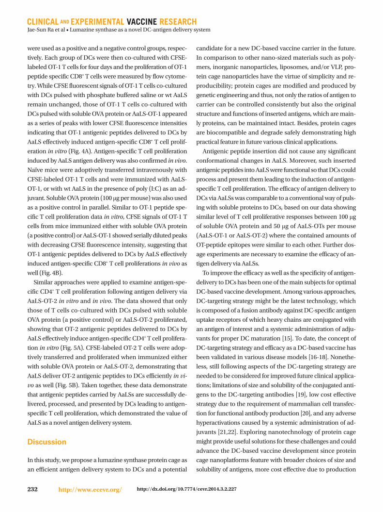
Jae-Sun Ra et al • Lumazine synthase as a novel DC-antigen delivery system
228 http://www.ecevr.org/ http://dx.doi.org/10.7774/cevr.2014.3.2.227
and fabrication of uniform layer-by-layer assemblies using
non-covalent interaction between surface displayed hexa-
hisitidine and Ni-NTA of the AaLS [11].
Dendritic cells (DCs) are the most potent professional an-
tigen-presenting cells and key regulator of antigen-specific
immune responses [12]. Immature DCs continously uptake
and process antigens. Upon danger signal, the antigen-load-
ed DCs migrate into draining lymph nodes where DCs pres-
ent antigens to naïve T cells leading to their proliferation and
differentiation into effector cells; CD8+ cytotoxic T cells to kill
infected target cells or CD4+ helper T cells to secrete cytokines
and facilitate diverse forms of cellular and humoral immuni-
ty. Therefore, based on such seminal roles of DCs in the im-
mune system, DC-based vaccine development has been a
promising approach to direct antigen-specific adaptive im-
munity [13].
Efficient antigen delivery to DCs is the first step for DC-based
vaccine development and simple and controllable antigen
delivery system is required. Up to date, nano-sized materials,
including polymers, inorganic nanoparticles, liposomes, and/
or virus-like particle (VLP), have been extensively used as an-
tigen delivery nanoplatforms for studying antigen-specific
immune responses and further applied them to follow-up
vaccine development. However, there is a limited amount of
study utilizing protein cage nanoparticles to deliver antigens
to DCs for immunization and vaccination. In our previous
study, we utilized ferritin protein cage nanoparticles as versa-
tile antigen delivery nanoplatforms for DC-based vaccine de-
velopment and demonstrated DC-mediated antigen specific
CD4+ and CD8+ T cell immune responses [14]. In the current
study, we introduce lumazine synthase protein cage nano-
particles (AaLS) for carrying antigenic peptides to DCs and
investigate DC-mediated antigen-specific T cell proliferation.
Our data demonstrate the potential of AaLS as a novel anti-
gen delivery system for DC-based vaccine development in
future clinical applications.
Materials and Methods
Antigenic peptide addition and protein cage purificationThe OT-1 (SIINFEKL) and OT-2 (ISQAVHAAHAEINEAGR)
peptides were added at the N-terminal and the C-terminal
ends of AaLS subunit, respectively, by an established poly-
merase chain reaction protocol using primers containing spe-
cific enzyme restriction sites and pETDuet based plasmids
containing genes encoding AaLS protein as a template. The
amplified DNAs were used to transform the competent Esch-
erichia coli strain BL21 (DE), resulting in the over-expression
in E. coli of the AaLS protein cages containing added antigen-
ic peptides. The resultant protein was purified as describ ed
previously [11].
Mass spectrometry The subunit masses of AaLS-OT-2, AaLS-OT-1, and wt AaLS
protein cages were analyzed using an electrospray ionization
time-of-flight mass (ESI-TOF) mass spectrometer (Xevo G2
TOF, Waters, Milford, MA, USA) interfaced to a Waters UPLC
and an autosampler. Samples were loaded onto the MassPREP
Micro desalting column (Waters) and eluted with a gradient
of 5-95% (v/v) acetonitrile containing 0.1% formic acid with a
flow rate of 300 µL/min [11]. Mass spectra were acquired in
the range of m/z 500-3,000 and processed using MaxEnt 1
and MaxEnt 3 from MassLynx version 4.1 to obtain the aver-
age mass from multiple charge state distributions.
MiceC57BL/6 mice were purchased from Taconic, and CD45.2+
OT-1 and CD45.2+ OT-2 mice were purchased from the Jack-
son Laboratory. All mice were maintained under specific patho-
gen-free conditions and used at 6-8 weeks with Institutional
Animal Care and Use Committee guidelines.
Isolation of splenic CD11c+ DCs and ovalbumin-specific T cellsSingle cell suspensions of splenocytes were prepared with
400 U/mL collagenase D (Roche, Basel, Switzerland) and
CD11c+ cells were positively enriched with magnetic-activat-
ed cell sorting (MACS) sorting (Miltenyl Biotech, Bergisch
Gladbach ,Germany). Ovalbumin (OVA)-specific CD8+ or
CD4+ T cells were prepared from OT-1 or OT-2 mice, respec-
tively. Briefly, single cell suspensions from lymph nodes and
spleens were prepared and CD8+ T cells or CD4+ T cells were
positively enriched by MACS sorting. Sorted T cells showed
>98% purity as detected by flow cytometry. All flow cytome-
try data were acquired by BD FACS Calibur and analyzed by
FlowJo software (TreeStar, San Carlos, CA, USA).
Carboxyfluorescein succinimidyl ester-dilution assay to detect antigen-specific T cell proliferation in vitro and in vivoFor in vitro OVA-specific T cell proliferation assay, OT-1 or
OT-2 T cells were stained with 0.5 μM of carboxyfluorescein
succinimidyl ester (CFSE) (Invitrogen, Carlsbad, CA, USA) at
1×10⁷ cells/mL for 10 minutes at 37°C. CD11c+ DCs were pulsed

Jae-Sun Ra et al • Lumazine synthase as a novel DC-antigen delivery system
229http://www.ecevr.org/http://dx.doi.org/10.7774/cevr.2014.3.2.227
with indicated proteins for 3 hours, washed and then co-cul-
tured with the CFSE-labeled T cells at a ratio of 1:3 (DC, 1×10⁵:
T cell, 3×10⁵) in a 96-well U-bottom plate at 37°C. After 4 days,
cells were harvested and then stained for Vβ5.1/5.2 (Bioleg-
end, San Diego, CA, USA), Vα2 and CD8 or CD4 (all from BD,
San Jose, CA, USA). For in vivo OVA-specific T cell prolifera-
tion assay, naïve C57BL/6 mice were adoptively transferred
with 5 μM CFSE-labeled OT-1 or OT-2 T cells intravenously at
day -1. At day 0, mice were injected with indicated proteins in
the footpads subcutaneously in the presence of 50 μg of poly
(I:C) (Invivogen, San Diego, CA, USA) as an adjuvant. At day
3, single cell suspensions were prepared from lymph nodes.
OVA-specific T cells (Vβ5.1/5.2+Vα2+CD8+ or Vβ5.1/ 5.2+Vα2+CD4+)
were gated and progressive halving of CFSE fluorescence per
cell was measured by flow cytometry.
Fig. 2. A schematic diagram showing dendritic cell (DC)-mediated antigen-specific T cell proliferation induced by lumazine synthase protein cage nanoparticles (AaLS) carrying OT peptides.
AaLS
Antigen delivery
DC
Processing and presentation
T cell
Antigen-specific T cell proliferation
Antigenpeptide
DC CD8+ T cell
CD4+ T cell
MHC I
MHC II
Fig. 1. Surface and ribbon diagram representations of AaLS (PDB: 1HQK) looking down the five-fold symmetry axis (A) and clipped view show-ing the interior space (9 nm interior and 15.4 nm exterior diameters) of the protein cage (B). Five subunits were shown as ribbon diagram with individual colors.
9 nm
15.4
nm
A B

Jae-Sun Ra et al • Lumazine synthase as a novel DC-antigen delivery system
230 http://www.ecevr.org/ http://dx.doi.org/10.7774/cevr.2014.3.2.227
Results
Surface and ribbon diagram representations of lumazine syn-
thase (AaLS) were depicted (Fig. 1) and a schematic diagram
showing DC-mediated antigen-specific T cell proliferation
induced by AaLS carrying OT peptides was shown in Fig. 2.
To adapt AaLS protein cage as antigen delivery nanoplat-
forms, we genetically introduced OT-1 (SIINFEKL) and OT-2
(ISQAVHAAHAEINEAGR) peptides to the N-terminal and
the C-terminal ends of AaLS subunit, respectively. Antigenic
peptide insertions were confirmed by DNA sequencing and
molecular mass measurement of subunits of OT-1 or OT-2
peptide added AaLS protein cages (AaLS-OT-1 or AaLS-OT-2).
AaLS-OT-1 and AaLS-OT-2 were individually over-expressed
in E. coli as a soluble protein without noticeable amounts of
precipitation. OT-peptide inserted AaLS protein cages were
purified by the same method used to purify wild-type AaLS
(wt AaLS), which do not have any OT peptide insertions. ESI-
TOF mass spectrometry analysis indicated that the subunit
masses of AaLS-OT-2, AaLS-OT-1, and wt AaLS protein cages
were 18,458.0 Da, 17,576.5 Da, and 16,705.0 Da, respectively,
which are well matched with their predicted masses (18,458.1
Da, 17,576.2 Da, and 16,706.3 Da, respectively) (Fig. 3A). Trans-
mission electron microscopic images of stained AaLS-OT-2,
AaLS-OT-1, and wt AaLS protein cages confirmed an intact
cage architecture with a uniform size distribution (~16 nm)
(Fig. 3B). These results indicate that AaLS-OT-2 and AaLS-
OT-1 protein cages form an intact protein cage architecture
100 nm
AaLS-OT-2
AaLS-OT-1
wt AaLS
100 nm
100 nmA B
Rela
tive
abun
danc
e (%
)
AaLS-OT-2Calc. 18458.1 Da
AaLS-OT-1Calc. 17576.2 Da
wt AaLSCalc. 16706.3 Da
100
%
0
18458 0
10,000 15,000 20,000 25,000 30,000Mass
100
%
0
17576 5
10,000 15,000 20,000 25,000 30,000Mass
100
%
0
16705 0
10,000 15,000 20,000 25,000 30,000Mass
Fig. 3. Characterization of OT-peptide added AaLS protein cages. (A) Molecular masses of dissociated subunits of AaLS-OT-2 (top), AaLS-OT-1 (middle), and wt AaLS (bottom) protein cages. Calculated (Calc.) and measured molecular masses were indicated. (B) Transmission electron micrographic image of 2% uranyl acetate stained AaLS-OT-2 (top), AaLS-OT-1 (middle), and wt AaLS (bottom) protein cages confirmed an intact cage architecture with a uniform size distribution.

Jae-Sun Ra et al • Lumazine synthase as a novel DC-antigen delivery system
231http://www.ecevr.org/http://dx.doi.org/10.7774/cevr.2014.3.2.227
PBS
OVA protein
wt AaLS
PBS
OVA protein
wt AaLS
AaLS-OT-1 AaLS-OT-1A B
Fig. 4. OT-1 antigenic peptides delivered by AaLS-OT-1 induce OT-1-specific CD8+ T cell proliferation in vitro and in vivo. (A) CD11c+ dendritic cells (DCs) were pulsed with phosphate buffered saline (PBS), soluble ovalbumin (OVA) protein, wt AaLS, or AaLS-OT-1 at 2 mg/mL for 3 hours. The DCs were washed and then co-cultured with carboxyfluorescein succinimidyl ester (CFSE)-labeled OT-1 T cells at a ratio of 1:3. Four days later, the proliferation of OT-1-specific CD8+ T cells was measured by flow cytometry. (B) Mice were adoptively transferred with CFSE-labeled OT-1 T cells and on the next day, they were immunized subcutaneously with PBS, 100 μg of OVA protein, 50 μg of wt AaLS, AaLS-OT-1 in the presence of an adjuvant. Three days later, the proliferation of OT-1-specific CD8+ T cells was measured by flow cytometry.
PBS
OVA protein
wt AaLS
PBS
OVA protein
wt AaLS
AaLS-OT-2 AaLS-OT-2A B
Fig. 5. OT-2 antigenic peptides delivered by AaLS-OT-2 induce OT-2-specific CD4+ T cell proliferation in vitro and in vivo. (A) CD11c+ dendritic cells (DCs) were pulsed with phosphate buffered saline (PBS), soluble ovalbumin (OVA) protein, wt AaLS, or AaLS-OT-2 at 2 mg/mL for 3 hours. The DCs were washed and then co-cultured with carboxyfluorescein succinimidyl ester (CFSE)-labeled OT-2 T cells at a ratio of 1:3. Four days later, the proliferation of OT-2-specific CD4+ T cells was measured by flow cytometry. (B) Mice were adoptively transferred with CFSE-labeled OT-2 T cells and on the next day, they were immunized subcutaneously with PBS, 100 μg of OVA protein, 50 μg of wt AaLS, AaLS-OT-2 in the presence of an adjuvant. Three days later, the proliferation of OT-2-specific CD4+ T cells was measured by flow cytometry.
without significant changes in size and composition.
To study antigen-specific CD8+ T cell proliferation induced
by DCs following antigen delivery by AaLS-OT-1 in vitro, DCs
were pulsed with either wt AaLS or AaLS-OT-1 for three hours
and were washed to ensure the presentation of only processed
antigens. DCs pulsed with soluble OVA protein and wt AaLS
Jae-Sun Ra et al • Lumazine synthase as a novel DC-antigen delivery system
232 http://www.ecevr.org/ http://dx.doi.org/10.7774/cevr.2014.3.2.227
were used as a positive and a negative control groups, respec-
tively. Each group of DCs were then co-cultured with CFSE-
labeled OT-1 T cells for four days and the proliferation of OT-1
peptide specific CD8+ T cells were measured by flow cytome-
try. While CFSE fluorescent signals of OT-1 T cells co-cultured
with DCs pulsed with phosphate buffered saline or wt AaLS
remain unchanged, those of OT-1 T cells co-cultured with
DCs pulsed with soluble OVA protein or AaLS-OT-1 appeared
as a series of peaks with lower CFSE fluorescence intensities
indicating that OT-1 antigenic peptides delivered to DCs by
AaLS effectively induced antigen-specific CD8+ T cell prolif-
eration in vitro (Fig. 4A). Antigen-specific T cell proliferation
induced by AaLS antigen delivery was also confirmed in vivo.
Naïve mice were adoptively transferred intravenously with
CFSE-labeled OT-1 T cells and were immunized with AaLS-
OT-1, or with wt AaLS in the presence of poly (I:C) as an ad-
juvant. Soluble OVA protein (100 μg per mouse) was also used
as a positive control in parallel. Similar to OT-1 peptide spe-
cific T cell proliferation data in vitro, CFSE signals of OT-1 T
cells from mice immunized either with soluble OVA protein
(a positive control) or AaLS-OT-1 showed serially diluted peaks
with decreasing CFSE fluorescence intensity, sug gesting that
OT-1 antigenic peptides delivered to DCs by AaLS effectively
induced antigen-specific CD8+ T cell proliferations in vivo as
well (Fig. 4B).
Similar approaches were applied to examine antigen-spe-
cific CD4+ T cell proliferation following antigen delivery via
AaLS-OT-2 in vitro and in vivo. The data showed that only
those of T cells co-cultured with DCs pulsed with soluble
OVA protein (a positive control) or AaLS-OT-2 proliferated,
showing that OT-2 antigenic peptides delivered to DCs by
AaLS effectively induce antigen-specific CD4+ T cell prolifera-
tion in vitro (Fig. 5A). CFSE-labeled OT-2 T cells were adop-
tively transferred and proliferated when immunized either
with soluble OVA protein or AaLS-OT-2, demonstrating that
AaLS deliver OT-2 antigenic peptides to DCs efficiently in vi-
vo as well (Fig. 5B). Taken together, these data demonstrate
that antigenic peptides carried by AaLSs are successfully de-
livered, processed, and presented by DCs leading to antigen-
specific T cell proliferation, which demonstrated the value of
AaLS as a novel antigen delivery system.
Discussion
In this study, we propose a lumazine synthase protein cage as
an efficient antigen delivery system to DCs and a potential
candidate for a new DC-based vaccine carrier in the future.
In comparison to other nano-sized materials such as poly-
mers, inorganic nanoparticles, liposomes, and/or VLP, pro-
tein cage nanoparticles have the virtue of simplicity and re-
producibility; protein cages are modified and produced by
genetic engineering and thus, not only the ratios of antigen to
carrier can be controlled consistently but also the original
structure and functions of inserted antigens, which are main-
ly proteins, can be maintained intact. Besides, protein cages
are biocompatible and degrade safely demonstrating high
practical feature in future various clinical applications.
Antigenic peptide insertion did not cause any significant
conformational changes in AaLS. Moreover, such inserted
antigenic peptides into AaLS were functional so that DCs could
process and present them leading to the induction of antigen-
specific T cell proliferation. The efficacy of antigen delivery to
DCs via AaLSs was comparable to a conventional way of puls-
ing with soluble proteins to DCs, based on our data showing
similar level of T cell proliferative responses between 100 μg
of soluble OVA protein and 50 μg of AaLS-OTs per mouse
(AaLS-OT-1 or AaLS-OT-2) where the contained amounts of
OT-peptide epitopes were similar to each other. Further dos-
age experiments are necessary to examine the efficacy of an-
tigen delivery via AaLSs.
To improve the efficacy as well as the specificity of antigen-
delivery to DCs has been one of the main subjects for optimal
DC-based vaccine development. Among various approaches,
DC-targeting strategy might be the latest technology, which
is composed of a fusion antibody against DC-specific antigen
uptake receptors of which heavy chains are conjugated with
an antigen of interest and a systemic administration of adju-
vants for proper DC maturation [15]. To date, the concept of
DC-targeting strategy and efficacy as a DC-based vaccine has
been validated in various disease models [16-18]. Nonethe-
less, still following aspects of the DC-targeting strategy are
needed to be considered for improved future clinical applica-
tions; limitations of size and solubility of the conjugated anti-
gens to the DC-targeting antibodies [19], low cost effective
strategy due to the requirement of mammalian cell transfec-
tion for functional antibody production [20], and any adverse
hyperactivations caused by a systemic administration of ad-
juvants [21,22]. Exploring nanotechnology of protein cage
might provide useful solutions for these challenges and could
advance the DC-based vaccine development since protein
cage nanoplatforms feature with broader choices of size and
solubility of antigens, more cost effective due to production

Jae-Sun Ra et al • Lumazine synthase as a novel DC-antigen delivery system
233http://www.ecevr.org/http://dx.doi.org/10.7774/cevr.2014.3.2.227
in E. coli, as well as with co-insertion of selective adjuvants
and antigens together. Furthermore, various protein cages
containing selective antigens and adjuvants can be mixed
and matched with known DC-targeting antibodies, and such
interdisciplinary synergy will open a new avenue of DC-based
vaccine design and development.
In conclusion, this study sets the stage for the analysis of
functional roles of lumazine synthase protein cages as effi-
cient DC-based vaccine carrier in various disease models.
ORCID
Jae-Sun Ra http://orcid.org/0000-0002-4525-7171
Hyun-Hee Shin http://orcid.org/0000-0003-4945-1059
Sebyung Kang http://orcid.org/0000-0001-7394-3550
Yoonkyung Do http://orcid.org/0000-0001-7947-4767
References
1. Kang HJ, Kang YJ, Lee YM, Shin HH, Chung SJ, Kang S.
Developing an antibody-binding protein cage as a molec-
ular recognition drug modular nanoplatform. Biomateri-
als 2012;33:5423-30.
2. Uchida M, Klem MT, Allen M, et al. Biological containers:
protein cages as multifunctional nanoplatforms. Adv Ma-
ter 2007;19:1025-42.
3. Kang S, Uchida M, O’Neil A, Li R, Prevelige PE, Douglas T.
Implementation of p22 viral capsids as nanoplatforms.
Biomacromolecules 2010;11:2804-9.
4. Min J, Jung H, Shin HH, Cho G, Cho H, Kang S. Implemen-
tation of p22 viral capsids as intravascular magnetic reso-
nance T1 contrast conjugates via site-selective attachment
of Gd(III)-chelating agents. Biomacromolecules 2013;14:
2332-9.
5. Kang YJ, Park DC, Shin HH, Park J, Kang S. Incorporation
of thrombin cleavage peptide into a protein cage for con-
structing a protease-responsive multifunctional delivery
nanoplatform. Biomacromolecules 2012;13:4057-64.
6. Uchida M, Kang S, Reichhardt C, Harlen K, Douglas T. The
ferritin superfamily: supramolecular templates for mate-
rials synthesis. Biochim Biophys Acta 2010;1800:834-45.
7. Zhang X, Meining W, Fischer M, Bacher A, Ladenstein R.
X-ray structure analysis and crystallographic refinement
of lumazine synthase from the hyperthermophile Aquifex
aeolicus at 1.6 A resolution: determinants of thermostabil-
ity revealed from structural comparisons. J Mol Biol 2001;
306:1099-114.
8. Worsdorfer B, Pianowski Z, Hilvert D. Efficient in vitro en-
capsulation of protein cargo by an engineered protein con-
tainer. J Am Chem Soc 2012;134:909-11.
9. Worsdorfer B, Woycechowsky KJ, Hilvert D. Directed evo-
lution of a protein container. Science 2011;331:589-92.
10. Shenton W, Mann S, Colfen H, Bacher A, Fischer M. Syn-
thesis of Nanophase Iron Oxide in Lumazine Synthase
Capsids This work was supported by the BBSRC (W.S.).
We thank A. M. Seddon for help with transmission elec-
tron microscopy and analytical ultracentrifufation studies
and G. D. Ruggiero for the generation of computer imag-
es. Angew Chem Int Ed Engl 2001;40:442-5.
11. Moon H, Kim WG, Lim S, et al. Fabrication of uniform lay-
er-by-layer assemblies with complementary protein cage
nanobuilding blocks via simple His-tag/metal recogni-
tion. J Mater Chem B 2013;1:4504-10.
12. Steinman RM. Decisions about dendritic cells: past, pres-
ent, and future. Annu Rev Immunol 2012;30:1-22.
13. Steinman RM, Banchereau J. Taking dendritic cells into
medicine. Nature 2007;449:419-26.
14. Han JA, Kang YJ, Shin C, et al. Ferritin protein cage nano-
particles as versatile antigen delivery nanoplatforms for
dendritic cell (DC)-based vaccine development. Nano-
medicine 2014;10:561-9.
15. Trumpfheller C, Longhi MP, Caskey M, et al. Dendritic
cell-targeted protein vaccines: a novel approach to induce
T-cell immunity. J Intern Med 2012;271:183-92.
16. Do Y, Koh H, Park CG, et al. Targeting of LcrV virulence
protein from Yersinia pestis to dendritic cells protects mice
against pneumonic plague. Eur J Immunol 2010;40:2791-6.
17. Flynn BJ, Kastenmuller K, Wille-Reece U, et al. Immuniza-
tion with HIV Gag targeted to dendritic cells followed by
recombinant New York vaccinia virus induces robust T-
cell immunity in nonhuman primates. Proc Natl Acad Sci
U S A 2011;108:7131-6.
18. Wang B, Zaidi N, He LZ, et al. Targeting of the non-mutat-
ed tumor antigen HER2/neu to mature dendritic cells in-
duces an integrated immune response that protects against
breast cancer in mice. Breast Cancer Res 2012;14:R39.
19. Trumpfheller C, Finke JS, Lopez CB, et al. Intensified and
protective CD4+ T cell immunity in mice with anti-den-
dritic cell HIV gag fusion antibody vaccine. J Exp Med 2006;
203:607-17.
20. Wright A, Morrison SL. Effect of glycosylation on antibody
function: implications for genetic engineering. Trends Bio-

Jae-Sun Ra et al • Lumazine synthase as a novel DC-antigen delivery system
234 http://www.ecevr.org/ http://dx.doi.org/10.7774/cevr.2014.3.2.227
technol 1997;15:26-32.
21. Lampkin BC, Levine AS, Levy H, Krivit W, Hammond D.
Phase II trial of a complex polyriboinosinic-polyribocyti-
dylic acid with poly-L-lysine and carboxymethyl cellulose
in the treatment of children with acute leukemia and neu-
roblastoma: a report from the Children’s Cancer Study
Group. Cancer Res 1985;45(11 Pt 2):5904-9.
22. Batista-Duharte A, Lindblad EB, Oviedo-Orta E. Progress
in understanding adjuvant immunotoxicity mechanisms.
Toxicol Lett 2011;203:97-105.