Embed Size (px)
Citation preview
CZECH AND SLOVAK OPHTHALMOLOGY 2/201762
on the type or effectiveness of the CS, the method of admini-stration and the place of application, the length of exposure, also on a positive family anamnesis of intraocular hypertensi-on or glaucoma, and the presence of other ocular or systemic pathologies (3, 5). Individuals with a positive family anamnesis of intraocular hypertension or glaucoma are at increased risk, as are patients younger than the age of 10 years and patients of higher age, as well as myopic and diabetic patients (1).
An increase of IOP is generally recorded within a few days to weeks after the commencement of steroid therapy, but may occur even at an interval of several months or years. Ocular complaints usually develop gradually, slowly and very inconspi-cuously. In their character they are similar to the symptoms in the case of primary chronic open-angle glaucoma, i.e. misty, blurred vision, feelings of pressure in the eye, headache. Often patients first visit an ophthalmologist with an already advan-ced finding of glaucomatous changes on the ocular fundus and pronounced, irreversible restriction of the visual field (VF).
In the majority of patients IOP is normalised spontaneously within days to weeks after the discontinuation of steroid the-rapy (3). In rare cases it persists even after the termination of application of steroids, and is resistant to anti-glaucomatous pharmacotherapy.
CASE REPORTS
In 2016 two young women (aged 31 and 36 years) repor-ted to the outpatient Department of Ophthalmology at the University Hospital in Hradec Králové due to indistinct sub-jective ocular complaints. Upon their arrival, high values of
INTRODUCTION
Steroid-induced glaucoma is an iatrogenic secondary open-angle glaucoma, occurring in predisposed individuals fo-llowing local or systemic application of corticosteroids (CS) (12). The effect of CS leads to changes in the outflow path-ways, with a subsequent elevation of intraocular pressure (IOP). Ocular hypertension induced by CS was first described in 1950 by McLean upon systemic administration of adreno-corticotropic hormone (ACTH), and four years later by Fran-cois upon local application of cortisone. These first reports on the effect of CS on IOP values led to further intensive study of the issue of steroid-induced glaucoma (8).
Corticosteroids play an important role in the therapy of inflammatory or immunity-mediated chronic, recurring skin diseases such as atopic dermatitis (AD), contact dermatitis, rosacea and perioral dermatitis (3). Despite the fact that this therapy is highly effective and mostly well tolerated in the acute phase of skin diseases, its long-term, uncontrolled application, especially in the periocular region, may lead not only to an irreversible increase of IOP, but also to the sub-sequent development of subcapsular cataract and secondary steroid-induced glaucoma, with permanent damage to the visual functions and even loss of sight (4, 6, 10).
The precise etiopathogenesis of the pathology is as yet not entirely clarified. It is assumed that it is multifactorial, a com-bination of genetically conditioned individual sensitivity of in-dividuals to CS and numerous mutually independent external factors. The probability of the development of steroid-induced increase of IOP and the speed of its onset depends primarily
STEROID-INDUCED GLAUCOMA AS A COMPLICATION OF ATOPIC ECZEMA LOCAL TREATMENT
SUMMARYSTEROID-INDUCED GLAUCOMA AS A COMPLICATION OF ATOPIC ECZEMA LOCAL TREATMENTThe authors present case reports of two young women, who visited the outpatient Depart-ment of Ophthalmology clinic, University Hospital in Hradec Králové with lacklustre subjec-tive eye complaints lasting over few weeks. It the beginning high values of intraocular pres-sure in both eyes and severe glaucomatous damage of the optic nerve head were find out, which was confirm using perimetry and OCT (optical coherence tomography). The anamnesis has shown that both patients have been treated for atopic eczema since their childhood. The skin disease is controlled by local application of corticosteroids preparations.The aim of this report is to highlight the issue of steroid induced glaucoma during local ster-oid therapy of chronical skin diseases with the maximum of expression in the face. Wrong and long-term using of corticosteroids can lead to the distinct and permanent reduction of visual field or even the loss of vision. The relevance of this eye disease is also depends on the initial, inconspicuous development of eye complaints. However, regular ophthalmology checks can prevent serious deteriorating of visual functions in patients with steroid therapy.
Key words: secondary open-angle glaucoma, corticosteroids, atopic eczema
Čes. a slov. Oftal., 73, 2017, No. 2, p. 64–68
ORIGINAL ARTICLE
Řeháková T., Stepanov A., Jirásková N.
Department of Ophthalmology, University Hospital Hradec Králové, Head prof. MUDr. Naďa Jirásková Ph.D., FEBO
The article was presented in abbreviated form at Futurum Ophthalmologicum 2017.
The authors of the study declare that no conflict of interest exists in the compilation, theme and subsequent publication of this professional communication, and that it is not supported by any pharmaceuticals company.
MUDr. Tereza ŘehákováOční klinika Fakultní nemocnice Sokolská 581 500 05 Hradec Králové e-mail: [email protected]
CZECH AND SLOVAK OPHTHALMOLOGY 2/2017 63
the age of 15, is prescribed only by an optician.Upon arrival at our outpatient clinic we measured IOP RLE
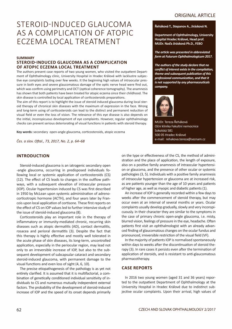
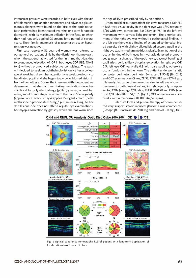
44/55 torr, visual acuity in the right eye was 1/50 naturally, 6/10 with own correction: -6.0-0.5cyl ax 78°, in the left eye movement with correct light projection. The anterior seg-ment of the right eye was without a pathological finding, in the left eye there was a finding of extended conjunctival blo-od vessels, iris with slightly dilated blood vessels, pupil in the right eye was in medium mydriasis plegic. Examination of the ocular fundus of both eyes in mydriasis detected pronoun-ced glaucoma change of the optic nerve, bayonet bending of capillaries, peripapillary atrophy, excavation in right eye C/D 0.5, left eye C/D vertically 0.8 with pale papilla, otherwise ocular fundus within the norm. The patient underwent static computer perimetry (perimeter Zeiss, test T 30-2) (fig. 2, 3) and OCT examination (Cirrus, ZEISS) RNFL RLE was 87/49 µm, bilaterally flat curve of neuroretinal rim, in left eye also with decrease to pathological values, in right eye only in upper sector, C/Da (average C/D ratio), RLE 0.60/0.78 and C/Dv (ver-tical C/D ratio) RLE 0.54/0.79 (fig. 1), OCT of macula was bila-terally within the norm (CRT RLE 267/263 µm).
Intensive local and general therapy of decompensa-ted very suspect steroid-induced glaucoma was commenced (Cosopt gtt – dorzolamide 20.0 mg and timolol 5.0 mg), Dilu-
intraocular pressure were recorded in both eyes with the aid of Goldmann's applanation tonometry, and advanced glauco-matous changes were found on the disc of the optic nerve. Both patients had been treated over the long term for atopic dermatitis, with its maximum affliction in the face, to which they had regularly applied CS creams for a period of several years. Their family anamnesis of glaucoma or ocular hyper-tension was negative.
First case report: A 31 year old woman was referred to our general outpatient clinic by the district ophthalmologist, whom the patient had visited for the first time that day, due to pronounced elevation of IOP in both eyes (IOP RLE: 43/48 torr) without pronounced subjective complaints. The pati-ent decided to seek an ophthalmologist only after a collea-gue at work had drawn her attention one week previously to her dilated pupil, and she began to perceive blurred vision in front of her left eye. During the interview with the patient we determined that she had been taking medication since her childhood for polyvalent allergy (pollen, grasses, animal fur, mites, mould) and atopic eczema in the face. She regularly (approx. once every 3 days) applies Belogent cream (beta-methasone dipropionate 0.5 mg / gentamincin 1 mg) to her skin lesions. She does not attend regular eye examinations, her myopia correction by glasses, which she has worn since
Fig. 1 Optical coherence tomography RLE of patient with long-term application of local corticosteroid cream to face
CZECH AND SLOVAK OPHTHALMOLOGY 2/201764
acetonide 0.1 and salicylic acid 3%) according to requirement.When the patient reported to us in 2016 the values of IOP
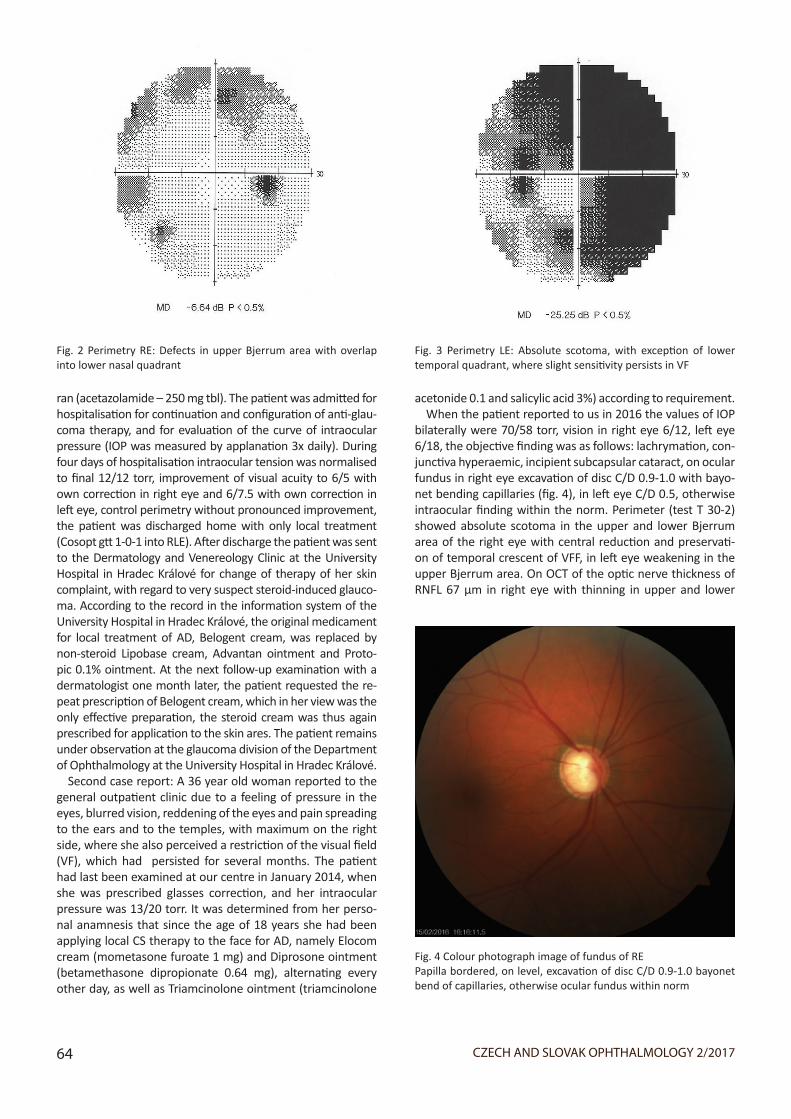
bilaterally were 70/58 torr, vision in right eye 6/12, left eye 6/18, the objective finding was as follows: lachrymation, con-junctiva hyperaemic, incipient subcapsular cataract, on ocular fundus in right eye excavation of disc C/D 0.9-1.0 with bayo-net bending capillaries (fig. 4), in left eye C/D 0.5, otherwise intraocular finding within the norm. Perimeter (test T 30-2) showed absolute scotoma in the upper and lower Bjerrum area of the right eye with central reduction and preservati-on of temporal crescent of VFF, in left eye weakening in the upper Bjerrum area. On OCT of the optic nerve thickness of RNFL 67 µm in right eye with thinning in upper and lower
ran (acetazolamide – 250 mg tbl). The patient was admitted for hospitalisation for continuation and configuration of anti-glau-coma therapy, and for evaluation of the curve of intraocular pressure (IOP was measured by applanation 3x daily). During four days of hospitalisation intraocular tension was normalised to final 12/12 torr, improvement of visual acuity to 6/5 with own correction in right eye and 6/7.5 with own correction in left eye, control perimetry without pronounced improvement, the patient was discharged home with only local treatment (Cosopt gtt 1-0-1 into RLE). After discharge the patient was sent to the Dermatology and Venereology Clinic at the University Hospital in Hradec Králové for change of therapy of her skin complaint, with regard to very suspect steroid-induced glauco-ma. According to the record in the information system of the University Hospital in Hradec Králové, the original medicament for local treatment of AD, Belogent cream, was replaced by non-steroid Lipobase cream, Advantan ointment and Proto-pic 0.1% ointment. At the next follow-up examination with a dermatologist one month later, the patient requested the re-peat prescription of Belogent cream, which in her view was the only effective preparation, the steroid cream was thus again prescribed for application to the skin ares. The patient remains under observation at the glaucoma division of the Department of Ophthalmology at the University Hospital in Hradec Králové.
Second case report: A 36 year old woman reported to the general outpatient clinic due to a feeling of pressure in the eyes, blurred vision, reddening of the eyes and pain spreading to the ears and to the temples, with maximum on the right side, where she also perceived a restriction of the visual field (VF), which had persisted for several months. The patient had last been examined at our centre in January 2014, when she was prescribed glasses correction, and her intraocular pressure was 13/20 torr. It was determined from her perso-nal anamnesis that since the age of 18 years she had been applying local CS therapy to the face for AD, namely Elocom cream (mometasone furoate 1 mg) and Diprosone ointment (betamethasone dipropionate 0.64 mg), alternating every other day, as well as Triamcinolone ointment (triamcinolone
Fig. 2 Perimetry RE: Defects in upper Bjerrum area with overlap into lower nasal quadrant
Fig. 3 Perimetry LE: Absolute scotoma, with exception of lower temporal quadrant, where slight sensitivity persists in VF
Fig. 4 Colour photograph image of fundus of REPapilla bordered, on level, excavation of disc C/D 0.9-1.0 bayonet bend of capillaries, otherwise ocular fundus within norm
CZECH AND SLOVAK OPHTHALMOLOGY 2/2017 65
with elements of the fibrillary extracellular matrix, which sub-sequently again increase the resistance of the outflow path-ways. Nevertheless, to date the connection between change of the MYOC gene and the origin of open-angle glaucoma has not been reliably confirmed (5, 13).
Another important parameter influencing the degree of risk of occurrence of CS-related ocular complications is the method of administration of CS. Most frequently an increase of IOP is brought about by locally applied CS, mainly in the form of eye drops and ointments, as well as ointments applied to the pe-riocular region and depot forms of CS. In our case we recorded the occurrence of secondary CS-related glaucoma following local treatment of AD in the form of an ointment.
Local CS with a stronger anti-inflammatory effect than dexamethasone, betamethasone or prednisolone, or CS with a higher glucocorticoid activity and greater capacity for pene-tration, have a greater probability of generating an increase in IOP than less effective loteprednol or fluorometholone, i.e. risk of preparations: dexamethasone 0.1% > prednisolone 1.0% > fluorometholone 0.1% > hydrocortisone 0.5% (4, 13). Usually an increase of IOP is manifested at the earliest 5 days after the commencement of application, and in rare cases after 14 days, nevertheless late onset is not uncommon. In the case of intravi-treal administration of Triamcinolone, Ozurdex, for example in therapy of cystoid macular edema, macular edema due to cent-ral retinal vein occlusion or earlier, before the commencement of anti-vascular endothelial growth factor preparations in the tre-atment of exudative form of age-related macular degeneration, the degree of increase of IOP is dependent upon the presence of factors facilitating the penetration of CS into the anterior seg-ment such as dose, aphakia or pseudophakia, previously perfor-med pars plana vitrectomy. Here an increase takes place at the earliest 1-4 weeks after application, and the IOP values return to the normal parameters several months after application. Serious and long-term increase of IOP occurs upon periocular adminis-tration (subconjunctival, sub-Tenon’s or retrobulbar application of Triamcinolone injection), because depot CS are formed with a prolonged effect. By contrast, systemically administered CS, pe-rorally, inhaled, in the form of nasal spray, manifest the lowest probability of influencing the value of IOP. Nevertheless, systemic therapy is often combined with a local method of administrati-on, and it is therefore necessary to keep in mind the potentiation of the steroid effect. In addition to exogenously administered CS and their endogenous increased production, it may generate an increase in IOP, for example in the case of Cushing's syndrome or adrenal hyperplasia (5).
Because steroid-induced glaucoma belongs to the group of open-angle glaucomas, an increase of IOP is not manifested suddenly in the form of an acute glaucoma attack. The IOP value increases gradually over time, and patients are asymp-tomatic for a long time. Ocular complications of CS therapy are therefore often not manifested until the moment of alre-ady advanced and irreversible damage to visual functions (6).
In our case also, both patients were examined already du-ring the time of advanced glaucomatous changes on the optic nerve.
Steroid-induced glaucoma usually responds well to the termi-nation of CS therapy. IOP typically returns to its initial values be-
sector, RNFL 101 µm in left eye, where the curve was within the norm.
Local and general therapy was commenced immediately in order to reduce intraocular tension (peroral glycerol 50 ml, Di-luran 250 mg tbl and local Cosopt gtt), and subsequently the patient was admitted for hospitalisation, where Manitol 20% 20 ml i.v. (mannitol) was administered, as well as locally Xalatan gtt (latanoprost 50 mcg), while IOP monitoring was simultane-ously commenced. Due to insufficient compensation of IOP in maximal local and general pharmacotherapy, a decision was taken with the consent of the patient to perform selective la-ser trabeculoplasty in both eyes. A reduction of IOP was then achieved to values of 10/8 torr, visual acuity and the perimeter remained without improvement. Due to the worsening of the skin complaint during hospitalisation upon discontinuation of steroid medication, a consultation was arranged with a der-matologist, who adjusted the AD therapy, newly administered Protopic 0.1% ung (tacrolimus, immunosuppresant), generally tetracycline 250 mg (tetracycline hydrochloride) in capsules. At a follow-up examination at the dermatological clinic one month later, the skin finding had improved to the extent that the patient is now without any AD therapy.
DISCUSSION
In the literature it is stated that approximately 1/3 of the population rank among medium or high steroid responders (increase of IOP by 6-15 torr, or up to more than 31 torr upon local steroid application). Nevertheless, in only a small per-centage of patients with steroid therapy a clinically significant increase of IOP occurs, specifically in 5-6% of the population IOP increases to more than 31 torr after 4-6 weeks of local CS therapy, in 33% of the population by 6-15 torr (5).
The assumed mechanism of origin of steroid-induced glau-coma is CS induced functional and morphological changes of the outflow pathways, causing an increase of their resistance (3, 10). One of the theories refers to the corticosteroid effect on glycosaminoglycans (GAG) in the extracellular matrix of meshwork, specifically via inhibition of hydrolases responsi-ble for the depolymerisation of GAG. The accumulated GAG balance water and subsequently constrict up to the point of closing outflow pathways (5). Another hypothesis assumes the effect of CS on reducing the phagocyte capacities of the endothelial cells of the meshwork, or inhibition of synthesis of prostaglandins improving the outflow capacities of the meshwork. The result is an accumulation of debris and an in-crease in the resistance of the outflow pathways (5, 13). Evi-dence is available about genetic predisposition to this type of glaucoma, which may explain why normalisation of IOP does not take place in certain patients after the discontinuation of steroid therapy, although the manner of heredity is not yet known (3). Genetic mapping demonstrated a change in the expression of the MYOC gene (myocilin gene) to chromosome 1 induced by CS, known also as TIGR (Trabecular meshwork Inducible Glucocortoid Response). Increased expression of the MYOC gene leads to increased formation of the glyco-protein myocilin, which was found in the cytoplasma of the meshwork cells and the juxtacanalicular region in connection
CZECH AND SLOVAK OPHTHALMOLOGY 2/201766
ment of glaucomatous changes has been known for over half a century, this issue remains current.
Chronic skin diseases, in particular in the area of the face, which are treated by the long-term local application of CS, primarily in the form of ointments and creams, are of high risk for the development of ocular hypertension or seconda-ry steroid-induced glaucoma and subcapsular cataract. A role is played also by the unwillingness of patients to discontinue CS over the long term due to their rapid and beneficial effect on alleviating skin complaints. The severity of the problem is further increased also by the initial inconspicuous manifesta-tion of ocular complications.
It continues to apply that CS are active substances with a good safety profile, and are invaluable in the treatment of a wide spectrum of disorders, though only if they are used co-rrectly and over the short term in individuals without predis-position. Patients must be notified of their side effects upon their long-term application, and must be thoroughly and ca-refully instructed with regard to their correct use and applica-tion, including adherence to the period for discontinuation of their use. It is of fundamental importance to attend regular eye checks in order to ensure timely detection of incipient glaucomatous changes and to avert the development of cata-strophic permanent damage to visual functions.
fore the commencement of therapy spontaneously within days to weeks, or following the administration of anti-glaucomatous pharmacotherapy, which we also recorded in our patients.
A number of cases of resistant increase of IOP have been described, in which the further therapeutic procedure is si-milar as in patients with primary open-angle glaucoma upon uncorrectable IOP, i.e. selective laser trabeculoplasty or filtra-tion operation (5, 10). It has also been recorded that in pa-tients younger than 20 years old with high IOP and greater glaucomatous neuropathy of the optics, a surgical solution was applied more frequently (12). In the case of uncontro-llable glaucoma upon maximum pharmacotherapy following periocular injection of triamcinolone, IOP was successfully compensated by surgical removal of the su-Tenon’s depot of CS. IOP subsequently returned to the initial values after 1 week, and remained within the norm also after 5 weeks fo-llowing the discontinuation of local anti-glaucomatous medi-cation (1). With regard to the fact that compensation of IOP occurred in our patients, a surgical solution was not indicated.
CONCLUSION
In our case reports we showed that despite the fact that the corticosteroid effect on increasing IOP and the develop-
5. Cohen, A.: Steroid Induced Glaucoma. In: RUMELT, Shimon (Ed.). Glaucoma – Basic and Clinical Concepts. InTech, 2011: 559–568
6. Greenwood, M., Grigorian, F.: Glau-comatous Damage Secondary to Long Term Topical Corticosteroid Use in a 7-Year-Old Female. Open J Pediat, (March): 2014: 62–66.
7. Haeck, I., Rouwen, T., Timmer-de Mik, L. et al.: Risk for Ocular Complications From Topical Corticosteroids. Indian J Ophthalmol, 64: 2011: 275–281.
8. Jones, R., Rhee, D.: Corticosteroid-in-duced intraocular hypertension and glaucoma: A brief review and update of the literature. Curr Opin Ophthal-mol, 2006: 163–167.
1. Akduman, L., Kolker, A., Black, D. et al.: Treatment of Persitent Glaucoma Secondary to Periocular Corticoste-roids. Am J Ophthalmol, 1996: Vol.122 (August): 275–277.
2. Armaly, M.: Effect of Corticosteroids on Intraocular Pressure and Fluid Dy-namics: I. The Effect of Dexamethaso-ne in the Normal Eye. JAMA Ophthal-mol, 70: 1963: 88–97.
3. Branco, N., C. Branco, B., Mallon, J. et al.: Cutaneous corticosteroid-indu-ced glaucoma. Journal of Toxicology: Cutaneous and Ocular Toxicology. 21 (March): 2002: 1–2.
4. Chen, S: Monitoring and management of steroid-induced glaucoma. Opto-metry Pharma. (March): 2013: 6–7.
LITERATURE
9. Mandapati, J., Metta, A.: Intraocular pressure variation in patients on lon-g-term corticosteroids. Indian Derma-tology Online Journal. 2 (Jul), 2011: 67–69.
10. Mohan, R., Muralidharan, A.: Steroid induced glaucoma and cataract. Ind J Ophthalmol, 37: 1989: 13–16.
11. Munjal, V., Dhir, S., Jain, I.: Steroid induced glaucoma. Ind J Ophthalmol, 1982, 30: 379–382.
12. Sihota, R., Konkal, V., Dada, T. et al.: Prospective, long-term evaluation of steroid-induced glaucoma. Nature Pu-blishing Group. 2008: 25–30.
13. Sowka, J., Kabat, A.: A Potent New Steroid. Review of Optometry. (July): 2010.
![[PPT]Steroid Induced Diabetes - Healing, Teaching & … Induced... · Web viewACE/ADA Task Force on Inpatient Diabetes. Diabetes Care. 2006;29(8):1955-1962. Bolus insulin to keep](https://img.pdfslide.net/doc/110x75/5ab7ace77f8b9a684c8bcb29/pptsteroid-induced-diabetes-healing-teaching-inducedweb-viewaceada.jpg)