Embed Size (px)
Citation preview
Int J Clin Exp Med 2015;8(10):18185-18190www.ijcem.com /ISSN:1940-5901/IJCEM0013752
Original ArticleThe neuroprotective effects of aspirin following crush injury to rat sciatic nerve
Yi Cui, Jun Li, Yueliang Zhu, Hui Tang, Xiaoqing He, Yongqing Xu
Department of Orthopedic Surgery, Kunming General Hospital of Chengdu Military Command, Kunming 650032, China
Received July 31, 2015; Accepted October 3, 2015; Epub October 15, 2015; Published October 30, 2015
Abstract: Aspirin has been reported to be neuroprotective and produce some benefits for central nervous system diseases. However, the possibility of using aspirin as a neuroprotective agent for peripheral nerve injuries has rarely been reported thus far. The aim of the present study was to investigate the possibly beneficial effects of aspirin on sciatic nerve crush injury therapy in rats. Crush injury animal model was prepared with Sprague-Dawley rats. The animals were evenly divided into high-dose aspirin group, low-dose aspirin group, and vehicle group. Aspirin solution or normal saline were intraperitoneally injected once a day for 28 days after sciatic nerve crush injury. A sham-operative group was also added as normal control. The results from walking track analysis and electrophysiologi-cal assessment indicated that motor functional recovery in the aspirin groups were better than that in the vehicle group. Morphometric analysis of regenerated nerves and Fluoro-Gold retrograde tracing demonstrated that axonal regeneration in the aspirin groups was superior to that in the vehicle group. Our findings suggest that aspirin might be used as a neuroprotective agent for treating peripheral nerve injuries.
Keywords: Aspirin, neuroprotection, nerve crush injury, nerve regeneration
Introduction
During the past decades, aspirin has been reported to have various pharmacological prop-erties and multiple sites of action [1-3]. Recently, aspirin has gained much attention because it has been shown to be neuroprotec-tive in treating central nervous system diseas-es. For example, aspirin has been shown to exert protective actions against focal ischemia injury, dopamine quinone-induced neurotoxici-ty, and cerebral white matter lesions [4-6]. To date, neuroprotective effects of aspirin on peripheral nerve injuries have been rarely investigated.
Currently, surgical intervention, drug therapy, and physical rehabilitation are typical choices for treating peripheral nerve injuries. Among the abovementioned strategies, drug therapy is a promising approach for promoting axonal regeneration and functional recovery. There- fore, in the present study, we investigated the possibly neuroprotective effects of aspirin for promoting nerve regeneration after sciatic crush injury in rats.
Methods
Animals and surgical procedures
Male Sprague-Dawley rats (220-250 g body weight) were provided by Laboratory Animal Center of Kunming Medical University, and all animal surgeries were approved ethically by Institutional Ethical Committee of Kunming Medical University. The use of animals followed the ethical guidelines of the Care and Use of Laboratory Animals (NIH publication No. 85-23, revised 1985).
The preparation of crush injury animal model followed the protocol reported previously [7]. In brief, rats were anesthetized with an intraperi-toneal injection of pentobarbital solution (1.5%, 30 mg/kg body weight). The right sciatic nerve was exposed using the gluteal muscle splitting incision under a surgical microscope. Using a pair of forceps, the exposed nerve was com-pressed at the proximal segment 5 mm from the bifurcation for 3 times (10 s/each time, 10 s interval). Four groups (n=9) were investigated, and aspirin solution or normal saline were intra-
Neuroprotective effects of aspirin on rat sciatic nerve
18186 Int J Clin Exp Med 2015;8(10):18185-18190
peritoneally injected once a day for 28 days after surgery.
High-dose aspirin group: 10 mg aspirin (40 mg/kg body weight), given at each predefined time point. Low-dose aspirin group: 5 mg aspirin (5 mg/kg body weight), given at each predefined time point. Vehicle group: normal saline, given at each predefined time point. Sham-operative group: normal saline, the sciatic nerve was exposed without being compressed.
Subsequently, the skin was closed with 6-0 stitches and all rats were kept in animal rooms under standard housing conditions.
Behavioral analysis
Walking track analysis was performed at 7, 10, 13, 16, 19, 22, 25, and 28 days following sur-gery, and the sciatic functional index (SFI) was calculated according to the equation [8].
SFI 38.3 NPLEPL NPL 109.5 NTS
ETS NTS
13.3 NITSEITS NITS 8.8
=--
+-
+- -
# #
#
All rats were trained to walk along a standard track (100 cm long and 7 cm wide) before sur-gery. The hind paws were painted with red ink. Thus, changes of paw prints were recorded on white papers. The following measurements were taken from the footprints.
PL (print length): distance from the heel to the top of the third toe. ITS (intermediary toe spread): distance from the second to the fourth toe. TS (toe spread): distance between the first and the fifth toe. N (normal): for the non-operat-ed foot. E (experimental): for the experimental foot.
The SFI value varies from -100 to 0. The value near 0 reflects normal function, while a value of approximately -100 reflects complete dysfun- ction.
Electrophysiological assessment
Following walking tests, animals were subject-ed to electrophysiological studies at 7, 13, 22 and 28 days after surgery. Under anesthesia, the right sciatic nerve was exposed as men-tioned above. A bipolar stimulating electrode was applied at the proximal portion of the nerve
trunk. Compound muscle action potentials (CMAPs) were recorded on the gastrocnemius belly.
Fluoro-gold retrograde tracing
Twenty-two days following surgery, retrograde labeling was performed. In brief, the right sci-atic nerve was exposed and 3 μl of 4% Fluoro-gold (Biotium, Hayward, CA) solution was inject-ed into the nerve trunk at the bifurcation by a Microliter Syringe (10 μl; Hamilton Co.). Then, the incision was sutured with 6-0 stitches and rats were return to their cages. 6 days later, the rats were intracardially perfused with parafor-maldehyde (4% w/v)-phosphate buffer (0.1 M). The L4, 5, 6 segments of the lumbar spinal cord were harvested. The sample was then subject-ed to a standard procedure and was sectioned on a cryostat. 25 μm-thick transverse sections of the sample were then mounted on glass slides. These slides were photographed by a fluorescent microscope (BX-60; Olympus), and the number of Fluoro-gold-labeled neurons was counted by an investigator who was blinded to the experiment design.
Nerve morphometric analysis
The right sciatic nerve was removed and fixed in glutaraldehyde (3 wt%), postfixed in osmium tetroxide (1%)-sodium cacodylate buffer (0.1 M, pH=7.3). The samples were dehydrated in grad-ed acetone, which was then replaced with ace-tone, and embedded in epoxy resin for section-ing. The transverse semi-thin sections (2 μm) were stained with a 1% toluidine blue/1% borax solution for light microscope. Ultrathin sections (50 nm) were stained with uranyl acetate-lead citrate for transmission electron microscope. The parameters measured included the total number of myelinated axons, the mean diame-ter of the nerve fibers, and the relation of axon diameter to total fiber diameter (G-ratio). All evaluations were completed by an investigator blinded to the experimental design.
Data analysis
SPSS13.0 software package (SPSS Inc., Chicago, IL) was used for statistical analysis. All data were expressed as the mean ± standard error of the mean (SEM), and were analyzed using one-way ANOVA with subsequent Bonferroni test for pairwise comparisons. P<0.05 were considered statistically signi- ficant.
Neuroprotective effects of aspirin on rat sciatic nerve
18187 Int J Clin Exp Med 2015;8(10):18185-18190
Results
Effect of aspirin on motor functional recovery
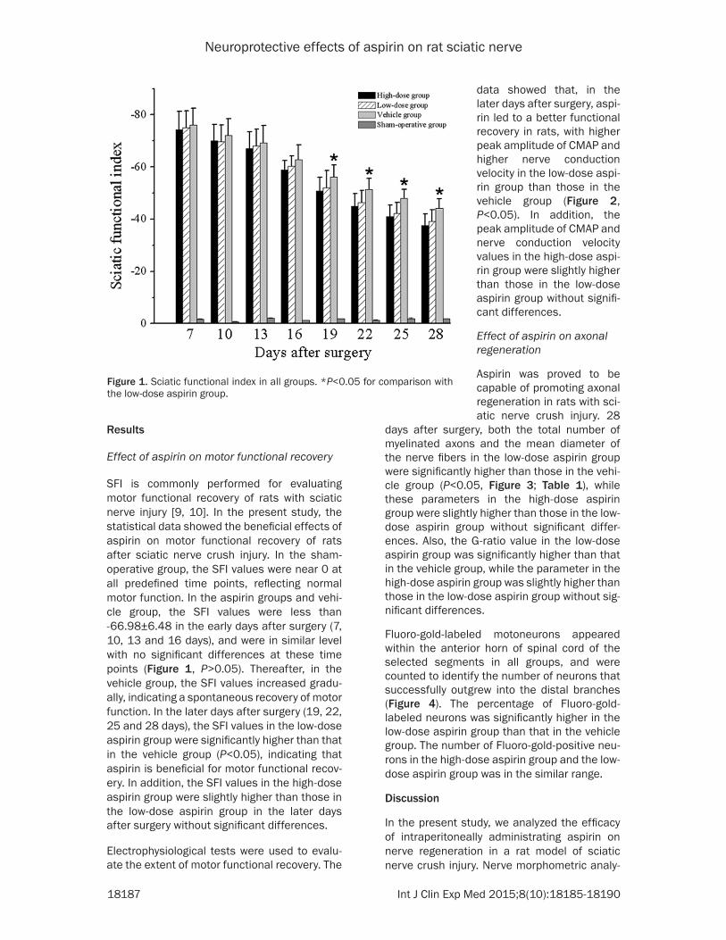
SFI is commonly performed for evaluating motor functional recovery of rats with sciatic nerve injury [9, 10]. In the present study, the statistical data showed the beneficial effects of aspirin on motor functional recovery of rats after sciatic nerve crush injury. In the sham-operative group, the SFI values were near 0 at all predefined time points, reflecting normal motor function. In the aspirin groups and vehi-cle group, the SFI values were less than -66.98±6.48 in the early days after surgery (7, 10, 13 and 16 days), and were in similar level with no significant differences at these time points (Figure 1, P>0.05). Thereafter, in the vehicle group, the SFI values increased gradu-ally, indicating a spontaneous recovery of motor function. In the later days after surgery (19, 22, 25 and 28 days), the SFI values in the low-dose aspirin group were significantly higher than that in the vehicle group (P<0.05), indicating that aspirin is beneficial for motor functional recov-ery. In addition, the SFI values in the high-dose aspirin group were slightly higher than those in the low-dose aspirin group in the later days after surgery without significant differences.
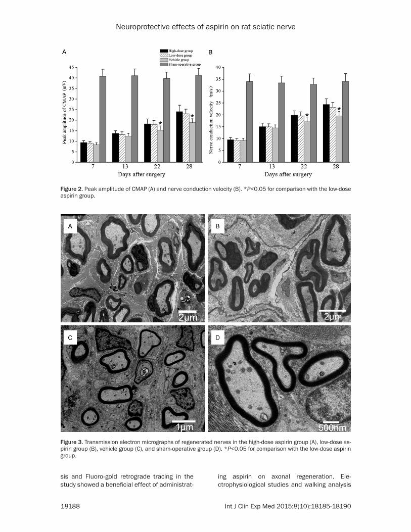
Electrophysiological tests were used to evalu-ate the extent of motor functional recovery. The
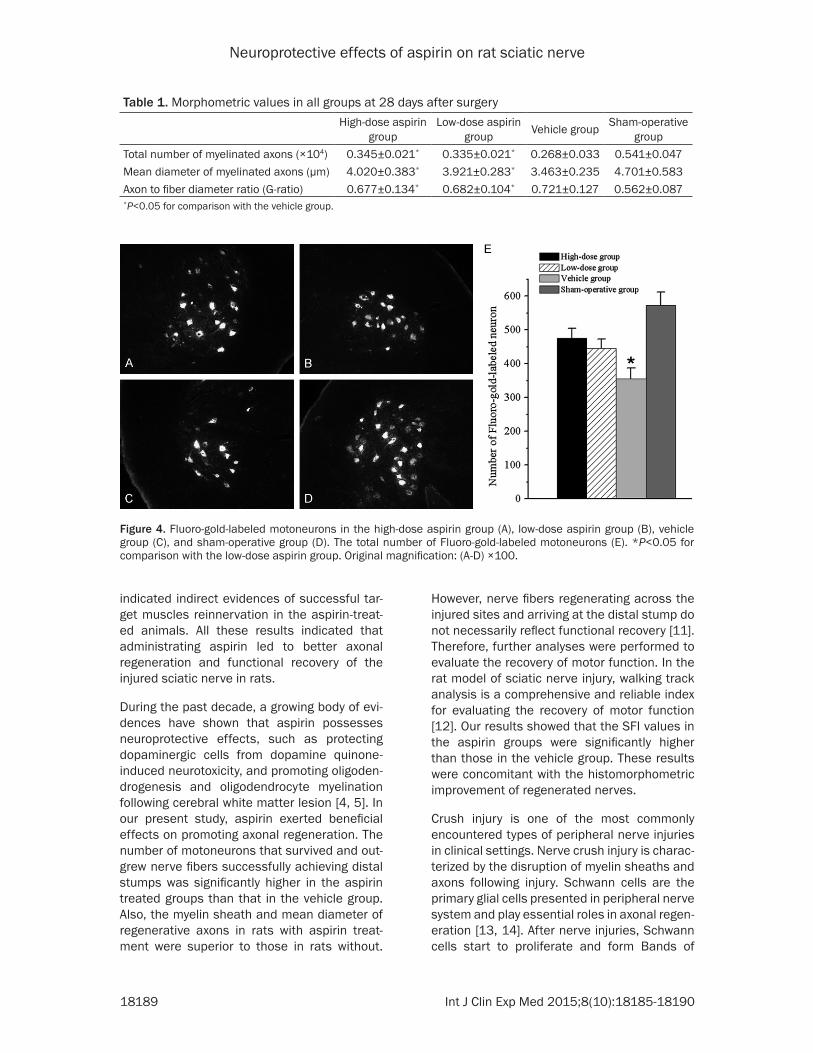
days after surgery, both the total number of myelinated axons and the mean diameter of the nerve fibers in the low-dose aspirin group were significantly higher than those in the vehi-cle group (P<0.05, Figure 3; Table 1), while these parameters in the high-dose aspirin group were slightly higher than those in the low-dose aspirin group without significant differ-ences. Also, the G-ratio value in the low-dose aspirin group was significantly higher than that in the vehicle group, while the parameter in the high-dose aspirin group was slightly higher than those in the low-dose aspirin group without sig-nificant differences.
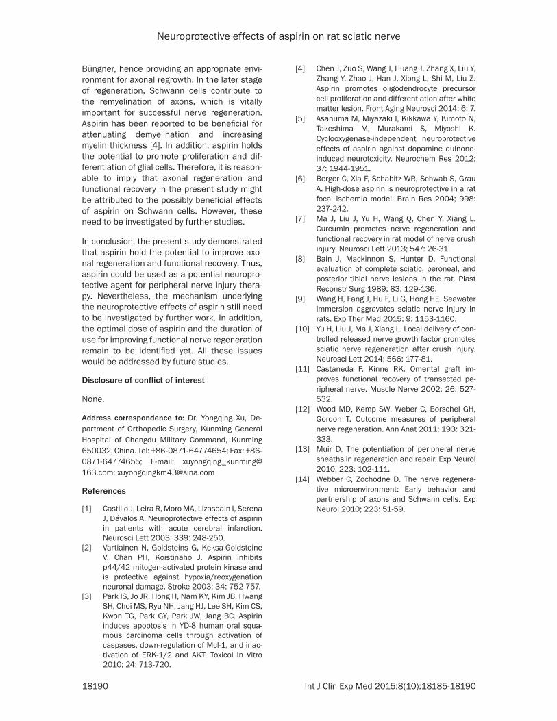
Fluoro-gold-labeled motoneurons appeared within the anterior horn of spinal cord of the selected segments in all groups, and were counted to identify the number of neurons that successfully outgrew into the distal branches (Figure 4). The percentage of Fluoro-gold-labeled neurons was significantly higher in the low-dose aspirin group than that in the vehicle group. The number of Fluoro-gold-positive neu-rons in the high-dose aspirin group and the low-dose aspirin group was in the similar range.
Discussion
In the present study, we analyzed the efficacy of intraperitoneally administrating aspirin on nerve regeneration in a rat model of sciatic nerve crush injury. Nerve morphometric analy-
Figure 1. Sciatic functional index in all groups. *P<0.05 for comparison with the low-dose aspirin group.
data showed that, in the later days after surgery, aspi-rin led to a better functional recovery in rats, with higher peak amplitude of CMAP and higher nerve conduction velocity in the low-dose aspi-rin group than those in the vehicle group (Figure 2, P<0.05). In addition, the peak amplitude of CMAP and nerve conduction velocity values in the high-dose aspi-rin group were slightly higher than those in the low-dose aspirin group without signifi-cant differences.
Effect of aspirin on axonal regeneration
Aspirin was proved to be capable of promoting axonal regeneration in rats with sci-atic nerve crush injury. 28
Neuroprotective effects of aspirin on rat sciatic nerve
18188 Int J Clin Exp Med 2015;8(10):18185-18190
sis and Fluoro-gold retrograde tracing in the study showed a beneficial effect of administrat-
ing aspirin on axonal regeneration. Ele- ctrophysiological studies and walking analysis
Figure 2. Peak amplitude of CMAP (A) and nerve conduction velocity (B). *P<0.05 for comparison with the low-dose aspirin group.
Figure 3. Transmission electron micrographs of regenerated nerves in the high-dose aspirin group (A), low-dose as-pirin group (B), vehicle group (C), and sham-operative group (D). *P<0.05 for comparison with the low-dose aspirin group.
Neuroprotective effects of aspirin on rat sciatic nerve
18189 Int J Clin Exp Med 2015;8(10):18185-18190
indicated indirect evidences of successful tar-get muscles reinnervation in the aspirin-treat-ed animals. All these results indicated that administrating aspirin led to better axonal regeneration and functional recovery of the injured sciatic nerve in rats.
During the past decade, a growing body of evi-dences have shown that aspirin possesses neuroprotective effects, such as protecting dopaminergic cells from dopamine quinone-induced neurotoxicity, and promoting oligoden-drogenesis and oligodendrocyte myelination following cerebral white matter lesion [4, 5]. In our present study, aspirin exerted beneficial effects on promoting axonal regeneration. The number of motoneurons that survived and out-grew nerve fibers successfully achieving distal stumps was significantly higher in the aspirin treated groups than that in the vehicle group. Also, the myelin sheath and mean diameter of regenerative axons in rats with aspirin treat-ment were superior to those in rats without.
However, nerve fibers regenerating across the injured sites and arriving at the distal stump do not necessarily reflect functional recovery [11]. Therefore, further analyses were performed to evaluate the recovery of motor function. In the rat model of sciatic nerve injury, walking track analysis is a comprehensive and reliable index for evaluating the recovery of motor function [12]. Our results showed that the SFI values in the aspirin groups were significantly higher than those in the vehicle group. These results were concomitant with the histomorphometric improvement of regenerated nerves.
Crush injury is one of the most commonly encountered types of peripheral nerve injuries in clinical settings. Nerve crush injury is charac-terized by the disruption of myelin sheaths and axons following injury. Schwann cells are the primary glial cells presented in peripheral nerve system and play essential roles in axonal regen-eration [13, 14]. After nerve injuries, Schwann cells start to proliferate and form Bands of
Table 1. Morphometric values in all groups at 28 days after surgeryHigh-dose aspirin
groupLow-dose aspirin
group Vehicle group Sham-operative group
Total number of myelinated axons (×104) 0.345±0.021* 0.335±0.021* 0.268±0.033 0.541±0.047Mean diameter of myelinated axons (μm) 4.020±0.383* 3.921±0.283* 3.463±0.235 4.701±0.583Axon to fiber diameter ratio (G-ratio) 0.677±0.134* 0.682±0.104* 0.721±0.127 0.562±0.087*P<0.05 for comparison with the vehicle group.
Figure 4. Fluoro-gold-labeled motoneurons in the high-dose aspirin group (A), low-dose aspirin group (B), vehicle group (C), and sham-operative group (D). The total number of Fluoro-gold-labeled motoneurons (E). *P<0.05 for comparison with the low-dose aspirin group. Original magnification: (A-D) ×100.
Neuroprotective effects of aspirin on rat sciatic nerve
18190 Int J Clin Exp Med 2015;8(10):18185-18190
Büngner, hence providing an appropriate envi-ronment for axonal regrowth. In the later stage of regeneration, Schwann cells contribute to the remyelination of axons, which is vitally important for successful nerve regeneration. Aspirin has been reported to be beneficial for attenuating demyelination and increasing myelin thickness [4]. In addition, aspirin holds the potential to promote proliferation and dif-ferentiation of glial cells. Therefore, it is reason-able to imply that axonal regeneration and functional recovery in the present study might be attributed to the possibly beneficial effects of aspirin on Schwann cells. However, these need to be investigated by further studies.
In conclusion, the present study demonstrated that aspirin hold the potential to improve axo-nal regeneration and functional recovery. Thus, aspirin could be used as a potential neuropro-tective agent for peripheral nerve injury thera-py. Nevertheless, the mechanism underlying the neuroprotective effects of aspirin still need to be investigated by further work. In addition, the optimal dose of aspirin and the duration of use for improving functional nerve regeneration remain to be identified yet. All these issues would be addressed by future studies.
Disclosure of conflict of interest
None.
Address correspondence to: Dr. Yongqing Xu, De- partment of Orthopedic Surgery, Kunming General Hospital of Chengdu Military Command, Kunming 650032, China. Tel: +86-0871-64774654; Fax: +86- 0871-64774655; E-mail: xuyongqing_kunming@ 163.com; [email protected]
References
[1] Castillo J, Leira R, Moro MA, Lizasoain I, Serena J, Dávalos A. Neuroprotective effects of aspirin in patients with acute cerebral infarction. Neurosci Lett 2003; 339: 248-250.
[2] Vartiainen N, Goldsteins G, Keksa-Goldsteine V, Chan PH, Koistinaho J. Aspirin inhibits p44/42 mitogen-activated protein kinase and is protective against hypoxia/reoxygenation neuronal damage. Stroke 2003; 34: 752-757.
[3] Park IS, Jo JR, Hong H, Nam KY, Kim JB, Hwang SH, Choi MS, Ryu NH, Jang HJ, Lee SH, Kim CS, Kwon TG, Park GY, Park JW, Jang BC. Aspirin induces apoptosis in YD-8 human oral squa-mous carcinoma cells through activation of caspases, down-regulation of Mcl-1, and inac-tivation of ERK-1/2 and AKT. Toxicol In Vitro 2010; 24: 713-720.
[4] Chen J, Zuo S, Wang J, Huang J, Zhang X, Liu Y, Zhang Y, Zhao J, Han J, Xiong L, Shi M, Liu Z. Aspirin promotes oligodendrocyte precursor cell proliferation and differentiation after white matter lesion. Front Aging Neurosci 2014; 6: 7.
[5] Asanuma M, Miyazaki I, Kikkawa Y, Kimoto N, Takeshima M, Murakami S, Miyoshi K. Cyclooxygenase-independent neuroprotective effects of aspirin against dopamine quinone-induced neurotoxicity. Neurochem Res 2012; 37: 1944-1951.
[6] Berger C, Xia F, Schabitz WR, Schwab S, Grau A. High-dose aspirin is neuroprotective in a rat focal ischemia model. Brain Res 2004; 998: 237-242.
[7] Ma J, Liu J, Yu H, Wang Q, Chen Y, Xiang L. Curcumin promotes nerve regeneration and functional recovery in rat model of nerve crush injury. Neurosci Lett 2013; 547: 26-31.
[8] Bain J, Mackinnon S, Hunter D. Functional evaluation of complete sciatic, peroneal, and posterior tibial nerve lesions in the rat. Plast Reconstr Surg 1989; 83: 129-136.
[9] Wang H, Fang J, Hu F, Li G, Hong HE. Seawater immersion aggravates sciatic nerve injury in rats. Exp Ther Med 2015; 9: 1153-1160.
[10] Yu H, Liu J, Ma J, Xiang L. Local delivery of con-trolled released nerve growth factor promotes sciatic nerve regeneration after crush injury. Neurosci Lett 2014; 566: 177-81.
[11] Castaneda F, Kinne RK. Omental graft im-proves functional recovery of transected pe-ripheral nerve. Muscle Nerve 2002; 26: 527-532.
[12] Wood MD, Kemp SW, Weber C, Borschel GH, Gordon T. Outcome measures of peripheral nerve regeneration. Ann Anat 2011; 193: 321-333.
[13] Muir D. The potentiation of peripheral nerve sheaths in regeneration and repair. Exp Neurol 2010; 223: 102-111.
[14] Webber C, Zochodne D. The nerve regenera-tive microenvironment: Early behavior and partnership of axons and Schwann cells. Exp Neurol 2010; 223: 51-59.