Embed Size (px)
Citation preview
Original Article
Treatment of cervical oesophageal rupture in horsesC. M. Whitfield-Cargile*, P. C. Rakestraw† and J. HardyDepartment of Large Animal Clinical Sciences, College of Veterinary Medicine, Texas A&M University, CollegeStation, Texas, USA.*Corresponding author email: [email protected]. Present addresses: *Texas Equine Hospital, 13688 S.State Hwy, 6 Bryan, Texas 77807, USA; and †Dubai Equine Hospital, Dubai, UAE.
Keywords: horse; oesophagus; oesophagostomy; cervical fasciotomy
SummaryOesophageal rupture in horses has only been previouslydescribed in detail in isolated case reports. The objectives of thisstudy were to describe the clinical findings, specific treatmentand outcome of oesophageal rupture in horses. Medicalrecords of horses diagnosed with oesophageal rupturebetween 1994–2008 were reviewed. Clinical findings, treatmentand outcome were recorded. Seven horses with cervicaloesophageal perforations were included in the study. Twohorses were subjected to euthanasia without treatment and 5were treated surgically. Treatment involved a fasciotomy ofthe cervical musculature and oesophageal tube placement.Three of 5 horses survived long-term (>one year). Our studyshowed that surgical treatment of cervical oesophagealrupture involving fasciotomy and oesophagostomy tubeplacement can be successful with 3/5 of treated horsessurviving more than one year.
IntroductionRupture of the oesophagus in horses is an uncommonlyencountered disorder (Risnes and Mair 2003) usuallyassociated with a poor prognosis (Lane 2002). Causes ofoesophageal rupture include external trauma (Lunn and Peel1985), nasogastric intubation (Hardy et al. 1992), perforation ofchronic oesophageal ulcers (Dechant et al. 1998), extensionof surrounding infection and mural necrosis secondary tolong-standing oesophageal obstructions (Craig et al. 1989;Read et al. 2002). Treatment of oesophageal rupture variesand depends on the location and extent of the perforation,the duration of perforation and the degree of surroundinginfection. In one report examining 61 cases of oesophagealdisorders in horses, 11 of which were oesophageal ruptures,medical management resulted in a survival rate of 0% (0 of 7)while surgical management resulted in a 50% (2 of 4) survivalrate; therefore, the authors concluded that surgicalmanagement was preferable (Craig et al. 1989). Surgicaltreatment consists of primary closure if there is minimalcontamination of the tissues (Stick 2006). This technique haspreviously been used successfully (Demoor et al. 1979;Wingfield-Digby and Burguez 1982). However, minimalcontamination of the surrounding soft tissues is unlikelyfollowing a rupture of the oesophagus making primary closureunlikely to be successful. In these cases, ventral drainage ofthe affected area, antimicrobials and oesophageal rest isrecommended to allow the oesophagus to heal by secondintention (Craig et al. 1989; Fubini 2002).
Complications associated with rupture of the oesophagusin horses include extension of the infection to the thorax
(Demoor et al. 1979), damage to the vagosympathetic trunkor recurrent laryngeal nerve (Craig et al. 1989), rupture of thecommon carotid artery (Risnes and Mair 2003), electrolytederangements due to loss of saliva (Stick et al. 1981b),endotoxaemia and peritonitis (Hardy et al. 1992) anddevelopment of nonhealing oesophageal fistulas (Craig et al.1989).
There have previously been isolated case reports ofoesophageal ruptures in horses (Raker and Sayers 1958;Demoor et al. 1979; Wingfield-Digby and Burguez 1982; Lunnand Peel 1985; Dechant et al. 1998; Read et al. 2002; Risnesand Mair 2003) and one report of 61 horses with oesophagealdisorders (Craig et al. 1989) that briefly describe treatment. Tothe authors’ knowledge, there are no published reportsdetailing the clinical signs, diagnosis, treatment and outcomeof horses with cervical oesophageal rupture. The objectives ofthis paper were to describe the clinical findings, complications,treatment and outcome of horses with cervical oesophagealrupture.
Materials and methodsThe records of all horses that presented to Texas A&MVeterinary Medical Teaching Hospital between April 1994 andJanuary 2008 were reviewed. Horses of all ages were includedin this study if a clinical diagnosis of ruptured cervicaloesophagus was made and confirmed by surgical explorationof the affected area or by necropsy. Horses were excludedfrom this study if the oesophagus ruptured secondary to asurgical procedure, if the clinical diagnosis was not confirmedby necropsy or surgical evaluation, or if the ruptured portion ofthe oesophagus was in the thorax.
Data collectionInformation obtained from the medical records includedcase details, history, physical examination findings uponpresentation, diagnostics, treatment and outcome.Information obtained from the history included, cause of therupture, duration of any oesophageal obstruction andtreatment prior to arrival at the referral clinic. Informationobtained upon presentation included physical examinationabnormalities, findings of oesophageal endoscopicexamination, radiographic findings of the oesophageal regionand thorax, treatment, surgical findings, complications,outcome and necropsy findings.
Long-term outcome of at least one year was obtained bytelephone interview with the owner or referring veterinarian orby re-evaluation at the clinic. Outcome was consideredsuccessful if the horse was discharged from the hospital andresumed a normal lifestyle.
456
© 2013 EVJ Ltd
EQUINE VETERINARY EDUCATION / AE / SEPTEMBER 2013
Technique of fasciotomy and oesophagostomytube placementPrior to surgical drainage and debridement, all horses wereadministered nonsteroidal anti-inflammatory drugs (flunixinmeglumine 1.1 mg/kg bwt i.v.), broad-spectrum antimicrobials(procaine penicillin G 22,000 iu/kg bwt i.m., gentamicin6.6 mg/kg bwt i.v.) and tetanus toxoid (1 ml i.m. once). Horseswere sedated with detomidine (0.01–0.04 mg/kg bwt i.v.) andbutorphanol (0.01–0.02 mg/kg bwt i.v.). Local anaesthesia wasachieved by infiltrating 10–20 ml of mepivacaine over thepalpable areas of cellulitis and fluid accumulation beneath theskin, usually on the ventral midline of the neck or ventral to thejugular vein. Skin incisions of appropriate lengths to allowadequate drainage were made over the area of palpablecellulitis and oedema. Blunt dissection was continued deeperbetween the sternocephalicus and jugular vein to allowadequate access to the contaminated areas. All feed materialwas flushed from the neck and necrotic tissue debrided asneeded. Dissection and debridement were performeddiligently avoiding the carotid artery, vagosympathetic trunkand recurrent laryngeal nerve. Throughout the course ofhospitalisation, the procedure was repeated as needed tofurther debride the necrotic tissues or to allow access todeveloping pockets of cellulitis and infection.
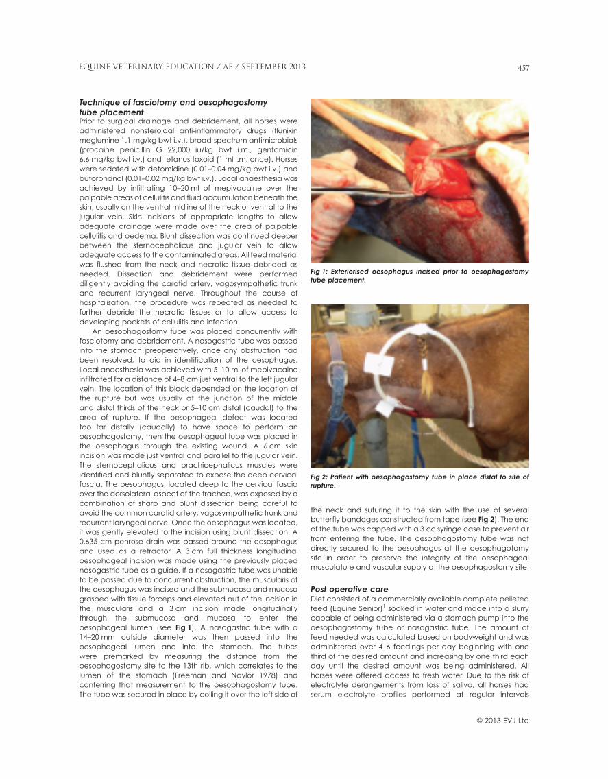
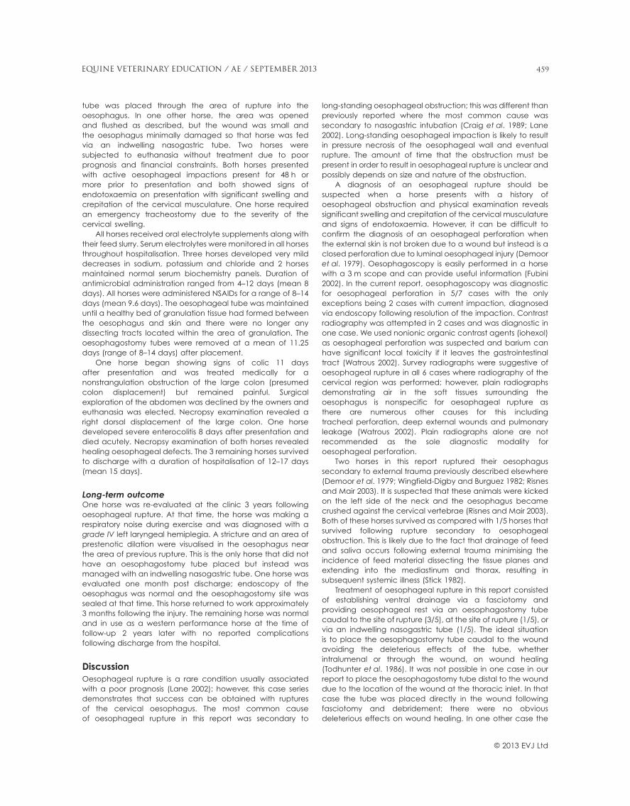
An oesophagostomy tube was placed concurrently withfasciotomy and debridement. A nasogastric tube was passedinto the stomach preoperatively, once any obstruction hadbeen resolved, to aid in identification of the oesophagus.Local anaesthesia was achieved with 5–10 ml of mepivacaineinfiltrated for a distance of 4–8 cm just ventral to the left jugularvein. The location of this block depended on the location ofthe rupture but was usually at the junction of the middleand distal thirds of the neck or 5–10 cm distal (caudal) to thearea of rupture. If the oesophageal defect was locatedtoo far distally (caudally) to have space to perform anoesophagostomy, then the oesophageal tube was placed inthe oesophagus through the existing wound. A 6 cm skinincision was made just ventral and parallel to the jugular vein.The sternocephalicus and brachicephalicus muscles wereidentified and bluntly separated to expose the deep cervicalfascia. The oesophagus, located deep to the cervical fasciaover the dorsolateral aspect of the trachea, was exposed by acombination of sharp and blunt dissection being careful toavoid the common carotid artery, vagosympathetic trunk andrecurrent laryngeal nerve. Once the oesophagus was located,it was gently elevated to the incision using blunt dissection. A0.635 cm penrose drain was passed around the oesophagusand used as a retractor. A 3 cm full thickness longitudinaloesophageal incision was made using the previously placednasogastric tube as a guide. If a nasogastric tube was unableto be passed due to concurrent obstruction, the muscularis ofthe oesophagus was incised and the submucosa and mucosagrasped with tissue forceps and elevated out of the incision inthe muscularis and a 3 cm incision made longitudinallythrough the submucosa and mucosa to enter theoesophageal lumen (see Fig 1). A nasogastric tube with a14–20 mm outside diameter was then passed into theoesophageal lumen and into the stomach. The tubeswere premarked by measuring the distance from theoesophagostomy site to the 13th rib, which correlates to thelumen of the stomach (Freeman and Naylor 1978) andconferring that measurement to the oesophagostomy tube.The tube was secured in place by coiling it over the left side of
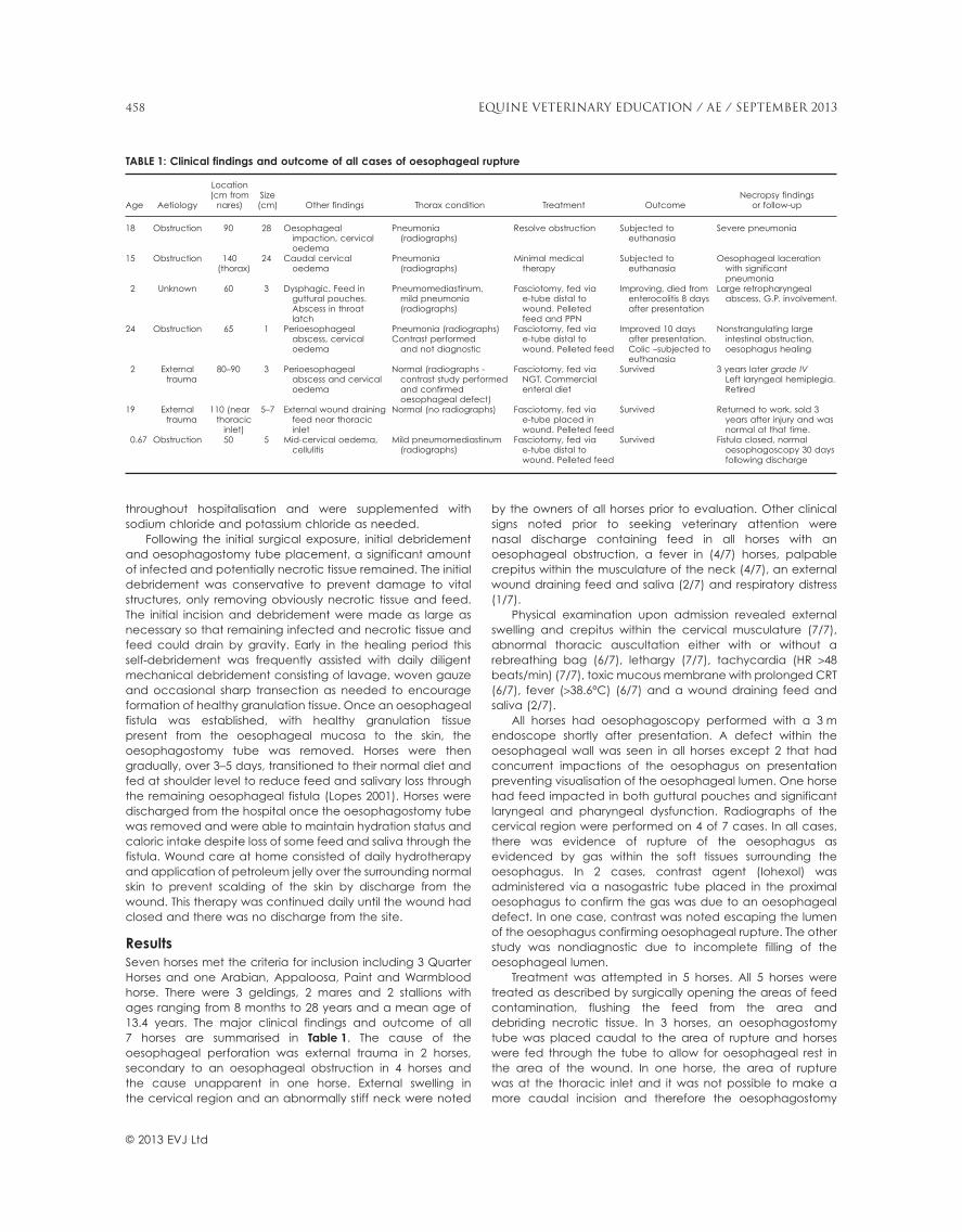
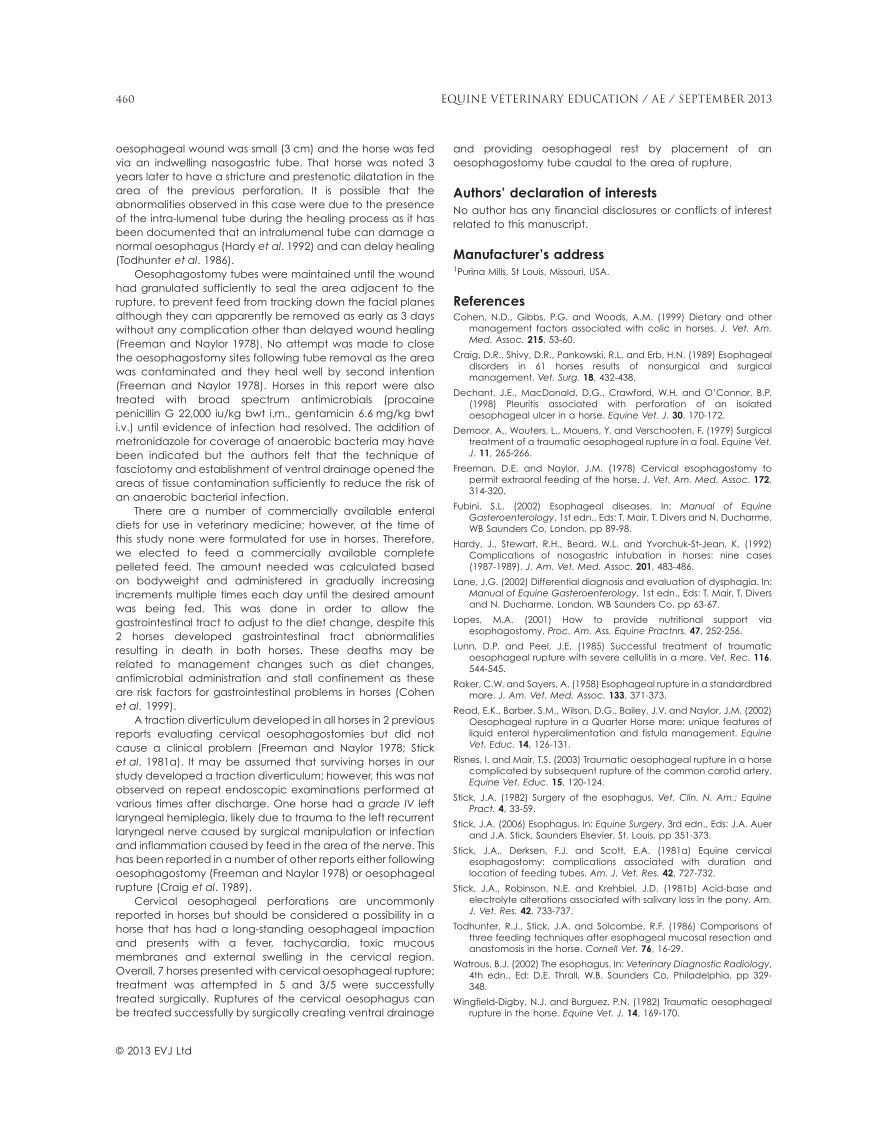
the neck and suturing it to the skin with the use of severalbutterfly bandages constructed from tape (see Fig 2). The endof the tube was capped with a 3 cc syringe case to prevent airfrom entering the tube. The oesophagostomy tube was notdirectly secured to the oesophagus at the oesophagotomysite in order to preserve the integrity of the oesophagealmusculature and vascular supply at the oesophagostomy site.
Post operative careDiet consisted of a commercially available complete pelletedfeed (Equine Senior)1 soaked in water and made into a slurrycapable of being administered via a stomach pump into theoesophagostomy tube or nasogastric tube. The amount offeed needed was calculated based on bodyweight and wasadministered over 4–6 feedings per day beginning with onethird of the desired amount and increasing by one third eachday until the desired amount was being administered. Allhorses were offered access to fresh water. Due to the risk ofelectrolyte derangements from loss of saliva, all horses hadserum electrolyte profiles performed at regular intervals
Fig 1: Exteriorised oesophagus incised prior to oesophagostomytube placement.
Fig 2: Patient with oesophagostomy tube in place distal to site ofrupture.
© 2013 EVJ Ltd
457EQUINE VETERINARY EDUCATION / AE / SEPTEMBER 2013
throughout hospitalisation and were supplemented withsodium chloride and potassium chloride as needed.
Following the initial surgical exposure, initial debridementand oesophagostomy tube placement, a significant amountof infected and potentially necrotic tissue remained. The initialdebridement was conservative to prevent damage to vitalstructures, only removing obviously necrotic tissue and feed.The initial incision and debridement were made as large asnecessary so that remaining infected and necrotic tissue andfeed could drain by gravity. Early in the healing period thisself-debridement was frequently assisted with daily diligentmechanical debridement consisting of lavage, woven gauzeand occasional sharp transection as needed to encourageformation of healthy granulation tissue. Once an oesophagealfistula was established, with healthy granulation tissuepresent from the oesophageal mucosa to the skin, theoesophagostomy tube was removed. Horses were thengradually, over 3–5 days, transitioned to their normal diet andfed at shoulder level to reduce feed and salivary loss throughthe remaining oesophageal fistula (Lopes 2001). Horses weredischarged from the hospital once the oesophagostomy tubewas removed and were able to maintain hydration status andcaloric intake despite loss of some feed and saliva through thefistula. Wound care at home consisted of daily hydrotherapyand application of petroleum jelly over the surrounding normalskin to prevent scalding of the skin by discharge from thewound. This therapy was continued daily until the wound hadclosed and there was no discharge from the site.
ResultsSeven horses met the criteria for inclusion including 3 QuarterHorses and one Arabian, Appaloosa, Paint and Warmbloodhorse. There were 3 geldings, 2 mares and 2 stallions withages ranging from 8 months to 28 years and a mean age of13.4 years. The major clinical findings and outcome of all7 horses are summarised in Table 1. The cause of theoesophageal perforation was external trauma in 2 horses,secondary to an oesophageal obstruction in 4 horses andthe cause unapparent in one horse. External swelling inthe cervical region and an abnormally stiff neck were noted
by the owners of all horses prior to evaluation. Other clinicalsigns noted prior to seeking veterinary attention werenasal discharge containing feed in all horses with anoesophageal obstruction, a fever in (4/7) horses, palpablecrepitus within the musculature of the neck (4/7), an externalwound draining feed and saliva (2/7) and respiratory distress(1/7).
Physical examination upon admission revealed externalswelling and crepitus within the cervical musculature (7/7),abnormal thoracic auscultation either with or without arebreathing bag (6/7), lethargy (7/7), tachycardia (HR >48beats/min) (7/7), toxic mucous membrane with prolonged CRT(6/7), fever (>38.6°C) (6/7) and a wound draining feed andsaliva (2/7).
All horses had oesophagoscopy performed with a 3 mendoscope shortly after presentation. A defect within theoesophageal wall was seen in all horses except 2 that hadconcurrent impactions of the oesophagus on presentationpreventing visualisation of the oesophageal lumen. One horsehad feed impacted in both guttural pouches and significantlaryngeal and pharyngeal dysfunction. Radiographs of thecervical region were performed on 4 of 7 cases. In all cases,there was evidence of rupture of the oesophagus asevidenced by gas within the soft tissues surrounding theoesophagus. In 2 cases, contrast agent (Iohexol) wasadministered via a nasogastric tube placed in the proximaloesophagus to confirm the gas was due to an oesophagealdefect. In one case, contrast was noted escaping the lumenof the oesophagus confirming oesophageal rupture. The otherstudy was nondiagnostic due to incomplete filling of theoesophageal lumen.
Treatment was attempted in 5 horses. All 5 horses weretreated as described by surgically opening the areas of feedcontamination, flushing the feed from the area anddebriding necrotic tissue. In 3 horses, an oesophagostomytube was placed caudal to the area of rupture and horseswere fed through the tube to allow for oesophageal rest inthe area of the wound. In one horse, the area of rupturewas at the thoracic inlet and it was not possible to make amore caudal incision and therefore the oesophagostomy
TABLE 1: Clinical findings and outcome of all cases of oesophageal rupture
Age Aetiology
Location(cm from
nares)Size(cm) Other findings Thorax condition Treatment Outcome
Necropsy findingsor follow-up
18 Obstruction 90 28 Oesophagealimpaction, cervicaloedema
Pneumonia(radiographs)
Resolve obstruction Subjected toeuthanasia
Severe pneumonia
15 Obstruction 140(thorax)
24 Caudal cervicaloedema
Pneumonia(radiographs)
Minimal medicaltherapy
Subjected toeuthanasia
Oesophageal lacerationwith significantpneumonia
2 Unknown 60 3 Dysphagic. Feed inguttural pouches.Abscess in throatlatch
Pneumomediastinum,mild pneumonia(radiographs)
Fasciotomy, fed viae-tube distal towound. Pelletedfeed and PPN
Improving, died fromenterocolitis 8 daysafter presentation
Large retropharyngealabscess, G.P. involvement.
24 Obstruction 65 1 Perioesophagealabscess, cervicaloedema
Pneumonia (radiographs)Contrast performed
and not diagnostic
Fasciotomy, fed viae-tube distal towound. Pelleted feed
Improved 10 daysafter presentation.Colic –subjected toeuthanasia
Nonstrangulating largeintestinal obstruction,oesophagus healing
2 Externaltrauma
80–90 3 Perioesophagealabscess and cervicaloedema
Normal (radiographs -contrast study performedand confirmedoesophageal defect)
Fasciotomy, fed viaNGT. Commercialenteral diet
Survived 3 years later grade IVLeft laryngeal hemiplegia.Retired
19 Externaltrauma
110 (nearthoracic
inlet)
5–7 External wound drainingfeed near thoracicinlet
Normal (no radiographs) Fasciotomy, fed viae-tube placed inwound. Pelleted feed
Survived Returned to work, sold 3years after injury and wasnormal at that time.
0.67 Obstruction 50 5 Mid-cervical oedema,cellulitis
Mild pneumomediastinum(radiographs)
Fasciotomy, fed viae-tube distal towound. Pelleted feed
Survived Fistula closed, normaloesophagoscopy 30 daysfollowing discharge
© 2013 EVJ Ltd
458 EQUINE VETERINARY EDUCATION / AE / SEPTEMBER 2013
tube was placed through the area of rupture into theoesophagus. In one other horse, the area was openedand flushed as described, but the wound was small andthe oesophagus minimally damaged so that horse was fedvia an indwelling nasogastric tube. Two horses weresubjected to euthanasia without treatment due to poorprognosis and financial constraints. Both horses presentedwith active oesophageal impactions present for 48 h ormore prior to presentation and both showed signs ofendotoxaemia on presentation with significant swelling andcrepitation of the cervical musculature. One horse requiredan emergency tracheostomy due to the severity of thecervical swelling.
All horses received oral electrolyte supplements along withtheir feed slurry. Serum electrolytes were monitored in all horsesthroughout hospitalisation. Three horses developed very milddecreases in sodium, potassium and chloride and 2 horsesmaintained normal serum biochemistry panels. Duration ofantimicrobial administration ranged from 4–12 days (mean 8days). All horses were administered NSAIDs for a range of 8–14days (mean 9.6 days). The oesophageal tube was maintaineduntil a healthy bed of granulation tissue had formed betweenthe oesophagus and skin and there were no longer anydissecting tracts located within the area of granulation. Theoesophagostomy tubes were removed at a mean of 11.25days (range of 8–14 days) after placement.
One horse began showing signs of colic 11 daysafter presentation and was treated medically for anonstrangulation obstruction of the large colon (presumedcolon displacement) but remained painful. Surgicalexploration of the abdomen was declined by the owners andeuthanasia was elected. Necropsy examination revealed aright dorsal displacement of the large colon. One horsedeveloped severe enterocolitis 8 days after presentation anddied acutely. Necropsy examination of both horses revealedhealing oesophageal defects. The 3 remaining horses survivedto discharge with a duration of hospitalisation of 12–17 days(mean 15 days).
Long-term outcomeOne horse was re-evaluated at the clinic 3 years followingoesophageal rupture. At that time, the horse was making arespiratory noise during exercise and was diagnosed with agrade IV left laryngeal hemiplegia. A stricture and an area ofprestenotic dilation were visualised in the oesophagus nearthe area of previous rupture. This is the only horse that did nothave an oesophagostomy tube placed but instead wasmanaged with an indwelling nasogastric tube. One horse wasevaluated one month post discharge; endoscopy of theoesophagus was normal and the oesophagostomy site wassealed at that time. This horse returned to work approximately3 months following the injury. The remaining horse was normaland in use as a western performance horse at the time offollow-up 2 years later with no reported complicationsfollowing discharge from the hospital.
DiscussionOesophageal rupture is a rare condition usually associatedwith a poor prognosis (Lane 2002); however, this case seriesdemonstrates that success can be obtained with rupturesof the cervical oesophagus. The most common causeof oesophageal rupture in this report was secondary to
long-standing oesophageal obstruction; this was different thanpreviously reported where the most common cause wassecondary to nasogastric intubation (Craig et al. 1989; Lane2002). Long-standing oesophageal impaction is likely to resultin pressure necrosis of the oesophageal wall and eventualrupture. The amount of time that the obstruction must bepresent in order to result in oesophageal rupture is unclear andpossibly depends on size and nature of the obstruction.
A diagnosis of an oesophageal rupture should besuspected when a horse presents with a history ofoesophageal obstruction and physical examination revealssignificant swelling and crepitation of the cervical musculatureand signs of endotoxaemia. However, it can be difficult toconfirm the diagnosis of an oesophageal perforation whenthe external skin is not broken due to a wound but instead is aclosed perforation due to luminal oesophageal injury (Demooret al. 1979). Oesophagoscopy is easily performed in a horsewith a 3 m scope and can provide useful information (Fubini2002). In the current report, oesophagoscopy was diagnosticfor oesophageal perforation in 5/7 cases with the onlyexceptions being 2 cases with current impaction, diagnosedvia endoscopy following resolution of the impaction. Contrastradiography was attempted in 2 cases and was diagnostic inone case. We used nonionic organic contrast agents (iohexol)as oesophageal perforation was suspected and barium canhave significant local toxicity if it leaves the gastrointestinaltract (Watrous 2002). Survey radiographs were suggestive ofoesophageal rupture in all 6 cases where radiography of thecervical region was performed; however, plain radiographsdemonstrating air in the soft tissues surrounding theoesophagus is nonspecific for oesophageal rupture asthere are numerous other causes for this includingtracheal perforation, deep external wounds and pulmonaryleakage (Watrous 2002). Plain radiographs alone are notrecommended as the sole diagnostic modality foroesophageal perforation.
Two horses in this report ruptured their oesophagussecondary to external trauma previously described elsewhere(Demoor et al. 1979; Wingfield-Digby and Burguez 1982; Risnesand Mair 2003). It is suspected that these animals were kickedon the left side of the neck and the oesophagus becamecrushed against the cervical vertebrae (Risnes and Mair 2003).Both of these horses survived as compared with 1/5 horses thatsurvived following rupture secondary to oesophagealobstruction. This is likely due to the fact that drainage of feedand saliva occurs following external trauma minimising theincidence of feed material dissecting the tissue planes andextending into the mediastinum and thorax, resulting insubsequent systemic illness (Stick 1982).
Treatment of oesophageal rupture in this report consistedof establishing ventral drainage via a fasciotomy andproviding oesophageal rest via an oesophagostomy tubecaudal to the site of rupture (3/5), at the site of rupture (1/5), orvia an indwelling nasogastric tube (1/5). The ideal situationis to place the oesophagostomy tube caudal to the woundavoiding the deleterious effects of the tube, whetherintralumenal or through the wound, on wound healing(Todhunter et al. 1986). It was not possible in one case in ourreport to place the oesophagostomy tube distal to the wounddue to the location of the wound at the thoracic inlet. In thatcase the tube was placed directly in the wound followingfasciotomy and debridement; there were no obviousdeleterious effects on wound healing. In one other case the
© 2013 EVJ Ltd
459EQUINE VETERINARY EDUCATION / AE / SEPTEMBER 2013
oesophageal wound was small (3 cm) and the horse was fedvia an indwelling nasogastric tube. That horse was noted 3years later to have a stricture and prestenotic dilatation in thearea of the previous perforation. It is possible that theabnormalities observed in this case were due to the presenceof the intra-lumenal tube during the healing process as it hasbeen documented that an intralumenal tube can damage anormal oesophagus (Hardy et al. 1992) and can delay healing(Todhunter et al. 1986).
Oesophagostomy tubes were maintained until the woundhad granulated sufficiently to seal the area adjacent to therupture, to prevent feed from tracking down the facial planesalthough they can apparently be removed as early as 3 dayswithout any complication other than delayed wound healing(Freeman and Naylor 1978). No attempt was made to closethe oesophagostomy sites following tube removal as the areawas contaminated and they heal well by second intention(Freeman and Naylor 1978). Horses in this report were alsotreated with broad spectrum antimicrobials (procainepenicillin G 22,000 iu/kg bwt i.m., gentamicin 6.6 mg/kg bwti.v.) until evidence of infection had resolved. The addition ofmetronidazole for coverage of anaerobic bacteria may havebeen indicated but the authors felt that the technique offasciotomy and establishment of ventral drainage opened theareas of tissue contamination sufficiently to reduce the risk ofan anaerobic bacterial infection.
There are a number of commercially available enteraldiets for use in veterinary medicine; however, at the time ofthis study none were formulated for use in horses. Therefore,we elected to feed a commercially available completepelleted feed. The amount needed was calculated basedon bodyweight and administered in gradually increasingincrements multiple times each day until the desired amountwas being fed. This was done in order to allow thegastrointestinal tract to adjust to the diet change, despite this2 horses developed gastrointestinal tract abnormalitiesresulting in death in both horses. These deaths may berelated to management changes such as diet changes,antimicrobial administration and stall confinement as theseare risk factors for gastrointestinal problems in horses (Cohenet al. 1999).
A traction diverticulum developed in all horses in 2 previousreports evaluating cervical oesophagostomies but did notcause a clinical problem (Freeman and Naylor 1978; Sticket al. 1981a). It may be assumed that surviving horses in ourstudy developed a traction diverticulum; however, this was notobserved on repeat endoscopic examinations performed atvarious times after discharge. One horse had a grade IV leftlaryngeal hemiplegia, likely due to trauma to the left recurrentlaryngeal nerve caused by surgical manipulation or infectionand inflammation caused by feed in the area of the nerve. Thishas been reported in a number of other reports either followingoesophagostomy (Freeman and Naylor 1978) or oesophagealrupture (Craig et al. 1989).
Cervical oesophageal perforations are uncommonlyreported in horses but should be considered a possibility in ahorse that has had a long-standing oesophageal impactionand presents with a fever, tachycardia, toxic mucousmembranes and external swelling in the cervical region.Overall, 7 horses presented with cervical oesophageal rupture;treatment was attempted in 5 and 3/5 were successfullytreated surgically. Ruptures of the cervical oesophagus canbe treated successfully by surgically creating ventral drainage
and providing oesophageal rest by placement of anoesophagostomy tube caudal to the area of rupture.
Authors’ declaration of interestsNo author has any financial disclosures or conflicts of interestrelated to this manuscript.
Manufacturer’s address1Purina Mills, St Louis, Missouri, USA.
ReferencesCohen, N.D., Gibbs, P.G. and Woods, A.M. (1999) Dietary and other
management factors associated with colic in horses. J. Vet. Am.Med. Assoc. 215, 53-60.
Craig, D.R., Shivy, D.R., Pankowski, R.L. and Erb, H.N. (1989) Esophagealdisorders in 61 horses results of nonsurgical and surgicalmanagement. Vet. Surg. 18, 432-438.
Dechant, J.E., MacDonald, D.G., Crawford, W.H. and O’Connor, B.P.(1998) Pleuritis associated with perforation of an isolatedoesophageal ulcer in a horse. Equine Vet. J. 30, 170-172.
Demoor, A., Wouters, L., Mouens, Y. and Verschooten, F. (1979) Surgicaltreatment of a traumatic oesophageal rupture in a foal. Equine Vet.J. 11, 265-266.
Freeman, D.E. and Naylor, J.M. (1978) Cervical esophagostomy topermit extraoral feeding of the horse. J. Vet. Am. Med. Assoc. 172,314-320.
Fubini, S.L. (2002) Esophageal diseases. In: Manual of EquineGasteroenterology, 1st edn., Eds: T. Mair, T. Divers and N. Ducharme,WB Saunders Co, London. pp 89-98.
Hardy, J., Stewart, R.H., Beard, W.L. and Yvorchuk-St-Jean, K. (1992)Complications of nasogastric intubation in horses: nine cases(1987-1989). J. Am. Vet. Med. Assoc. 201, 483-486.
Lane, J.G. (2002) Differential diagnosis and evaluation of dysphagia. In:Manual of Equine Gasteroenterology, 1st edn., Eds: T. Mair, T. Diversand N. Ducharme, London, WB Saunders Co. pp 63-67.
Lopes, M.A. (2001) How to provide nutritional support viaesophagostomy. Proc. Am. Ass. Equine Practnrs. 47, 252-256.
Lunn, D.P. and Peel, J.E. (1985) Successful treatment of traumaticoesophageal rupture with severe cellulitis in a mare. Vet. Rec. 116,544-545.
Raker, C.W. and Sayers, A. (1958) Esophageal rupture in a standardbredmare. J. Am. Vet. Med. Assoc. 133, 371-373.
Read, E.K., Barber, S.M., Wilson, D.G., Bailey, J.V. and Naylor, J.M. (2002)Oesophageal rupture in a Quarter Horse mare: unique features ofliquid enteral hyperalimentation and fistula management. EquineVet. Educ. 14, 126-131.
Risnes, I. and Mair, T.S. (2003) Traumatic oesophageal rupture in a horsecomplicated by subsequent rupture of the common carotid artery.Equine Vet. Educ. 15, 120-124.
Stick, J.A. (1982) Surgery of the esophagus. Vet. Clin. N. Am.: EquinePract. 4, 33-59.
Stick, J.A. (2006) Esophagus. In: Equine Surgery, 3rd edn., Eds: J.A. Auerand J.A. Stick, Saunders Elsevier, St. Louis. pp 351-373.
Stick, J.A., Derksen, F.J. and Scott, E.A. (1981a) Equine cervicalesophagostomy: complications associated with duration andlocation of feeding tubes. Am. J. Vet. Res. 42, 727-732.
Stick, J.A., Robinson, N.E. and Krehbiel, J.D. (1981b) Acid-base andelectrolyte alterations associated with salivary loss in the pony. Am.J. Vet. Res. 42, 733-737.
Todhunter, R.J., Stick, J.A. and Solcombe, R.F. (1986) Comparisons ofthree feeding techniques after esophageal mucosal resection andanastomosis in the horse. Cornell Vet. 76, 16-29.
Watrous, B.J. (2002) The esophagus. In: Veterinary Diagnostic Radiology,4th edn., Ed: D.E. Thrall, W.B. Saunders Co, Philadelphia. pp 329-348.
Wingfield-Digby, N.J. and Burguez, P.N. (1982) Traumatic oesophagealrupture in the horse. Equine Vet. J. 14, 169-170.
© 2013 EVJ Ltd
460 EQUINE VETERINARY EDUCATION / AE / SEPTEMBER 2013