Embed Size (px)
Citation preview
Int J Anat Res 2016, 4(1):2073-75. ISSN 2321-4287 2073
Original Research Article
STUDY OF BRANCHING PATTERN OF SPLENIC ARTERYAshok K R *1, Kiran T V 2.
ABSTRACT
Address for Correspondence: Dr. Ashok. K. R, Assistant Professor, Department of Anatomy, BGSGlobal Institute of Medical Sciences, Kengeri, Bengaluru, Karnataka – 560060, India.Phone No: 91 – 9743006759 E-Mail: [email protected]
Background: Surgeons and radiologists need detailed anatomic vascular information. The color doppler flowimaging is a major diagnostic tool in preoperative assessment for laparoscopic splenectomy which helps inselecting the operative procedures and treatment modalities. Knowledge of splenic artery anatomy coupled withpreoperative evaluation helps to reduce the operation time and intraoperative complications.Materials and Methods: 76 cadavers have been dissected to study the branching pattern of the splenic artery.Results: Splenic artery divides in distributed pattern or bundled pattern before entering the hilum of spleen.However in few cases splenic artery enters the spleen without dividing into terminal branches. In our studydistributed pattern was seen in 55.3%cases, bundled type in 34.2% cases and splenic artery entered the hilumwithout branching in remaining 10.5% cases.Conclusion: As sepsis occurs as sequel in post splenectomy patients the spleen preserving surgeries are gainingimportance. Spleen preserving surgeries can be performed in distributed and bundled type of splenic artery.KEY WORDS: Splenic artery, splenectomy, spleen preserving surgery.
INTRODUCTION
International Journal of Anatomy and Research,Int J Anat Res 2016, Vol 4(1):2073-75. ISSN 2321-4287
DOI: http://dx.doi.org/10.16965/ijar.2016.145
Access this Article online
Quick Response code Web site:
Received: 12 Feb 2016 Accepted: 03 Mar 2016Peer Review: 12 Feb 2016 Published (O): 31 Mar 2016Revised: None Published (P): 31 Mar 2016
International Journal of Anatomy and ResearchISSN 2321-4287
www.ijmhr.org/ijar.htm
DOI: 10.16965/ijar.2016.145
*1 Assistant Professor, Department of Anatomy, BGS Global Institute of Medical Sciences, Bengaluru,Karnataka, India.2 Associate Professor, Department of Anatomy, BGS Global Institute of Medical Sciences, Bengaluru,Karnataka, India.
the spleen. Based on terminal branchingpattern, the splenic artery pedicle can beclassified into distributed type where the splenicartery divides away from the hilum of the spleenwith long terminal branches that enters throughthe hilum and bundled/marginal type where theartery divides at the hilum with short terminalbranches. The number of terminal branchesvaries from 2 to 6 or even more [2]. In someindividuals the splenic artery enters through thehilum without giving any terminal branches.Incidence of splenic injury is on the rise owing
Historically spleen was considered as avestigial organ and was surgically removed evenfor minor ailments and injuries. After under-standing the fact that the spleen is called uponprimarily to eliminate senescent and damagedcells from the circulation, to filter the antigensand its role in immunological response; spleenpreserving surgeries gathered importance [1].The Splenic artery gives numerous branches thatsupplies nutrition to the stomach, pancreas, andgreater omentum and finally ends by supplying
Int J Anat Res 2016, 4(1):2073-75. ISSN 2321-4287 2074
Ashok K R, Kiran T V. STUDY OF BRANCHING PATTERN OF SPLENIC ARTERY.
to increase in road traffic accidents. Nowsplenectomy is not the first choice of treatmentmodality which is replaced by non operativemanagement and spleen preserving surgerieslike partial splenic artery embolization, ligationof terminal branches of the splenic artery.Splenic artery aneurysm is the most commonaneurysm in the abdomen next to aortoiliacaneurysm [3]. Knowledge of the branchingpattern of splenic artery helps to plan spleenpreserving surgeries.Keeping in mind the applied importance and toadd up some more knowledge to the alreadyexisting ones, the present study was undertakento know the branching pattern of splenic artery.
MATERIALS AND METHODS
76 embalmed cadavers from Department ofAnatomy in M.R. Medical College, HKE’s Homeo-pathic College, Kalburgi; Navodaya Institute ofMedical Sciences, Raichur; JSS Medical College,Mysuru; KVG Medical College Sullia and BGSGlobal Institute of Medical sciences were usedfor this study.Peritoneal cavity was explored. Stomach wasturned superiorly. As the pancreas was uncov-ered the celiac trunk was identified and thedense autonomic plexus around it was cleared.Then the splenic artery was traced distally. Theterminal branching pattern of splenic artery wasnoted. The splenic artery and its terminalbranches were painted with red color and pho-tographed.
RESULTS AND DISCUSSION
artery without terminal branches in 2.8 % cases.The disparity between Pandey S K et al and ourstudy might be due to variation in the numberof cadavers dissected. Pandey S K didn’tclassify the branching pattern, together it is 97%.XU wei – li et al [5] retrospectively evaluatedthe color doppler imaging for the determinationof splenic vascular pattern in patients whounderwent laparoscopic splenectomy. Theyobserved distributed pattern of splenic pediclein 69.6 % cases and bundled pattern in theremaining 30.4 % cases. They didn’t observesplenic artery without terminal branches.
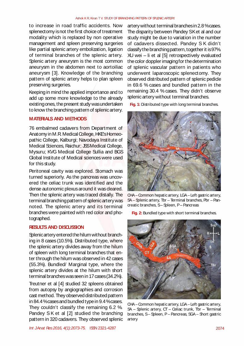
Splenic artery entered the hilum without branch-ing in 8 cases (10.5%). Distributed type, wherethe splenic artery divides away from the hilumof spleen with long terminal branches that en-ter through the hilum was observed in 42 cases(55.3%). Bundled/ Marginal type, where thesplenic artery divides at the hilum with shortterminal branches was seen in 17 cases (34.2%).Treutner et al [4] studied 32 spleens obtainedfrom autopsy by angiographies and corrosioncast method. They observed distributed patternin 84.4 % cases and bundled type in 9.4 % cases.They couldn’t classify the remaining 6.2 %.Pandey S K et al [2] studied the branchingpattern in 320 cadavers. They observed splenic
Fig. 1: Distributed type with long terminal branches.
CHA – Common hepatic artery, LGA – Left gastric artery,SA – Splenic artery, Tbr – Terminal branches, Pbr – Pan-creatic branches, S – Spleen, P – Pancreas
Fig. 2: Bundled type with short terminal branches.
CHA – Common hepatic artery, LGA – Left gastric artery,SA – Splenic artery, CT – Celiac trunk, Tbr – Terminalbranches, S – Spleen, P – Pancreas, SGA – Short gastricartery
Int J Anat Res 2016, 4(1):2073-75. ISSN 2321-4287 2075
Ashok K R, Kiran T V. STUDY OF BRANCHING PATTERN OF SPLENIC ARTERY.
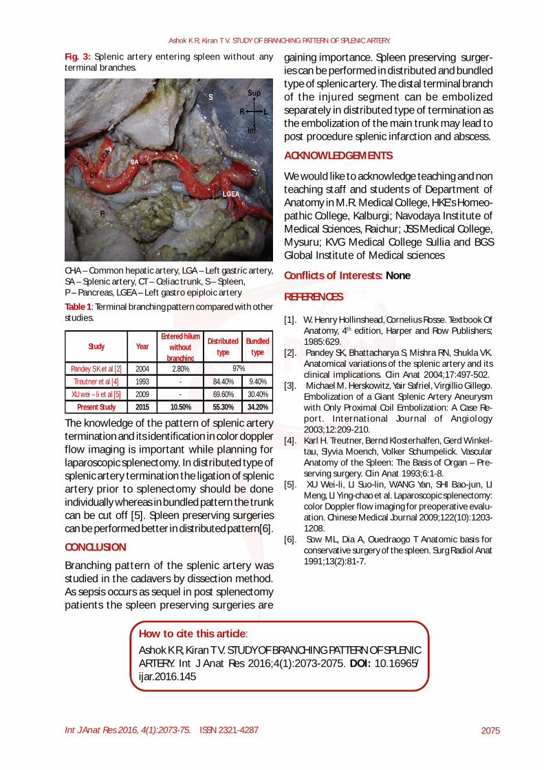
Fig. 3: Splenic artery entering spleen without anyterminal branches.
CHA – Common hepatic artery, LGA – Left gastric artery,SA – Splenic artery, CT – Celiac trunk, S – Spleen,P – Pancreas, LGEA – Left gastro epiploic arteryTable 1: Terminal branching pattern compared with otherstudies.
Study YearEntered hilum
without branching
Distributed type
Bundled type
Pandey S K et al [2] 2004 2.80%Treutner et al [4] 1993 - 84.40% 9.40%
XU wei – li et al [5] 2009 - 69.60% 30.40%Present Study 2015 10.50% 55.30% 34.20%
97%
The knowledge of the pattern of splenic arterytermination and its identification in color dopplerflow imaging is important while planning forlaparoscopic splenectomy. In distributed type ofsplenic artery termination the ligation of splenicartery prior to splenectomy should be doneindividually whereas in bundled pattern the trunkcan be cut off [5]. Spleen preserving surgeriescan be performed better in distributed pattern[6].
CONCLUSION
Branching pattern of the splenic artery wasstudied in the cadavers by dissection method.As sepsis occurs as sequel in post splenectomypatients the spleen preserving surgeries are
ACKNOWLEDGEMENTS
We would like to acknowledge teaching and nonteaching staff and students of Department ofAnatomy in M.R. Medical College, HKE’s Homeo-pathic College, Kalburgi; Navodaya Institute ofMedical Sciences, Raichur; JSS Medical College,Mysuru; KVG Medical College Sullia and BGSGlobal Institute of Medical sciences
Conflicts of Interests: None
REFERENCES
[1]. W. Henry Hollinshead, Cornelius Rosse. Textbook OfAnatomy, 4th edition, Harper and Row Publishers;1985:629.
[2]. Pandey SK, Bhattacharya S, Mishra RN, Shukla VK.Anatomical variations of the splenic artery and itsclinical implications. Clin Anat 2004;17:497-502.
[3]. Michael M. Herskowitz, Yair Safriel, Virgillio Gillego.Embolization of a Giant Splenic Artery Aneurysmwith Only Proximal Coil Embolization: A Case Re-port. International Journal of Angiology2003;12:209-210.
[4]. Karl H. Treutner, Bernd Klosterhalfen, Gerd Winkel-tau, Slyvia Moench, Volker Schumpelick. VascularAnatomy of the Spleen: The Basis of Organ – Pre-serving surgery. Clin Anat 1993;6:1-8.
[5]. XU Wei-li, LI Suo-lin, WANG Yan, SHI Bao-jun, LIMeng, LI Ying-chao et al. Laparoscopic splenectomy:color Doppler flow imaging for preoperative evalu-ation. Chinese Medical Journal 2009;122(10):1203-1208.
[6]. Sow ML, Dia A, Ouedraogo T Anatomic basis forconservative surgery of the spleen. Surg Radiol Anat1991;13(2):81-7.
gaining importance. Spleen preserving surger-ies can be performed in distributed and bundledtype of splenic artery. The distal terminal branchof the injured segment can be embolizedseparately in distributed type of termination asthe embolization of the main trunk may lead topost procedure splenic infarction and abscess.
How to cite this article:Ashok K R, Kiran T V. STUDY OF BRANCHING PATTERN OF SPLENICARTERY. Int J Anat Res 2016;4(1):2073-2075. DOI: 10.16965/ijar.2016.145