Embed Size (px)
Citation preview
V. Moraru et al. Moldovan Medical Journal. December 2018;61(4):3-9
ORIGINAL RESEARCH
Introduction
Acute appendicitis (AA) is the most common surgical abdominal emergency and its lifetime risk is 8.6% for men and 6.7% for women, yet the risk of undergoing appendec-tomy is higher for women (12 vs. 23%) [1-3]. In developed countries the incidence of AA is 400-520 cases per 100000 population, while in poorly developed countries it is 100-320 cases per 100000 population [4, 5]. In the Republic of Moldova the AA frequency denotes 220 cases per 100000 inhabitants [6].
Apparently simple as a pathology, AA does not always find an easy solution, and by its complications it can some-times generate situations requiring complex therapeutic fea-tures. Although surgical treatment is well tolerated by most patients, it is associated with a risk of postoperative com-plications in 2%- 23% of cases [7, 8]. In addition, notwith-standing the implementation of miniinvasive techniques, it is noted that about 3% of patients who underwent an appen-dectomy with or without laparoscopy [9] were repeatedly admitted to hospital with a diagnosis of intestinal occlusion, cataloged as a tardive post-operative complication, often a long time after primary surgery [10].
DOI: 10.5281/zenodo.1456892UDC: 616.346.2-002.1
Risk factors in the development of acute appendicitis complications*1Viorel Moraru, MD, PhD, Associate Professor; 1Petru Bujor, MD, PhD, Professor;
1Galina Pavliuc, MD, PhD, Associate Professor; 2Sergiu Bujor, MD, PhD, Researcher Fellow1Department of Surgery No 2, 2Laboratory of Liver Surgery
Nicolae Testemitsanu State University of Medicine and Pharmacy, Chisinau, the Republic of Moldova
*Corresponding author: [email protected] received September 02, 2018; revised manuscript December 03, 2018
AbstractBackground: To study the risk factors of the development of acute appendicitis (AA) complications in adults in order to improve the results of surgical treatment.Material and methods: The research included 449 patients with AA treated surgically during the years 2015-2017 divided into 2 groups: 117 patients who were admitted with complicated appendicitis (intra- and extraabdominal complications) and 332 patients with non-complicated AA were randomly selected from the same period. The rate and characteristic of the complications evolved during the pre- and postoperative period in these two groups were specified and analyzed. Results: In the acute complicated appendicitis group (CAA), there was a predominance of women with a ratio of 1.60 versus 1.26 in the uncomplicated acute appendicitis group (NAA). The proportion of people aged> 60 years was significantly higher in the case of CAA-23.1% (n=27), while in uncomplicated AA it was only 3.9% (n=13). In the case of AA complications, there was an emphasis on late addressing, the debut-addressing term being higher compared to uncomplicated AA. The low socio-economic status has a significant negative impact on the evolution of AA and its complications, as well as on the results of appendectomy. Thus, uninsured patients (n=59, 49.6%) formed almost half of CAA group. Associated comorbidities were established in 76 or 16.9% of cases, respectively in CAA-21.4% vs 15.4% in NAA group. In summary we note that the presence of associated uncorrected comorbidities has an obvious negative impact on the development of AA.Conclusions: Our findings suggest that clinical assessment is most important for identifying individuals at risk of developing complications of AA and the above-mentioned risk factors are useful for emergency surgical decisions.Key words: Complications of acute appendicitis, risk factors.
AA complications may evolve either as a natural stage in the pathophysiological process of vermicular appendicitis with plastron formation or depending on wall integrity with its perforation and triggering of generalized or localized ap-pendicular peritonitis [11,12]. This type of complications can be called intraabdominal complications, according to the literature; they have an incidence of about 5-7% cases in the developed countries and up to 30% in the case of coun-tries with poor socio-economic status [13]. Notwithstand-ing the general decrease in the morbidity rate through AA, an impressive number of studies demonstrate the stability of these rates over the past decades [14-16].
Another group of complications are caused by the pu-rulent processes in the postoperative wound (suppuration, abscess, ligature fistula) at a rate of 18-20%, they do not have a certain tendency to diminish [17,18]. Even despite the fre-quent and prolonged use of antibacterial drugs for prophy-laxis of postoperative wound complications, the frequency of appearance remains at a constant level [19]. In general, post-appendectomy complications rates are usually with-in 10%-19% range for acute AA without perforation and reaches to 12%-30% for perforated AA [20-24]. Perforation
3
4
ORIGINAL RESEARCHV. Moraru et al. Moldovan Medical Journal. December 2018;61(4):3-9
increases the AA mortality rate from 0.0002% to 3% and causes an increase in morbidity from 3% to 47% [25-27].
Thus, we can see that, despite all the surgical progress achieved, the AA complications remain a problem that still requires increased attention. This determines the need to specify risk and prognostic factors in the development of AA complications for their prophylaxis and improvement of surgical treatment results.
The purpose of the paper: to study the risk factors of the development of acute appendicitis complications in adults in order to improve the results of surgical treatment.
Material and methods
Study Design. The study includes a retrospective analy-sis of clinical material focused on the estimation of the inci-dence, character and risk factors of the development of AA complications. We designed a case-control study to compare different perceived risk factors among patients with com-plicated or uncomplicated AA. The point of reference was the analysis and evaluation of the anamnestic disease, the clinical picture, the laboratory and instrumental data estab-lished preoperatively in connection with the morphological changes of the vermicular appendix performed by morpho-pathological examination of the operative piece. The rate and characteristic of the complications evolved during the pre- and postoperative period in the analyzed patients were specified. For the purpose of assessing the microbial etio-logical factor, the patients of the study group were subjected to the bacteriological examination which included seeding on aerobic culture media as a source of collected material for samples collected during diagnostic laparoscopy, surgi-cal intervention, pathological leakage from the safety drains or postoperative wound.
Participants and data collection. The research inclu-ded 449 AA patients treated surgically by classical approach during the years 2015-2017 at Surgical Clinic Nr. 2 and Nr. 3 of Surgery Department Nr. 2 of Nicolae Testemitsanu State University of Medicine and Pharmacy. The analyzed data were extracted from: clinical observation sheets, op-erative protocols, histopathological examination bulletins and electronic database Sfinta Treime Municipial Hospital (code K35.0-3, K35.9). An individual clinical research file was complemented for each patient. The terms of evolution of the disease were analyzed, the studied cases being classi-fied in 3 time intervals: addressing up to 6 hours after the onset of clinical manifestations, 7-24 hours; more than 24 hours. At the same time, attention was drawn to the socio-economic status of the patient (insured or uninsured).
The Charlson co-morbidity index (CCI) was used in or-der to establish the aggressive synergistic action of chron-ic co-morbidities on AA evolution and its complications (tab. 1).
This score was evaluated in each patient with the consec-utive specification in the following groups according to the stage of compensation of the associated condition: √CCI – I (lack of co-morbidity) – 0 points; √CCI- II (compensated co-morbidity) – 1 point, √CCI III (uncompensated co-mor-
bidity) 2-3 points, √CCI-IV (decompensated co-morbidity) >4 points, √CCI-V (decompensated co-morbidity refrac-tory to any treatment).
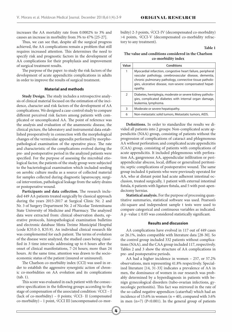
Table 1The value and conditions considered in the Charlson
co-morbidity index
Value Conditions
1 Myocardial infarction, congestive heart failure, peripheral vascular pathology, cerebrovascular disease, dementia, chronic pulmonary pathology, connective tissue patholo-gies, ulcerative disease, non-severe compensated hepat-opathy.
2 Diabetes, hemiplegia, moderate or severe kidney patholo-gies, complicated diabetes with internal organ damage, leukemia, lymphoma.
3 Moderate or severe hepatopathy.
6 Non-metastatic solid tumors, Metastatic tumors, AIDS.
Definitions. In order to standardize the results we di-vided all patients into 2 groups: Non-complicated acute ap-pendicitis (NAA) group, consisting of patients without the progression of complications of cataract and phlegmonous AA without perforation; and complicated acute appendicitis (CAA) group, consisting of patients with complications of acute appendicitis. It included phlegmonous with perfora-tion AA, gangrenous AA, appendicular infiltration or peri-appendicular abscess, local, diffuse or generalized peritoni-tis; septic complications of postoperative wound. The same group included 4 patients who were previously operated for AA, who at distant point had acute adherent intestinal oc-clusion, treated surgically, 1 patient with external intestinal fistula, 6 patients with ligature fistula, and 5 with post-apen-dectomy hernias.
Statistical analysis. For the purpose of processing quan-titative summaries, statistical software was used. Pearson’s chi-square and independent sample t tests were used to compare categorical and continuous variables as indicated. A p -value ≤ 0.05 was considered statistically significant.
Results and discussion
AA complications have evolved in 117 out of 449 cases or 26.1%, index compatible with literature data [28-30]. So the control group included 332 patients without complica-tions (NAA), and the CAA group included 117, respectively. Tables 2 and 3 show the structure of AA complications in pre- and postoperative periods.
AA had a higher incidence in women – 257, or 57.2% observations, men representing 41.8% respectively. Special-ized literature [14, 31-33] indicates a prevalence of AA in men, the dominance of women in our research was prob-ably determined by a hyperdiagnosis in patients with be-nign gynecological disorders (tubo-ovarian infections, gy-necologic peritonitis). This fact was mirrored in the rate of the so-called negative appendices (catarrhal) which had an incidence of 15.6% in women (n = 40), compared with 3.6% in men (n=7) (P<0.001). In the general group of patients

ORIGINAL RESEARCH V. Moraru et al. Moldovan Medical Journal. December 2018;61(4):3-9
undergoing appendectomy catarrhal rate was 10.5%. We found a higher incidence of postoperative complications of AA in this group compared to phlegmonous appendicitis and a lower incidence of complications compared to gan-grenous appendicitis (tab. 4).
Figure 2 shows the stratification of patients with AA ac-cording to age groups and sex.
Fig. 2. The incidence of acute appendicitis according to age and sex (n=449).
Data analysis denotes the highest incidence of AA in the age group of 18-30 years, – 202 patients (45%), therefore, practically half of the total number of patients. The decrease in morbidity through AA over the next 3 decades of life (age 31-60 years) is noted, while in patients> 60 years old we have confirmed changes in the epidemiological trend, namely: the trend of AA growth in the general group, es-pecially in women (marked with a red arrow on fig. 3.3). We consider this remark to be important because elderly pa-tients with associated comorbidities are at risk of developing AA complications either on the basis of delayed referral to a physician or because of difficulties or mistakes in establish-ing the diagnosis and respectively the withdrawal of surgical treatment.
In the complicated acute appendicitis group there was a predominance of women with a ratio of 1.60 (F/B=72/45) comparing to 1.26 (F/B=185/147) in the non-complicated acute appendicitis. The comparative analysis also recorded statistical differences in age, which was 33.5 ± 13.4 years in the NAA group compared to 39.4 ± 16.1 years in the CAA group (P<0.001). Moreover, the share of persons >60 years old was higher in the case of complications of AA – 23.1% (n=27), while in uncomplicated AA it only constituted 3.9% (n=13). Table 5 shows the patients’ profile by age and gender.
Table 2Intra-abdominal pre- and postoperative complications
of acute appendicitis in CAA group (n=117)
IntraabdomInal ComplICatIons n= %
Perforated appendicitis 21 17,9
Appendicular infiltration 11 9,4
Appendicular infiltration with abscessing 7 5,9
Periappendicular abscess 19 16,2
Interintestinal abscess 1 0,9
Typhlitis 9 7,7
Local and diffuse peritonitis 73 62,4
Generalized peritonitis 1 0,9
Acute dynamic intestinal occlusion 31 26,5
Intestinal occlusion by post-appendecto-my adhesion
4 3,4
External intestinal fistula 1 0,9
Table 3Pre-and postoperative extra-abdominal complications of
acute appendicitis in the CAA group (n=117)
WoUnd ComplICatIons n= %
Suppuration 15 12,8
Abscess 7 5,9
Infiltration 12 10,2
Seroma 14 11,9
Ligature fistula 6 5,1
Hernia post-appendicectomy 5 4,3
Table 4The structure of postoperative complications according to the morpho-pathological form of AA
ComplicationsCatarrhal
(n=47, 10.5%)
the morpho-pathological form of acute appendicitis (n=449)
phlegmonous (n=330, 73.5%)
Gangrenous(n=72, 16.0%)
Complications of the postoperative wound 5 (10.6%) 17 (5.2%) 26 (36.1%)
Intestinal fistula - 1 (0.3%) -
Ligature fistula 1 (2.2%) 2 (0.6%) 3 (4.2%)
Postappendectomy hernia 1 (0.3%) 4 (5.6%)
Intestinal occlusion by postappendectomy adhesion 1 (2.2%) 2 (0.6%) 1 (1.4%)
Interintestinal abscess 1 (1.4%)
Total 7 (14.9%)* 23 (7.0%) 35 (48.6%)**
*- siginificant statistic difference between catarrhal and phlegmonous AA ; P<0.05**- siginificant statistic difference between cataral and gangrenous AA; P<0.001
5
6
ORIGINAL RESEARCHV. Moraru et al. Moldovan Medical Journal. December 2018;61(4):3-9
Table 5Distribution of complications (CAA) and absence
of complications (NAA) of acute appendicitis by age group and sex
INDEX 18-30 31-40 41-50 51-60 >60 Total
AAC M 8 14 7 8 8 45
W 19 10 13 11 19 72
AAN M 82 42 9 11 3 147
W 93 47 15 20 10 185
In this context we can conclude that advanced age is a risk factor in the development of complications of acute ap-pendicitis.
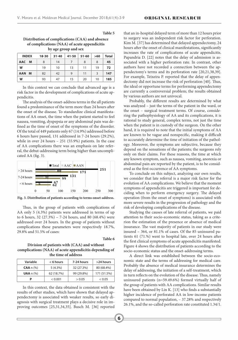
The analysis of the onset-address terms in the all patients found a predominance of the term more than 24 hours after the onset of the disease. To standardize clinical manifesta-tions of AA onset, the time when the patient started to feel nausea, vomiting, dyspepsia or any abdominal pain was de-fined as the time of onset of the symptoms of the disorder. Of the total of 449 patients only 67 (14.9%) addressed before 6 hours have passed, 131 addressed in 7-24 hours (29.2%), while in over 24 hours – 251 (55.9%) patients. In the case of AA complications there was an emphasis on late refer-ral, the debut-addressing term being higher than uncompli-cated AA (fig. 3).
62
99
171
5
32
80
67
131
251
0 50 100 150 200 250 300
< 6 hours
7-24 hours
> 24 hoursTotal AAC AAN
Fig. 3. Distribution of patients according to terms onset-address.
Thus, in the group of patients with complications of AA only 5 (4.3%) patients were addressed in terms of up to 6 hours, 32 (27.3%) – 7-24 hours, and 80 (68.4%) were addressed over 24 hours. In the group of patients without complications these parameters were respectively 18.7%, 29.8% and 51.5% of cases:
Table 6Division of patients with (CAA) and without
complications (NAA) of acute appendicitis depending of the time of address
Variable < 6 hours 7-24 hours >24 hours
CAA n (%) 5 (4.3%) 32 (27.3%) 80 (68.4%)
UAA n (%) 62 (18.7%) 99 (29.8%) 171 (51.5%)
P < 0.001 > 0.05 < 0.05
In this context, the data obtained is consistent with the results of other studies, which have shown that delayed ap-pendectomy is associated with weaker results, so early di-agnosis with surgical treatment plays a decisive role in im-proving outcomes [25,31,34,35]. Busch M. [36] reported
that an in-hospital delayed term of more than 12 hours prior to surgery was an independent risk factor for perforation. Kim M. [37] has determined that delayed appendectomy, 24 hours after the onset of clinical manifestations, significantly increases the rate of complications of acute appendicitis. Papandria D. [22] notes that the delay of admission is as-sociated with a higher perforation rate. In contrast, other authors have not recorded a connection between the ap-pendectomy's terms and its perforation rate [20,21,38,39]. For example, Teixeira P. reported that the delay of appen-dectomy did not increase the risk of perforation [40]. Thus, the ideal or opportune terms for performing appendectomy are currently a controversial problem; the results obtained by various authors are not univocal.
Probably, the different results are determined by what was analyzed – just the terms of the patient in the ward, or the onset – surgical treatment terms. Of course, conside-ring the pathophysiology of AA and its complications, it is rational to study general, complex terms, not just the time when the patient is in custody of the surgeon. On the other hand, it is required to note that the initial symptoms of AA are known to be vague and nonspecific, making it difficult to accurately determine the time of initiation of the pathol-ogy. Moreover, the symptoms are subjective, because they depend on the sensations of the patients; the surgeons rely only on their claims. For these reasons, the time at which any known symptom, such as nausea, vomiting, anorexia or abdominal pain are reported by the patient, is to be consid-ered as the first occurrence of AA symptoms.
To conclude on this subject, analyzing our own results, we consider that late referral is a major risk factor for the evolution of AA complications. We believe that the moment symptoms of appendicitis are triggered is important for de-ciding when to perform emergency surgery. The delayed operation (from the onset of symptoms) is associated with more severe results in the progression of pathology and the risk of developing complications of the disease.
Studying the causes of late referral of patients, we paid attention to their socio-economic status, taking as a crite-rion the estimation of the presence or absence of medical insurance. The vast majority of patients in our study were insured – 364, or 81.1% of cases. Of the 85 uninsured pa-tients 61 (71.%) went to hospital late, over 24 hours after the first clinical symptoms of acute appendicitis manifested. Figure 4 shows the distribution of patients according to the socio-economic status and the onset-addressing terms.
A direct link was established between the socio-eco-nomic state and the terms of addressing for medical care. Probably the absence of medical insurance determines the delay of addressing, the initiation of a self-treatment, which in turn reflects on the evolution of the disease. Thus, namely uninsured patients (n=59.49.6%) formed virtually half of the group of patients with AA complications. Similar results have been obtained by Lin K. [13] who finds a substantially higher incidence of perforated AA in low-income patients compared to normal population, – 37.28% and respectively 26.1%, and the so-called perforation rate constituted 1.34/1.

ORIGINAL RESEARCH V. Moraru et al. Moldovan Medical Journal. December 2018;61(4):3-9
This study, as well as the results of our research, confirmed that low socio-economic status has a significant negative impact on the evolution of acute appendicitis and its com-plications, as well as on appendectomy results.
As regards the influence of associated co-morbidities on the natural evolution of the disease, some connections have been noted, namely: with the increase of the co-morbidity score increases the rate of complications as well as the gen-eral ones, as well as the abdominal or post-appendectomy wound. This is probably due to the difference in the biologi-cal conditions on the background of which the inflammation process occurs in patients with associated co-morbidities, as well as the inflammatory response of the macro-organism itself to the pathogen. Thus, these suggestions are in favor of alternative theories in the field of AA pathophysiology which place the local immunological response made by the vermiform appendix (favored by the richness of lymphoid tissue in the appendix submucosa) and the general release of pro- and antiinflammatory cytokines in response to the pathogenic microbial agent.
Associated co-morbidities were established in 76 or 16.9% of cases, most frequently – cardiovascular (hyperten-sion, ischemic heart disease, angina pectoris, atherosclerotic or postinfarct cardiosclerosis, paroxysmal arrhythmia) – 26 (34.2%) cases. Digestive tract disorders (chronic cholecys-titis, chronic gastroduodenitis, chronic viral hepatitis B and C, chronic gastro-duodenal disease in remission) was marked in 12 (15.8%) patients. Type II diabetes was pres-ent in 15 (19.7%) patients, and respiratory tract disorders (bronchial asthma, pneumonia, chronic bronchitis) were noted in 6 (7.9%). Urogenital diseases (salpingitis, salpin-goophoritis, ovarian cyst, urolithiasis, chronic renal failure) were diagnosed in 7 (9.2%) cases. In 10 (13.2%) patients, co-morbidities were represented by various causes of cerebral infarction, dysmetabolic or atherosclerotic encephalopathy, feripritis anemia, narcotics, varicose veins, chest cancer che-motherapy.
In case of co-morbid score «0» we noticed significant differences, 84.6% of patients being in the NAA group com-pared to 78.6% of the patients in the CAA group (P <0.001). In other words, the majority of patients who had a favorable affection did not show associated co-morbidities. The asso-ciation of compensated co-morbidity did not show a severe impact on disease progression, at 13.3% with NAA, com-pared with 15.4% of patients with CAA (P>0.05). In the case of undercompensated or decompensated associated diseas-es, or the presence of more co-morbidities, the picture was radically different: 1.5% compared to 3.4% in CCI = 2; and 0.6% vs. 2.6% in CCI ≥ 3 in the respective groups.
Table 7Characteristics of patients with complications (CAA)
and without complications (NAA) of acute appendicitis depending on Charlson Co-morbidity Index (CCI)
Parameter Total (N= 449)
CAA (n= 117)
NAA (n= 332)
P
CCI ”0” (%) 83.1 78.6 84.6 <0.001
CCI ”1” (%) 13.8 15.4 13.3 >0.05
CCI ”2” (%) 2.0 3.4 1.5 <0.01
CCI ”≥3” (%) 1.1 2.6 0.6 <0.001
In brief, we note that the presence of associated co-morbidities has a clear negative impact on AA evolution. The failure to diagnose secondary pathologies or to perform surgical treatment in a patient with insufficiently corrected subcompensated or decompensated co-morbidity and the associations of several co-morbid pathologies are significant risk factors for the development of AA complications.
One of the cardinal problems discussed in the literature is the specification of the pathophysiological mechanisms that underlie the pre- and postoperative complications of AA. It is considered that all septic complications after ap-pendectomy evolved within the surgical wards are due to nosocomial infection. However, the presence of an exoge-nous source is necessary for its appearance, or otherwise the patient who underwent the operation must contact either a supposed wound of another patient or with contaminated dressing material, instrumentation and so on. The most frequent phenomenon occurs when a septic department is absent in the clinic and the patients with septic complica-tions are not isolated. Another reason could be due to the non-observance of the profile of the hospitalized surgical patients. Such situations may result in outbreaks of in-hos-pital infection.
In the group of CAA patients, bacteriological cultures were culled intraoperatively in order to analyze whether there is a correlation between the bacterial species normally present in the digestive tract and those isolated from the peritoneal fluid in the appendicular peritonitis. Another ob-jective was the comparative analysis of microbial germs in the case of postoperative wound complications and isolated germs in the peritoneal cavity.
Although peritoneal fluid response was noted in all cas-es, only 78 patients (66.7%) identified microbes. Constantly, the germs were present in all observations accompanied by diffuse peritonitis and partly in AA with localized perito-nitis. In 33.3% observations, the bacteriological laboratory response was negative, probably due to the impossibility of determining the anaerobic flora. This moment is a weak point of the bacteriological research analyzed in our study, because the absence of flora identification does not neces-sarily signify its absence. The aforementioned increases its importance, because the microbial flora is decisive in the evolution of the disease in the etiopathogenesis of AA, most of the postoperative complications are septic. The aggres-siveness of microbial flora in AA is due to the colon, which
Fig. 4. The distribution of the patients (%) according to the onset-addressing and the socio-economic status.
0 20 40 60 80
< 6 hours 7-24 hours > 24 hours
17.3 30.5
52.2
4.7 23.5
71.8
Uninsured Insured
7
8
ORIGINAL RESEARCHV. Moraru et al. Moldovan Medical Journal. December 2018;61(4):3-9
in the healthy individual contains anaerobic flora with the predominance of gram negative bacteria Bacterioidis. On the other hand, it is known that the most commonly found in the colon is E.Coli, which is an aerobic germ, and is also an anaerobic microbe (optional) and therefore can also de-velop under anaerobiosis conditions. These considerations are probably the explanations of a large number of negative bacteriological outcomes in our research.
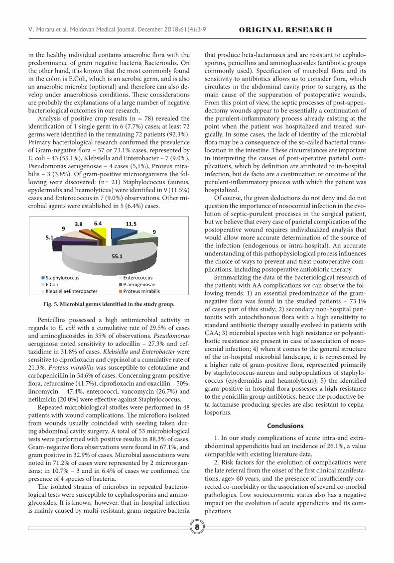
Analysis of positive crop results (n = 78) revealed the identification of 1 single germ in 6 (7.7%) cases; at least 72 germs were identified in the remaining 72 patients (92.3%). Primary bacteriological research confirmed the prevalence of Gram-negative flora – 57 or 73.1% cases, represented by E. coli – 43 (55.1%), Klebsiella and Enterobacter – 7 (9.0%), Pseudomonas aerugenosae – 4 cases (5,1%), Proteus mira-bilis – 3 (3.8%). Of gram-positive microorganisms the fol-lowing were discovered: (n= 21) Staphylococcus (aureus, epydermidis and heamolyticus) were identified in 9 (11.5%) cases and Enterococcus in 7 (9.0%) observations. Other mi-crobial agents were established in 5 (6.4%) cases.
11.5 9
55.1
5.1
9 3.8 6.4
Staphylococcus Enterococcus E.Coli P.aerugenosae Klebsiella+Enterobacter Proteus mirabilis
Fig. 5. Microbial germs identified in the study group.
Penicillins possessed a high antimicrobial activity in regards to E. coli with a cumulative rate of 29.5% of cases and aminoglucosides in 35% of observations. Pseudomonas aeruginosa noted sensitivity to azlocillin – 27.3% and cef-tazidime in 31.8% of cases. Klebsiella and Enterobacter were sensitive to ciprofloxacin and cyprinol at a cumulative rate of 21.3%. Proteus mirabilis was susceptible to cefotaxime and carbapenicillin in 34.6% of cases. Concerning gram-positive flora, cefuroxime (41.7%), ciprofloxacin and oxacillin – 50%; lincomycin – 47.4%, enterococci, vancomycin (26.7%) and netilmicin (20.0%) were effective against Staphylococcus.
Repeated microbiological studies were performed in 48 patients with wound complications. The microflora isolated from wounds usually coincided with seeding taken dur-ing abdominal cavity surgery. A total of 53 microbiological tests were performed with positive results in 88.3% of cases. Gram-negative flora observations were found in 67.1%, and gram positive in 32.9% of cases. Microbial associations were noted in 71.2% of cases were represented by 2 microorgan-isms; in 10.7% – 3 and in 6.4% of cases we confirmed the presence of 4 species of bacteria.
The isolated strains of microbes in repeated bacterio-logical tests were susceptible to cephalosporins and amino-glycosides. It is known, however, that in-hospital infection is mainly caused by multi-resistant, gram-negative bacteria
that produce beta-lactamases and are resistant to cephalo-sporins, penicillins and aminoglucosides (antibiotic groups commonly used). Specification of microbial flora and its sensitivity to antibiotics allows us to consider flora, which circulates in the abdominal cavity prior to surgery, as the main cause of the suppuration of postoperative wounds. From this point of view, the septic processes of post-appen-dectomy wounds appear to be essentially a continuation of the purulent-inflammatory process already existing at the point when the patient was hospitalized and treated sur-gically. In some cases, the lack of identity of the microbial flora may be a consequence of the so-called bacterial trans-location in the intestine. These circumstances are important in interpreting the causes of post-operative parietal com-plications, which by definition are attributed to in-hospital infection, but de facto are a continuation or outcome of the purulent-inflammatory process with which the patient was hospitalized.
Of course, the given deductions do not deny and do not question the importance of nosocomial infection in the evo-lution of septic-purulent processes in the surgical patient, but we believe that every case of parietal complication of the postoperative wound requires individualized analysis that would allow more accurate determination of the source of the infection (endogenous or intra-hospital). An accurate understanding of this pathophysiological process influences the choice of ways to prevent and treat postoperative com-plications, including postoperative antiobiotic therapy.
Summarizing the data of the bacteriological research of the patients with AA complications we can observe the fol-lowing trends: 1) an essential predominance of the gram-negative flora was found in the studied patients – 73.1% of cases part of this study; 2) secondary non-hospital peri-tonitis with autochthonous flora with a high sensitivity to standard antibiotic therapy usually evolved in patients with CAA; 3) microbial species with high resistance or polyanti-biotic resistance are present in case of association of noso-comial infection; 4) when it comes to the general structure of the in-hospital microbial landscape, it is represented by a higher rate of gram-positive flora, represented primarily by staphylococcus aureus and subpopulations of staphylo-coccus (epydermidis and heamolyticus); 5) the identified gram-positive in-hospital flora possesses a high resistance to the penicillin group antibiotics, hence the productive be-ta-lactamase-producing species are also resistant to cepha-losporins.
Conclusions
1. In our study complications of acute intra-and extra-abdominal appendicitis had an incidence of 26.1%, a value compatible with existing literature data.
2. Risk factors for the evolution of complications were the late referral from the onset of the first clinical manifesta-tions, age> 60 years, and the presence of insufficiently cor-rected co-morbidity or the association of several co-morbid pathologies. Low socioeconomic status also has a negative impact on the evolution of acute appendicitis and its com-plications.

ORIGINAL RESEARCH V. Moraru et al. Moldovan Medical Journal. December 2018;61(4):3-9
3. There was a higher incidence of AA parietal postop-erative complications in catarrhal appendicitis compared to phlegmonous appendicitis and a lower incidence of compli-cations compared to gangrenous appendicitis. Thus, more extensive application of diagnostic laparoscopy is necessary in uncertain clinical situations.
4. Specification of microbial flora and its susceptibility to antibiotics allows us to consider the main cause of post-operative wound sedation of native flora susceptible to the usual antibiotic therapy. From this point of view, the sep-tic processes of the post-appendectomy wound appear to be essentially a continuation of the purulent-inflammatory process with which the patient was hospitalized and surgi-cally treated. Any suspicion of postoperative complication through nosocomial infection requires wide-spectrum des-iccation antibiotic therapy, including one covering the an-aerobic flora.
5. Our findings suggest that clinical assessment is most important for identifying individuals at risk of developing complications of acute appendicitis and the above-mentio-ned risk factors are useful for emergency surgical decisions.
References1. Ferris M, Quan S, Kaplan BS, et al. Global incidence of appendi-
citis: a systematic review of population-based studies. Ann Surg. 2017;266(2):237-241.
2. Morrow SE, Newman KD. Current management of appendicitis. Semin Pediatr Surg. 2007;16(1):34-40.
3. Temple CL, Huchcroft SA, Temple WJ. The natural history of appen-dicitis in adults. A prospective study. Ann Surg. 1995;221(3):278-81.
4. Kong VY, Bulajic B, Allorto NL, et al. Acute appendicitis in a developing country. World J Surg. 2012;36(4):2068-73.
5. Leppäniemi A. Acute appendicitis – we thought we knew it all? Scand J Surg. 2014;103(1):3–4.
6. Maloman E, Gladun N, Ungureanu S, Lepadatu C. Apendicita acută - ghid practic bazat pe evidenţa clinică [Acute appendicitis – a practical guide based on clinical evidence]. Jurnalul de Chirurgie (Iasi, Romania). 2006;2(3):305-15. Romanian.
7. Konstantinidis KM, Anastasakou KA, Vorias MN, et al. A decade of lapa-roscopic appendectomy: presentation of 1,026 patients with suspected appendicitis treated in a single surgical department. J Laparoendosc Adv Surg Tech. 2008;18:248-58.
8. Ming PC, Yan TY, Tat LH. Risk factors of postoperative infections in adults with complicated appendicitis. Surg Laparosc Endosc Percutan Tech. 2009;19(5):244-8.
9. Guller U, Hervey S, Purves H, et al. Laparoscopic versus open appendec-tomy: outcomes comparison based on a large administrative database. Ann Surg. 2004;239(4):43-52.
10. Leung TT, Dixon E, Gill M, et al. Bowel obstruction following appendec-tomy: what is the true incidence? Ann Surg. 2009;250(2):51-3.
11. Bhangu A, Søreide K, Saverio S, et al. Emergency surgery. Acute ap-pendicitis: modern understanding of pathogenesis, diagnosis, and management. Lancet. 2015;386(5):1278-87.
12. Tannoury J, Abboud B. Treatment options of inflammatory appendiceal masses in adults. World J Gastroenterol. 2013;19(25):3942-50.
13. Lin KB, Lai KR, Yang NP, et al. Epidemiology and socio- economic features of appendicitis in Taiwan: a 12-year population-based study. World J Emerg Surg. 2015;10:42-50.
14. Buckius MT, McGrath B, Monk J, et al. Changing epidemiology of acute appendicitis in the United States: study period 1993-2008. J Surg Res. 2012;175:185-90.
15. Darwazeh G, Cunningham SC, Kowdley GC. A systematic review of perforated appendicitis and phlegmon: interval appendectomy or wait-and-see? Am Surg. 2016;82(1):11-5.
16. Hale DA, Molloy M, Pearl RH, et al. Appendectomy: a contemporary appraisal. Ann Surg. 1997;225(3):252-61.
17. Bhangu A. Systemic review and meta-analysis of randomized clinical trials comparing primary vs delayed primary skin closure in contami-nated and dirty abdominal incisions. JAMA Surg. 2013;148(8):779-86.
18. Kim JK, Ryoo S, Oh HK, et al. Management of appendicitis presenting with abscess or mass. J Korean Soc Coloproctol. 2010;26(6):413-9.
19. Teo AT, et al. Institutional review of patients presenting with suspected appendicitis. ANZ J Surg. 2015;85(6):420-4.
20. Ansaloni L, Catena F, Coccolini F, et al. Surgery versus conservative antibiotic treatment in acute appendicitis: a systematic review and meta-analysis of randomized controlled trials. Dig Surg. 2011;28:210-21.
21. Boomer LA, Cooper JN, Anandalwar S, et al. Delaying appendec-tomy does not lead to higher rates of surgical site infections: A multi-institutional analysis of children with appendicitis. Ann Surg. 2016;264(1):164-8.
22. Papandria D, Goldstein SD, Rhee D, et al. Risk of perforation increases with delay in recognition and surgery for acute appendicitis. J Surg Res. 2013;184(2):723-9.
23. Paquette IM, Zuckerman R, Finlayson SR. Perforated appendicitis among rural and urban patients: implications of access to care. Ann Surg. 2011;253(3):534-8.
24. Park HC, Yang DH, Lee BH. The laparoscopic approach for perforated appendicitis, including cases complicated by abscess formation. J Lapa-roendosc Adv Surg Tech. 2009;19(6):727-30.
25. Ditillo MF, Dziura JD, Rabinovici R. Is it safe to delay appendectomy in adults with acute appendicitis? Ann Surg. 2006;244(5):656-60.
26. Drake FT, Mottey NE, Castelli AA, et al. Time-of-day and appendicitis: Impact on management and outcomes. Surgery. 2017;161(2):405-14.
27. Parks NA, Schroeppel TJ. Update on imaging for acute appendicitis. Surg Clin North Am. 2011;91(1):141-54.
28. Körner H, Söndenaa K, Söreide JA, et al. Incidence of acute nonperfo-rated and perforated appendicitis: age-specific and sex-specific analysis. World J Surg. 1997;21(3):313-7.
29. Livingston EH, Woodward WA, Sarosi GA, Haley RW. Disconnect between incidence of nonperforated and perforated appendicitis: implications for pathophysiology and management. Ann Surg. 2007;245(6):886-92.
30. Naderan M, Babaki A, Shoar S, et al. Risk factors for the development of complicated appendicitis in adults. Ulus Cerrahi Derg. 2016;32(2):37-42.
31. Andersson RE. Short and long-term mortality after appendectomy in Sweden 1987 to 2006. Influence of appendectomy diagnosis, sex, age, co-morbidity, surgical method, hospital volume, and time period. A national population-based cohort study. World J Surg. 2013;37(5):974-81.
32. Coward S, Kareemi H, Clement F, et al. Incidence of appendicitis over time: a comparative analysis of an administrative healthcare database and a pathology proven appendicitis registry. PLoS One. 2016 Nov 7;11(11):e0165161. doi: 10.1371/journal.pone.0165161.
33. Luckmann R, Davis P. The epidemiology of acute appendicitis in California: racial, gender, and seasonal variation. Epidemiology. 1991;2(5):323-30.
34. Ingraham AM, Cohen ME, Bilimoria KY, et al. Effect of delay to operation on outcomes in adults with acute appendicitis. Arch Surg. 2010;145(9):886-92.
35. Menclová K, Traboulsi E, Nikov A, et al. Treatment of acute appendicitis: Retrospective analysis. Rozhl Chir Fall. 2016;95(8):317-21.
36. Busch M, Gutzwiller FS, Aellig S, et al. In-hospital delay increases the risk of perforation in adults with appendicitis. World J Surg. 2011;35(7):1626-33.
37. Kim M, Kim SJ, Cho HJ. Effect of surgical timing and outcomes for ap-pendicitis severity. Ann Surg Treat Res. 2016;91(2):85-9.
38. Augustin T, Cagir B, Vandermeer TJ. Characteristics of perforated ap-pendicitis: effect of delay is confounded by age and gender. J Gastrointest Surg. 2011;15(7):1223-31.
39. Bickell NA, Aufses AH, Rojas M, Bodian C. How time affects the risk of rupture in appendicitis. J Am Coll Surg. 2006;202(3):401-6.
40. Teixeira PG, Demetriades D. Appendicitis: changing perspectives. Adv Surg. 2013;47(2):119-40.
9