Embed Size (px)
Citation preview
© 2015 Sneha and Sundaram. This work is published by Dove Medical Press Limited, and licensed under Creative Commons Attribution – Non Commercial (unported, v3.0) License. The full terms of the License are available at http://creativecommons.org/licenses/by-nc/3.0/. Non-commercial uses of the work are permitted without any further
permission from Dove Medical Press Limited, provided the work is properly attributed. Permissions beyond the scope of the License are administered by Dove Medical Press Limited. Information on how to request permission may be found at: http://www.dovepress.com/permissions.php
International Journal of Nanomedicine 2015:10 (Suppl 1: Challenges in biomaterials research) 99–106
International Journal of Nanomedicine Dovepress
submit your manuscript | www.dovepress.com
Dovepress 99
O r I g I N a l r e S e a r C h
open access to scientific and medical research
Open access Full Text article
http://dx.doi.org/10.2147/IJN.S79985
Preparation and characterization of an iron oxide-hydroxyapatite nanocomposite for potential bone cancer therapy
Murugesan SnehaNachiappan Meenakshi SundaramDepartment of Biomedical engineering, PSg College of Technology, Tamil Nadu, India
Abstract: Recently, multifunctional magnetic nanostructures have been found to have potential
applications in biomedical and tissue engineering. Iron oxide nanoparticles are biocompatible
and have distinctive magnetic properties that allow their use in vivo for drug delivery and
hyperthermia, and as T2 contrast agents for magnetic resonance imaging. Hydroxyapatite is
used frequently due to its well-known biocompatibility, bioactivity, and lack of toxicity, so
a combination of iron oxide and hydroxyapatite materials could be useful because hydroxy-
apatite has better bone-bonding ability. In this study, we prepared nanocomposites of iron
oxide and hydroxyapatite and analyzed their physicochemical properties. The results suggest
that these composites have superparamagnetic as well as biocompatible properties. This type
of material architecture would be well suited for bone cancer therapy and other biomedical
applications.
Keywords: iron oxide, hydroxyapatite, nanocomposite, superparamagnetic, bone cancer
IntroductionCancer is the most dreaded disease, along with heart disease. Hyperthermia has attracted
much interest in the treatment of cancer due to its advantages over chemotherapy and
radiotherapy. Using hyperthermia, cancer cells are killed directly within a short period
of time, whereas normal cells are unaffected.1 Magnetic hyperthermia is practiced by
applying an external alternating magnetic field which in turn oscillates the magnetic
moment of each particle, converting magnetic energy into heat. Superparamagnetic
materials for magnetic hyperthermia are promising candidates for antitumor therapy
because they have the capacity to destroy deep tumors and are controlled by an exter-
nal magnetic field.2 Magnetic nanoparticles have attractive features that could be used
effectively in nanomedicine. First, they have a controllable particle size (from a few
nanometers to tens of nanometers). Second, they are magnetic, so can be manipulated
by an external magnetic field. Third, they can be made to heat up, so can be used as
hyperthermia agents, delivering large amounts of thermal energy to tumor cells and
destroying them.3 If an iron oxide nanoparticle is below 45 nm in size, it is classified
as superparamagnetic due to its line-type hysteresis loop iron oxide nanoparticles.
The magnetite (Fe3O
4) phase of superparamagnetic iron oxide nanoparticles has
numerous in vivo applications, since it can respond to an external stimulus and heat
up, and does not retain any magnetism after removal of the external magnetic field.4,5
Materials that are compatible with bone tissue are preferred for bone repair and hard
tissue engineering.6 Hydroxyapatite (HAp) is widely used in this setting because of
its exceptional biocompatibility, bioactivity, and osteoconductivity.7 A number of
Correspondence: Nachiappan Meenakshi SundaramDepartment of Biomedical engineering, PSg College of Technology, Coimbatore-04, Tamil Nadu, IndiaTel +91 42 2257 2177email [email protected]
Journal name: International Journal of NanomedicineArticle Designation: Original ResearchYear: 2015Volume: 10 (Suppl 1: Challenges in biomaterials research)Running head verso: Sneha and SundaramRunning head recto: Iron oxide-hydroxyapatite nanocomposites for bone cancerDOI: http://dx.doi.org/10.2147/IJN.S79985
Point your SmartPhone at the code above. If you have a QR code reader the video abstract will appear. Or use:
http://youtu.be/rRFw0_DBkBU
Video abstract
International Journal of Nanomedicine 2015:10 (Suppl 1: Challenges in biomaterials research)submit your manuscript | www.dovepress.com
Dovepress
Dovepress
100
Sneha and Sundaram
researchers have developed a variety of HAp-based magnetic
materials for hyperthermia-based treatment of cancer.8–11
Murakami et al prepared a porous Fe3O
4-HAp compos-
ite using a hydrothermal method. The composite holds 30%
Fe3O
4 in cages of rod-shaped HAp particles.8 Fe
3O
4 nano-
particles are prepared by a coprecipitation method, and a
horizontal tumbling ball mill is used to mechanochemically
synthesize submicron-sized HAp particles. According to
Iwasaki et al9 this process promotes the dispersion of Fe3O
4
nanoparticles in the HAp matrix. The magnetic Fe3O
4 particles
are coated with HAp by spray-drying. Donadel et al found
that the spray-drying technique is an efficient and inexpensive
method for creating spherical particles with a core/shell struc-
ture.10 Tampieri et al prepared iron-doped HAp endowed with
superparamagnetic properties for hyperthermic application.11
These composites can be synthesized conventionally by
mixing HAp nanopowder with Fe3O
4 nanoparticles that are
prepared separately. In this work, Fe3O
4 nanoparticles were
prepared by alkaline coprecipitation of ferric and ferrous
chloride in aqueous solution. Nanocrystalline HAp was
prepared using an optimized sol-gel method. Dry (zirconia
ball) milling was used to blend the Fe3O
4-HAp (0.70 w/w)
nanoparticles. The Fe3O
4-HAp nanoparticles and their
composite were analyzed by Fourier transform infrared
spectroscopy, diffuse reflectance spectroscopy, scanning
electron microscopy, thermogravimetric analysis, differen-
tial scanning calorimetry, vibrating sample magnetometry,
and cytotoxicity tests. The method proposed in this paper is
easy and cost-effective for preparing these nanocomposites.
The synthesis and characterization of this biocompat-
ible magnetic biomaterial is explained in detail, and the
nanostructured Fe3O
4-HAp composite is ideal for bone
cancer therapy.
Materials and methodsSynthesis of iron oxide nanoparticlesFe
3O
4 magnetic nanoparticles were synthesized by the alka-
line coprecipitation method. The molar ratio of Fe3+:Fe2+
chloride was maintained at 2:1. The prepared magnetite was
black in color and the pH was maintained between 9 and 14.12
The overall reaction is written as:
Fe2+ + 2Fe3+ + 8OH-→Fe3O
4 + 4H
2O (1)
Synthesis of hap nanoparticlesThe HAp nanoparticles were prepared by the wet chemical
route method. First, 0.25 M phosphoric acid was prepared in
distilled water. Ammonia (NH3) was added to this solution,
with stirring to maintain the pH at 10. Next, a 1 M calcium
nitrate tetrahydrate solution was prepared by dissolving in
double-distilled water, which was then added slowly to the
above phosphoric acid-ammonia solution. The solution was
stirred vigorously for 1 hour and allowed to age at room tem-
perature for 24 hours. The gel obtained after aging was dried
at 80°C for 48 hours in a dry oven. The resulting mixture
were washed repeatedly using distilled water. After wash-
ing, the powder was sintered for 2 hours at a temperature
of 900°C.
Synthesis of Fe3O4-hap nanocompositesThe Fe
3O
4-HAp nanocomposites were prepared using a
wet-type ball mill (Figure 1A). The ratio of HAp to Fe3O
4
nanoparticles was 1.5:1 (w/w). Ball milling was carried out
for 5 hours using a zirconia bowl and ball at 300 rpm, with
a ball to sample powder ratio of 10:1 (w/w).
CharacterizationFourier transform infrared spectra were taken using an
8400S spectrophotometer (Shimadzu, Tokyo, Japan). KBr
pellets were used, and the spectra were recorded in aqueous
medium.
The morphology of the Fe3O
4-HAp nanostructures was
observed using a scanning electron microscope (ICON,
Quanta 200 Mark II Environmental scanning electron micro-
scope) with an acceleration voltage of 0.2–30 kV.
The thermal properties of the prepared nanocomposites
were investigated using a thermal analyzer (STA 449 F3
Jupiter, Netzsch Gerätebau GmbH, Selb, Germany) along
with thermogravimetry and differential scanning calorimetry
in the temperature range of 28°C–1,100°C at a heating rate
of 20°C per minute in a dry air atmosphere. Al2O
3 was used
as the reference material.
Diffuse reflectance spectroscopy was performed using a
Specord 210 Plus (Analytik Jena, The Woodlands, TX, USA)
between 190 and 1,100 nm at room temperature.
The superparamagnetic properties of the Fe3O
4-HAp
nanocomposites were studied using a 7410 vibrating sample
magnetometer (Lake Shore, Westerville, OH, USA), in
atmospheric air at room temperature.
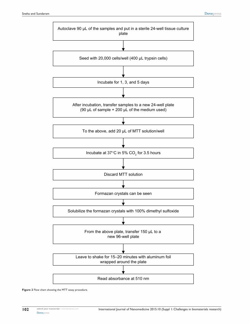
The cytotoxic effects of the Fe3O
4-HAp nanocompos-
ites were evaluated using MG63 cells. The MTT assay
was performed according to the procedure shown in
Figure 2. The formazan was dissolved in dimethyl sulfox-
ide and the absorbance of the solution was quantified at
510 nm.
International Journal of Nanomedicine 2015:10 (Suppl 1: Challenges in biomaterials research) submit your manuscript | www.dovepress.com
Dovepress
Dovepress
101
Iron oxide-hydroxyapatite nanocomposites for bone cancer
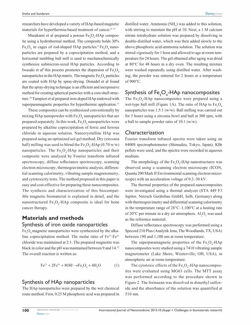
Figure 1 (A) Fe3O4-hap nanoparticles in powder form. (B) Fourier transform infrared spectra of hap, Fe3O4, and Fe3O4-hap nanoparticles. (C) Diffuse reflectance spectra of magnetite and the composite. (D) Scanning electron micrograph of Fe3O4-hap nanoparticles. Abbreviations: Fe3O4, iron oxide; hap, hydroxyapatite.
Results and discussionFourier transform infrared spectroscopyThe Fourier transform infrared spectral assignments for pure
Fe3O
4, HAp, and the Fe
3O
4-HAp nanoparticles are shown
in Figure 1B and tabulated in Table 1. From these data, the
functional groups of prepared samples confirms the presence
of pure Fe3O
4 and pure HAp. IN Fe
3O
4-HAp nanoparticles,
a slight variation in spectral assignments confirms the close
interaction between Fe3O
4 and HAp.
Diffuse reflectance spectroscopyDiffuse reflectance spectroscopy provides the electron tran-
sition of pure and mixed samples. Figure 1C represents the
diffuse reflectance spectra for pure Fe3O
4 and Fe
3O
4-HAp.
Pure Fe3O
4 shows a wavelength (4T
l) transition in the range
of 780–830 nm and an electron pair transition of 510 nm.
This shows a higher position of the ligand-to-metal charge
transfer transition and pair transition band of red Fe3O
4.
Scanning electron microscopyThe micromorphology and texture of the Fe
3O
4-HAp nano-
composites are shown in Figure 1D. This scanning electron
microscopic image shows that pure Fe3O
4 has an irregular
approximately spherical-like morphology with an average
particle size in the range of 50–70 nm and that HAp has
a particle size in the range of 30 – 40 nm. The aggregated
Fe3O
4-HAp clusters have a size range of 100–350 nm and
both phases distributed uniformly, as shown in Figure 1D.
The characteristic dark and light gray represents the Fe3O
4 and
HAp, respectively.
Thermogravimetric analysisThe Fe
3O
4-HAp nanoparticles were studied by thermogravimet-
ric analysis in a nitrogen atmosphere at a heating rate of 20°C
per minute. Figure 3 shows the thermogravimetric analysis
curves, depicting the variations in residual mass of the samples
with increasing temperature. The absolute weight loss from
International Journal of Nanomedicine 2015:10 (Suppl 1: Challenges in biomaterials research)submit your manuscript | www.dovepress.com
Dovepress
Dovepress
102
Sneha and Sundaram
°
Figure 2 Flow chart showing the MTT assay procedure.
International Journal of Nanomedicine 2015:10 (Suppl 1: Challenges in biomaterials research) submit your manuscript | www.dovepress.com
Dovepress
Dovepress
103
Iron oxide-hydroxyapatite nanocomposites for bone cancer
Table 1 Fourier transform infrared spectral assignments of pure Fe3O4, hap, and Fe3O4-hap nanoparticles
S No Wave number (cm-1) Spectral assignments
Fe3O4 HAp Fe3O4-HAp
1 – 3,639 3,639 Oh- group, CaO2 – 3,561 3,570 Stretching mode of Oh- group3 3,420 – 3,420 h2O, Oh- group4 – 2,080 – Phosphate group (PO4
3-)/absorbed CO32-
5 – 2,003 – Phosphate group (PO43-)/absorbed CO3
2-
6 – – 1,638 O-h in-plane bending7 1,625 – – (h-O-h) adsorbed water8 – 1,473 1,473 CO3
2-
9 1,383 – – N-O nitro compounds10 1,151 – – C-O11 – – 1,091 Phosphate group (PO4
3-)12 1,062 – – C-O13 – 1,046 1,046 Phosphate group (PO4
3-)14 – – 960 Phosphate group (PO4
3-)15 891 – – C-h16 – – 875 C-h17 797 – – C-h18 – – 629 C-Cl19 – – 601 O-P-O bending20 588 – – Fe-O-Fe bond (Fe ions in tetrahedral and octahedral site)21 – 566 566 O-P-O bending
Abbreviations: Fe3O4, iron oxide; hap, hydroxyapatite.
°
Figure 3 Weight loss from the Fe3O4-hap nanoparticles. Abbreviations: Fe3O4, iron oxide; hap, hydroxyapatite; NPs, nanoparticles.
the uncoated Fe3O
4 is nearly 17% for the whole temperature
range due to removal of adsorbed physical and chemical water.
Another 5% weight loss was seen from 160°C to 300°C, and
this was associated with decomposition of residual chemical
compounds. No significant weight loss was observed at higher
temperatures. Thermogravimetric analysis of the Fe3O
4-HAp
powder showed weight loss up to a temperature of 800°C and
thereafter weight remained constant.
The first stage of weight loss is between 90°C and 390°C,
which corresponds to dehydration of the precipitating
complex and loss of physically adsorbed water molecules
from the HAp powder. The weight loss in this region is 10%.
International Journal of Nanomedicine 2015:10 (Suppl 1: Challenges in biomaterials research)submit your manuscript | www.dovepress.com
Dovepress
Dovepress
104
Sneha and Sundaram
°
°
°
°
°
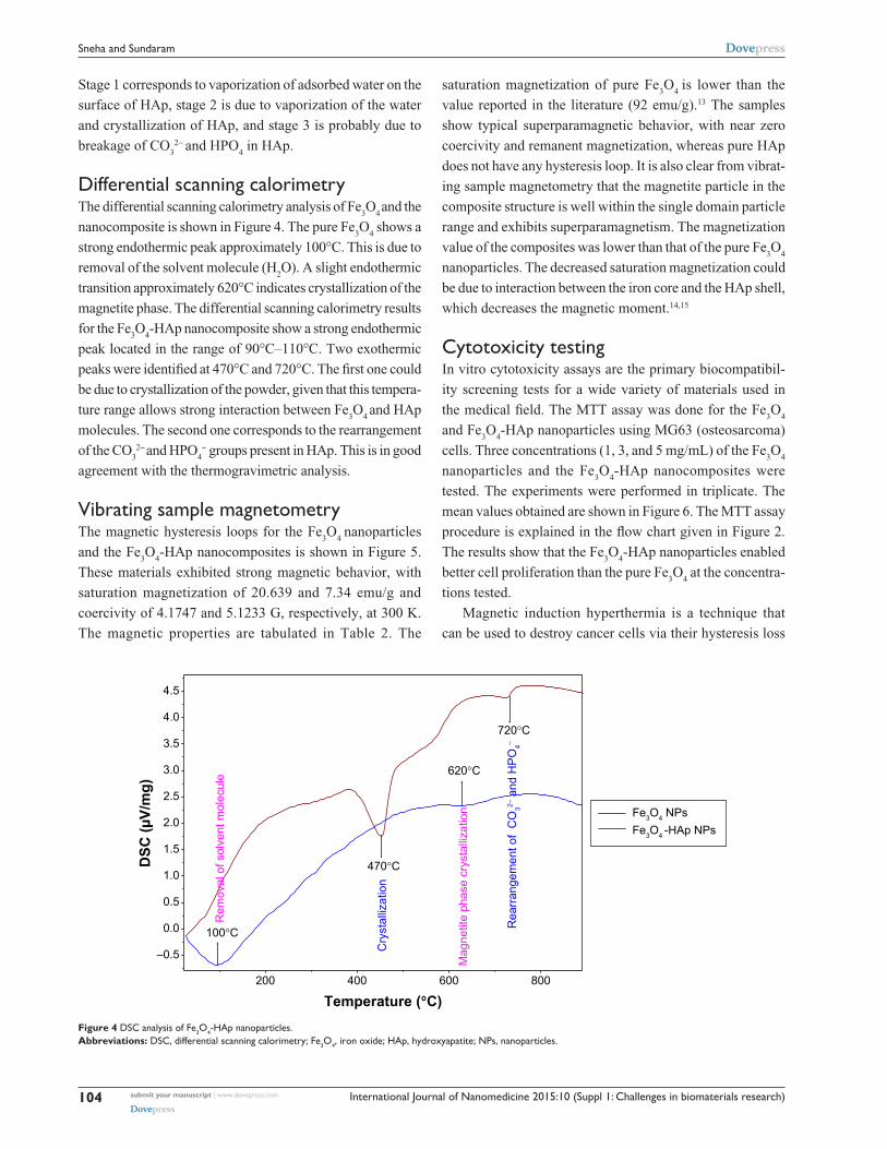
Figure 4 DSC analysis of Fe3O4-hap nanoparticles. Abbreviations: DSC, differential scanning calorimetry; Fe3O4, iron oxide; hap, hydroxyapatite; NPs, nanoparticles.
Stage 1 corresponds to vaporization of adsorbed water on the
surface of HAp, stage 2 is due to vaporization of the water
and crystallization of HAp, and stage 3 is probably due to
breakage of CO3
2- and HPO4 in HAp.
Differential scanning calorimetryThe differential scanning calorimetry analysis of Fe
3O
4 and the
nanocomposite is shown in Figure 4. The pure Fe3O
4 shows a
strong endothermic peak approximately 100°C. This is due to
removal of the solvent molecule (H2O). A slight endothermic
transition approximately 620°C indicates crystallization of the
magnetite phase. The differential scanning calorimetry results
for the Fe3O
4-HAp nanocomposite show a strong endothermic
peak located in the range of 90°C–110°C. Two exothermic
peaks were identified at 470°C and 720°C. The first one could
be due to crystallization of the powder, given that this tempera-
ture range allows strong interaction between Fe3O
4 and HAp
molecules. The second one corresponds to the rearrangement
of the CO32- and HPO
4- groups present in HAp. This is in good
agreement with the thermogravimetric analysis.
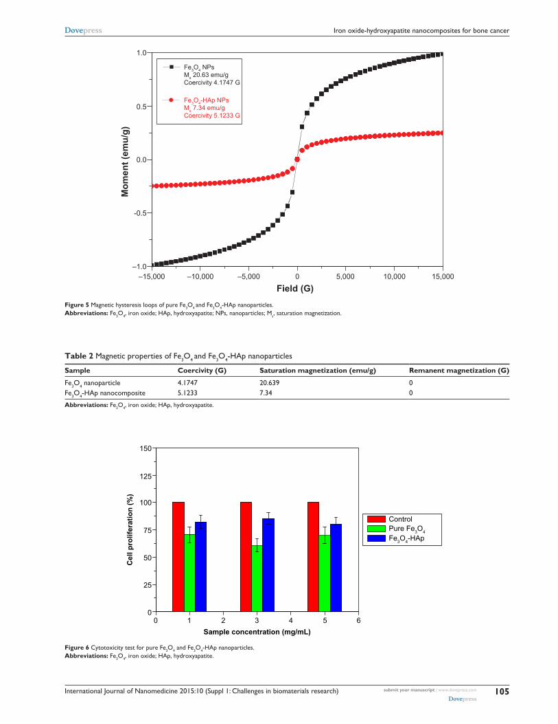
Vibrating sample magnetometryThe magnetic hysteresis loops for the Fe
3O
4 nanoparticles
and the Fe3O
4-HAp nanocomposites is shown in Figure 5.
These materials exhibited strong magnetic behavior, with
saturation magnetization of 20.639 and 7.34 emu/g and
coercivity of 4.1747 and 5.1233 G, respectively, at 300 K.
The magnetic properties are tabulated in Table 2. The
saturation magnetization of pure Fe3O
4 is lower than the
value reported in the literature (92 emu/g).13 The samples
show typical superparamagnetic behavior, with near zero
coercivity and remanent magnetization, whereas pure HAp
does not have any hysteresis loop. It is also clear from vibrat-
ing sample magnetometry that the magnetite particle in the
composite structure is well within the single domain particle
range and exhibits superparamagnetism. The magnetization
value of the composites was lower than that of the pure Fe3O
4
nanoparticles. The decreased saturation magnetization could
be due to interaction between the iron core and the HAp shell,
which decreases the magnetic moment.14,15
Cytotoxicity testingIn vitro cytotoxicity assays are the primary biocompatibil-
ity screening tests for a wide variety of materials used in
the medical field. The MTT assay was done for the Fe3O
4
and Fe3O
4-HAp nanoparticles using MG63 (osteosarcoma)
cells. Three concentrations (1, 3, and 5 mg/mL) of the Fe3O
4
nanoparticles and the Fe3O
4-HAp nanocomposites were
tested. The experiments were performed in triplicate. The
mean values obtained are shown in Figure 6. The MTT assay
procedure is explained in the flow chart given in Figure 2.
The results show that the Fe3O
4-HAp nanoparticles enabled
better cell proliferation than the pure Fe3O
4 at the concentra-
tions tested.
Magnetic induction hyperthermia is a technique that
can be used to destroy cancer cells via their hysteresis loss
International Journal of Nanomedicine 2015:10 (Suppl 1: Challenges in biomaterials research) submit your manuscript | www.dovepress.com
Dovepress
Dovepress
105
Iron oxide-hydroxyapatite nanocomposites for bone cancer
Table 2 Magnetic properties of Fe3O4 and Fe3O4-hap nanoparticles
Sample Coercivity (G) Saturation magnetization (emu/g) Remanent magnetization (G)
Fe3O4 nanoparticle 4.1747 20.639 0Fe3O4-hap nanocomposite 5.1233 7.34 0
Abbreviations: Fe3O4, iron oxide; hap, hydroxyapatite.
Figure 5 Magnetic hysteresis loops of pure Fe3O4 and Fe3O4-hap nanoparticles. Abbreviations: Fe3O4, iron oxide; hap, hydroxyapatite; NPs, nanoparticles; Ms, saturation magnetization.
Figure 6 Cytotoxicity test for pure Fe3O4 and Fe3O4-hap nanoparticles. Abbreviations: Fe3O4, iron oxide; hap, hydroxyapatite.
International Journal of Nanomedicine
Publish your work in this journal
Submit your manuscript here: http://www.dovepress.com/international-journal-of-nanomedicine-journal
The International Journal of Nanomedicine is an international, peer-reviewed journal focusing on the application of nanotechnology in diagnostics, therapeutics, and drug delivery systems throughout the biomedical field. This journal is indexed on PubMed Central, MedLine, CAS, SciSearch®, Current Contents®/Clinical Medicine,
Journal Citation Reports/Science Edition, EMBase, Scopus and the Elsevier Bibliographic databases. The manuscript management system is completely online and includes a very quick and fair peer-review system, which is all easy to use. Visit http://www.dovepress.com/testimonials.php to read real quotes from published authors.
International Journal of Nanomedicine 2015:10 (Suppl 1: Challenges in biomaterials research)submit your manuscript | www.dovepress.com
Dovepress
Dovepress
Dovepress
106
Sneha and Sundaram
when placed in an alternating magnetic field. The prepared
nanocomposites have the capacity to generate heat in the
presence of an alternating magnetic field. The temperature
of the cancer cells is raised between 42°C and 46°C via the
heat produced by our prepared composite materials. Although
the heating efficiency of the prepared superparamagnetic
Fe3O
4-HAp nanocomposite is not demonstrated here, Hou
et al16 showed that a similar Fe3O
4-HAp nanocomposite was
capable of magnetically inducing effective thermal destruc-
tion of cancer cells in vivo.
ConclusionIn this work, pure Fe
3O
4 was prepared by alkaline copre-
cipitation and HAp nanoparticles were prepared using an
optimized sol-gel method. Fe3O
4-HAp (0.7 w/w) nanocom-
posites were developed by wet milling. The prepared Fe3O
4-
HAp nanoparticles and its composite were characterized
and confirmed by scanning electron microscopy, Fourier
transform infrared spectroscopy, and diffuse reflectance
spectroscopy. Their superparamagnetic nature was con-
firmed by vibrating sample magnetometry, and their ther-
mal stability was confirmed by thermogravimetric analysis
and differential scanning calorimetry. This nanostructured
Fe3O
4-HAp composite would be ideal for use in bone cancer
therapy.
AcknowledgmentsThe authors are grateful to their head of department, dean,
principal, and management for providing the facilities to
carry out this research. MS is grateful to TEQIP-II for provid-
ing a PhD research assistantship. This work was financially
supported by DST (SB/S2/CMP-106/2013), New Delhi, and
UGC (major, 42-907/2013SR), New Delhi.
DisclosureThe authors report no conflicts of interest in this work.
References 1. Jiang QL, Zheng SW, Hong RY, et al. Folic acid conjugated Fe
3O
4
magnetic nanoparticles for hyperthermia and MRI in vitro and in vivo. Appl Surf Sci. 2014;307:224–233.
2. Di Corato R, Espinosa A, Lartigue L, et al. Magnetic hyperthermia efficiency in the cellular environment for different nanoparticle designs. Biomaterials. 2014;35:6400–6411.
3. Pankhurst QA, Connolly J, Jones SK, Dobson J. Applications of magnetic nanoparticles in biomedicine. J Phys D Appl Phys. 2003;36: R167–R181.
4. Bear JC, Yu B, Blanco-Andujar C, et al. A low cost synthesis method for functionalised iron oxide nanoparticles for magnetic hyperthermia from readily available materials. Faraday Discuss. 2014;175:83–95.
5. Bonnemain B. Superparamagnetic agents in magnetic resonance imag-ing: physicochemical characteristics and clinical applications – a review. J Drug Target. 1998;6:167–174.
6. Singh RK, El-Fiqi AM, Patel KD, Kim HW. A novel preparation of magnetic hydroxyapatite nanotubes. Mater Lett. 2012;75:130–133.
7. Karunamoorthi R, Kumar GS, Prasad AI, Vatsa RK, Thamizhavel A, Girija EK. Fabrication of a novel biocompatible magnetic biomaterial with hyperthermia potential. J Am Ceram Soc. 2014;97:1115–1122.
8. Murakami S, Hosono T, Jeyadevan B, Kamitakahara M, Ioku K. Hydrothermal synthesis of magnetite/hydroxyapatite composite mate-rial for hyperthermia therapy for bone cancer. J Am Ceram Soc. 2008; 116:950–954.
9. Iwasaki T, Nakatsuka R, Murase K, Takata H, Nakamura H, Watano S. Simple and rapid synthesis of magnetite/hydroxyapatite composites for hyperthermia treatments via a mechanochemical route. Int J Mol Sci. 2013;14:9365–9378.
10. Donadel K, Felisberto MD, Laranjeira MC. Preparation and character-ization of hydroxyapatite-coated iron oxide particles by spray-drying technique. An Acad Bras Cienc. 2009;81:179–186.
11. Tampieri A, D’Alessandro T, Sandri M, et al. Intrinsic magnetism and hyperthermia in bioactive Fe-doped hydroxyapatite. Acta Biomater. 2012;8:843–851.
12. Gupta AK, Gupta M. Synthesis and surface engineering of iron oxide nanoparticles for biomedical applications. Biomaterials. 2005;26: 3995–4021.
13. Bretcanu O, Spriano S, Brovarone C, Verne E. Synthesis and char-acterization of coprecipitation-derived ferromagnetic glass-ceramic. J Mater Sci. 2006;41:1029–1037.
14. Cheng J, Ni X, Zheng H, Li B, Zhang X, Zhan D. Preparation of Fe (core)/SiO
2 (shell) composite particles with improved oxidation resis-
tance. Mater Res Bull. 2006;41:1424–1429. 15. Ramanujan RV, Yeow YY. Synthesis and characterization of polymer-
coated metallic magnetic materials. Mater Sci Eng C. 2005;25:39–41. 16. Hou CH, Hou SM, Hsueh YS, Lin J, Wu HC, Lin FH. The in vivo
performance of biomagnetic hydroxyapatite nanoparticles in cancer hyperthermia therapy. Biomaterials. 2009;30:3956–3960.