Embed Size (px)
Citation preview

ORTHODONTICS LEGEND
Major Topic Abbreviation
Appliances Appl
Cephalometries Ceph
Crossbites & Open bites Cb & Ob
General Information Gen Info
Malocclusion MalOccl
Miscellaneous Misc.
Space Maintainers Space Mnt
Copyright © 2001 - DENTAL DECKS
ORTHODONTICSAppl
Which of the following is the best method for tipping maxillary and mandibular anterior teeth?
• Finger springs• Z-springs• Both are equally efficient
Copyright © 2001 - DENTAL DECKS

• Finger springs
These finger springs are attached to a removable appliance. The most commonproblems associated with these simple removable appliances are lack of patient cooperation, poor design leading to lack of retention, and improper activation . An undesirable common side effect of a finger spring is the tendency for the root apex to move inthe direction opposite from the crown.
Z-springs can also be used but they deliver excessively heavy forces and lack rangeof motion.
.M~iII!ry incisor rotation is not commonly treated during the stage of mixed dentition. It IS best treated after all permanent teeth have erupted (early permanent dentition). This is usually accomplished with a simple removable appliance. H oweyer, if the
inCIS or is in crossbite. it should be corrected as soon as possible (while it is erupting).
Note: When using buccal coil springs to try and regain space by pushing a toothmesially or distally, be careful because rotation of that tooth instead of actual movement commonly occurs.

ORTHODONTICSAppl
Which of the following types of headgear produces a distal and upward force on themaxillary teeth and maxilla?
• Straight-pull headgear• Reverse-pull headgear
Cervical-pull headgear• High-pull headgear
Copyright © 2001 - DENTAL DECKS

• High-pull headgear
High- ull headgear consists of a head cap connected to a face-bow. This appliance places adistal and upward forc~.on the maxillary teeth and maxilla. These types of headgear have a moredirect effect on the anterior segment of the arch. Indications: Class II, Division I malocclusion -t!!lat have anropen bitK
Cervical-Pull head ear is made up of a neck strap connected to a face bow. This applianceproduces a distal and downward force against the maxillary teeth and the maxilla. A major disadvantage of treatment using cervical headgear is possible extrusion of the maxillary molarsLikely results include: opening the bite , f irst molars will move distally and forward growthof the maxilla will decrease. Indications: Class II, Division I malocclusions. ~
Straigh t-pu ll headgear is similar to the cervical-pull headgear. However, this appliance placesa force in strai ht distal direction form the maxillary molar. Like cervical-pull headgear, the indications are Class , ivision I malocclusion (when bite opening is undesirable).
Reverse-pull headgear unlike all the other headgears above, the extraoral component is supported by the chin , cheeks, forehead or a combination of these structures. Indications:Class III malocclusions (where protraction of the maxilla is desirable).
ORTHODONTICSAppl
Which of the following is usually used as an etching agent before direct bonding oforthodontic bracket?
• 10-15 % unbuffered phosphoric acid• 35·50 % unbuffered phosphoric acid• 75-85 % unbuffered phosphoric acid• 100 % unbuffered phosphoric acid
Copy right © 2001 - DENTAL DECKS

• 35-50 % unbuffered phosphoric acid
"""For 1 minute
Remember: After etch ing , the tooth surface should have a frosted appearance.
The tooth surface must not be contaminated with saliva, which promotes immediate remineralization, until bonding is completed; otherwise re-etching is required.Topical fluoride should not be used before etching because fluoride decreasesthe solubility of enamel.
Indicat ions for using bands instead of bonded brackets:• To provide better anchorage for greater tooth movement• For teeth that will need both lingual and labial attachment• Teeth with short clinical crowns• Tooth surfaces that are incompatible with successful bonding
Cementation of bands: Generally, a heavier mix is required with a longer setting timeto permit seating and adaptation of the band. Zinc phosphate cements are routinelymixed over a large area on a cold mixing slab to extend the working and setting time.
ORTHODONTICS
Orthodont ic appliances can:
• Cause irritation to the gingiva• Act as plaque harbors• Make proper oral hygiene difficult to perform• All of the above
Copyright © 2001 - DENTAL DECKS
Appl
• All of the above
Orthodontic appliances may be irritating or interfere with the performance of goodoral hygiene. Prolonged orthodontic treatment has long been associated with the causation of inflammatory periodontal diseases. However, if meticulous oral hygiene ismaintained at all times during orthodontic treatment, the periodontal health can bemaintained.
Note: When a patient (young or old) is in active orthodontic treatment and the gingivais inflamed, the dentist should encourage better oral hygiene. It may be useful to recommend the use of water irrigation devices to help flush food debris away from thebrackets.
ORTHODONTICSAppl
Which of the following determines the effect of a face-bow headgear on the molarsto which it is attached?
• The relation of the outer bow to the center of resistance of the tooth• The direction of pull of the headgear strap• The length and position of the outer bow in relation to the inner bow• All of the above
Copyright © 2001 - DENTAL DECKS

• All of the above
Remember:1. There are four basic types of headgear (cervical-pull, straight-pull, high-pull and
reverse-pull). Each type has two major components: intraoral (face-bow) and extraoral (neck strap, head cap, chin cup, etc.). The extraoral component is what generally categorizes the type of headgear.
2. One of the greatest advantages of using extraoral anchorage (i.e., headgear) isthat it permits posterior movement of teeth in one arch without adversely disturbingthe opposite arch.
3. Headgear is often employed for the maintenance of anchorage. Anchorage is theresistance to unwanted tooth movement.
ORTHODONTICSAppl
Which of the following is the most common removable retainer used in orthodontics?
• Tooth positioner• Hawley retainer• Functional retainer• Anter ior spring retainer
Copyright © 2001 - DENTAL DECKS
• Hawley retainer
It incorporates clasps on molar teeth and a characteristic outer bow with adjustment loops, spanning from canine to canine.
The palatal coverage of a removable plate like a Hawley retainer makes it possible to incorporate a bite plane lingual to the upper incisors to control the bite depth. This design consideration is important for any patient who once had an excessive overbite. This palatal coverage(acrylic) is the major source of anchorage in the Hawley appliance.
A Hawley retainer can be made for the upper or lower arch. The lower retainer is somewhatfragile and may be difficult to insert because of undercuts in the premolar region. A patient mayhave difficulty pronouncing linguoalveolar consonants for a few days after receiving a maxillary Hawley appliance until the tongue adapts to the palatal coverage.
Any removable appliance has three major components:1. Retentive com mrumt: consisting of various clasps to hold it in place2. A framework or base late: usually acrylic. This provides anchorage.3. To.ath-movi ng EllimJents.: typically either springs or screws
Indications for removable appliances:• Retention after comprehensive treatment• Limited tipping movements• Growth modification during the mixed dentition

ORTHODONTICSAppl
Which of the following alloys are used for archwires in orthodontics?
• Stainless steel• Chromium-cobalt• Titanium• All of the above
Copyright © 2001 - DENTAL DECKS
• All of the above
The properties of stainless steel wires used for archwires can be controlled over awide range by varying the amount of cold working annealing during manufacturi ng.Steel is softened by annealing and hardened by cold working (work hardening).
Chromium-cobalt alloys have the advantage that they can be supplied in a softerand therefore more formable state , and that they can be hardened by heat treatmentafter being shaped. The heat treatment increases strength significantly.
Titanium alloys offer a highly desirable combinat ion of strength and springiness andreasonably good formabil ity.
The properties of an ideal wire material for orthodontic purposes can bedescribed largely in terms of the following criteria. It should possess : 1) high strength,2) low stiffness, 3) high range and 4) high formability. In addition, the material shouldbe weldable or solderab le, so that hooks or stops can be attached to the wire. 1.QQJ1s..and helices are incorporated into archwire to increase the activation range.
The stiffness of orthodontic wires is a function of the length of the wire, the diameter of the wire and alloy compos ition.

ORTHODONTICS
All of the following are fixed orthodont ic appliances except
• Lingual archwire• Fixed space maintainers• Palate-separating devices• Lip bumpers• Edgewise mechanisms• Light-wire appliances
Copyright © 2001 - DENTAL DECKS
. Appl
• Lip bumpers
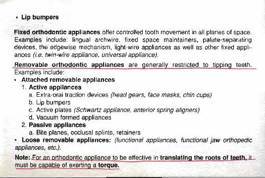
Fixed orthodontic appliances offer controlled tooth movement in all planes of space.Examples include: lingual archwire, fixed space maintainers, palate-separatingdevices, the edgewise mechanism, light-wire appliances as well as other fixed appliances (i.e. twin-wire appliance, universal appliance).
Removable orthodontic appliances are generally restricted to tipping teeth.Examples include:• Attached removable appliances
1. Active appliancesa. Extra-oral traction devices (head gears, face masks, chin cups)b. Lip bumpersc. Active plates (Schwartz appliance, anterior spring aligners)d. Vacuum formed appliances
2. Passive appliancesa. Bite planes, occlusal splints, retainers
• Loose removable appliances: (functional appliances, functional jaw orthopedicappliances, etc.).
Note: .For an orthodontic appliance to be effective in translating the roots of teeth, itmust be capable of exerting a torque.

ORTHODONTICSAppl
A seven-year-old child with an otherwise good occlusion has a lingually locked maxillary permanent central incisor. There is sufficient room for the tooth. In order to treatthis condition properly, the dentist should do what?
• Wait until all permanent teeth have erupted• Surgically reposit ion the central incisor• Correct the condition immediately with a simple appliance• None of the above
Copyright © 2001 - DENTAL DECKS

• Correct the condition immediately with a simple appliance
Ideally, this anterior crossbite should have been corrected befo.r~ It reached theocclusal plane (while it was erupting). The most probable etiologic factor for this happening is prolonged retention of the primary maxillary incisors.
Cross-elastics from the maxillary lingual to the mandibular labial can be used to correct a single-tooth crossbite. A maxillary removable appliance can also be used. Whenelastics are used to move teeth they should be attached directly to the appliancecomponents.
Anterior crossbite, particularly crossbite of the incisors, is rarely found in childrenwho do not have a skeletal Class III jaw relationship. A crossbite relationship of one ortwo anterior teeth, however, may develop in a child who has good facial proportions.The maxillary lateral incisors tend to erupt to the lingual and may become trapped inthat location, especially in the presence of severe crowding. In this situation, extractingthe adjacent primary canines usually leads to spontaneous correction of the crossbite.It is important to evaluate the space situation before attempting to correct any anteriorcrossbite. If enough space is available to accomplish the movement, a maxillaryremovable appliance is usually the best mechanism to correct a simple anterior crossbite that requires a tipping movement.
ORTHODONTICSAppl
Which appliance listed below is probably the most widely used today by orthodontists?
• The universal appliance• The edgewise appliance• Neither of the above
Copyright © 2001 - DENTAL DECKS

• The edgewise appliance
"", 1"trJ
In its essential form, mechanism consists of bands on all of the teeth, tubes on the lastmolar and brackets on all other teeth. One labial arch is used at a time. The ultimatelabial arch wire is .01 25 x .028 in diameter and the narrow dimension or edge fits precisely into the bracket slot, which is .:022 inches wide from top to bottom. It finds itsgreatest application in the treatment of comprehensive malocclusions of theadolescent permanent dentitions.
Variations of the basic edgewise appliance include the use of double or tandembrackets (Siamese twin brackets) and narrow slotted brackets, .018 from top to bottom.A straight wire appliance is a version of the edgewise appliance With several featuresthat allow placement of an ideal rectangular archwire without bends.
Components of an edgewise appliance:• Siamese twin bracket: for use on maxillary anterior teeth• Broussard buccal tube: to allow the use of the segmented arch technique, which
is necessary to intrude teeth• Straight wire bracket• A bracket with a .0222 x .028 rectangular slot
Note: A first order bend in an orthodontic wire is in the horizontal plane

ORTHODONTICS
A steep mand ibu lar plane angle correlates with a:
Ceph
• Short anterior facial vertical dimensions and anterior open bite malocclusion• Long anterior facial vertical dimensions and anterior open bite malocclusion• Short anterior facial vertical dimensions and anterior deep bite malocclusion• Long anterior facial vertical dimensions and anterior deep bite malocclusion
Copyright © 2001 - DENTAL DECKS
• Long anterior facial vertical dimensions and anterior open bite malocclusion
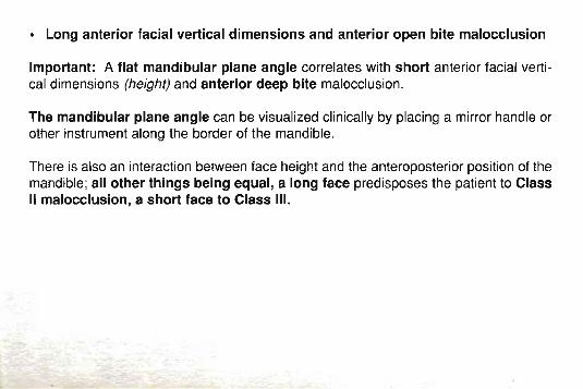
Important: A flat mandibular plane angle correlates with short anterior facial vertical dimensions (height) and anterior deep bite malocc lusion .
The mandibular plane angle can be visualized clinically by placing a mirror handle orother instrument along the border of the mandible.
There is also an interaction between face height and the anteroposterior position of themandible ; all other things being equal, a long face predisposes the patient to ClassII malocclusion, a short face to Class III.

ORTHODONTICSCeph
Which of the following would be indicative of maxillary retrognathism ?
• An SNA angle of approximately 82°• An SNA angle greater than 82°• An SNA angle less than 82°
Copyright © 2001 - DENTAL DECKS
• An SNA angle less than 82°
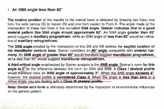
The relative position of the maxilla to the cranial base is obtained by drawing two lines; onefrom the sella turcica (S) to nasion (N) and one from nasion to Point A. The angle made at theintersection of these two lines is the so-called SNA angle. Steiner indicates that in a goodskeletal pattern this SNA angle should approximate 82°. An SNA angle greater than 82°would suggest a maxillary prognathism, while an SNA angle of less than 82° would be indicative of maxillary retrognathism.
The SNB angle created by the intersection of line SN and NB defines the sagittal location ofthe mandibular denture base. Steiner considers an J!Q0 angle compatible with skeletal harmony. An SNB angle of greater than 80° would therefore suggest mandibular prognathism,while less than 800 would suggest mandibular retrognathism.
A third critical angle emphasized by Steiner analysis is the ANB angle. Steiner's norm for thisangle i~r the difference between the norm for SNA and SNB. A Class I skeletal profilewould therefore have an ANB angle of approximately 2°. When the ANB angle exceeds 4°,~owever, the skeletal profile is considered Class II. When the angle is less than zero or aminus angle, you are dealing with a Class III skeletal-profile.
Note: Dental arch form is ultimately determined by the interaction of environmental influenceson the genetic pattern.

ORTHODONTICSCeph
Identify and define the following cephalometric landmarks labeled 1-15.
Copyright © 2001 - DENTAL DECKS
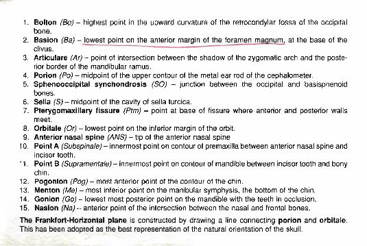
1. Bolton (80) - highest point in the upward curvature of the retrocondylar fossa of the occipitalbone.
2. Basion (8a) - lowest point on the anterior margin of the foramen magnum, at the base of theclivus.
3. Articulare (Ar) - point of intersection between the shadow of the zygomatic arch and the posterior border of the mandibular ramus.
4. Porion (Po) - midpoint of the upper contour of the metal ear rod of the cephalometer.5. Sphenooccipital synchondrosis (SO) - junction between the occipital and basisphenoid
bones.6. Sella (5) - midpoint of the cavity of sella turcica.7. Pterygomaxillary fissure (Ptm} - point at base of fissure where anterior and posterior walls
meet.8. Orbitale (Or) - lowest point on the inferior margin of the orbit.9. Anterior nasal spine (ANS) - tip of the anterior nasal spine
10. Point A (Subspinale) - innermost point on contour of premaxilla between anterior nasal spine andincisor tooth.
11. Point B (Supramentale) - innermost point on contour of mandible between incisor tooth and bonychin.
12. Pogonion (Pog) - most anterior point of the contour of the chin.13. Menton (Me) - most inferior point on the manibular symphysis, the bottom of the chin.14. Gonion (Go) - lowest most posterior point on the mandible with the teeth in occlusion.15. Nasion (Na) - anterior point of the intersection between the nasal and frontal bones.
The Frankfort-Horizontal plane is constructed by drawing a line connecting porion and orbitale.This has been adopted as the best representation of the natural orientation of the skull.

ORTHODONTICSCeph
Which of the following are uses for cephalometries in orthodontics?
• Diagnosis• Analysis of treatment results• Longitudinal study of growth• All of the above
Copyright © 2001 - DENTAL DECKS

• All of the above
The lateral head radiograph (cephalometric x-ray) must be compared with the "normal" lateral radiographs from an accepted norm. Linear and angular measurementsare obtained utilizing known anatomical landmarks in the lateral head radiography ofthe patient. These measurements are then compared with those considered within normal limits and in that way enable the orthodontist to assess aberration in the dentitionand jaw structures, which result in malocclusion.
Analysis of cephalometric radiographs is not limited to the hard structures such asbone and teeth, but also includes measurements of soft tissue structures such as thenose, lips and soft tissue chin.
Superimposition in longitudinal cephalometric studies is generally on a referenceplane and a registration point. This will best demonstrate the growth of structures furthest from the plane and the point. The most stable area from which to evaluatecraniofacial growth is the anterior cranial base because of its early cessation ofgrowth.
Cephalometries is useful in assessing tooth-to-tooth, bone-to-bone and tooth-to-bonerelationships. Serial cephalometric films can show the amount and direction of growth.

Cb &ObORTHODONTICS
An anterior crossbite in the primary dent ition is often indicative of which two of the following?
• Impacted permanent maxillary canines• A skeletal growth problem• A developing Class II malocclus ion• A developing Class III malocclusion
Copyright © 2001 - DENTAL DECKS
• A skeletal growth problem· A developing Class III malocclusion
It can be the result of:• A labially situated supernumerary tooth• Traumatic injury• Arch length discrepancy
Anterior crossbite of one or more of the permanent incisors , however, may be evidence of a localized discrepancy and a condition that almost always shou ld be treatedin the mixed dentition state or as soon as it's discovered. It is most often associatedwith prolonged retention of a primary tooth. Delayed treatment can lead to serious complications, such as loss of arch length. The most essential factor related to correctionof anterior crossbite is the space available mesiodistally. It is easily retained once itis corrected.
Note: The premature exfoliation of a primary canine may indicate an arch lengthdeficiency. The premature loss of a primary mandibular canine may cause a lingual collapse of the mandibular anterior teeth .

Cb & ObORTHODONTICS
Displaced teeth related to functional shifts are usually seen in which two of the following circumstances?
• Posterior crossbite after prolonged thumb sucking• Class II, Division I malocclusion• Anterior crossbite in mildly prognathic children• An anterior open bite after prolonged thumb sucking
Copyright © 2001 - DENTAL DECKS
• Posterior crossbite after prolonged thumb sucking• Anterior crossbite In mildly prognathic children
Prolonged sucking habits often produce a mildly narrow maxillary arch and a tendency toward bilateral crossbite. Children with this condition usually shift the mandibleto one side on closure to ain better function, which can guide permanent molars, ora er, premolars into a crossbite relationship.
A young child who has a tendency toward Class III malocclusion will have end-toend contact of the primary incisors. A true e.rioLcrossbite in the p-rlma dentition Is quite.rare beesllsamandibuJar-growth-lagS-behincLmaxillarY-9rowtb.Jhe primary incisors wear down rapidly, and an anterior shift of the mandible to escapeocclusal interferences rarely occurs until the permanent incisors begin to erupt. A pattern of anterior displacement of the mandible may develop when the permanent incisors come into contact , however, producing an anterior crossbite from the shift.
Notes:1. An anatomic crossbite (skeletal) , as contrasted with a functional crossbite (from
thumb sucking), usually demonstrates a smooth closure to centric occlusion.2. A corrected anterior crossbite is best retained by the normal incisor relationship that
is achieved through treatment (the overbite), not appliances.
Cb & DbORTHODONTICS
Which of the following are true concerning a posterior crossbite in the mixed dentition?
• It should be corrected as soon as possible• It should be thoroughly diagnosed as to whether it is of a dental, functional or
skeletal or igin• May be corrected with palatal expansion• May be associated with a mandibular shaft• All of the above are true
Copyright © 2001 - DENTAL DECKS

• All of the above are true
It is important to correct posterior crossbites (which are related to the transverseplane of space) and mild anterior crossb ites in the first stage of treatment, even if permanent first molars have not yet erupted. Severe anterior crossbites, in contrast, areusually not corrected until the second stage of convent ional treatment.
The most common type of active tooth movement in the primary dentition is to correcta posterior crossbite (transverse problem).
Remember: A skeletal crossbite, as contrasted with a functional crossbite, usually demonstrates a smooth closure to centric occlusion.
After palatal expansion, the following are observed:• Diastema formation between central incisors• Expansion of the nasal floor
Note: Tooth movement and skeletal expansion are inevitable when the midpalatalsuture is widened.
ORTHODONTICS
Which of the following can cause an open bite?
• Tongue-thrusting• Thumb-sucking• Genetics• Speech impediments (i.e. lisping)• All of the above
Copyr ight © 2001 - DENTAL DECKS
Cb&Ob
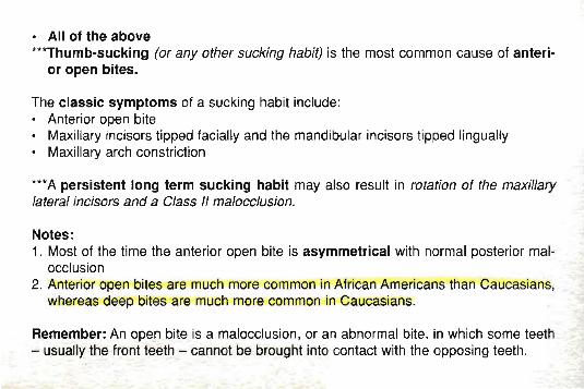
• All of the above"'Thumb-sucking (or any other sucking habit) is the most common cause of anteri
or open bites.
The classic symptoms of a sucking habit include:• Anterior open bite• Maxillary incisors tipped facially and the mandibular incisors tipped lingually• Maxillary arch constriction
" ' A persistent long term sucking habit may also result in rotation of the maxillarylateral incisors and a Class /I malocclusion .
Notes:1. Most of the time the anterior open bite is asymmetrical with normal posterior mal
occlusion2. Anterior open bites are much more common in African Americans than Caucasians,
whereas deep bites are much more common in Caucasians.
Remember: An open bite is a malocclusion, or an abnormal bite, in which some teeth- usually the front teeth - cannot be brought into contact with the opposing teeth.

Cb &ObORTHODONTICS
There are many reasons why crossbites occur. Which one of the following is not oneof them?
• Jaw size• Heredity• Faulty restorations• Mouth breathing• Prolonged retention of primary teeth
Copyright © 2001 ~ DENTAL DECKS

• Faulty restorations
A crossbite occurs when some of the teeth wind up on the "wrong side of the track".A crossbite can be unilateral (on one side) or bilateral (on both sides). It also can occuranteriorly or posteriorly.
Orthodontic treatment tOSQrrect a cm.ssbiteinchildren should begin sari as . ossible. The first ste "maxilla ex ansion" broadens the maxilla with an appliancecaifed an "expander". Fixed to the roof of the mouth, .the expander is widened eachnight for about 1 to 2 months with the turn of a key. The expander remains in the mouthfor about 3 more months to allow the bone to harden in its new position.
Note: Braces may be put on the maxillary teeth while expansion is going on to eventually close the "gap-tooth grin" that will develop as the maxilla is being expanded.Once the expansion is complete, the child may need to wear a f~II set of braces for 1to 2 years to achieve an ideal occlusion.
Important: The main reason to correct a crossbite in children is to prevent TMJ disorders.
MalOcclORTHODONTICS
Which type of malocclusion listed below is most often associated with mouth breathing?
• Dental open bite• Skeletal open bite• Dental cross bite• Skeletal cross bite
Copyright © 2001 - DENTAL DECKS

• Skeletal open bite (sometimes called the "Long Face Syndrome'?
The following factors are associated with chronic mouth breathing:Narrow faceNarrow oropharyngeal spaceChronic rhinitis - inflammation of the mucous membranes of the noseChronic tonsillitisAllergiesDeviated nasal septum
Note: The earliest possible diagnosis of this open bite is essential because the condition is not self-correcting and usually worsens with time. Anterior open bites canbe classified as a form of apertognathism (which means open bite deformity).

ORTHODONTICS
Class III malocclusion is also referred to as:
• Retrognathism or overbite• Prognathism or underbite• Neither of the above
Copyright © 2001 - DENTAL DECKS
MalOccl

• Prognathism or underbite
****It is the least common (compared to Class I and II). It occurs when the mandibleprotrudes forward and the mandibular teeth extend over the maxillary teeth.
Class III malocclusions are those in which the body of the mandible and its superimposed dental arch are in a mesial relationship to the skull base and maxilla. The maxillary first molar therefore occludes distal to the mandibular first molar, while the maxillary canine is an exaggerated distal relationship to the mandibular canine. Themandibular incisors are forward to the maxillary incisors. Also characteristic of the"true" Class III malocclusion is the prognathic mandible. Class III subd ivision is aClass III relationshi e.leeth-on-one-side-witR-a-etass I relationship on the other
~
A pseudo-class III malocclusion is one in which the mandibular incisors are forwardof the maxillary incisors when in centric occlusion, however, the patient has the ability to bring the mandible back without strain so that the mandibular Incisors cantouch the maxillary incisors (this ability is often considered diagnostic). This type istherefore a milder form of the "true" Class III malocclusion and more amenable toconservative orthodontic movement than the "true" Class III malocclusion which oftenrequires surgical correction.

MalOcclORTHODONTICS
A severe malocclusion may compromise which of the following aspects of oral function?
• Mastication• Swallowing• Speech• All of the above
Copyright © 2001 - DENTAL DECKS
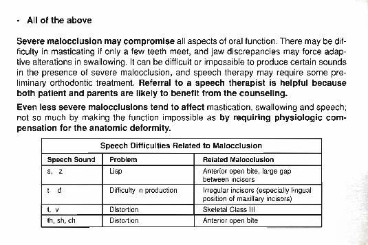
• All of the above
Severe malocclusion may compromise all aspects of oral function. There may be difficulty in masticating if only a few teeth meet, and jaw discrepancies may force adaptive alterations in swallowing. It can be difficult or impossible to produce certain soundsin the presence of severe malocclusion, and speech therapy may require some preliminary orthodontic treatment. Referral to a speech therapist is helpful becauseboth patient and parents are likely to benefit from the counseling.
Even less severe malocclusions tend to affect mastication, swallowing and speech;not so much by making the function impossible as by requiring physiologic compensation for the anatomic deformity.
Speech Difficulties Related to Malocclusion
Speech Sound Problem Related Malocclusion
s, z Lisp Anterior open bite, large gapbetween incisors
t, d Difficulty in production Irregular incisors (especially lingualposition of maxillary incisors)
f, v Distort ion Skeletal Class III
th, sh, ch Distort ion Ante rior open bite

ORTHODONTICS
Class I malocclusions share what basic characteristic?
• The midface is concave• A harmonious skeletal profi le• The midface is convex• None of the above
Copyright © 2001 - DENTAL DECKS
MalOccl
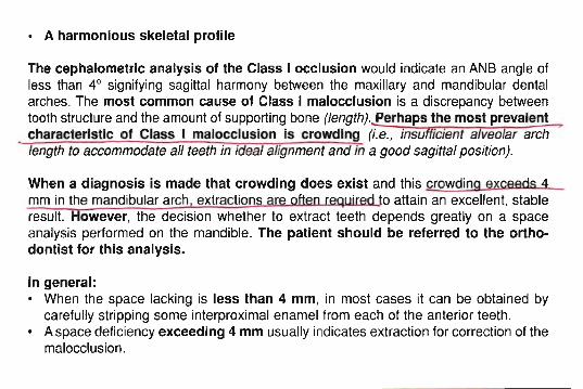
• A harmonious skeletal profile
The cephalometric analysis of the Class I occlusion would indicate an ANB angle ofless than 4° signifying sagittal harmony between the maxillary and mandibular dentalarches. The most common cause of Class I malocclusion is a discrepancy betweentooth structureand the amountof supporting bone (length).Y erhaps the most prevalent .....,.characteristic of Class I malocclusion is crowding (i.e., insufficient alveolar archlength to accommodate all teeth in ideal alignment and in a good sagittal position).
When a diagnosis is made that crowding does exist and this crowding exceeds 4mm in the mandibular arch extractions ar . to attain an excellent, stableresult. owever, the decision whether to extract teeth depends greatly on a spaceanalysis performed on the mandible. The patient should be referred to the orthodontist for this analysis.
In general:• When the space lacking is less than 4 mm , in most cases it can be obtained by
carefully stripping some interproximal enamel from each of the anterior teeth. ,• A space deficiency exceeding 4 mm usually indicates extraction for correction of the
malocclusion. .
ORTHODONTICS
Class II malocclusion is also referred to as:
• Retrognathism or overbite• Prognathism or underbite• Neither of the above
Copyright © 2001 - DENTAL DECKS
MalOccl
• Retrognathism or overbite
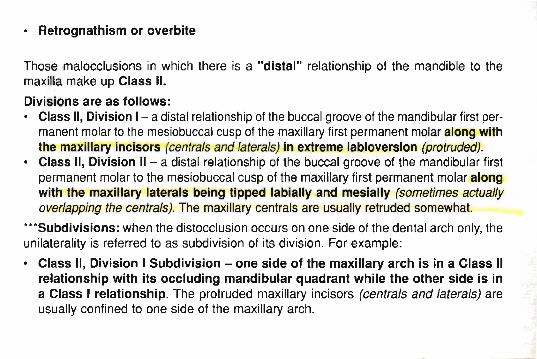
Those malocclusions in which there is a "distal" relationship of the mandible to themaxilla make up Class II.
Divisions are as fo llows:• Class II, Division 1- a distal relationship of the buccalgroove of the mandibularfirst per
manentmolar to the mesiobuccalcusp of the maxillaryfirst permanent molar along withthe maxilla ry incisors (centrals and laterals) in extreme labioversion (protruded).
• Class II, Div ision II - a distal relationship of the buccal groove of the mandibular firstpermanent molar to the mesiobuccal cusp of the maxillary first permanent molar alongwith the maxillary laterals being ti pped labially and mesially (sometimes actuallyoverlapping the centrals). The maxillary centrals are usually retruded somewhat.
***Subdlvislons: when the distocclusion occurs on one side of the dental arch only, theunilaterality is referred to as subdivision of its division. For example:
• Class II, Div is ion I Subdivision - one side of the maxillary arch is in a Class IIrelationship with Its occluding mandibular quadrant while the other side is ina Class I relationship. The protruded maxillary incisors (centrals and laterals) areusually confined to one side of the maxillary arch.

MalOcclORTHODONTICS
Which cusp listed below of the maxillary first permanent molar serves as a referencepoint in identifying Angle's Class I, II and III occlus ions?
• Distobuccal• Mesiobuccal• Mesiolingual• Distolingua l
Copyright © 2001 - DENTAL DECKS

• Mesiobucca l
Class ification of human occlusion (Angle 's):• Class I - most common (about 70 % of the population). The mesiobuccal cusp of the max
illary first molar lines up approximately with the buccal groove of the mandibular first molar.The maxillary central incisors overlap the mandibulars. Maxillary canine lies between themandibular canine and first premolar.
• Class II - less common (about 25 %). The mesiobuccal cusp of the maxillary first molar fallsapproximately between the mandibular first molar and the second premolar. The lower jawand chin may also appear small and withdrawn . The mandibular incisors occlude even moreposterior to the maxillary incisors so they may not touch at all. Maxillary canine is mesial tomandibular canine.
• Class III - the least common (less than 5 %). The mesiobuccal cusp of the maxillary firstmolar falls approximately between the mandibular first molar and second molar. The chin mayalso protrude like a bUlldog's does. The mandibular incisors overlap anterior to the maxillary incisors. The maxillary canine is distal to mandibular canine.
Planes of space used to classify malocclusion :• Antero-posterior• Transverse• Vertical

Mal DcclORTHODONTICS
The existence of a forward shift of the mandible during closure is found in:
• "True" Class III malocclusions• "Pseudo" Class III malocclusions
Copyright © 2001 - DENTAL DECKS

• "Pseudo" Class III malocclusions
Remember: Pseudo-Class III malocclusion is therefore a milder form of the "t rue"Class III malocclusion and more amenable to conservative orthodontic movement thanthe "true" Class III malocclusion which often requires surgical correction.
Notes :• Hypertrophy of the mandibular condyle can result in a unilateral Class III mal
occ lus ion as well as an anterior cross bite and an ipsilateral posterior open bite.• For a patient that is a skeletal Class III, the SNB angle will increase as the patient
gets olde r.
ORTHODONTICS
Malocclusion is most often:
• Acquired from a friend• Hereditary• Caused by antibiotics• Caused by bad habits
Copyright © 2001 - DENTAL DECKS
MalOccl

• Hereditary
There may be a disproportion between the size of the maxilla and mandible or between the jaws andtooth size resulting in overcrowding of teeth or in abnormal bite patterns. Supernumerary teeth, malformed teeth, impacted or lost teeth and teeth that erupt in an abnormal direction may contribute tomalocclusion. Less frequent causes of malocclusion include habits such as thumb sucking or tonguethrusting.
Signs of incipient malocclusion include:• I he lack of interdental spacing in the primary dentition• The crowding of the permanent incisors in the mixed dentition• The premature loss of the primary canines, particularly in the mandibular arch
Notes :1. The sign ificance of the lack of spac ing relates to the increased mesio-distal width of the per
manent teeth2. Since arch per imeter increases after the eruption of the incisors and is small in the maxilla and
essentially non-existent in the mandible, arch growth can not usually contribute to further dentalalignment
3. Th emature loss of the mandibular rima canine eflects insufficient.arch..size in.the ante;.rim region. As such, the crowns of the lateral incisors, during eruption, impinge on the roots of theprimary canines causing them to resorb. Wher1JlllLc.mJin..ejl sb~d lJ.e.J11idline-will -shift in the
_dim ' of the lost tooth. You will have lateral and lingual migration of the mandibular incisors.4. Remember : An anterior crossbite in a primary dentition usually indicates a skeletal growth
problem.
MalOcclORTHODONTICS
Which of the following are common characteristics of a Class II, Division II malocclusion?
• The maxillary centrals are near normal anteroposteriorly or slightly in linguoversion• The maxillary lateral incisors are usually labiomesially flared and overlap the central
incisors• Impinging overbite• All of the above
Copyright © 2001 - DENTAL DECKS
• All of the above
Class II, Division II is a malocclusion in which the body of the mandible and its superimposed dental arch are also in distal realationship to the maxilla, and the molar andcanine occlusion are the same as Class II, Division I type. The distobuccal cusp ofthe maxillary first molar occludes in the buccal developmental groove of the mandibular first molar, and the maxillary canines occlude mesial to the mandibular canines. Thebig difference between Division I and Division II is in Division II the maxillary laterals have tipped labially and mesially.
Remember: Class II, Division I = maxillary incisors (centrals and laterals) are inextreme labioversion.
Notes :• There is no set rule as to when a malocclusion should be treated. The age at
which it is treated depends on the problem involved.• Malocclusions are more identifiable in children 7 to 9 years old because the
eruption of permanent incisors reveals tooth-arch length discrepancies.

MalOcclORTHODONTICS
In most Class II, Division I malocclusions, the body of the mandible and its superimposed dental arch are in a:
• Mesial relationship to the maxilla and the maxillary incisors are usually in a labialaxial inclination
• Distal relationship to the maxilla and the maxillary incisors are usually in a lingualaxial inclination
• Mesial relationship to the maxilla and the maxillary incisors are usually in a lingualaxial inclination
• Distal relationship to the maxilla and the maxillary incisors are usually in a labialaxial inclination
Copyright © 2001 - DENTAL DECKS

• Distal relationship to the maxilla and the maxillary incisors are usually in alabial axial inclination
In addition, the relationship of the maxillary first molars and canines to the mandibularfirst molars are such that the distobuccal cusp of the maxillary first molar occludes inthe buccal developmental groove of the mandibular first molar and the maxillarycanines occlude mesial to the mandibular canines. Besides the labial axial inclination of the maxillary incisors (overjet), various aberrations in the individual alignment ofthe teeth (for example , crowding) can be superimposed upon this class.
Class II, Division I Subdivision includes malocclusions, which have one side of themaxillary arch in a Class II relationship with its occluding mandibular quadrant , whilethe other side is in a Class I relationship. The maxillary overjet or other anterior aberrations are usually confined to one side of the maxillary arch.
Note: Relative to a heterogeneous population, the incidence of malocclusion in ahomogeneous population generally is lower.

MalOcclORTHODONTICS
Which of the following facial profiles is usually accompan ied by a Class II malocclusion?
• An orthognathic profile• A retrognathic profile• A prognathic profile• None of the above
Copyright © 2001 - DENTAL DECKS

• A retrognathic profile
The convexity is due to the relative prominence of the maxilla compared to themandible. The mandibular incisors will most likely be tipped forward.
An orthognathic profile is one in which the nose, lips and chin are harmoniously related. This relationship is usually accompanied by a Class I dental occlusion.
A prognathic profile is one in which the mandible is markedly forward of the maxillagiving a concave midfacial appearance. This is often indicative of a Class III malocclusion. The maxillary incisors will most likely be tipped lingually.
Important: Abimaxilla ry dentoalveolar protrusion means that in both jaws the teeth~protrude.. In this condition, you will see severe dental and lip protrusion accompaniedby severe lip strain, which is needed to bring the lips into closure.
Note: As children mature their profiles become less convex

ORTHODONTICSSpace Mnt
A 13-year-old patient has all teeth present and normal occlusion; molar root apexes arenot totally closed. The mandibular left first molar has been extracted. The idealtreatment at this time is what?
• Place a fixed bridge• Place a space maintainer• Place a removable partial denture• Do nothing and observe
Copyright © 2001 - DENTAL DECKS

• Place a space maintainer
Notes:1. Premature loss !larJJ....sually-produces.a.CJassJI
ationshi on the affected side. A distal shoe s ace maintainer may helpalleviate this potential problem. This appliance extends backwards from a crown onthe primary first molar and subgingivally to the mesial line of the unerupted first permanent molar, thus preventing mesial migration.
2. With the "lingual arch" space maintainer, the primary second molars or permanentfirst molars are banded. Typically, the "lingual arch" space maintainer is comprisedof two bands which are cemented to the primary second molars or permanent firstmolars with a loop of wire that rests on the cingula of the incisors.
ORTHODONTICSSpace Mnt
Which space maintainer is mostoften usedwhen the primaryfirst molarneeds to beprematurely extracted?
• "Band and loop" space maintainer• "Distal shoe" space maintainer• "Lingual arch" appliance• "Nance" appliance
Copyright © 2001 - DENTAL DECKS
• "Band and loop" space maintainer
Space maintainers that are used to replace one prematurely missing primary tooth include:• The "band and loop" space maintainer is a fixed unilateral appliance. The band is usually
cemented to the second primary molar while a loop extends to the primary canine tooth. Note :This type of space maintainer must be restricted to holding the space of one tooth because ofit has limited strength.The "distal shoe" space maintainer is also a fixed unilateral appliance that is used when aprimary second molar is lost before the eruption of the permanent first molar (typically children under the age of 5 or 6).
Space maintainers that are used to replace multiple prematurely missing primary teeth include:The " lingual arch " which can be a fixed or removable appliance that is used to maintainspace when multiple primary teeth are missing and the permanent incisors have erupted.Note : Does not restore function and should be made completely passive.
• The "Nance" appliance or transpalatal appliance is used for bilateral loss of primary maxillarymolars. An acrylic button rests on the palate and the appliance prevents the mesial rotationand mesial drifting of the permanent maxillary molars to which it is attached.
• Part ial denture space maintainers are most useful for bilateral posterior space maintenancewhen the permanent incisors have not erupted. Note : Also used for missing anterior teethwhen esthetics is a concern.
Space MntORTHODONTICS
A child with a pulpally involved primary tooth comes into your office. Which of the following ideally is the best space maintainer for this child?
• A "band and loop" space maintainer• A "distal shoe" space maintainer• The pulpally involved primary tooth• A "lingual arch" space maintainer
Copyright © 2001 - DENTAL DECKS
• The pulpally involved primary tooth
The key point here is that no prefabricated space maintainer is as good as the natural tooth. Obviously in this case proper pulpal therapy followed by a restorative procedure would be needed on this tooth for it to function as a space maintainer.
The natural tooth will preserve arch length and integrity better than any prefabricated space maintainer.
If a primary tooth is lost, an orthodontic evaluation is indicated to determine whetheror not space maintenance is necessary. The decision is based on the patient's skeletaland dental development. For example , if a child is dental age 10 and loses the primaryfirst molar, no treatment is usually needed. Remember: The permanent first premolarusually erupts between 10-12 years old.

Gen InfoORTHODONTICS
The most rapid losses in the perimeter of the arch usually are due to a:
• Distal tipping and rotation of the permanent second molar after removal of the permanent third molar
• Mesial tipping and rotation of the permanent first molar after removal of the primarysecond molar
• Mesial tipping and rotation of the permanent canine after removal of the primary lateral incisor
• Distal tipping and rotation of the permanent second premolar after removal of thepermanent first molar
Copyright © 2001 - DENTAL DECKS

· Mesial tipping and rotation of the permanent first molar after removal of theprimary second molar
Very Important : When the primary second molar is lost, always maintain space untilthe arrival of the second premolar.
Notes:1. If a permanent first molar is extracted on a child before the eruption of the per
manent second molar, the best approach is to allow the eruption of the secondmolar and the mesial drifting to occur naturally. This will fill in the space most ofthe time.
2. A space maintainer can be removed as soon as the permanent tooth begins toerupt through the gingiva.
Gen InfoORTHODONTICS
An appropriate candidate for post-orthodontic circumferential supracrestal flbrotomy is:
• An intruded mandibu lar second molar• A rotated maxillary lateral incisor• An extruded maxillary second premolar• A mandibular first molar that is in crossbite
Copyright © 2001 - DENTAL DECKS
• A rotated maxillary lateral incisor
One of the most important aspects of orthodontic therapy is retention . After malposed teeth havebeen moved into the desired position, they must be mechanically supported until the hard andsoft tissues have been thoroughly modified - both in structure and in function - to meet thedemands of the new position. Once the desired occlusal results are achieved and the hard tissues are in normal function, the next step is to maintain or to modify the soft tissues in the retention phase. Important : Most clinicians believe that the collagen fibers in the supra-alveolartissue are significantly responsible for the relapse of orthodontically rotated teeth as well as theredevelopment of spaces between orthodontically moved teeth.
Remember: Collagen fibers are the primary components of the attached gingiva. When teeth areorthodontically moved, the fibers stretch like rubber bands to adjust to the new position.However, like rubber bands, they have a strong tendency to return to their former position,pulling teeth with them as they go.
The circumferential supracrestal fibrotomy is a minor surgical procedure. A simple incision inthe sulcus is made to the crest of the bone. This incises all of the collagen fibers that are inserted into the root of the tooth. By cutting the collagen fibers, two things are accomplished:1. Eliminate the potential for relapse due to collagen fiber retraction.2. Allow new fibers to form that will help retain the tooth in its new position.
ORTHODONTICS
The flat bones of the skull and part of the clavicle are formed by:
• Intramembranous ossificat ion• Endochond ral ossification• Erythropoies is• Epiphysea l formation
Copyright © 2001 - DENTAL DECKS
Gen Info

• Intramembranous ossification
Bone formation begins in the embryo where mesenchymal cells differentiate intoeither fibrous membrane or cartilage. This leads to two paths of bone development:1. Intramembranous ossification is so called because it takes place within mem
branes of connective tissue. Osteoprogenitor cells in the membrane differentiateinto osteoblasts; a collagen matrix is formed which undergoes ossification. Note:The maxilla and mandible are formed this way.
2. Endochondral ossification is how the remainder of the skeleton forms and takesplace within a hyaline cartilage model. Cartilage cells are replaced by bone cells(osteocytes replace chondrocytes) , organic matrix is laid down and calcium andphosphate are deposited. This type of ossification is principally responsible for theformation of short and long bones. Note: The ethmoid, sphenoid and temporal bonesform this way.
Remember : Once bone is formed, it grows by appositional growth (which is growthby the addition of new layers on those previously formed).

Gen InfoORTHODONTICS
Bone deposition in which region listed below is responsible for the lengthening of themaxillary arch?
• Palate• Tuberosity• Incisor• Zygomatic
Copyright © 2001 - DENTAL DECKS

• Tuberosity
The maxillary arch elongates, moves in a posterior direction , and increases in height.Bone deposition in the tuberosity region is responsible for the lengthening of thearch (elongation). The movement in a poster ior direct ion is a result of resorption of thelabio-alveolar surface and apposition of the lingual surface. Alveolar growth is responsible for an increase in height of maxillary bones.
Posterior movement predominates in the area of the tuberosity. The principalmovement of the alveolar region and palate is downward , the nasal region moves forward and the zygomatic process moves posteriorly and laterally.
Growth of the maxilla and its associated structures occurs from a combinat ion ofgrowth at sutures and direct remodeling of the surface of the bone.
Gen InfoORTHODONTICS
In which direction do the permanent teeth move during eruption?
• Mesially and occlusally• Occlusally and buccally• Buccally and mesially• Occlusally and lingually
Copyright © 2001 - DENTAL DECKS
• Occlusally and buccally
Permanent teeth move occlusally and buccally while erupting. Also, during activetooth eruption there is apposition of bone on all surfaces of the alveolar crest and onthe walls of the bony socket.
Remember: The maxillary arch is slightly longer in length compared to themandibular arch. The reason is the sum of the M-D diameter of maxillary permanentteeth is approximatel~ whereas t~e M-D diameter of themandibular permanent teeth is approximatel
Gen InfoORTHODONTICS
Displacement of a tooth from the socket in the direction of eruption is referred to as:
• Tipping• Translation• Extrusion• Intrusion• Torque• Rotation
Copyright © 2001 - DENTAL DECKS

• Extrusion
Six types of tooth movement that can be accomplished with orthodontics:1. Tipping - The crown moves in one direction while the root tip is displaced in the opposite
direction due to rotation or pivoting of the tooth around the axis of resistance or axis of rotation (located somewhere in the apical one-third of the root). Most readily accomplished with aremovable appliance. Accomplished most easily with anterior incisor teeth.
2. Translation (bodily movement) - Coupled force is applied to the crown to control root movement in the same direction as crown movement (force is applied through the tooth 's center ofresistance). Very difficult to accomplish.
3. Extrusion - Displacement of the tooth from the socket in the direction of the eruption.4. Intrusion - Movement into the socket along the long axis of the tooth. Very difficult to
accomplish.5. Torque - Controlled root movement labiolingually or mesiodistally while the crown is held rel
atively stable (mesial-distal root movement is also termed "uprighting'').6. Rotation - Revolving the tooth around its long axis. Recurring tooth rotations after orthodon
tic correction occur because of the persistence of the elastic supracrestal gingival fibers(mainly free gingival and transseptal fibers). Need adequate retention to prevent relapse.
Note: On the side toward which the tooth is being moved, you will find "osteoclasts" (breakdown bone) and on the side of the root from which the tooth moves, you will find "osteoblasts"(bone-forming cells).
ORTHODONTICS
A major site of growth of the mandible is the:
• Angle• Condyle• Ramus• Chin
Copyright © 2001 - DENTAL DECKS
Gen Info

• Condyle
Growth of the mandible occurs by both illoliferation of cartilage at the condyles and. . esor tion of bone at the surfaces of the mandible itself. Resor tion
_occurs alene.the antenor.surtace of the ramus andJiPP-QsiJloa.o.L Dne_occurS-.aIDn~
the osterior su e·of..ths-ffimus...-T-he main growth site, however, is in the condylar cartilage. Note: The "V" principle of growth is best illustrated by the growth of themandibular ramus.
Mandibular growth involves a synchronous and selective deposition and resorptionof bone from membrane surfaces as well as interstitial and appositional growthchanges in the condyle. The main growth thrust appears to be in an upward andbackward direction causing the body of the mandible to move downward and forward. In this process, bone is deposited along the posterior aspects of the ramus andin the condylar area.
Important: Growth at the mandibular condyle during puberty usually results in anincrease in posterior facial height.

Gen InfoORTHODONTICS
In a young child. which structure listed below grows in height and length to accommodate the developing dentition?
• The tuberosity• The ramus• The condyles• The alveolar process
Copyright © 2001 - DENTAL DECKS

• The alveolar process
The bone of the alveolar process exists only to support the teeth. If a tooth fails toerupt, alveolar bone never forms in that area; and if a tooth is extracted , the alveolusresorbs after the extraction until finally the alveolar ridge completely atrophies .
The space between the jaws into which the teeth erupt is generally considered to beprovided by growth at the mandibular condyles (especially the molars). Thecondyle is a major site of vertical growth in the mandible. Many arguments havebeen made about the condyle 's function in mandibular growth. Most authorities agreethat~-liss ' l e de\Lelopment carries the mandible forward and downward. while condylar growth fills in the resultant space to maintain contact with the base of the skull.
In infancy, the ramus is located at about the spot where the primary first molar willerupt. Progressive posterior remodeling creates space for the second primary molarand then for the sequential eruption of the permanent molar teeth. More often than not,however, this growth ceases before enough space has been created for eruption of thethird permanent molar, which becomes impacted in the ramus. Note: After age 6, thegreatest increase in size of the mandible occurs distal to the first molars.
Remember: Resorption occurs along the anterior surface of the ramus (creates spacefor mandibular molars). Apposition occurs along the posterior surface of the ramus.

Gen InfoORTHODONTICS
A diastema between erupting permanent maxillary inc isors may indicate which ofthe following?
• A normal stage of development before eruption of canines• It may be related to a tooth size discrepancy or a mesiodens• It may indicate an abnormal frenum attachment• All of the above
Copyright © 2001 - DENTAL DECKS

• All of the above
The spaces tend to close as the permanent canines erupt. The greater the amount ofspacing, the less the likelihood that a maxillary central diastema will totally close on itsown. As a general guideline, a maxillary central diastema of 2 mm or less will probably close spontaneously, while total closure of a diastema initially greater than 2 mm isunlikely. If the space is 2 mm or less and the maxillary laterals are in good position , itis most likely the result of a normal developmental process.
If it is caused by an abn mal frenum , it is best to~n the teeth orthQdontically.and then do a f re nectQmy...Ysual4Ub' is not done until the ermanent canines erup!:...
Accepted methods of closing a diastema:• Using a lingual arch with finger springs• Using a Hawley appliance with finger springs• Using cemented orthodontic bands with intertooth traction

Gen InfoORTHODONTICS
Serial extraction procedures involve:
• The orderly removal of selected permanent teeth only in a predeterminedsequence
• The orderly removal of selected primary teeth only in a predetermined sequence• The orderly removal of selected primary and permanent teeth in a predetermined
sequence• The orderly removal of selected wisdom teeth only
Copyright © 2001 - DENTAL DECKS

• The orderly removal of selected -t:.:.::.:.:.:.:~..:e~~__~~teeth in a predetermined sequence
Serial extraction is indicated primarily in severe Class I malocclusion in the mixeddentition that has insuffi ie This procedure primarily benefits children
o emonstrate an arch-length discrepancy.
Stages in serial extraction: The rimar canine are the first to be removed, followedby the Imary first mo ai'S and then the ermanen Irs remo usually). §ix to fif:teen mon s i e i een extraction. To aid in support and retention duringthis time, a lingual arch should be used in the mandible and a Hawley appliance in themaxilla. This is usually followed by full orthodontic treatment. Note: The key to suc-A-cess is extraction of the first re e the ermanent canines e pt,
In serial extraction procedures, concerns about eruption sequence are usuallyrelated to the eruption pattern of the permanent mandibular canines and first premolars. Note:.-After extraetiGJl-Of the maxillary first premolar in a serial extraction procedure, the maxillary canines path of eruptipn will usually be ~ownwar~ and backward.
Remember: Severe arch space deficiency in the permanent dentition (over 10mm) will almost always require extractions to properly align teeth.
ORTHODONTICS
The most commonly impacted teeth are:
• Maxillary canines• Maxillary centra l incisors• Mandibular first premolars• Mandibular lateral incisors
Copyright © 2001 - DENTAL DECKS
Gen Info
• Maxillary canines
Failure of a permanent tooth to erupt may cause damage to roots of other teeth andalso create a severe orthodontic problem . Orthodontic consultation is indicatedwhen first observed on x-ray. An impacted canine or other tooth in a teenage patientcan usually be brought into the arch by orthodontic traction after being surgicallyexposed. In older patients, there is an increasing risk that the impacted tooth hasbecome ankylosed. Even adolescents have a risk that surgical exposure of a tooth willlead to ankylosis.
In treatment planning for an impacted tooth, three principles should be followed:1. The prognosis should be based on the extent of displacement and the surgical
trauma required for exposure.2. During surgical exposure, flaps should be reflected so that the tooth is ultimately
pulled into the arch through keratinized tissue, not through alveolar mucosa.3. Adequate space should be provided in the arch before attempting to pull the
impacted tooth into position.

Gen InfoORTHODONTICS
Which of the following is most important to the orthodontist with regard to the timetreatment should begin?
• The chronologie age of the patient• The physiologic age of the patient• Both are the same
Copyright © 2001 - DENTAL DECKS

• The physiologic age of the patient
Also called developmental age. This indicates the degree of physical maturation.Persons with the same chronologie age can vary greatly in their physical maturity.Therefore , a child of 12 years chronologically may be commencing his adolescentgrowth spurt while another child of the same chronologie age could be months or evenyears away from the same physical development. Note: An early prepubertal growthspurt indicates a fast maturing child.
Indicators of physiologic age include assessments of bone age and height-weightdata. Dental age is another indicator of physiologic age and can be assessed byradiographs of the jaws to determine the degree of crown and root formation of eachtooth.
Since treatment of orthodontic problems, particularly those related to jaw rnalrelationships, often requires concomitant favorable growth of the jaw and dentition, indicators of developmental age are valuable to the orthodontist in his timing andapproach to treatment. Chronologie age is a poor substitute for physiologic age whengrowth of the individual is related to one's objectives. Therefore , the orthodontist reliesgreatly on parameters of physiologic age in his efforts to attain treatment goals.

Gen InfoORTHODONTICS
Ectopic eruption of a permanent maxillary first molar is frequently treated by:
• Disking the dista l of the primary first molar• An appliance incorporating a finger spring to move the primary second molar mesially• A brass wire placed between the primary second molar and permanent first molar• Extraction of the primary second molar
Copyright © 2001 - DENTAL DECKS
• A brass wire placed between the primary second molar and permanent firstmolar
This separating device (brass wire) will cause the permanent first molar to be tippeddistally.
Ectopic eruption occurs when a tooth erupts in the wrong place. It is most like1¥to occur in ption-oLmaxiUaryjirsLJnQla nd mandibular incisors. Itsoccurrence is much more common in the maxilla and is often associated with a eveloping skeleta l Class II pattern. It is seen in about 2-6% of the population and spontaneously corrects itself in about 60% of cases.
If the eruption path of the maxillary first molar carries far too mesially at an earlystage, the permanent molar is unable to erupt and the root of the primary molar maybe damaged . The mesial position of the permanent molar means that the arch will becrowded unless the child receives treatment.
Ectopic eruption of mandibular lateral incisors, which occurs more frequently thanmandibular first molars, may lead to transposition of the lateral incisor and canine. Apoor eruption direction of the canine , sometimes leading to impaction, is observed oftenbut usually is due to the eruption path being altered by a lack of space .

ORTHODONTICS
Continuous heavy orthodontic forces may cause :
• The POL to become crushed• Resorption of cementum• Resorption of the alveolar bone• All of the above
Copyright © 2001 - DENTAL DECKS
Gen Info
• All of the above
Root resorption of permanent teeth often occurs after orthodontic tooth movements.However, if forces are light, serious root resorption is only rarely seen in orthodontictherapy.
Notes:1. Resorption of the alveolar bone is termed "undermining resorption" since the
attack is from the underside of the lamina dura.2. The greater resistance to resorption of the teeth (specifically cementum) as com
pared to that of the alveolar bone is explained by the fact that the teeth are permanent depositories of mineral salt, with a continuous apposition, while the bonysystem (of which the alveolar bone is part) is a mineral reservoir for the wholeorganism, with the physiologic resorption and apposition going on all the time.
The most common site for a supernumerary tooth is:
• Distal to the mandibular third molar• Between the maxillary premolars• Between the mandibular central incisors• Between the maxillary central incisors
Copyright © 2001 - DENTAL DECKS
Gen Info

• Between the maxillary central incisors
···Supernumerary teeth have a 2:1 predilection for males
Extra teeth or those that develop in excess of normal complement are called supernumeraryteeth. They can occur in either jaw, but are seen most frequently in the maxilla in the midline ofthe anterior teeth and sometimes distal to the molar teeth. When an extra tooth occurs betweenthe maxillary central incisors, it is called a mesiodens. These teeth usually are small teeth(microdontia), are peg-shaped and do not resemble the teeth normal to the site. A mesiodensthat is impacted can cause a diastema or spacing between the maxillary central incisors.Supernumerary teeth may cause crowding of the normal teeth and may delay the eruption ofpermanent teeth. Treatment involves removing surgically and observing the progress ofthe permanent teeth.
An inverted mesiodens may cause delayed eruption of the maxillary central incisors.
To localize a supernumerary tooth or impacted tooth and its relationship to other teeth, youshould take two or more periapical x-rays at different angles and an occlusal view film.
Conditions associated with multiple supernumerary teeth:• Gardener's syndrome • Down syndrome• Cleidocranial dysplasia • Sturge-Weber syndrome

Gen InfoORTHODONTICS
Conditions that may compl icate molar uprighting include:
• A high mandibu lar plane and open bite• The presence of periodonta l disease• Poor crown-to-root ratio and/or short roots• The presence of root resorption• A significant centric relation-to-maximum intercuspation discrepancy• A severe lingual inclination of the tooth in addition to the mesial tipping• Occlusal plane disharmony (i.e., extruded maxillary and mandibular molars)• Severe skeletal discrepancies• All of the above
Copyright © 2001 - DENTAL DECKS
A common dental condition that can benefit from orthodontic treatment prior to prosthetic treatment is the long-term loss of a mandibular permanent first molar. The loss of the first molarresults in tipping, migration and rotation of the adjacent teeth into the edentulous space. Note :The best way to upright a second molar that had drifted mesially is by tipping its crown distally and opening up space for a pontic to replace the missing first molar, rather than attempting tomove the second molar mesially to close the space.
A normal angulation of a molar is desirable since it:Improves the direction and distribution of occlusal forcesDecreases the amount of tooth reduction required for parallelism of the abutmentsDecreases the possibility of endodontic , periodont ic or more complex prosthodont ic proceduresIncreases the durability of the restorations , due to better force distribut ionImproves the periodontal environment by eliminating plaque-retentive areasImproves the alveolar contourImproves crown-to-root ratio

ORTHODONTICS
The time required to upright a molar can vary from:
• 2-3 weeks• 1-2 months• 6-12 months• 2-3 years
Copyright © 2001 - DENTALDECKS
Gen Info
• 6-12 months••• A severely tipped molar or one that requires mesial movement to shorten the pontic spacerequires a longer treatment time
A fixed edgewise orthodontic appliance is usually used for molar upright ing. The bracket slotsize of 0.022 inch allows a wide range of wire sizes to be used. The alternate slot size is 0.018inch, which can also upright the molar, but limits the wire sizes available. The tipped secondmolar should be banded because of the considerable posterior masticatory forces producedcan easily shear off bonded brackets.
Facts about molar uprighting:• A severely lingually tipped mandibular molar is more difficult to control and upright prop
erly.• Molar uprighting treatment in high angle cases will tend to result in excessive bite opening

ORTHODONTICSGen Info
Hyaline cartilage differs from bone in that hyaline cartilage may grow:
• By appositional growth• By interstitial growth• Neither of the above
Copyright © 2001 - DENTAL DECKS
• By interstitial growth
Growth of cartilage occurs in two ways:1. Appositional by the recruitment of fresh cells, chondroblasts, from perichondral
stem cells and the addition of new matrix to the surface. Note: The perichondriumconsists of a fibrous outer layer and a chondroblastic inner layer.
2. Interstitial by the mitotic division of, and deposition of more matrix around, chondrocytes already established in the cartilage. Examples of sites that grow by interstitial growth include the mandibular cond Ie, nasal septum and spheno-occipitalsynchondrosis. .
Growth of bone:Appositional: below the covering periosteal layer of bone. Periosteum consists of afibrous outer layer and a cellular inner layer of osteoblasts, which lay down bone.Because of its rigid structure, interstitial growth is not possible.
... (Do not confuse bone growth with bone formation. Bone forms by either endochondral ossification or intramembranous ossification).
Gen InfoORTHODONTICS
Which of the following theories most likely explains why there is a strong tendency formandibular anterior crowding in the late teens and early twenties?
• Lack of leeway space• Pressure from third molars• Late mandibular growth• Late maxillary growth
Copyright © 2001 - DENTAL DECKS
• Late mandibular growth
The current concept is that late incisor crowding develops as the mandibular incisors , and perhaps the entire mandibular dentition , move distally relative to the body ofthe mandible late in mandibular growth.
Late incisor crowding does occur in individuals with no third molars at all, and so thepresence of these teeth is not a critical variable. Important: The extent of latemandibular growth is. The mandible can and does undergo more growth in the lateteens than does the maxilla.

~ >-",'"~-
ORTHODONTICS
The rationale for retention in orthodontics is to:
Gen Info
• Allow for reorganizat ion of the gingival and periodontal tissues• Minimize changes due to growth• Permit neuromuscular adaptation to the corrected tooth position• Maintain teeth in unstable conditions• All of the above
Copy right © 2001 - DENTAL DECKS

• All of the above
Maintaining the treatment result following orthodontic treatment is one of the most difficultaspects of the entire treatment process. Retention is necessary in orthodontics for the followingreasons:1. The gingival and periodontal tissues are affected by orthodontic tooth movement and
require time for reorganization when the appliances are removed.2. Changes produced by growth may alter the orthodontic treatment result.3. The teeth may be in an inherentl y unstable position after the treatment. so that the soft
tissue ressures constantly produce a tendency for relapse.
In the last situation, gradual withdrawal of an orthodontic appliance is of no value. The only possibilities are accepting relapse or using permanent retention. Fortunately, only the first two reasons apply to most orthodontic patients, and maintaining the position of the teeth until remodeling of the supporting tissues is completed and growth has essentially ceased allows a stableorthodontic result without further retention. Note: Retention is accomplished with either fixedor removable retainers.
Remember:• Anterior crossbite is easily retained after orthodontic correction by the overbite achieved
during treatment.• Su racrestal fibers mmcmz.assoclated with relapse following orthodontic rotation of
teeth.
e,
ORTHODONTICSMisc.
Which of the following statements are true concerning a mixed dentition analysis?
• It is used to predict the amount of crowd ing after the permanent teeth come in• It is performed during the mixed dentition• It is performed with a boley gauge, study models and a prediction table• All of the above statements are true concerning a mixed dent ition analys is
Copyright © 2001 - DENTAL DECKS

• All of the above statements are true concerning a mixed dentition analysis
Procedure for mixed dentition analysis :1. Measure the mesial-distal diameter of the mandibular incisors and add them together2. Measure the space available for the mandibular incisors3. Subtract # 1 from # 2•...A..negative-numbeLlndicates..crowding jn the incisor regi4. Measure the space available for the canine and premolars on each side of the arch5. Calculate from the prediction table the size of the canine and premolars6. Subtract #6 from #5 on each side"'Once again, a negative number indicates crowding
··· ·At this point, there will be 3 numbers• The number for incisor crowding or excess space• The number for the right canine and premolar crowding or excess space• The number for the left canine and premolar crowding or excess space
"'Add the three numbers: A negative number = crowdingA positive number = space
Note: For the maxill mandibular in i ors to redict the size of the maxillacanines and premolars. Follow the same steps as described for mandibular teeth.
Misc.ORTHODONTICS
Relative to the primary mandibular canines, the permanent mandibular canineserupt:
• Lingually• Facially• Distally• Mesially
Copyright © 2001 - DENTAL DECKS

• Facially (labially)
However, often they are right in line with the primary canines. If there are problemsin eruption , these teeth can be displaced either lingually or labially, but usually they aredisplaced labially if there is not enough room to accommodate them within the arch.
Note: The mesial inclined plane of the primary maxillary canine articulates with the distal inclined plane of the primary mandibular canine. This is the normal relationship.
In both the maxillary and mandibular arches, the permanent incisor tooth buds lielingual as well as apical (inferior) to the primary incisors. The result is a tendency forthe mandibular permanent incisors to erupt somewhat lingually and in a slightly irregular position. This occurs even in children who have normal dental arches and normalspacing within the arches.

Misc.ORTHODONTICS
Which of the following is the normal relationship of the primary molars in the deciduous dentition?
• Distal step• Flush terminal plane• Mesial step• None of the above
Copyright © 2001 - DENTAL DECKS

Flush
~TerminalPlane
Distal
~Step
Mesial
~Step
The normal relationship of the primary molar teeth is the flush terminal plane.
The primary dentition equivalent of Angle's Class II is the distal step
The primary dentition equivalent of Angle's Class I is the mesial step
An equivalent of Class III is almost never seen in the primary dentitionbecause of the normal pattern of craniofacial growth in which themandible lags behind the maxilla.
The edge-to-edge position of the cusps of permanent maxillary and mandibular firstmolars is the most frequent initial relationship (when primary molars are in a flush terminal plane). This will most likely become a Class I molar relationship by both molarsdrifting forward (called ''early mesial shift ") with the mandibular molar drifting abouttwice as far as the maxillary molar.
• Flush terminal plane
Note: The terminal plane relationship of primary second molars determines thefuture antero-posterior position of the permanent first molars.
I
ORTHODONTICSMisc.
A phase of dentit ion during which some of the teeth present in the oral cavity are permanent and some are primary is referred to as what?
• Intermediate dent ition• Succedaneous dent ition• Mixed dent ition• Non-succedaneous dentition
Copyright © 2001 - DENTALDECKS

• Mixed dentition
The earliest Indicat ion of a mixed dentition consists of the primary dentition and the permanent mandibular f irst molars
A mixed dentition analysis (transitional dentition analysis) determines space available versusspace required , The analysis is based on a correlation of tooth size; one may measure a toothor a group of teeth and predict accurately the size of the other teeth in the same mouth.
In the Moyers' mixed dentition analyses, the size of the unerupted canines and premolars ispredicted from knowledge of the size (mesiodistal width) of the mandibular incisors that havealready erupted into the mouth early in the mixed dentition. The maxillary incisors are not usedin any of the predictive procedures, since they show too much variability in size. Note: Themand ibula r incisors are measured to predict the size of maxillary as well as mandibular posterior teeth.
If mandibular anterior crowding is noted during the mixed dentition phase, the most appropriateapproach to management is to take study models and perform an arch length analysis. Thismandibular incisor crowding usually results from a tooth size-arch length discrepancy.
Remember: Supervision of a child's development of occlusion is most critical at ages 7-10years (mixed dentition).
Misc.
"Primate spaces" in the primary dentition are found in which two locations?
• In the maxillary arch , the primate space is located between the central incisorsand lateral incisors
• In the maxillary arch, the primate space is located between the lateral incisorsand canines
• In the mandibular arch , the primate space is located between the canines andfirst molars
• In the mandibular arch, the primate space is located between the lateral incisorsand canines
Copyright © 2001 - DENTAL DECKS

• In the~h, the primate space is located between the I teral incisorsand canines
• In the mili dihular arc , the primate space is located between the anines andfirst molars
····Spacing is normal throughout the anterior part of the primary dentition, but is most noticeable in these two locations.
These primate spaces are normally present from the time the teeth erupt. Developmentalspaces between the incisors are often present from the beginning, but become somewhat larger as the child grows and the alveolar processes expand. Generalized spacing of the primaryteeth is a requirement for proper alignment of the permanent incisors. This spacing is most frequently caused by the growth of the dental arches.
If spacing is present, there is a possibility that drifting of the adjacent teeth will occur if there isa loss of primary incisor. However, if there is no spacing present and primary anterior teethwere in contact before the loss, a collapse in the arch after the loss of one of the primary incisorsis almost certain.
This is not true in the case of a lost permanent incisor. Space closure occurs rapidly whetherspacing is present or not prior to the loss. Space maintenance would be indicated.
Remember: One of the most common causes of malocclusion is inadequate space management following the early loss of primary teeth .

.:, ORTHODONTICS
Misc.
Which of the following is the difference in the total of the mesiodistal widths betweenthe primary canine , first molar and second molar, and the permanent canine , first premolar and second premolar?
• Primate space• Leeway space• Moyer's space• Anatomic space
Copyright © 2001 - DENTAL DECKS

• Leeway space
Note: The permanent successors are most often smaller than their primary predecessors.
The mandibular leeway space averages 3-4 m~ while the ~iIIary leeway spaceaverages 2-2.5 mm. The important factor is that some space will be available In
lhe posterior part of the mouth. This leeway space serves to at least accommodatethe permanent canines, which are generally larger than the primary canines .
During the canine-premolar transition period, the permanent first molars generally move mesially into the leeway space after the prima~d-molars-are shed, thuscausing a loss in arch length. (This is referred to a~te mesial shift of a perma-nent first motet"}. .-/

I
ORTHODONTICS
Overbite is:
Misc.
• The horizonta l projection of the maxillary anterior teeth beyond the mandibular anterior teeth
• The vertical overlapping of the maxillary anterior teeth over the mandibular anteriorteeth
• A malocclusion in which the anter ior teeth do not close or come together• A malocclusion where some of the maxillary teeth are inside of the mandibular teeth
when they occlude
Copyright © 2001 - DENTAL DECKS

• The vertical overlapping of the maxillary anterior teeth over the mandibularanterior teeth
···Overjet is the horizontal projection of the maxillary anterior teeth beyond themandibular anterior teeth
Other terms to know include:• Physiologic occlus ion - although not necessarily an ideal or Class I occlusion, it
is one that adapts to the stress of function and can be maintained indefinitely.• Pathologic occlusion - cannot function without contributing to it's own destruc
tion. It may manifest itself by any combination of:- excessive wear of the teeth without sufficient compensatory mechanisms- TMJ problems- pulpal changes ranging from pulpitis to necrosis- periodontal damage
Note: Tooth movement caused by pathologic conditions is termed pathologic toothmovement.
An example of physiologic tooth movement would be the mesial drifting of a permanent molar into a space created by the premature loss of a primary molar.(Remember: Permanent molars have a natural tendency to drift mesially).


• A =Class I:Molar relation - The mesiobuccal cusp of the maxillary first permanent molaroccludes with the buccal groove of the mandibular first permanent molar.Canine relation - The maxillary permanent canine occludes with the distal half ofthe mandibular canine and the mesial half of the mandibular first premolar.
• B =Class II:Molar relation - The buccal groove of the mandibular first perman