Embed Size (px)
Citation preview
7/27/2019 OS Spinal Cord
http://slidepdf.com/reader/full/os-spinal-cord 1/6
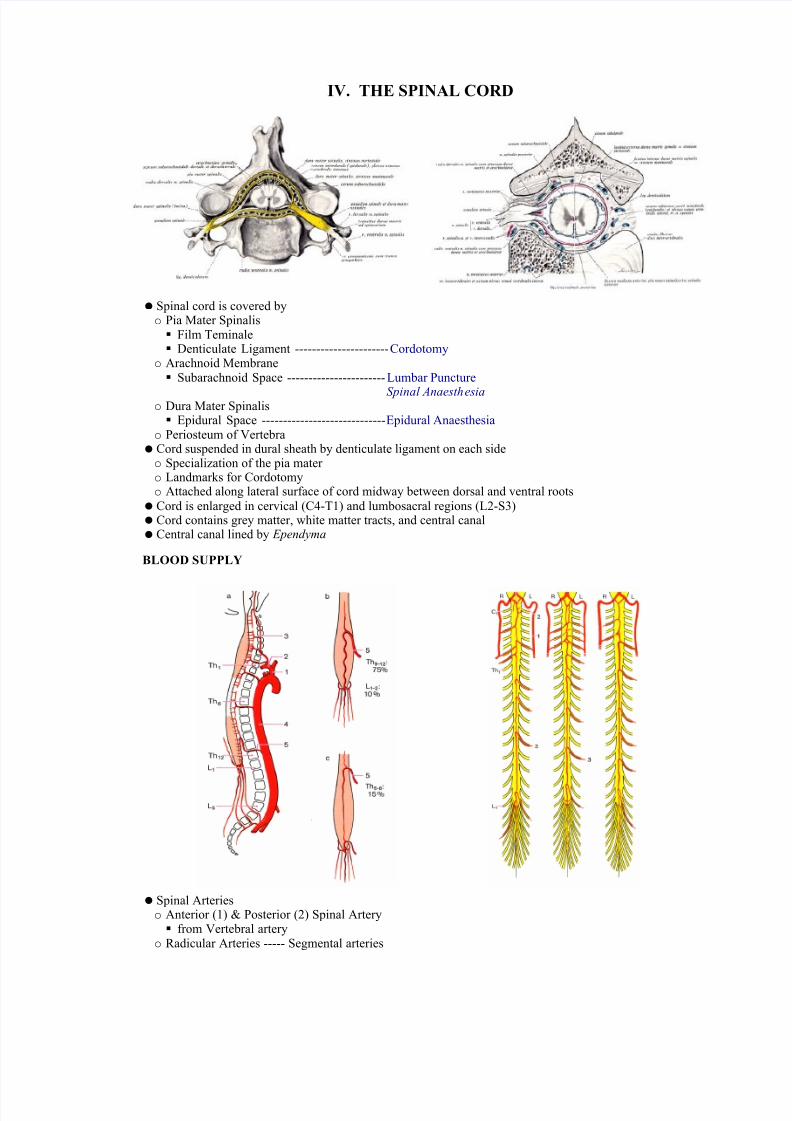
IV. THE SPINAL CORD
Spinal cord is covered byo Pia Mater Spinalis Film Teminale Denticulate Ligament ---------------------- Cordotomy
o Arachnoid Membrane Subarachnoid Space ----------------------- Lumbar Puncture
Spinal Anaesthesia o Dura Mater Spinalis Epidural Space ----------------------------- Epidural Anaesthesia
o Periosteum of Vertebra Cord suspended in dural sheath by denticulate ligament on each sideo Specialization of the pia mater o Landmarks for Cordotomyo Attached along lateral surface of cord midway between dorsal and ventral roots Cord is enlarged in cervical (C4-T1) and lumbosacral regions (L2-S3) Cord contains grey matter, white matter tracts, and central canal Central canal lined by Ependyma
BLOOD SUPPLY
Spinal Arterieso Anterior (1) & Posterior (2) Spinal Artery from Vertebral artery
o Radicular Arteries ----- Segmental arteries
7/27/2019 OS Spinal Cord
http://slidepdf.com/reader/full/os-spinal-cord 2/6
from Vertebral, Ascending Cervical, Intercostal and Lumbar Artery Venous Drainageo Longitudinal & Radicular Veins to Intervertebral veins to Internal Vertebral Venous Plexus to external vertebral venous plexus to segmental veins
o External plexus has anterior part (anterior to vertebral body) and posterior part (over posterior elements including laminae and spinous processes) Anterior and posterior parts freely anastomose
o Internal plexus: anterior part is on each side of PLL, posterior to vertebral body; posterior part isinterior to ligamentum flavum Vertebral body drained by basivertebral veins which enter anterior external plexus
o Veins of cord mirror related arteries in distributiono Venules drain into anterior and posterior veins, which drain into two median longitudinal veins, and
into anterolateral and posterolateral longitudinal veins lying adjacent to the nerve rootso Radicular veins join branches from internal plexus forming intervertebral veins (have valves), which
exit intervertebral foramina and join their respective segmental veins
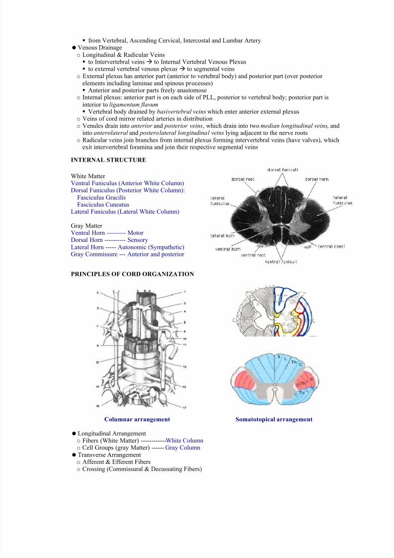
INTERNAL STRUCTURE
White Matter Ventral Funiculus (Anterior White Column)Dorsal Funiculus (Posterior White Column):
Fasciculus GracilisFasciculus Cuneatus
Lateral Funiculus (Lateral White Column)
Gray Matter Ventral Horn --------- Motor Dorsal Horn ---------- SensoryLateral Horn ----- Autonomic (Sympathetic)Gray Commissure --- Anterior and posterior
PRINCIPLES OF CORD ORGANIZATION
Columnar arrangement Somatotopical arrangement
Longitudinal Arrangemento Fibers (White Matter) ------------White Columno Cell Groups (gray Matter) ------ Gray Column Transverse Arrangemento Afferent & Efferent Fiberso Crossing (Commissural & Decussating Fibers)
7/27/2019 OS Spinal Cord
http://slidepdf.com/reader/full/os-spinal-cord 3/6
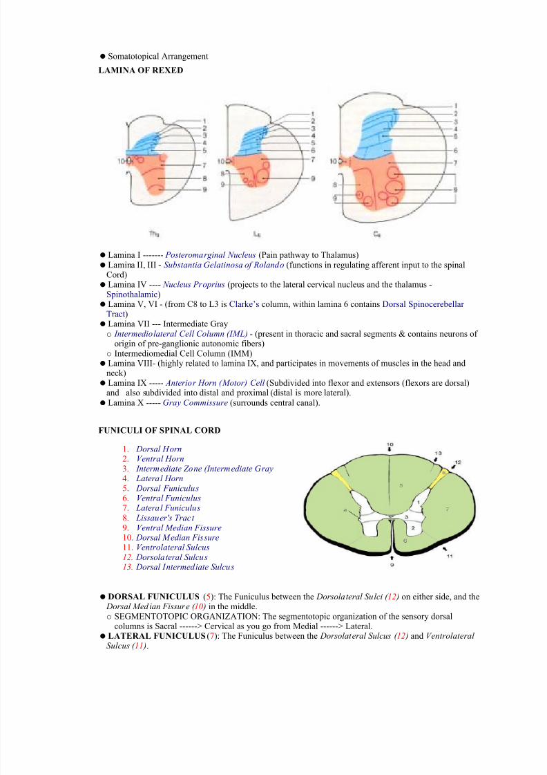
Somatotopical Arrangement
LAMINA OF REXED
Lamina I ------- Posteromarginal Nucleus (Pain pathway to Thalamus) Lamina II, III - Substantia Gelatinosa of Rolando (functions in regulating afferent input to the spinal
Cord) Lamina IV ---- Nucleus Proprius (projects to the lateral cervical nucleus and the thalamus -
Spinothalamic) Lamina V, VI - (from C8 to L3 is Clarke’s column, within lamina 6 contains Dorsal Spinocerebellar
Tract) Lamina VII --- Intermediate Grayo Intermediolateral Cell Column (IML) - (present in thoracic and sacral segments & contains neurons of
origin of pre-ganglionic autonomic fibers)o Intermediomedial Cell Column (IMM) Lamina VIII- (highly related to lamina IX, and participates in movements of muscles in the head and
neck) Lamina IX ----- Anterior Horn (Motor) Cell (Subdivided into flexor and extensors (flexors are dorsal)
and also subdivided into distal and proximal (distal is more lateral). Lamina X ----- Gray Commissure (surrounds central canal).
FUNICULI OF SPINAL CORD
1. Dorsal Horn2. Ventral Horn 3. Intermediate Zone (Intermediate Gray 4. Lateral Horn5. Dorsal Funiculus 6. Ventral Funiculus7. Lateral Funiculus 8. Lissauer's Tract
9. Ventral Median Fissure 10. Dorsal Median Fissure 11. Ventrolateral Sulcus 12. Dorsolateral Sulcus13. Dorsal Intermediate Sulcus
DORSAL FUNICULUS (5): The Funiculus between the Dorsolateral Sulci ( 12 ) on either side, and the Dorsal Median Fissure ( 10 ) in the middle.o SEGMENTOTOPIC ORGANIZATION: The segmentotopic organization of the sensory dorsal
columns is Sacral ------> Cervical as you go from Medial ------> Lateral. LATERAL FUNICULUS(7): The Funiculus between the Dorsolateral Sulcus ( 12 ) and Ventrolateral
Sulcus ( 11 ).
7/27/2019 OS Spinal Cord
http://slidepdf.com/reader/full/os-spinal-cord 4/6
VENTRAL FUNICULUS(6): The Funiculus between the Ventrolateral Sulci ( 11 ) on either side, andthe Ventral Median Fissure (9) in the middle.
SENSORY (ASCENDING) TRACTS: Sensory Tracts are Three- Neuron Chains.
Fasciculus Gracilis: Median half of Dorsal Funiculus.o MODALITY: Discriminative touch and proprioception. The fasciculus gracilis consists of large
myelinated fibers.o LESION: Ipsilateral loss of discriminative touch for all levels below (distal) to the lesion.
o SEGMENTOTOPIC ORGANIZATION: Sacral is most medial and T7 is most lateral. As you continuelaterally from there, you get into the Fasciculus Cuneatus.
Fasciculus Cuneatus: Lateral half of Dorsal Funiculuso MODALITY: Discriminative touch and proprioception. o LESION: Ipsilateral loss of discriminative touch for all levels below (distal) to the lesion, down to
level T7.o SEGMENTOTOPIC ORGANIZATION: T6 is the most medially placed in this tract, while Cervical
levels are most lateral. Segmentotopically, the Fasciculus Cuneatus is simply an extension of theGracilis above.
The two Fasciculi (Gracilis et Cuneatus) constitute the Posterior Columns, hence the name Posterior
Column System.
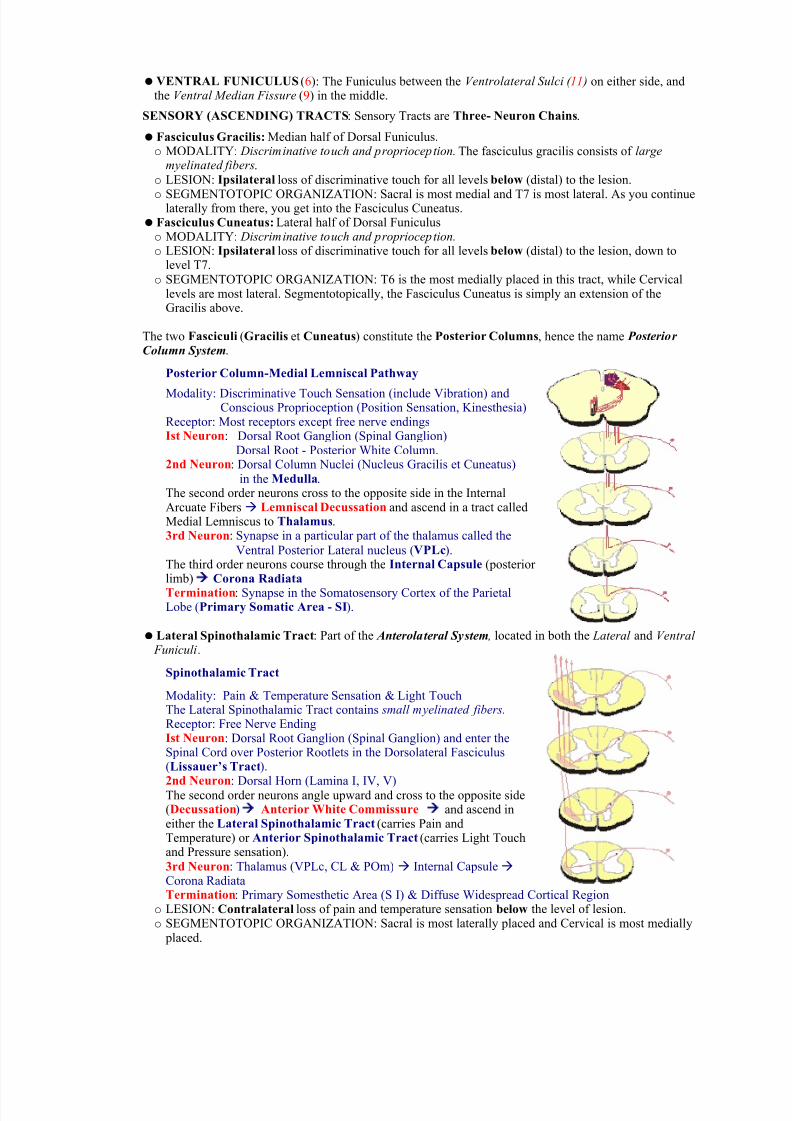
Posterior Column-Medial Lemniscal Pathway
Modality: Discriminative Touch Sensation (include Vibration) andConscious Proprioception (Position Sensation, Kinesthesia)
Receptor: Most receptors except free nerve endingsIst Neuron: Dorsal Root Ganglion (Spinal Ganglion)
Dorsal Root - Posterior White Column.2nd Neuron: Dorsal Column Nuclei (Nucleus Gracilis et Cuneatus)
in the Medulla. The second order neurons cross to the opposite side in the InternalArcuate Fibers Lemniscal Decussation and ascend in a tract calledMedial Lemniscus to Thalamus.3rd Neuron: Synapse in a particular part of the thalamus called the
Ventral Posterior Lateral nucleus (VPLc). The third order neurons course through the Internal Capsule (posterior limb) Corona Radiata
Termination: Synapse in the Somatosensory Cortex of the ParietalLobe (Primary Somatic Area - SI).
Lateral Spinothalamic Tract: Part of the Anterolateral System , located in both the Lateral and Ventral Funiculi.
Spinothalamic Tract
Modality: Pain & Temperature Sensation & Light TouchThe Lateral Spinothalamic Tract contains small myelinated fibers. Receptor: Free Nerve EndingIst Neuron: Dorsal Root Ganglion (Spinal Ganglion) and enter theSpinal Cord over Posterior Rootlets in the Dorsolateral Fasciculus(Lissauer’s Tract).
2nd Neuron: Dorsal Horn (Lamina I, IV, V)The second order neurons angle upward and cross to the opposite side(Decussation) Anterior White Commissure and ascend ineither the Lateral Spinothalamic Tract (carries Pain andTemperature) or Anterior Spinothalamic Tract (carries Light Touchand Pressure sensation). 3rd Neuron: Thalamus (VPLc, CL & POm) Internal Capsule Corona RadiataTermination: Primary Somesthetic Area (S I) & Diffuse Widespread Cortical Region
o LESION: Contralateral loss of pain and temperature sensation below the level of lesion.o SEGMENTOTOPIC ORGANIZATION: Sacral is most laterally placed and Cervical is most medially
placed.
7/27/2019 OS Spinal Cord
http://slidepdf.com/reader/full/os-spinal-cord 5/6
o Note that in the posterior column system the somatotopic organization in the cord is: leg is medialand arm is lateral. The opposite is true for the anterolateral system: the leg is lateral or dorsolateraland arm is medial.
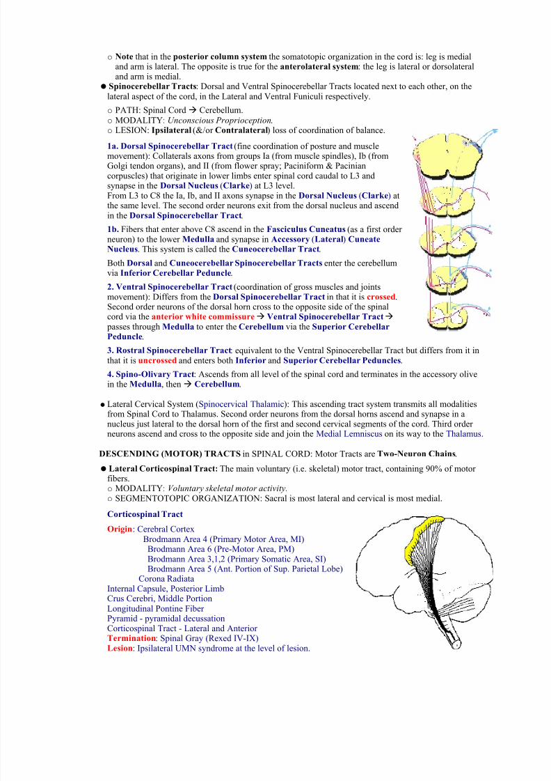
Spinocerebellar Tracts: Dorsal and Ventral Spinocerebellar Tracts located next to each other, on thelateral aspect of the cord, in the Lateral and Ventral Funiculi respectively.
o PATH: Spinal Cord Cerebellum.o MODALITY: Unconscious Proprioception.
o LESION: Ipsilateral (&/or Contralateral) loss of coordination of balance.
1a. Dorsal Spinocerebellar Tract (fine coordination of posture and musclemovement): Collaterals axons from groups Ia (from muscle spindles), Ib (fromGolgi tendon organs), and II (from flower spray; Paciniform & Paciniancorpuscles) that originate in lower limbs enter spinal cord caudal to L3 andsynapse in the Dorsal Nucleus (Clarke) at L3 level.From L3 to C8 the Ia, Ib, and II axons synapse in the Dorsal Nucleus (Clarke) atthe same level. The second order neurons exit from the dorsal nucleus and ascendin the Dorsal Spinocerebellar Tract.
1b. Fibers that enter above C8 ascend in the Fasciculus Cuneatus (as a first order neuron) to the lower Medulla and synapse in Accessory (Lateral) CuneateNucleus. This system is called the Cuneocerebellar Tract.
Both Dorsal and Cuneocerebellar Spinocerebellar Tracts enter the cerebellum
via Inferior Cerebellar Peduncle.2. Ventral Spinocerebellar Tract (coordination of gross muscles and jointsmovement): Differs from the Dorsal Spinocerebellar Tract in that it is crossed.Second order neurons of the dorsal horn cross to the opposite side of the spinalcord via the anterior white commissure Ventral Spinocerebellar Tract passes through Medulla to enter the Cerebellum via the Superior CerebellarPeduncle.
3. Rostral Spinocerebellar Tract: equivalent to the Ventral Spinocerebellar Tract but differs from it inthat it is uncrossed and enters both Inferior and Superior Cerebellar Peduncles.
4. Spino-Olivary Tract: Ascends from all level of the spinal cord and terminates in the accessory olivein the Medulla, then Cerebellum.
Lateral Cervical System (Spinocervical Thalamic): This ascending tract system transmits all modalitiesfrom Spinal Cord to Thalamus. Second order neurons from the dorsal horns ascend and synapse in anucleus just lateral to the dorsal horn of the first and second cervical segments of the cord. Third order neurons ascend and cross to the opposite side and join the Medial Lemniscus on its way to the Thalamus.
DESCENDING (MOTOR) TRACTSin SPINAL CORD: Motor Tracts are Two-Neuron Chains.
Lateral Corticospinal Tract: The main voluntary (i.e. skeletal) motor tract, containing 90% of motor fibers.o MODALITY: Voluntary skeletal motor activity. o SEGMENTOTOPIC ORGANIZATION: Sacral is most lateral and cervical is most medial.
Corticospinal Tract
Origin: Cerebral CortexBrodmann Area 4 (Primary Motor Area, MI)
Brodmann Area 6 (Pre-Motor Area, PM)Brodmann Area 3,1,2 (Primary Somatic Area, SI)Brodmann Area 5 (Ant. Portion of Sup. Parietal Lobe)
Corona RadiataInternal Capsule, Posterior LimbCrus Cerebri, Middle PortionLongitudinal Pontine Fiber Pyramid - pyramidal decussationCorticospinal Tract - Lateral and Anterior Termination: Spinal Gray (Rexed IV-IX)Lesion: Ipsilateral UMN syndrome at the level of lesion.
7/27/2019 OS Spinal Cord
http://slidepdf.com/reader/full/os-spinal-cord 6/6
Anterior Corticospinal Tract: Contains the 10% of motor fibers that did not cross in the PyramidalDecussation. Thus it is controlled by the Ipsilateral Motor Cortex throughout its path.
PYRAMIDAL MOTOR SYSTEM: The Lateral Corticospinal Tract, Anterior Corticospinal Tract, andCorticobulbar Tract. All other motor systems are called extrapyramidal. Within the pyramidal system:
UPPER MOTOR-NEURON LESIONS: You lose control over the lower (alpha-Motor) neurons, butthey can still fire spontaneously by themselves. Thus you get the classic triad of symptoms:o Spastic Paralysis: Rigid paralysis. No muscle wasting.
o Hyperreflexia: For patellar reflex.o Positive Babinski Sign: Dorsiflexion and flaring of toes when you stroke the sole of the foot.
LOWER (alpha-MOTOR) NEURON LESION: This is a peripheral lesion. Wallerian Degeneration of the nerve will occur leading to denervation of muscles.o SYMPTOMS: Flaccid paralysis Hyporeflexia Weakness & Muscle Wasting
o You can only lose lower motor innervation for one myotome at a time. If you cut the spinal cord, lower motor innervation will be lost at that level (Ipsilateral Lower-Motoneuron loss), and upper motor innervation will be lost at all levels distal to that level (Contralateral Upper-Motoneuron loss).
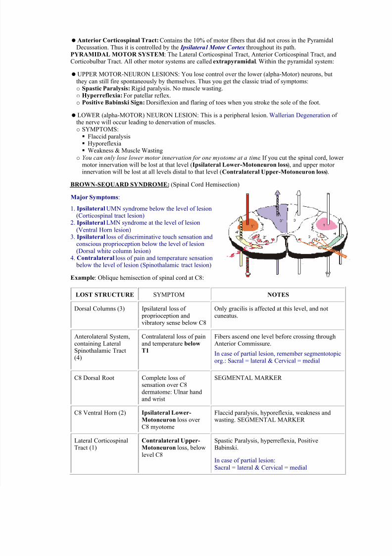
BROWN-SEQUARD SYNDROME:(Spinal Cord Hemisection)
Major Symptoms:
1. Ipsilateral UMN syndrome below the level of lesion (Corticospinal tract lesion)
2. Ipsilateral LMN syndrome at the level of lesion
(Ventral Horn lesion)3. Ipsilateral loss of discriminative touch sensation and
conscious proprioception below the level of lesion(Dorsal white column lesion)
4. Contralateral loss of pain and temperature sensation below the level of lesion (Spinothalamic tract lesion)
Example: Oblique hemisection of spinal cord at C8:
LOST STRUCTURE SYMPTOM NOTES
Dorsal Columns (3) Ipsilateral loss of proprioception andvibratory sense below C8
Only gracilis is affected at this level, and notcuneatus.
Anterolateral System,containing LateralSpinothalamic Tract(4)
Contralateral loss of painand temperature belowT1
Fibers ascend one level before crossing throughAnterior Commissure.
In case of partial lesion, remember segmentotopicorg.: Sacral = lateral & Cervical = medial
C8 Dorsal Root Complete loss of sensation over C8
dermatome: Ulnar handand wrist
SEGMENTAL MARKER
C8 Ventral Horn (2) Ipsilateral Lower-
Motoneuron loss over C8 myotome
Flaccid paralysis, hyporeflexia, weakness andwasting. SEGMENTAL MARKER
Lateral CorticospinalTract (1)
Contralateral Upper-
Motoneuron loss, belowlevel C8
Spastic Paralysis, hyperreflexia, PositiveBabinski.
In case of partial lesion:Sacral = lateral & Cervical = medial