Embed Size (px)
Citation preview

/ . Embryol. exp. Morph., Vol. 16, 3, pp. 581-390, December 1966 3 8 1With 5 plates
Printed in Great Britain
Osteogenesis in transplants of bone marrow cells
By A. J. FRIEDENSTEIN1,1. I. PIATETZKY-SHAPIRO1
& K. V. PETRAKOVA1
From the Laboratory of Immunomorphology, Gamaleya Institute ofEpidemiology and Microbiology, Academy of Medical
Sciences of the U.S.S.R., and Laboratory of MathematicalMethods in Biology, University of Moscow
After heterotopic (e.g. subcutaneous) transplantation of bone marrow,haemopoiesis in the graft ceases; reticular tissue develops instead, and later boneis formed (Denis, 1958). The result can be achieved by grafting either free piecesof bone marrow or those placed in diffusion chambers (Petrakova, Tolmacheva& Friedenstein, 1963; Rosin, Freiberg & Sajnek, 1963). In the case of free trans-plantation the bone formed is later filled with bone marrow. After transplanta-tion in diffusion chambers haemopoiesis does not recur despite the developmentof a considerable mass of bone in the chambers (Friedenstein, 1965).
The population of bone marrow cells is very heterogeneous, includinghaemopoietic cells, reticular cells and endosteum elements. According to generallyaccepted views this population is a mixture of individual cell lines capable ofmutual transformations within certain limits (Maximov, 1927; Burwell, 1964).After transplantation some of the pathways of differentiation open to bonemarrow tissue (formation of reticular and bone tissues) are stimulated, whileothers (haemopoiesis) are arrested. This effect could be ascribed to the death ofhaemopoietic stem cells and to proliferation of cells responsible for the develop-ment of reticular and bone tissue. It may, however, depend upon transformationof haemopoietic cells to reticular and osteogenic cells.
An analysis of these possibilities encounters difficulties as there are no reliabledata concerning the potentialities for transformation of different types of bonemarrow cells, and especially of the stem cells. There is no convincing evidencefor the dependence of various histogenetic pathways in bone marrow tissue(haemopoiesis, osteogenesis or the development of reticular tissue) upon in-dividual differences of the stem cells concerned. All of them could be providedby one common line of stem cells whose differentiation is controlled by condi-tions within the population.
Osteogenesis appearing in bone marrow grafts may serve as a model of
1 Authors' address: Gamaleya Institute of Epidemiology and Microbiology, U.S.S.R.of Medical Sciences, Gamaleya Street 2, Moscow D-182, U.S.S.R.
24 JEEM l6

382 A. J. FRIEDENSTEIN £T AL.
differentiation in a mixed cell population. Irrespective of whether all or some ofthe stem cells of bone marrow tissue are able to form bone, the following questionsmay be raised. In bone marrow transplants either bone and reticular tissue, orreticular tissue alone, develops. Does the difference depend upon whether or notosteogenic stem cells are included in the graft, their behaviour being unaffectedby other members of the cell population? Or is differentiation towards osteo-genesis a result of an interaction within a community of cells? These questionscan be answered when bone marrow cell suspensions are transplanted indiffusion chambers. Under these conditions bone tissue is formed in the chambers.This technique makes it possible to vary the number of transplanted cells andthe density of their initial population.
MATERIALS AND METHODS
Bone marrow of adult C57BL mice was isotransplanted intraperitoneally toadult recipients in diffusion chambers made of Millipore HA filters (pore size0-45 /i, thickness 150 /A) or of AUFS filters (pore size 0-6-0-9 /*, thickness 100 ju).The chambers were constructed according to the method of Algire (Algire,
1 1
Text-fig. 1. Construction of the diffusion chamber. A, Diffusion chamber type A;B, diffusion chamber type B; 1, millipore filter; 2, Plexiglass ring; 3, the cells.
Weaver & Prehn, 1957) and were of two sizes: A, chambers with filter diametersof 14 and 10 mm; and B, with those of 7 and 3-2 mm, respectively (cf. Text-fig. 1).Chambers were sterilized in 70° alcohol for 15 min, washed in distilled water andplaced into Hanks's solution.
Bone marrow was extracted from femur and chopped into fragments of about2 mm, which were placed into diffusion chambers, or bone marrow cell suspen-sion was prepared in Hanks's solution. After elimination of cell clumps byfiltration through Capron net the suspension was diluted to 2 x 10⁶ cells per ml.
To prepare lymphocyte suspensions cervical lymph nodes were used. Afterfiltration the concentration of lymphocytes was adjusted to 107 per ml. Tointroduce cells into a chamber the larger filter was put on glass rails over ahollow-ground slide into which Hanks's solution was poured until it touchedthe filter. A given volume of cell suspension was placed on the filter from above,the liquid passing across the filter and the cells precipitating on it. Filtration wasperformed in a Petri dish lined with cotton-wool soaked in saline. Then a filter

Osteogenesis in transplants 383
of smaller diameter (without cells) was placed over the larger one (with cells)and the chamber was stuck. The number of viable cells in the remaining cellsuspension was counted to determine that of the cells placed into chambers.
The chambers were fixed with 96° alcohol between 1 and 30 days after trans-plantation. They were freed from the surrounding tissue, the niters were separatedand put into a cooled fixative. In most cases the Gomori reaction for alkalinephosphatase was performed on filters (Gomori, 1939) to reveal foci of osteo-genesis. Filters then were counterstained with haematoxylin, dehydrated withalcohol, cleared with xylene and mounted in balsam as total preparations. Somefilters were tested for calcium as the control to the Gomori reaction. Afterfreeing from the plexiglass rings and fixation some chambers were embedded inparaffin and cut in serial sections that were stained by the Gomori method, forcalcium, with haematoxylin-eosin, for PAS (counterstained with haematoxylin).
The following experiments were performed:(1) Transplantation of a piece of bone marrow into chambers of types A
and B.(2) Transplantation of a suspension of 106 bone marrow cells into chambers
of type A.(3) Transplantation of a suspension of 105 bone marrow cells into chambers
of type A.(4) Transplantation of a mixture of bone marrow cells and lymphocytes
(1:9), the total number of cells being 107, into chambers of type A.(5) Transplantation of a suspension of 105 bone marrow cells into chambers
of type B.(6) Implantation of empty chambers.In series 2 and 5 the initial density of nucleated cells on the filter was approxi-
mately 210 and in series 3 approximately 30 per 0-05 mm2.
EXPERIMENTAL RESULTS
(1) Morphology of transplanted pieces of bone marrow {HA chambers)
When pieces of bone marrow were placed into chambers small blood vesselsand fragments of spongy bone were included with them (Plate 1, fig. 1). Usingthe Gomori method both these tissues are clearly stained. The residual bonemarrow tissue of mice showed a negative reaction for alkaline phosphatase.During the first days after transplantation the trabeculae of the bone necrosed.They ceased to show reaction for phosphatase, the osteocytes perished andempty cellular cavities remained in the bone. The surface of bone became de-nuded losing its covering layer of osteoblasts (Plate 1, fig. 2). At the same time alarge number of cells migrated from the graft and a broad zone of haemopoieticand reticular cells arose in the chamber. The relative amount of the formerrapidly declined, while that of the latter rose.
On the third day the filters were completely covered by cells usually arranged24-2

384 A. J. FRIEDENSTEIN£rAL.
in several layers in total preparations (Plate 1, fig. 3). The majority of these cellswere fibroblast-like elements, many in mitosis. Large spindle cells with densecytoplasm, large nuclei and clear-cut nucleoli could be easily distinguishedamong them; in these cells mitoses were seen particularly often (Plate 1, fig. 4).Within such tissue osteogenic foci appeared on the third day. When tested by theGomori reaction these foci looked like a phosphatase-positive network composedof elongated reticular cells with phosphatase-positive cytoplasm in a smallamount of phosphatase-positive matrix (Plate 1, fig. 5). The osteogenic cellswere bigger than the fibroblast-like cells on the filter. Meshes in the networkwere formed by one or, less frequently, two layers of adjacent cells between whichphosphatase-negative fibroblast-like and haemopoietic cells were distributed, i.e.elements that covered the remaining area of the filter.
Foci of osteogenic tissue were easily distinguished from other phosphatase-positive structures that could occur in these preparations, such as fragments ofmarrow blood vessels and the remnants of dead bone. The vessels possessed adistinct wall, their branching tubes were of regular shape with their contoursclearly outlined. The fragments of the spongy bone that got into the chamberduring implantation lost all signs of viability by the third day. It is significantthat in none of the cases investigated did foci of developing osteogenesis touchthe fragments of old bone. These two were always observed at a distance fromeach other; fragments of necrotic bone were located within the initial graft,while foci of osteogenesis were found in the zone of outgrowth on the filter.
Between the third and the eighth day after transplantation the number ofbone foci did not increase markedly but their structure changed: the amount ofbone matrix rose, trabeculae became thicker and composed of a greater numberof cells (Plate 1, fig. 6). The alveoli between trabeculae narrowed and the entirestructure became more compact. The osteogenic foci increased in size; on theeleventh day they were quite distinct even when stained with haematoxylin only.They showed the structure of typical bone trabeculae with osteoblasts embeddedin the bone matrix. In sections this bone tissue has also a peculiar appearance(Plate 2, fig. 7). By the twenty-fourth day bone occupied most of the chamber(Plate 2, figs. 8-10).
Table 1 presents the results of transplantation of pieces of bone marrow. Atthe time of transplantation such pieces consisted on the average of 8 x 10⁶ cells.
(2) Morphology of bone marrow transplanted inform of suspension of 106
cells into the chamber of type A (HA chambers)
When a suspension of bone marrow cells was placed in a chamber the initialpopulation consisted of: haemocytoblasts (11 %), myeloids (33 %), leucocytes(10 %), erythroids (8 %), monocytes (4 %), lymphocytes (34 %), reticular cells(0-5 %), only nucleated cells being taken into account. The cells were evenlydistributed over the whole surface of the filters; no phosphatase-positive cellscould be found.

J. Embryo!, ex p. Morph., Vol. 16, Part 3 PLATE t
Fig. 1. Transplantation of a piece of bone marrow in a chamber with HA filters. Three days.Alcohol. Gomori. Total preparation, x 200. Blood vessels chamber with bone marrow.Fig. 2. The same preparation, x 200. Fragment of dead bone in the chamber.Figs. 3,4. Transplantation of a piece of bone marrow in a chamber with HA filters. Three days.Alcohol. Haematoxylin. Total preparation, x 400. Areas of the zone of growth. M, mitosis.Fig. 5. Transplantation of a piece of bone marrow in a chamber with HA filters. Three days.Alcohol. Gomori. Total preparation, x 400. A focus of phosphatase activity in the zone ofgrowth.Fig. 6. Transplantation of a piece of bone marrow in a chamber with HA filters. Five days.Alcohol. Gomori. Total preparation, x 400. A focus of phosphatase activity in the zone ofgrowth.
A. J. FRIEDENSTEIN ET AL. facing p. 384

/ . Embryo!. exp. Morph., Vol. 16, Part 3 PLATE 2
i gnwur mi ^ "* ^ ^
Fig. 7. Transplantation of a piece of bone marrow in a chamber with HA filters. Eleven days.Alcohol. PAS-haematoxylin. A section, x 200. A focus of osteogenesis in the chamber underthe filter, a, Filter; b, bone tissue; c, a layer of osteoblasts.Figs. 8, 9. Transplantation of a piece of bone marrow in a chamber with HA filters. Fifteendays. Alcohol. PAS-haematoxylin. x 400. Bone tissue adjoining the filter, a, Filter, b, bonetissue.Fig. 10. Transplantation of a piece of bone marrow in a chamber with HA filters. Alcohol.Gomori. Section, x 200. Bone in the chamber, a, Filter, b, bone tissue.
A. J. FRIEDENSTEIN ET AL.

J. Embryol. exp. Morph., Vol. 16, Part 3 PLATE 3
11
.114
12
Fig. 11. Transplantation of 106 bone marrow cells in a chamber of type A with HA filters.Two days. Alcohol. Haematoxylin. Total preparation, x 400.Fig. 12. The same experiment. Three days. Alcohol. Haematoxylin. Total preparation, x 200.A focus of 49 myeloid cells with five mitoses.Fig. 13. The same experiment. Three days, x 200. Large basophilic cells. Mitosis in one.Figs. 14-16. The same experiment. Five days. Alcohol. Haematoxylin. Total preparations.x 280. Bands of large polygonal cells.
A. J. FR1EDENSTEIN ET AL.

J. Embryo/, exp. Morph., Vol. 16, Part 3 PLATE 4
Figs. 17, 18. The same experiment. Five days. Alcohol. Gomori. Total preparations. x400.Fig. 19. The same experiment. Five days. Alcohol. Haematoxylin. Total preparation. x400.A focus of myeloid cells with mitoses.Fig. 20. The same experiment. Seven days. Alcohol. Haematoxylin. Total preparation, x 400.A focus of osteoblasts with mitoses.
A. J. FRIEDENSTEIN ET AL. facing p. 385
Osteogenesis in transplants 385
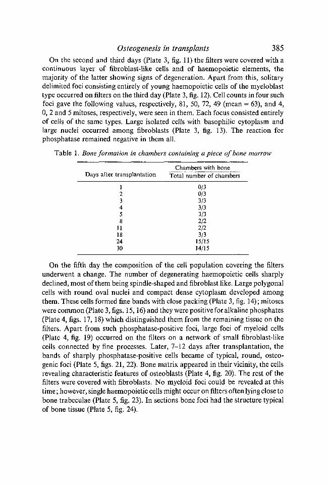
On the second and third days (Plate 3, fig. 11) the filters were covered with acontinuous layer of fibroblast-like cells and of haemopoietic elements, themajority of the latter showing signs of degeneration. Apart from this, solitarydelimited foci consisting entirely of young haemopoietic cells of the myeloblasttype occurred on filters on the third day (Plate 3, fig. 12). Cell counts in four suchfoci gave the following values, respectively, 81, 50, 72, 49 (mean = 63), and 4,0, 2 and 5 mitoses, respectively, were seen in them. Each focus consisted entirelyof cells of the same types. Large isolated cells with basophilic cytoplasm andlarge nuclei occurred among fibroblasts (Plate 3, fig. 13). The reaction forphosphatase remained negative in them all.
Table 1. Bone formation in chambers containing a piece of bone marrow
Days after transplantation
123458
11182430
Chambers with boneTotal number of chambers
0/30/33/33/33/32/22/23/3
15/1514/15
On the fifth day the composition of the cell population covering the filtersunderwent a change. The number of degenerating haemopoietic cells sharplydeclined, most of them being spindle-shaped and fibroblast like. Large polygonalcells with round oval nuclei and compact dense cytoplasm developed amongthem. These cells formed fine bands with close packing (Plate 3, fig. 14); mitoseswere common (Plate 3, figs. 15,16) and they were positive for alkaline phosphates(Plate 4, figs. 17, 18) which distinguished them from the remaining tissue on thefilters. Apart from such phosphatase-positive foci, large foci of myeloid cells(Plate 4, fig. 19) occurred on the filters on a network of small fibroblast-likecells connected by fine processes. Later, 7-12 days after transplantation, thebands of sharply phosphatase-positive cells became of typical, round, osteo-genic foci (Plate 5, figs. 21, 22). Bone matrix appeared in their vicinity, the cellsrevealing characteristic features of osteoblasts (Plate 4, fig. 20). The rest of thefilters were covered with fibroblasts. No myeloid foci could be revealed at thistime; however, single haemopoietic cells might occur on filters often lying close tobone trabeculae (Plate 5, fig. 23). In sections bone foci had the structure typicalof bone tissue (Plate 5, fig. 24).

386 A. J. FRIEDENSTEIN £r^L.
(3) The effect of the number of cells and of their packing upon bone formation
Different numbers of cells were placed in chambers of different size and ofeach type. The results in terms of the proportion of chambers in which bonewas formed are given in Table 2. The remaining chambers were filled withreticular tissue.
(4) Implantation of empty chambersEmpty chambers composed of HA filters sterilized for 15 min with 70°
alcohol before intraperitoneal implantation proved to be impermeable to cells:in none of five empty chambers were cells found 7 days after implantation. Nocells were detected in the filter material itself in sections of the many chamberscomposed of HA filters which were studied.
Chambers composed of AUFS filters were permeable to cells The averagenumbers of cells found after implantation of an empty chamber with 0 04 mm2
of filter surface were: 5-0 cells after 2 h; 18-5 cells after 6 h; 14-5 cells after 12 h;24-5 cells after 24 h, and 50-0 cells after 48 h. During the first 5 h leucocytespredominated among the cells, later lymphocytes and fibroblast-like cells orhistiocytes were predominant. Many cells were always found in the filtermaterial in sections of chambers composed of AUFS filters (Plate 5, fig. 25).
Table 2. Bone formation in chambers with suspension of bone marrow cells
Chambertype
A
B
Filtertype
AUFS
HA
AUFS
HAAUFS
AUFSHA
No. of cells at time oftransplantation
106 bone marrow cells
105 bone marrow cells
106 bone marrow cells+ 9xl06lymph.
105 bone marrow cells
Fixationtime
(days)
11912
f 1 124
1301115
1111
Chambers with bone
Total number of chambers
11/12]5/5 [24/278/10J2/122/60/50/6
•4/29
2/8 2/8
8 / 1 1 l 12/174/6 J
DISCUSSION
When bone marrow cells are placed into diffusion chambers reticular orbone tissue is formed instead of haemopoietic elements. This considerable changeof histogenesis occurs after free heterotopic transplantation of bone marrow aswell (Denis, 1958). Therefore, it does not depend upon special cultivationconditions in diffusion chambers. The cells transplanted in a chamber are innovel conditions by comparison with those in situ. Their interaction with sur-rounding tissues (with bone in particular) and their mutual arrangement (tissue

/ . Embryol. exp. Morph., Vol. 16, Part 3 PLATE 5
21
25
Fig. 21. The same experiment. Seven days. Alcohol. Gomori. Total preparation. x200.A focus of osteogenesis.Fig. 22. The same experiment. Ten days. Alcohol. Gomori. Total preparation, x 200. A focusof osteogenesis.Fig. 23. The same experiment. Eleven days. Alcohol. Haematoxylin. Total preparation, x 200.Bone trabeculae and haemopoietic cells.Fig. 24. The same experiments. Twelve days. Alcohol. PAS. A section, x 200. Bone in thechamber, a, Filter, b, bone tissue.Fig. 25. An empty chamber composed of AUFS chambers. Five days after implantation.Alcohol. Haematoxylin. Section, x 200. a, Filter; b, chamber contents.
A. J. FRIEDENSTEIN ET AL. facing p. 386

Osteogenesis in transplants 387
structure) are disturbed. In the case of transplantation of bone marrow frag-ments this holds true for the zone of cell outgrowth, while the original transplantitself usually degenerates. These disturbances seem to cause the main alterationin differentiation of transplanted cells, i.e. the extinction of haemopoiesis. Whichof the causes mentioned is decisive remains unknown.
The fact that osteogenesis occurs in cell suspension shows that the osteogenicpotency of the bone marrow cell population does not disappear after dissocia-tion of cells.
It is evident that not all the transplanted bone marrow cells can act as osteo-genic stem-cells. The results of the present work suggest that in a population ofbone marrow cells cultured in diffusion chambers no cells form osteogenic fociindividually. If this were otherwise, bone would form to the same extent in thechambers of different size after transplantation of the same number of cells(i.e. containing the same number of such precursor elements). However, thesame number of bone marrow cells, say 105, behaves in a different way in thechambers of different size. In those of type B this number of cells formed bone,as a rule, while in the chambers with an area tenfold larger (type A) it did not.The area of the chamber was determined by the area of the smaller filter.Bone formation in larger chambers (type A) requires more cells, namely 106, i.e.ten times more. A tenfold dilution of 106 bone marrow cells placed in type Achamber (by the addition of 9 x 10⁶ lymphocytes taken from lymph nodes)prevents osteogenesis. These data cannot be explained by the fact that generalmetabolic processes (e.g. those of metabolism, respiration, etc.) could themselvesinhibit or stimulate bone formation in the chambers containing a differentnumber of cells. When 106—108 bone marrow cells were placed into A-chambersosteogenesis usually occurred (Friedenstein, 1964). Yet, when 106 bone marrowcells + 9 x 10⁶ lymphocytes were placed in such chambers bone was rarely formed.A lesser dilution of the bone marrow cells does not affect the result of trans-plantation : penetration of a small number of cells from outside into the chamber(when the chambers consisted of AUFS filters) did not change the frequency ofosteogenesis when compared with the chambers composed of HA filtersimpermeable to cells.
The cells responsible for the formation of osteogenic foci seem to have a highmitotic activity. This is proved by the frequent occurrence of mitosis in the cellswhich, judging by morphological characters, are those which give rise toosteogenic structures in chambers.
It could be expected, therefore, that cells serving as precursors for osteogenictissue at transplantation of 105 bone marrow cells in type B chambers can formconsiderable cell clones during 30-day cultivation when placed in type Achambers. Thus, if the population density of 105 cells in type A chambers isinsufficient for osteogenesis, the cells of which bone foci could be formed eitherdo not multiply or they proliferate, but differentiation of the correspondingclone does not proceed towards osteogenesis. This latter possibility seems to be

388 A. J. FRIEDENSTEIN^TAL.
most likely. At any rate, the results presented demonstrate that osteogenesisrequires a certain initial density of bone marrow cells. Thus, the developmentof bone in a chamber differs from the formation of haemopoietic foci on thespleen of irradiated mice at transplantation of bone marrow cells (Till, Mc-Culloch & Siminovitch, 1964; Becker, McCulloch & Till, 1963; Lewis &Trobaugh, 1964). A haemopoietic stem-cell, if in contact with an appropriatestroma (e.g. in irradiated spleen) is able to create a haemopoietic clone inisolation (Becker et al. 1963). On the other hand, when bone is formed in achamber the differentiation of osteogenic stem-cells depends upon the interactionof cells at some initial period. It is characteristic that duration does not affectthe results of transplantation, i.e. if the initial packing does not result in boneformation during eleven days, no osteogenesis develops in 30 days either.
As is well known, bone marrow cells are mobile and are expected to migratewithin the chamber after transplantation. The fact that osteogenesis requires acertain density of the initial population of bone marrow cells implies thatosteogenesis arises when necessary cells meet to form a corresponding structure.Apparently it is easily established when the initial density corresponds to 210bone marrow cells per 0-05 mm2 being achieved but very rarely when the densityequals thirty bone marrow cells per 0-05 mm2.
In the case of transplantation of a piece of marrow, even if it is sufficientlysmall to avoid rapid necrosis, bone formation proceeds only in the zone ofoutgrowing cells. This implies that the initial structure of marrow is inappro-priate for bone formation, and the structure required for osteogenesis is createdanew. Foci of proliferating haemopoietic cells are found on the third to seventhday in chambers with cell suspension. It seems very likely that these are clonesoriginated from individual haemopoietic stem-cells similar to haemopoietic foci inthe spleen of irradiated mice (Till et al. 1964). In chambers these foci disappearon the tenth day probably due to the fact that haemopoiesis proceeds only in thepresence of a certain structure of haemopoietic tissue whose maintenancerequires contact with either bone or spleen stroma (Friedenstein, 1965).
In the presence of bone (or spleen stroma) the stem cells of haemopoietictissue differentiate towards haemopoiesis (Till et al. 1964). In the absence of bone,osteogenic potencies of marrow cell population are realized and bone is formedwhich in turn is necessary for the maintenance of haemopoiesis. This creates themechanism supporting differentiation of haemopoietic tissue and controlling thebehaviour of stem-cells in the bone marrow cell population. It should be bornein mind that bone tissue undergoes sustained reconstruction, i.e. it is resorbed insome places while being formed in others (Hattner, Epker & Frost, 1965). Forthis reason its influence upon the differentiation of stem cells can be expected inany individual area of marrow tissue.
There are grounds for believing that the formation of cells of marrow stromaand of blood cells is ensured by common stem cells, but no direct evidence isavailable. However, haemopoietic and stromal elements might have different

Osteogenesis in transplants 389
stem cells while control over the composition of the whole population of marrowcells provides selective effects upon each of these categories of stem cells. At thepresent time it cannot be ruled out that bone and reticular tissue too haveseparate lines of stem cells in bone marrow, although it seems more likely thatthey have a common line.
SUMMARY
1. In bone marrow fragments and bone marrow cell suspensions isotrans-planted to mice in diffusion chambers, reticular tissue develops and sometimesosteogenesis also occurs. No haemopoiesis takes place in the grafts.
2. Bone formation in the chambers requires a certain density of the initialpacking of bone marrow cells. This follows from the considerable differences inthe frequency of bone formation in the case of transplantation of the samenumber of cells (105) in chambers of different size, and from cases in whichdifferent cell numbers (105 and 106) are transplanted in the chambers of the samesize.
3. The results obtained show that differentiation of the stem cells towardsosteogenesis requires cell interaction within the cell community at a certaincrucial moment.
PE3I0ME
Bo (jyparMeirrax Kocraoro M03ra H B cycneH3HHx KOCTHOMO3:TO:BEIX KJIGTOK,
II3OTpaHCnJiaHTHpOBaHHLIX MbllliaM B HH$(|)y3H03HLIX KaMepaX, B03HHKaeTocTeorene3 HJIH me pa3BHBaeTCH TOJIBKO peraKyjiflpHaH TKaHb. KpoBeTBopeHneB TpaHcnjiaHTaTax He nponcxoAHT.
TpaHcnjiaHTauHH Oflnoro H Toro ?Ke KOJinnecTBa KOCTHOMO3roBbix KJieiOK BKanepax , HMeionpix pasnHHHyio ruiomajjb, RaeT pa3Hbie pe3yjn>TaTLi. /JjiflB03HiiKH0BeHHH KOCTii Heo6xo,n,HMa onpeflejiemiaynaKOBKH KOCTHOMO3roBbix KJieTOK B KaMepe.
npiiBefleHHbie pe3yjn>TaTbi noKa3HBaioT, HTOajibHO ocTeoreHHbix KJIGTOK B HanpaBjiemiH ocTeoreHe3a TpeSyeT B3anM0-
f B r p y n n e KJieTOK B HeKOTopbiii KpHTHHecKHii
The authors would like to express their sincere gratitude to Professor G. V. Lopashov forhis valuable advice and interest in this paper.
REFERENCES
ALGIRE, G. A., WEAVER, J. M. & PREHN, K. T. (1957). Studies on tissue homotransplantationin mice using diffusion chamber method. Ann. N.Y. Acad. Sci. 64, 1009-12.
BECKER, A. J., MCCULLOCH, E. A. & TILL, J. E. (1963). Cytological demonstration of theclonal nature of spleen colonies derived from transplanted mouse marrow cells. Nature, Lond.197, 452-4.
BUR WELL, R. (1964). Studies on the transplantation of bone. / . Bone Jt Surg. 46 B, 110-40.DENIS, A. (1958). Etude de Vossification dans les graftes de moelle osseuse. Bruxelles.FRIEDENSTEIN, A. J. (1964). In Press (Russian).FRIEDENSTEIN, A. J. (1965). Factors influencing differentiation of hematopoietic tissue in
diffusion chambers. Molecular and cellular basis of antibody formation. Praha, 1965,321-8.

390 A. J. FRIEDENSTEIN£T AL.
GOMORI, G. (1939). Microtechnical demonstration of phosphatase in tissue sections. Proc.Soc. exp. biol. Med. 42, 23-6.
HATTNER, R., EPKER, B. & FROST, H. (1965). Suggested sequential mode of control of changesin cell behaviour in adult bone remodelling. Nature, Lond. 206, 489-90.
LEWIS, J. P. & TROBAUGH, F. E. (1964). Haemopoietic stem-cells. Nature, Lond. 204, 589-90.MAXIMOV, A. A. (1927). Bindgewebe und blutbildende Gewebe. Handb. mikr. Anat. Menschen.
2, 232-426.PETRAKOVA, K. V., TOLMACHEVA, A. A. & FRIEDENSTEIN, A. J. (1963). Bone formation occurring
in bone marrow transplantation in diffusion chambers. Bull. exp. biol. med. (Russian),56(12), 87-91.
ROSIN, A., FREIBERG, H. & SAJNEK G. (1963). The fate of bone marrow, spleen and periosteumcultivated in vivo in the diffusion chamber with special reference to bone formation.Exp. Cell Res. 29, 176-87.
TILL, J. E., MCCULLOCH, E. A. & SIMINOVITCH, L. (1964). A stochastic model of stem-cellproliferation based on the growth of spleen colony-forming cells. Proc. natn. Acad. Sci.U.S.A. 51, 29-36.
(Manuscript received 26 February 1966)