Embed Size (px)
Citation preview
Introduction
Th e concept of osteoimmunology emerged more than a
decade ago and is based on rapidly growing insight into
the functional interdependence between the immune
system and bone at the anatomical, vascular, cellular, and
molecular levels [1]. In 1997, the receptor activator of the
nuclear factor-kappa-B ligand (RANKL)/RANK/osteo-
protegerin (OPG) pathway was identifi ed as a crucial
molecular pathway of the coupling between osteoblasts
and osteoclasts [2]. It appeared that not only osteoblasts
but also activated T lymphocytes, which play a crucial
role in the pathogenesis of rheumatoid arthritis (RA), and
many other infl ammatory cells can produce RANKL,
which stimulates the diff erentiation and activation of
osteoclasts [3]. Th ese fi ndings have contributed to the
birth of osteoimmunology as a discipline.
Because of the multiple interconnections and inter-
actions of bone and the immune system, bone is a major
target of chronic infl ammation in RA and ankylosing
spondylitis (AS). Infl ammation increases bone resorption
and results in suppressed local bone formation in RA and
locally increased bone formation in AS, causing a wide
spectrum of bone involvement in RA and AS [4,5].
Osteoporosis has been defi ned as a bone mineral
density (BMD) of lower than 2.5 standard deviations of
healthy young adults and in daily practice is measured by
dual-energy x-ray absorptiometry (DXA) at the spine and
hip [6]. However, the bone disease component in RA and
AS is much more complex, especially around the sites of
infl ammation. We reviewed the literature on the quanti-
fi cation of local and general bone changes and their
relation to the structural damage of bone, disease activity
parameters, and fracture risk in the context of osteo-
immunology, both in RA and AS. We have chosen to
focus on RA and AS since these infl ammatory rheumatic
diseases have the highest prevalence and since, in both
diseases, characteristic but diff erent types of bone
involve ment may occur.
Anatomical and molecular cross-talk between
bone and the immune system
Multiple anatomical and vascular contacts and overlap-
ping and interacting cellular and molecular mechanisms
are involved in the regulation of bone turnover and the
immune system, so that one can no longer view either
Abstract
The concept of osteoimmunology is based on
growing insight into the links between the immune
system and bone at the anatomical, vascular,
cellular, and molecular levels. In both rheumatoid
arthritis (RA) and ankylosing spondylitis (AS), bone
is a target of infl ammation. Activated immune cells
at sites of infl ammation produce a wide spectrum
of cytokines in favor of increased bone resorption
in RA and AS, resulting in bone erosions, osteitis,
and peri-infl ammatory and systemic bone loss.
Peri-infl ammatory bone formation is impaired in
RA, resulting in non-healing of erosions, and this
allows a local vicious circle of infl ammation between
synovitis, osteitis, and local bone loss. In contrast,
peri-infl ammatory bone formation is increased
in AS, resulting in healing of erosions, ossifying
enthesitis, and potential ankylosis of sacroiliac joints
and intervertebral connections, and this changes
the biomechanical competence of the spine. These
changes in bone remodeling and structure contribute
to the increased risk of vertebral fractures (in RA and
AS) and non-vertebral fractures (in RA), and this risk
is related to severity of disease and is independent
of and superimposed on background fracture risk.
Identifying patients who have RA and AS and are at
high fracture risk and considering fracture prevention
are, therefore, advocated in guidelines. Local peri-
infl ammatory bone loss and osteitis occur early and
precede and predict erosive bone destruction in RA
and AS and syndesmophytes in AS, which can occur
despite clinically detectable infl ammation (the so-
called ‘disconnection’). With the availability of new
techniques to evaluate peri-infl ammatory bone loss,
osteitis, and erosions, peri-infl ammatory bone changes
are an exciting fi eld for further research in the context
of osteoimmunology.
© 2010 BioMed Central Ltd
Osteoimmunology and osteoporosisPiet Geusens*1,2 and Willem F Lems3
R E V I E W
*Correspondence: [email protected] of Internal Medicine, Subdivision of Rheumatology, Maastricht
University Medical Center, P. Debyelaan 25 Postbus 5800, 6202 AZ Maastricht,
The Netherlands
Full list of author information is available at the end of the article
Geusens and Lems Arthritis Research & Therapy 2011, 13:242 http://arthritis-research.com/content/13/5/242
© 2011 BioMed Central Ltd
system in isolation but should consider bone and the
immune system to be an integrated whole [4,5].
Anatomical connections
Bone, by virtue of its anatomy and vascularization, is at
the inside and outside and is in direct and indirect and in
close and distant contact with the immune system. At the
inside, bones are the host for hematopoiesis, allowing
bone and immune cells to cooperate locally. At the
outside, bone is in direct contact with the periost, the
synovial entheses within the joints at the periost- and
cartilage-free bare area [7], the fi brous tendon entheses,
the calcifi ed component of cartilage and tendon inser-
tions, and the intervertebral discs.
Until recently, it was thought, on the basis of plain
radiographs of the hands, that there is only rarely a direct
anatomical connection between bone marrow and joint
space. Bone erosions have been found in hand joints of
presumably healthy controls in less than 1% with plain
radiology and in 2% with MRI [8]. However, exciting new
data have shown that, with the use of high-resolution
quantitative computer tomography (HRqCT), small
erosions (<1.9 mm) in the metacarpophalangeal (MCP)
joints can be found in 37% of healthy subjects without
any signs or symptoms of RA, indicating that small
erosions are not specifi c for RA [9]. Large erosions
(>1.9 mm) were found to be specifi c for RA. Interestingly,
58% of erosions detected by HRqCT in healthy volunteers
were not visible on plain radiographs [9]. In healthy
controls, the erosions in the MCP joints were not
randomly located but were located at the bare area and at
high-pressure points adjacent to ligaments, which are
erosion-prone sites in RA [10]. Bone erosions are also
extremely common in healthy controls in the entheses
[11] and in the vertebral cortices covered by periost and
the intervertebral discs (in AS) [12]. Th e immune system,
bone, and its internal and external surfaces not only are
connected by these local anatomical connections but also
are connected with the general circulation by the main
bone nutrition arteries and locally with the periost (by its
vasculature that perforates cortical bone) and within the
bone compartment by attachments of fi brous entheses
and the calcifi ed components of cartilage and fi bro-
cartilage up to the tidemark, which separates calcifi ed
from non-calcifi ed components of cartilage and tendons
[11].
Molecular connections
Bone cells exert major eff ects on the immune system.
Bone cells interact with immune cells and play an
essential role in the development of the bone marrow
space during growth [13] and during fracture healing
[14]. Osteoblasts play a central role in the regulation of
renewal and diff erentiation of hematopoietic stem cells
(HSCs) and of B cells in niches near the endosteum [15-
17]. Metabolic pathways of the osteoblast which are in-
volved in bone remodeling are also involved in the regu-
lation of HSCs by osteoblasts, such as the calcium
receptor, parathyroid hormone (PTH), bone morpho-
genetic proteins (BMPs), the Wnt signaling, and cell-cell
interactions by the NOTCH (Notch homolog, trans-
location-associated (Drosophila)) signaling pathway
[15-19]. On the other hand, multiple cytokines, chemo-
kines, and growth factors of immune cells such as T and
B cells, fi broblasts, dendritic cells, and macrophages
directly or indirectly regulate osteoblast and osteoclast
activity by producing or infl uencing the production of
the RANKL/RANK/OPG pathway, tumor necrosis
factor-alpha (TNFα), interferon-gamma (IFNγ), and
inter leukins (such as IL-1, IL-6, IL-15, IL-17, IL-18, and
IL-23) and the Wnt signaling with involvement of
Dikkoppf (DKK), sclerostin, and BMP [4,5,19-21].
In RA, bone loss and bone destruction are dependent
on the imbalance between osteoclastogenic and anti-
osteoclastogenic factors. T-cell infi ltration in the syno-
vium is a hallmark of RA. TH17 cells, whose induction is
regulated by dendritic cells that produce transforming
growth factor-beta, IL-6, and IL-23, secrete IL-17, which
induces RANKL in fi broblasts and activates synovial
macrophages to secrete TNFα, IL-1, and IL-6, which
directly or indirectly (via fi broblasts producing RANKL)
activate osteoclastogenesis [1]. Other direct or indirect
osteoclastogenic factors include monocyte/macrophage
colony-stimulating factor, IL-11, IL-15, oncostatin M,
leukemia inhibitor factor, and prostaglandins of the E
series (PGE) [22-24]. Inhibitors of osteoclastogenesis in
RA include TH1 (producing IFNγ) and TH2 (producing
IL-4) cells and possibly T helper regulatory (THREG) cells
[1].
In AS, increased bone formation, as refl ected by
syndesmophyte formation in the spine, is related to
decreased serum levels of DKK [25] and sclerostin [21],
both inhibitors of bone formation, and to serum levels of
BMP, which is essential for enchondral bone formation
[26], and of CTX-II [27], which refl ects cartilage destruc-
tion that occurs during enchondral bone formation in
syndesmophytes [26-28]. Th ere is, thus, increasing
evidence that immune cells and cytokines are critically
responsible for the changes in bone resorption and
forma tion and vice versa, resulting in changes in bone
quality in chronic infl ammatory conditions. Th ese condi-
tions include RA, spondylarthopathies (SpAs) (AS,
psoriatic arthritis, and infl ammatory bowel disease),
systemic lupus erythematosis, juvenile RA, periodontal
diseases, and even postmenopausal osteoporosis [29]. We
reviewed the literature on the quantifi cation of bone
involvement in RA and AS. For an in-depth discussion of
the underlying metabolic pathways, a topic that is beyond
Geusens and Lems Arthritis Research & Therapy 2011, 13:242 http://arthritis-research.com/content/13/5/242
Page 2 of 16
the scope of this review, the reader is referred to other
reviews [4,5].
Histology of bone in rheumatoid arthritis and
ankylosing spondylitis
Bone resorption
Bone resorption is increased in RA and AS. In RA, this
has been demonstrated histologically by the presence of
activated osteoclasts in the pannus at the site of bone
erosions [30,31], in the periarticular trabecular and
cortical bone [32,33], and, in a general way, in sites
distant from infl ammation [34]. In AS, osteoclastic bone
resorption has been demonstrated in the sacroiliac joints
[35-37].
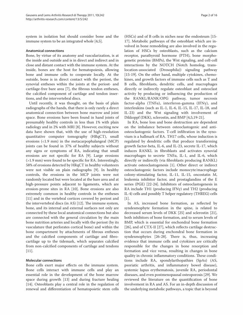
Th e introduction of MRI has shed new light on the
involvement of subchondral bone and bone marrow in
RA and AS (Figure 1). Periarticular MRI lesions have
been described technically as bone edema (on short T
inversion recovery (STIR), indicating that fatty bone
marrow is replaced by fl uid) and osteitis (on T1 after IV
gadolinium) [38] and histo logi cally as osteitis as
infl ammation has been demon strated on histological
examination of these lesions [33]. In joint specimens of
patients with RA and with MRI signs of bone edema,
histological correlates have been studied in specimens
obtained at the time of joint replacement and have shown
the presence of greater numbers of osteo clasts than in
controls and in patients with osteoarthritis and the
presence of T cells, B-cell follicles, plasma cells,
macrophages, decreased trabecular bone density, and
increased RANKL expression [33].
Osteitis is also a major component of AS [39-42].
Osteitis was described by histology of the vertebrae in
1956 [43] and occurs early in the disease and predicts the
occurrence of bone erosions [39]. It has been shown that,
as in RA, these lesions contain activated immune cells
and osteoclasts [44,45]. In contrast to RA, these lesions
diff er in their location: in the vertebrae, the entheses, the
periost of vertebrae and around the joints, the disco-
vertebral connections, the intervertebral joints and the
sacroiliac joints, and, to a lesser degree, the peripheral
joints, mainly hips and shoulders (Figure 1) [46,47].
Bone formation
In spite of the presence of cells with early markers of
osteoblasts in and around erosions in RA, bone formation
is locally suppressed [48]. Th is uncoupling of bone
resorption and bone formation contributes to the only
rare occurrence of healing bone erosions [49] and results
in persisting direct local connections between the joint
cavity and subchondral bone and thus between synovitis
and osteitis. In contrast, in AS, local peri-infl ammatory
bone formation is increased, resulting in healing of
erosions, ossifying enthesitis, and potential ankylosis of
sacroiliac joints and of intervertebral connections. Th e
ossifi cation of entheses and sacroiliac joints involves
calcifi cation of the fi brocartilage, followed by enchondral
bone formation; that is, calcifi ed cartilage is replaced by
bone through osteoclastic resorption of calcifi ed cartilage
and deposition of bone layers on the inside of the
resorption cavity with a very slow evolution and with
prolonged periods of arrest [50].
Bone biomarkers
In patients with RA, markers of bone resorption are
increased in comparison with controls [51]. Correlations
between bone markers, bone erosions, and bone loss in
RA varied according to study designs (cross-sectional or
longitudinal), patient selection, and study endpoints
(disease activity score, radiology, and MRI) [52]. Baseline
markers of bone and cartilage breakdown (CTX-I and
CTX-II) and the RANKL/OPG ratio were related to
Figure 1. Osteitis in rheumatoid arthritis (RA) (in metacarpophalangeal joint) and in ankylosing spondylitis (AS) (in the sacroiliac joint
and in vertebra).
Osteitis
:SA :SA :ARMetacarpophalingeal joint Sacroiliac joint Spine
Geusens and Lems Arthritis Research & Therapy 2011, 13:242 http://arthritis-research.com/content/13/5/242
Page 3 of 16
short- and long-term (up to 11 years for RANKL/OPG)
progression of joint damage in RA, independently of
other risk factors of bone erosions [53,54]. Increased
markers of bone resorption were related to increased
fracture risk [49]. Studies on markers of bone formation
in RA, such as osteocalcin, are scarce and show
contradictory results, except low serum values in
glucocorticoid (GC) users [55,56].
In AS, markers of bone resorption were increased
[27,57] and were related to infl ammation as measured by
serum IL-6 [58]. Increased serum levels of RANKL have
been reported [59] with decreased OPG [60,61], and
RANKL expression is increased in peripheral arthritis of
SpA [62]. Markers of bone formation (type I collagen N-
terminal propeptide, or PINP) were related to age,
disease duration, and markers of bone resorption
(CTX-I) but not with low BMD in the hip or spine [63].
Markers of cartilage breakdown (CTX-II) were related to
progression of the modifi ed Stoke Ankylosing Spondylitis
Spine Score (mSASSS) and the appearance of syn des-
pomphytes [27].
Imaging of bone in rheumatoid arthritis and
ankylosing spondylitis
Many methods, including histomorphometry, imaging
(Figure 2), and biomarkers, have been used to study the
eff ect of infl ammation on structural and functional
aspects of bone in RA and AS. Conventional radiology of
the peripheral joints and the spine is used for identifying
erosions, joint space narrowing, enthesitis, and syn des-
mo phytes for diagnosis; assessment of disease progres-
sion; and standardized scoring in clinical trials, but it is
estimated that bone loss of less than 20% to 40% cannot
be detected on plain radiographs [64].
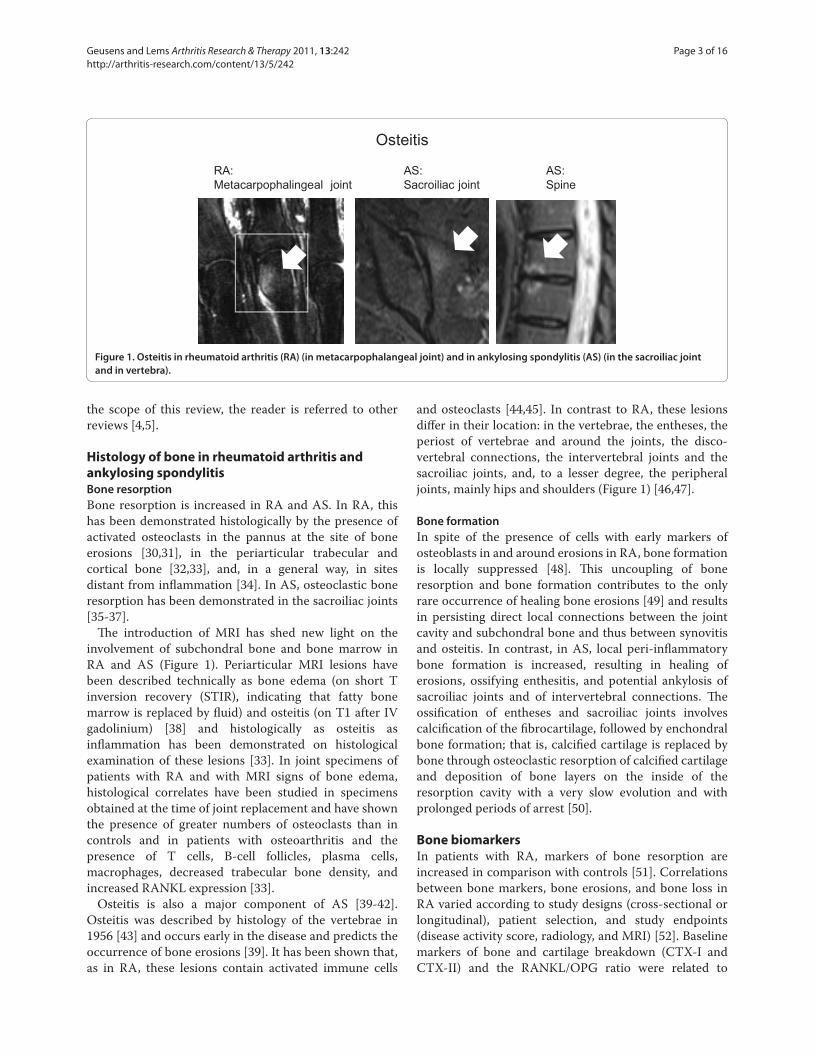
Methods that quantify changes in periarticular bone
include radiogrammetry, digitalized radiogrammetry
(DXR) [65], peripheral dual-energy x-ray absorptiometry
(pDXA) [66], quantitative ultrasound (QUS) [67], high-
resolution digital radiography [68], high-resolution peri-
pheral qCT [9], and MRI [8], and methods that quantify
changes in the vertebrae include DXA, qCT, MRI, and
morphometry by vertebral fracture assessment on x-rays
or DXA images [69] (Figure 2). At other sites of the
skeleton, single x-ray absorptiometry, qCT, MRI, DXA,
and QUS are available; of these, DXA is considered the
gold standard [70]. Semiquantitative scoring of osteitis
on MRI in the vertebrae has been standardized [40,42,71].
Local peri-infl ammatory bone formation can be evalu-
ated semiquantitatively in a standardized way on radio-
graphs for scoring of syndesmophytes [41,42,72]. Th ese
techniques diff er in regions of interest that can be
measured, in the ability to measure cortical and trabe-
cular bone separately or in combination, and in radiation
dose, cost, and precision [64,73] (Table 1).
Periarticular bone loss and osteitis in rheumatoid
arthritis
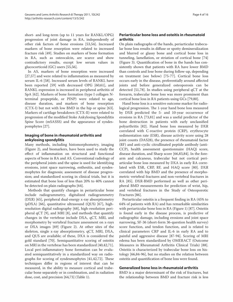
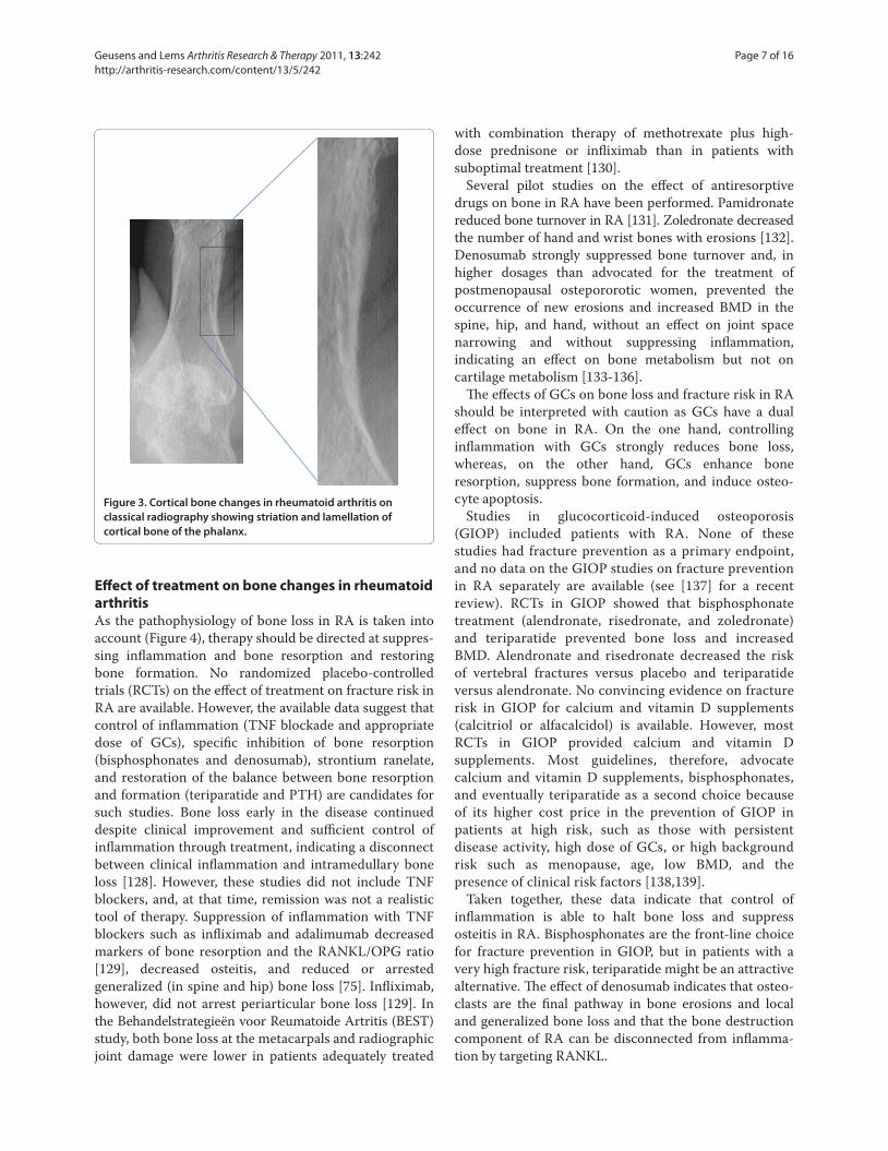
On plain radiographs of the hands, periarticular trabe cu-
lar bone loss results in diff use or spotty demineralization
and blurred or glassy bone and cortical bone loss in
tunneling, lamellation, or striation of cortical bone [74]
(Figure 3). Quantifi cation of bone in the hands has con-
sis tently shown that patients with RA have lower BMD
than controls and lose bone during follow-up, depending
on treatment (see below) [75-77]. Cortical bone loss
occurs early in the disease, preferentially around aff ected
joints and before generalized osteoporosis can be
detected [51,78]. In studies using peripheral qCT at the
forearm, trabecular bone loss was more prominent than
cortical bone loss in RA patients using GCs [79,80].
Hand bone loss is a sensitive outcome marker for radio-
logical progression. Th e 1-year hand bone loss measured
by DXR predicted the 5- and 10-year occurrence of
erosions in RA [73,81] and was a useful predictor of the
bone destruction in patients with early unclassifi ed
polyarthritis [82]. Hand bone loss measured by DXR
correlated with C-reactive protein (CRP), erythrocyte
sedimentation rate (ESR), disease activity score using 28
joint counts (DAS28), the presence of rheumatoid factor
(RF) and anti-cyclic citrullinated peptide antibody (anti-
CCP), health assessment questionnaire (HAQ) score,
disease duration, and Sharp score [66,83,84]. In the fore-
arm and calcaneus, trabecular but not cortical peri-
articular bone loss measured by DXA in early RA corre-
lated with ESR, CRP, RF, and HAQ score [80]. DXR
correlated with hip BMD and the presence of morpho-
metric vertebral fractures and non-vertebral fractures in
RA [85]. DXR-BMD performed as well as other peri-
pheral BMD measurements for prediction of wrist, hip,
and vertebral fractures in the Study of Osteoporotic
Fractures [86].
Periarticular osteitis is a frequent fi nding in RA (45% to
64% of patients with RA) and has remarkable similarities
with periarticular bone loss in RA (Figure 1) [87]. Osteitis
is found early in the disease process, is predictive of
radiographic damage, including erosions and joint space
narrowing, SF-36 (short-form 36-question health survey)
score function, and tendon function, and is related to
clinical parameters CRP and IL-6 in early RA and to
painful and aggressive disease [87-94]. Scoring of MRI
edema has been standardized by OMERACT (Outcome
Measures in Rheumatoid Arthritis Clinical Trials) [88].
Osteitis is characterized by trabecular bone loss on his-
tology [66,84-96], but no studies on the relation between
osteitis and quantifi cation of bone loss were found.
Generalized bone loss in rheumatoid arthritis
BMD is a major determinant of the risk of fractures, but
the relationship between BMD and fracture risk is less
Geusens and Lems Arthritis Research & Therapy 2011, 13:242 http://arthritis-research.com/content/13/5/242
Page 4 of 16
clear in RA than in postmenopausal osteoporosis,
indicat ing that factors other than those captured by
measuring BMD are involved in the pathophysiology of
fractures in RA.
Patients with RA have a decreased BMD in the spine
and hip and consequently have a higher prevalence of
osteoporosis [56,97-101]. However, this was not con-
fi rmed in the Canadian Multicentre Osteoporosis Study
(CaMos) [102]. In early untreated RA, BMD was related
to longer symptom duration, the presence of RF [103]
and anti-CCP [104], disease activity score [105], and the
presence and progression of joint damage [106].
Th e interpretation of longitudinal changes in RA is
complicated by the lack of untreated patients, and this
limits our insights into the natural evolution of bone
changes in RA to the above-mentioned studies. In one
study with early untreated RA, bone loss was found in
the spine and trochanter for a period of one year [107].
However, Kroot and colleagues [108] did not fi nd bone
loss over the course of a 10-year follow-up in RA patients
treated with disease-modifying anti rheumatic drugs,
except when these patients were treated with GCs.
Generalized bone loss was related to joint damage in
some studies [109,110], but this relation dis appeared after
multivariate adjustment [111]. No correla tion between
BMD and the presence of vertebral frac tures in RA
patients treated with GCs was found [112].
Fracture risk in rheumatoid arthritis
In the largest epidemiological study, patients with RA
were at increased risk for fractures of osteoporotic
fractures (relative risk (RR) 1.5), fractures of the hip (RR
2.0), clinical vertebral fractures (RR 2.4), and fractures of
the pelvis (RR 2.2) [113]. Th e risk of morphometric
vertebral fractures was also increased [114,115]. In some
but not all studies, the risk of fractures of the humerus
(RR 1.9), wrist (RR 1.2), and tibia/fi bula (RR 1.3) was
increased [75,116,117].
Th e etiology of increased fracture risk in RA is
multifactorial and superimposed on and independent of
Figure 2. Methods to quantify bone changes in the hands and vertebrae. (a) Methods to quantify periarticular bone changes. (b) Methods
to quantify vertebral bone changes. μCT, micro-computed tomography; DXA, dual-energy x-ray absorptiometry; DXR, digitalized radiogrammetry;
HRDR, high-resolution digital radiology; MRI, magnetic resonance imaging; QCT, quantitative computer tomography; QUS, quantitative ultrasound;
VFA, vertebral fracture assessment.
Radiogrammetry
DXA
DXR
QUS
HRDR
μCT
MRI
Morphometry(radiology, VFA)
DXA
QCT
MRI
(a)
(b)
Geusens and Lems Arthritis Research & Therapy 2011, 13:242 http://arthritis-research.com/content/13/5/242
Page 5 of 16
BMD and other clinical risk factors for fractures, includ-
ing the use of GCs. RA is included as an independent
clinical risk factor for 10-year fracture risk calculation for
major and hip fractures in the fracture risk assessment
tool (FRAX) case-fi nding algorithm [118]. Stress frac-
tures have been found in 0.8% of patients with RA, can be
diffi cult to diagnose, and were related to GC use but not
to BMD [119].
Fracture risk in RA was related to the duration of RA
[120], the severity of disease, and its musculoskeletal
conse quences, such as disability, HAQ score, lack of
physical activity, and impaired grip strength [120-122].
Vertebral fractures were related to disease duration and
severity [69]. In the general population, fracture risk was
related to serum levels of IL-6, TNF, and CRP [123] and
parameters of bone resorption [124], all of which can be
increased in RA. Extraskeletal risk factors that infl uence
fracture risk include increased risk of fall rates which
were related to number of swollen joints and impaired
balance tests [125].
Risk predictors of bone changes in rheumatoid
arthritis
Currently, the most widely used case-fi nding algorithm
for calculating the 10-year fracture risk for major and hip
fractures is the FRAX tool [118]. FRAX includes RA as a
risk for fractures, independently of and superimposed on
other risk factors, including BMD and use of GCs [118].
No fracture risk calculator that also includes other risk
factors that are related to RA, such as disease duration
and disease severity, is available. Th e Garvan fracture risk
calculator (GFRC) can be used to calculate the 5- and
10-year fracture risk which includes the number of recent
falls and the number of previous fractures but lacks RA
as a risk factor [126]. Fracture risk is higher with GFRC
than with FRAX in patients with recent falls [126]. In
view of the increased fracture risk in patients with RA,
systematic evaluation of fracture risk should be
considered using FRAX, disease severity, and duration,
and GFRC is helpful when patients report recent falls.
Risk of low BMD is diffi cult to estimate in RA [90], and
this suggests that bone densito metry should also be
considered in fracture risk calculation in patients with
active RA [127]. Many risk factors, including baseline
disease severity, RF, anti-CCP, baseline bone destruction,
the RANKL/OPG ratio, and CTX-I and CTX-II, have
been identifi ed for the prediction of bone erosions in RA.
Th is pallet of predictors can now be extended with
measurement of changes in periarticular bone (by DXR)
and osteitis (on MRI) early in the disease [73,81,82].
Additional studies will be necessary to study the relation
between osteitis and bone loss.
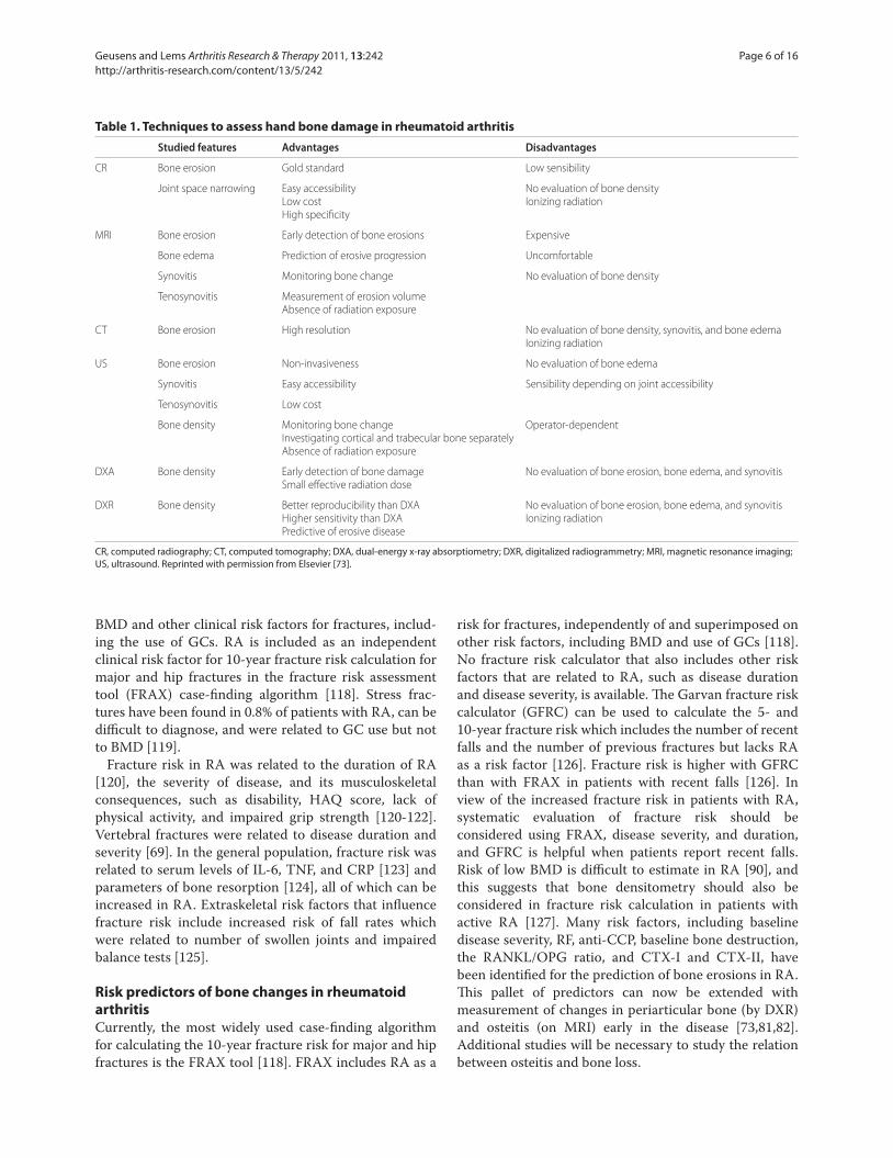
Table 1. Techniques to assess hand bone damage in rheumatoid arthritis
Studied features Advantages Disadvantages
CR Bone erosion Gold standard Low sensibility
Joint space narrowing Easy accessibility No evaluation of bone density
Low cost Ionizing radiation
High specifi city
MRI Bone erosion Early detection of bone erosions Expensive
Bone edema Prediction of erosive progression Uncomfortable
Synovitis Monitoring bone change No evaluation of bone density
Tenosynovitis Measurement of erosion volume
Absence of radiation exposure
CT Bone erosion High resolution No evaluation of bone density, synovitis, and bone edema
Ionizing radiation
US Bone erosion Non-invasiveness No evaluation of bone edema
Synovitis Easy accessibility Sensibility depending on joint accessibility
Tenosynovitis Low cost
Bone density Monitoring bone change Operator-dependent
Investigating cortical and trabecular bone separately
Absence of radiation exposure
DXA Bone density Early detection of bone damage No evaluation of bone erosion, bone edema, and synovitis
Small eff ective radiation dose
DXR Bone density Better reproducibility than DXA No evaluation of bone erosion, bone edema, and synovitis
Higher sensitivity than DXA Ionizing radiation
Predictive of erosive disease
CR, computed radiography; CT, computed tomography; DXA, dual-energy x-ray absorptiometry; DXR, digitalized radiogrammetry; MRI, magnetic resonance imaging; US, ultrasound. Reprinted with permission from Elsevier [73].
Geusens and Lems Arthritis Research & Therapy 2011, 13:242 http://arthritis-research.com/content/13/5/242
Page 6 of 16
Eff ect of treatment on bone changes in rheumatoid
arthritis
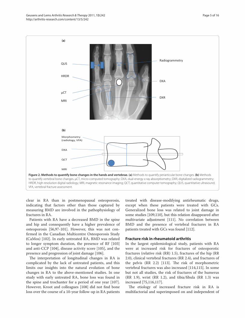
As the pathophysiology of bone loss in RA is taken into
account (Figure 4), therapy should be directed at suppres-
sing infl ammation and bone resorption and restoring
bone formation. No randomized placebo-controlled
trials (RCTs) on the eff ect of treatment on fracture risk in
RA are available. However, the available data suggest that
control of infl ammation (TNF blockade and appropriate
dose of GCs), specifi c inhibition of bone resorption
(bisphosphonates and denosumab), strontium ranelate,
and restoration of the balance between bone resorption
and formation (teriparatide and PTH) are candidates for
such studies. Bone loss early in the disease continued
despite clinical improvement and suffi cient control of
infl ammation through treatment, indicating a disconnect
between clinical infl ammation and intramedullary bone
loss [128]. However, these studies did not include TNF
blockers, and, at that time, remission was not a realistic
tool of therapy. Suppression of infl ammation with TNF
blockers such as infl iximab and adalimumab decreased
markers of bone resorption and the RANKL/OPG ratio
[129], decreased osteitis, and reduced or arrested
generalized (in spine and hip) bone loss [75]. Infl iximab,
however, did not arrest periarticular bone loss [129]. In
the Behandelstrategieën voor Reumatoide Artritis (BEST)
study, both bone loss at the metacarpals and radiographic
joint damage were lower in patients adequately treated
with combination therapy of methotrexate plus high-
dose prednisone or infl iximab than in patients with
suboptimal treatment [130].
Several pilot studies on the eff ect of antiresorptive
drugs on bone in RA have been performed. Pamidronate
reduced bone turnover in RA [131]. Zoledronate decreased
the number of hand and wrist bones with erosions [132].
Denosumab strongly suppressed bone turnover and, in
higher dosages than advocated for the treatment of
postmenopausal ostepororotic women, prevented the
occurrence of new erosions and increased BMD in the
spine, hip, and hand, without an eff ect on joint space
narrowing and without suppressing infl ammation,
indicating an eff ect on bone metabolism but not on
cartilage metabolism [133-136].
Th e eff ects of GCs on bone loss and fracture risk in RA
should be interpreted with caution as GCs have a dual
eff ect on bone in RA. On the one hand, controlling
infl am mation with GCs strongly reduces bone loss,
whereas, on the other hand, GCs enhance bone
resorption, suppress bone formation, and induce osteo-
cyte apoptosis.
Studies in glucocorticoid-induced osteoporosis
(GIOP) included patients with RA. None of these
studies had fracture prevention as a primary endpoint,
and no data on the GIOP studies on fracture prevention
in RA separately are available (see [137] for a recent
review). RCTs in GIOP showed that bisphosphonate
treatment (alendronate, risedronate, and zoledronate)
and teripara tide prevented bone loss and increased
BMD. Alendro nate and risedronate decreased the risk
of vertebral fractures versus placebo and teriparatide
versus alendro nate. No convincing evidence on fracture
risk in GIOP for calcium and vitamin D supplements
(calcitriol or alfacalcidol) is available. However, most
RCTs in GIOP provided calcium and vitamin D
supplements. Most guide lines, therefore, advocate
calcium and vitamin D supple ments, bisphosphonates,
and eventually teripara tide as a second choice because
of its higher cost price in the prevention of GIOP in
patients at high risk, such as those with persistent
disease activity, high dose of GCs, or high background
risk such as menopause, age, low BMD, and the
presence of clinical risk factors [138,139].
Taken together, these data indicate that control of
infl am mation is able to halt bone loss and suppress
osteitis in RA. Bisphosphonates are the front-line choice
for fracture prevention in GIOP, but in patients with a
very high fracture risk, teriparatide might be an attractive
alternative. Th e eff ect of denosumab indicates that osteo-
clasts are the fi nal pathway in bone erosions and local
and generalized bone loss and that the bone destruction
component of RA can be disconnected from infl am ma-
tion by targeting RANKL.
Figure 3. Cortical bone changes in rheumatoid arthritis on
classical radiography showing striation and lamellation of
cortical bone of the phalanx.
Geusens and Lems Arthritis Research & Therapy 2011, 13:242 http://arthritis-research.com/content/13/5/242
Page 7 of 16
Generalized bone loss in ankylosing spondylitis
Bone loss in the vertebrae occurs early in the disease, as
shown by DXA [140] and qCT [141]. In advanced disease,
the occurrence of syndesmophytes and periosteal and
discal bone apposition does not allow intravertebral bone
changes with DXA to be measured accurately. Combined
analyses of DXA and QCT in patients with early and
long-standing disease indicate that bone loss in the
vertebrae occurs early in the disease and can be measured
by DXA and QCT but that, in long-standing disease,
DXA of the spine can be normal, in spite of further
intravertebral bone loss as shown with qCT [142,143]. As
a result, in early disease, osteoporosis was found more
frequently in the spine than in the hip, whereas in
patients with long-standing disease, osteoporosis was
more frequent in the hip [75]. Hip BMD was related to
the presence of syndesmophytes and vertebral fractures,
to disease duration and activity [142,144], and to CRP
[145]. Osteitis in the vertebrae precedes the development
of erosions and syndesmophytes [41,42].
Fracture risk in ankylosing spondylitis
Morphometric vertebral fractures (with a deformation of
15% or 20%) have been reported to be 10% to 30% in
groups of patients with AS [146]. Th e odds ratios of
clinical vertebral fractures were 7.7 in a retrospective
population-based study [147] and 3.3 in a primary care-
based nested case control study [148]. In both studies,
the risk of non-vertebral fractures was not increased.
Th e risk of vertebral fractures is multifactorial and
inde pendent of and superimposed on other clinical risk
factors [118].
Vertebral fracture risk in AS was higher in men than in
women and was associated with low BMD, disease
activity, and the extent of syndesmophytes [144,149].
Vertebral fractures contributed to irreversible hyper-
kyphosis, which is characteristic in some patients with
advanced disease with extensive syndesmophytes (bamboo
spine) [150,151].
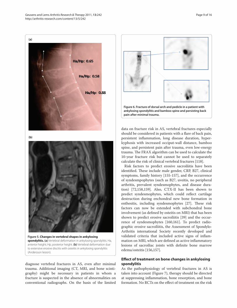
Apart from presenting with these ‘classical’ vertebral
fractures, patients with AS can present with vertebral
fractures that are specifi cally reported in AS. First,
erosions at the anterior corners and at the endplates of
vertebrae (Andersson and Romanus lesions) result in
vertebral deformities if erosions are extensive and the
results of such measurements should not be considered a
classical vertebral fracture (Figure 5) [75,152]. Second, in
a survey of 15,000 patients with AS, 0.4% reported
clinical vertebral fractures with major neuro logical
complications [153]. Th ird, owing to the stiff ening of the
spine by syndesmophytes, transvertebral fractures have
been described [153]. Fourth, fractures can occur in the
ossifi ed connections between the vertebrae [153]. In all of
these cases, CT, MRI, and eventually bone scintigraphy
are helpful to identify these lesions and the extent of
neurological consequences (Figure 6) [154].
Risk predictors of bone changes in ankylosing
spondylitis
Th e diagnosis of vertebral fractures is hampered by the
fi nding that only one out of three morphometric vertebral
fractures is accompanied by clinical signs and symptoms
of an acute fracture. Th is is probably even less in patients
with AS as fractures of the vertebrae and their annexes
can be easily overlooked when a fl are of back pain is
considered to be of infl ammatory origin without taking
into account the possibility of a fracture. In case of a fl are
of back pain, special attention, therefore, is necessary to
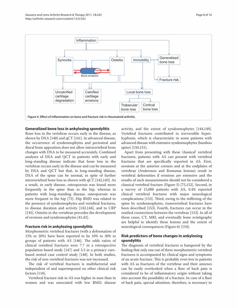
Figure 4. Eff ect of infl ammation on bone and fracture risk in rheumatoid arthritis.
Inflammation
Calcifiedcartilageerosions
Local bone loss
Trabecularbone loss
Corticalbone loss
Generalisedbone loss
Uncalcifiedcartilagedegradation
ImmobilitySynovitis Osteitis
Bone erosionsFracture risk
Geusens and Lems Arthritis Research & Therapy 2011, 13:242 http://arthritis-research.com/content/13/5/242
Page 8 of 16
diagnose vertebral fractures in AS, even after minimal
trauma. Additional imaging (CT, MRI, and bone scinti-
graphy) might be necessary in patients in whom a
fracture is suspected in the absence of abnormalities on
conventional radiographs. On the basis of the limited
data on fracture risk in AS, vertebral fractures especially
should be considered in patients with a fl are of back pain,
persistent infl ammation, long disease duration, hyper-
kyphosis with increased occiput-wall distance, bamboo
spine, and persistent pain after trauma, even low-energy
trauma. Th e FRAX algorithm can be used to calculate the
10-year fracture risk but cannot be used to separately
calculate the risk of clinical vertebral fractures [118].
Risk factors to predict erosive sacroiliitis have been
identifi ed. Th ese include male gender, CRP, B27, clinical
symptoms, family history [155-157], and the occurrence
of syndesmpophytes (such as B27, uveitis, no peripheral
arthritis, prevalent syndesmophytes, and disease dura-
tion) [72,158,159]. Also, CTX-II has been shown to
predict syndesmophytes, which could refl ect cartilage
destruc tion during enchondral new bone formation in
enthesitis, including syndesmophytes [27]. Th ese risk
factors can now be extended with subchondral bone
involvement (as defi ned by osteitis on MRI) that has been
shown to predict erosive sacroiliitis [39] and the occur-
rence of syndesmophytes [160,161]. To predict radio-
graphic erosive sacro iliitis, the Assessment of Spondylo-
Arthritis international Society recently developed and
validated criteria that included active signs of infl am-
mation on MRI, which are defi ned as active infl ammatory
lesions of sacroiliac joints with defi nite bone marrow
edema/osteitis [156,157].
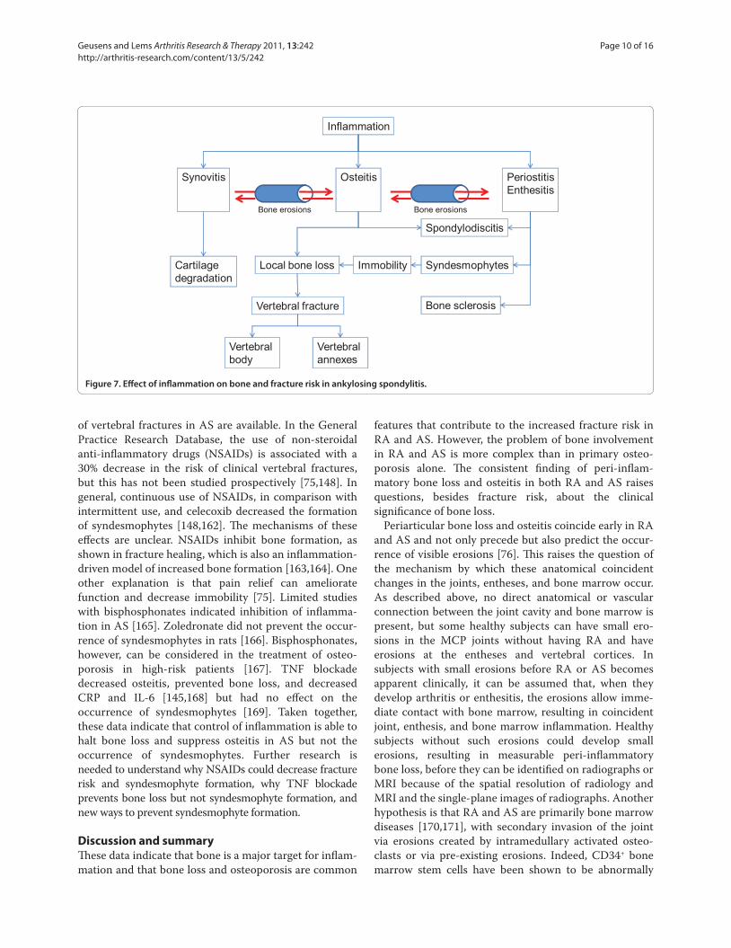
Eff ect of treatment on bone changes in ankylosing
spondylitis
As the pathophysiology of vertebral fractures in AS is
taken into account (Figure 7), therapy should be directed
at suppressing infl ammation, bone resorption, and bone
formation. No RCTs on the eff ect of treatment on the risk
Figure 5. Changes in vertebral shapes in ankylosing
spondylitis. (a) Vertebral deformation in ankylosing spondylitis. Ha,
anterior height; Hp, posterior height. (b) Vertebral deformation due
to extensive erosive discitis with osteitis in ankylosing spondylitis
(Andersson lesion).
(a)
(b)
Figure 6. Fracture of dorsal arch and pedicle in a patient with
ankylosing spondylitis and bamboo spine and persisting back
pain after minimal trauma.
Geusens and Lems Arthritis Research & Therapy 2011, 13:242 http://arthritis-research.com/content/13/5/242
Page 9 of 16
of vertebral fractures in AS are available. In the General
Practice Research Database, the use of non-steroidal
anti-infl ammatory drugs (NSAIDs) is associated with a
30% decrease in the risk of clinical vertebral fractures,
but this has not been studied prospectively [75,148]. In
general, continuous use of NSAIDs, in com parison with
intermittent use, and celecoxib decreased the formation
of syndesmophytes [148,162]. Th e mechanisms of these
eff ects are unclear. NSAIDs inhibit bone formation, as
shown in fracture healing, which is also an infl ammation-
driven model of increased bone formation [163,164]. One
other explanation is that pain relief can ameliorate
function and decrease immobility [75]. Limited studies
with bisphosphonates indicated inhibi tion of infl am ma-
tion in AS [165]. Zoledronate did not prevent the occur-
rence of syndesmophytes in rats [166]. Bisphosphonates,
however, can be considered in the treatment of osteo-
porosis in high-risk patients [167]. TNF blockade
decreased osteitis, prevented bone loss, and decreased
CRP and IL-6 [145,168] but had no eff ect on the
occurrence of syndesmophytes [169]. Taken together,
these data indicate that control of infl ammation is able to
halt bone loss and suppress osteitis in AS but not the
occurrence of syndesmophytes. Further research is
needed to understand why NSAIDs could decrease fracture
risk and syndesmophyte formation, why TNF blockade
prevents bone loss but not syndesmophyte formation, and
new ways to prevent syndesmophyte formation.
Discussion and summary
Th ese data indicate that bone is a major target for infl am-
mation and that bone loss and osteoporosis are common
features that contribute to the increased fracture risk in
RA and AS. However, the problem of bone involvement
in RA and AS is more complex than in primary osteo-
porosis alone. Th e consistent fi nding of peri-infl am-
matory bone loss and osteitis in both RA and AS raises
questions, besides fracture risk, about the clinical
signifi cance of bone loss.
Periarticular bone loss and osteitis coincide early in RA
and AS and not only precede but also predict the occur-
rence of visible erosions [76]. Th is raises the question of
the mechanism by which these anatomical coincident
changes in the joints, entheses, and bone marrow occur.
As described above, no direct anatomical or vascular
connection between the joint cavity and bone marrow is
present, but some healthy subjects can have small ero-
sions in the MCP joints without having RA and have
erosions at the entheses and vertebral cortices. In
subjects with small erosions before RA or AS becomes
apparent clinically, it can be assumed that, when they
develop arthritis or enthesitis, the erosions allow imme-
diate contact with bone marrow, resulting in coincident
joint, enthesis, and bone marrow infl ammation. Healthy
subjects without such erosions could develop small
erosions, resulting in measurable peri-infl ammatory
bone loss, before they can be identifi ed on radiographs or
MRI because of the spatial resolution of radiology and
MRI and the single-plane images of radiographs. Another
hypothesis is that RA and AS are primarily bone marrow
diseases [170,171], with secondary invasion of the joint
via erosions created by intramedullary activated osteo-
clasts or via pre-existing erosions. Indeed, CD34+ bone
marrow stem cells have been shown to be abnormally
Figure 7. Eff ect of infl ammation on bone and fracture risk in ankylosing spondylitis.
Inflammation
Synovitis Osteitis PeriostitisEnthesitis
Cartilagedegradation
Local bone loss
Vertebral fracture
Vertebral body
Vertebral annexes
Immobility Syndesmophytes
Bone sclerosis
Spondylodiscitis
Bone erosions Bone erosions
Geusens and Lems Arthritis Research & Therapy 2011, 13:242 http://arthritis-research.com/content/13/5/242
Page 10 of 16
sensitive to TNFα to produce fi broblast-like cells [172],
suggesting an underlying bone marrow stem cell
abnormality in RA.
In AS, the fi nding of early osteitis is even more
intriguing as osteitis is occurring in the vertebrae, where
no synovium but periost is present at the anterior sites
and discs between vertebrae. Local communication with
the periost is possible by the local vascular connections
or pre-existing erosions, leaving open the possibility that
periost is the primary location of infl ammation in AS.
Th e same applies for the intervertebral disc, which has no
direct vascular contact but can have pre-existing ero-
sions. Whether RA and AS are initialized in the joints,
enthesis, or the bone marrow is a growing fi eld of debate
[170], and such hypotheses will need much more study.
Regardless of these anatomical considerations, when
the size of bone edema that can be found by MRI and the
extent of early periarticular bone loss are taken into
account, it seems that infl ammation is as intense and
extensive inside bone marrow as in the synovial joint in
RA and AS and in the enthesis in AS. As bone loss and
bone edema occur early in the disease, these fi ndings
indicate that bone marrow infl ammation – and not just
joint or enthesis infl ammation – is a classical feature of
early RA and AS. To what degree impaired osteoblast
function is associated with loss of control of HSC and B-
cell diff erentiation in their subendosteal niches in RA is
unknown and needs further study as B-cell proliferation
is a feature of RA but not of AS [173-175].
Th e fi nding that bone involvement can be disconnected
from clinically detectable infl ammation is quite intrigu-
ing. In RA, bone erosions can progress even when the
infl ammatory process is adequately controlled (that is, in
clinical remission) [176], and progress of bone erosions
can be halted by denosumab in spite of persistent infl am-
mation [133-136]. In AS, the occurrence of syndesmo-
phytes can progress in spite of suppression of infl am-
mation by TNF blockade [160]. Th ese fi ndings have been
described as a disconnection between infl ammation and
bone destruction and repair.
Th e correlation and eventual disconnection between
osteitis and bone loss, parameters of disease activity, and
erosions suggest a dual time-dependent role for the
occurrence of erosions. Early in the disease process, the
primary negative eff ect of pre-existing or newly formed
erosions is the connection they create between the bone
marrow and the joints, periost, and entheses. In this way,
erosions contribute to local amplifi cation of infl ammation
by allowing bone marrow cells to have direct local
connection with extraosseous structures and creating a
vicious circle of infl ammation between joints, periost,
entheses, and bone marrow [177]. Only in a later stage do
erosions contribute to loss of function [178]. In this
hypothesis, the attack of infl ammation on bone by
stimulating osteoclasts has far-reaching consequences.
First, it would indicate that timely disease suppression
and the prevention of the development of a fi rst erosion
rather than halting erosion progression should be
considered a primary objective, both in RA and AS [179].
Second, periarticular bone loss and osteitis should be
considered, at least theoretically, an indication for the
presence of erosions, even when erosions cannot be
visualized on radiographs or MRI, and periarticular bone
loss and osteitis should be considered an indication for
early aggressive therapy [180]. Of course, the eff ectiveness
of antirheumatic treatment based on osteitis should be
demonstrated. Th ird, the fi nding of disconnection
between infl ammation and bone involvement indicates
that, even when infl ammation is clinically under control,
the degree to which bone-directed therapy is indicated
should be studied in order to prevent (further) progres-
sion of erosions and syndesmophytes. In conclusion, the
involve ment of bone as a major target of infl ammation in
RA and AS raises many questions [10,181-184], opening
perspectives for further research in the understanding
and treatment of the complex bone disease component of
RA and AS.
Abbreviations
anti-CCP, anti-cyclic citrullinated peptide antibody; AS, ankylosing
spondylitis; BMD, bone mineral density; BMP, bone morphogenetic protein;
CRP, C-reactive protein; CT, computed tomography; DKK, Dikkoppf; DXA,
dual-energy x-ray absorptiometry; DXR, digitalized radiogrammetry; ESR,
erythrocyte sedimentation rate; FRAX, fracture risk assessment tool; GC,
glucocorticoid; GFRC, Garvan fracture risk calculator; GIOP, glucocorticoid-
induced osteoporosis; HAQ, health assessment questionnaire; HRqCT, high-
resolution quantitative computer tomography; HSC, hematopoietic stem
cell; IFNγ, interferon-gamma; IL, interleukin; MCP, metacarpophalangeal; MRI,
magnetic resonance imaging; NSAID, non-steroidal anti-infl ammatory drug;
OPG, osteoprotegerin; PTH, parathyroid hormone; qCT, quantitative computer
tomography; QUS, quantitative ultrasound; RA, rheumatoid arthritis; RANK,
receptor activator of the nuclear factor-kappa-B; RANKL, receptor activator of
the nuclear factor-kappa-B ligand; RCT, randomized placebo-controlled trial;
RF, rheumatoid factor; RR, relative risk; SpA, spondylarthopathy; TNF, tumor
necrosis factor.
Competing interests
WFL has received speaking fees form Amgen, Eli Lilly, Merck, and Procter and
Gamble.
Author details1Department of Internal Medicine, Subdivision of Rheumatology, Maastricht
University Medical Center, P. Debyelaan 25 Postbus 5800, 6202 AZ Maastricht,
The Netherlands. 2Biomedical Research Institute, University of Hasselt, Belgium. 3Department of Rheumatology, 3A61 VU Medical Center, Postbox 7057, 1007
MB Amsterdam, The Netherlands.
Published: 30 September 2011
References
1. Takayanagi H: Osteoimmunology and the eff ects of the immune system on bone. Nat Rev Rheumatol 2009, 5:667-676.
This article is part of the series Osteoimmunology, edited by
Georg Schett. Other articles in this series can be found at
http://arthritis-research.com/series/osteoimmunology
Geusens and Lems Arthritis Research & Therapy 2011, 13:242 http://arthritis-research.com/content/13/5/242
Page 11 of 16

2. Fuller K, Wong B, Fox S, Choi Y, Chambers TJ: TRANCE is necessary and suffi cient for osteoblast-mediated activation of bone resorption in osteoclasts. J Exp Med 1998, 188:997-1001.
3. Lacey DL, Timms E, Tan HL, Kelley MJ, Dunstan CR, Burgess T, Elliott R,
Colombero A, Elliott G, Scully S, Hsu H, Sullivan J, Hawkins N, Davy E,
Capparelli C, Eli A, Qian YX, Kaufman S, Sarosi I, Shalhoub V, Senaldi G, Guo J,
Delaney J, Boyle WJ: Osteoprotegerin ligand is a cytokine that regulates osteoclast diff erentiation and activation. Cell 1998, 93:165-176.
4. Lorenzo J, Horowitz M, Choi Y: Osteoimmunology: interactions of the bone and immune system. Endocr Rev 2008, 29:403-40.
5. Lorenzo J, Choi Y, Horowitz M, Takayanagi H (Editors): Osteoimmunology.
London: Academic Press, Elsevier Inc.; 2011.
6. Kanis JA: Assessment of fracture risk and its application to screening for postmenopausal osteoporosis: synopsis of a WHO report. WHO Study Group. Osteoporos Int 1994, 4:368-381.
7. Sommer OJ, Kladosek A, Weiler V, Czembirek H, Boeck M, Stiskal M:
Rheumatoid arthritis: a practical guide to state-of-the-art imaging, image interpretation, and clinical implications. Radiographics 2005, 25:381-398.
8. Ejbjerg B, Narvestad E, Rostrup E, Szkudlarek M, Jacobsen S, Thomsen HS,
Østergaard M: Magnetic resonance imaging of wrist and fi nger joints in healthy subjects occasionally shows changes resembling erosions and synovitis as seen in rheumatoid arthritis. Arthritis Rheum 2004,
50:1097-1106.
9. Stach CM, Bäuerle M, Englbrecht M, Kronke G, Engelke K, Manger B, Schett G:
Periarticular bone structure in rheumatoid arthritis patients and healthy individuals assessed by high-resolution computed tomography. Arthritis
Rheum 2010, 62:330-339.
10. McGonagle D, Tan AL, Møller Døhn U, Ostergaard M, Benjamin M:
Microanatomic studies to defi ne predictive factors for the topography of periarticular erosion formation in infl ammatory arthritis. Arthritis Rheum
2009, 60:1042-1051.
11. Benjamin M, Toumi H, Suzuki D, Redman S, Emery P, McGonagle D:
Microdamage and altered vascularity at the enthesis-bone interface provides an anatomic explanation for bone involvement in the HLA-B27-associated spondylarthritides and allied disorders. Arthritis Rheum 2007,
56:224-233.
12. François RJ, Dhem A: Microradiographic study of the normal human vertebral body. Acta Anat (Basel) 1974, 89:251-265.
13. Raisz LG: What marrow does to bone. N Engl J Med 1981, 304:1485-1486.
14. Colburn NT, Zaal KJ, Wang F, Tuan RS: A role for gamma/delta T cells in a mouse model of fracture healing. Arthritis Rheum 2009, 60:1694-1703.
15. Calvi LM, Adams GB, Weibrecht KW, Weber JM, Olson DP, Knight MC, Martin
RP, Schipani E, Divieti P, Bringhurst FR, Milner LA, Kronenberg HM, Scadden
DT: Osteoblastic cells regulate the haematopoietic stem cell niche. Nature
2003, 425:841-846.
16. Frisch BJ, Porter RL, Calvi LM: Hematopoietic niche and bone meet. Curr
Opin Support Palliat Care 2008, 2:211-217.
17. Yin T, Li L: The stem cell niches in bone. J Clin Invest 2006, 116:1195-1201.
18. Adams GB, Chabner KT, Alley IR, Olson DP, Szczepiorkowski ZM, Poznansky
MC, Kos CH, Pollak MR, Brown EM, Scadden DT: Stem cell engraftment at the endosteal niche is specifi ed by the calciumsensing receptor. Nature 2006,
439:599-603.
19. Lories RJ, Luyten FP: Bone morphogenetic proteins in destructive and remodeling arthritis. Arthritis Res Ther 2007, 9:207.
20. Diarra D, Stolina M, Polzer K, Zwerina J, Ominsky MS, Dwyer D, Korb A, Smolen
J, Hoff mann M, Scheinecker C, van der Heide D, Landewe R, Lacey D, Richards
WG, Schett G: Dickkopf-1 is a master regulator of joint remodeling. Nat
Med 2007, 13:156-163.
21. Appel H, Ruiz-Heiland G, Listing J, Zwerina J, Herrmann M, Mueller R, Haibel
H, Baraliakos X, Hempfi ng A, Rudwaleit M, Sieper J, Schett G: Altered skeletal expression of sclerostin and its link to radiographic progression in ankylosing spondylitis. Arthritis Rheum 2009, 60:3257-3262.
22. Goldring SR, Schett G: The role of the immune system in bone loss of infl ammatory arthritis. In Osteoimmunology. Edited by Lorenzo J, Choi Y,
Horowitz M, Takayanagi H. London: Academic Press, Elsevier Inc.;
2011:301-324.
23. Herman S, Müller RB, Krönke G, Zwerina J, Redlich K, Hueber AJ, Gelse H,
Neumann E, Müller-Ladner U, Schett G: Induction of osteoclast-associated receptor, a key osteoclast costimulation molecule, in rheumatoid arthritis. Arthritis Rheum 2008, 58:3041-3050.
24. Nemeth K, Schoppet M, Al-Fakhri N, Helas S, Jessberger R, Hofbauer LC,
Goettsch C: The role of osteoclast-associated receptor in osteoimmunology. J Immunol 2011, 186:13-18.
25. Daoussis D, Liossis SN, Solomou EE, Tsanaktsi A, Bounia K, Karampetsou M,
Yiannopoulos G, Andonopoulos AP: Evidence that Dkk-1 is dysfunctional in ankylosing spondylitis. Arthritis Rheum 2010, 62:150-158.
26. Chen HA, Chen CH, Lin YJ, Chen PC, Chen WS, Lu CL, Chou CT: Association of bone morphogenetic proteins with spinal fusion in ankylosing spondylitis. J Rheumatol 2010, 37:2126-2132.
27. Vosse D, Landewé R, Garnero P, van der Heijde D, van der Linden S, Geusens
P: Association of markers of bone- and cartilage-degradation with radiological changes at baseline and after 2 years follow-up in patients with ankylosing spondylitis. Rheumatology (Oxford) 2008, 47:1219-1222.
28. Braun J, Baraliakos X: Imaging of axial spondyloarthritis including ankylosing spondylitis. Ann Rheum Dis 2011, 70 Suppl 1:i97-103.
29. Teitelbaum SL: Postmenopausal osteoporosis, T cells, and immune dysfunction. Proc Natl Acad Sci U S A 2004, 101:16711-16712.
30. Leisen JC, Duncan H, Riddle JM, Pitchford WC: The erosive front: a topographic study of the junction between the pannus and the subchondral plate in the macerated rheumatoid metacarpal head. J Rheumatol 1988, 15:17-22.
31. Pettit AR, Walsh NC, Manning C, Goldring SR, Gravallese EM: RANKL protein is expressed at the pannus-bone interface at sites of articular bone erosion in rheumatoid arthritis. Rheumatology (Oxford) 2006, 45:1068-1076.
32. Bywaters EG: The early radiological signs of rheumatoid arthritis. Bull
Rheum Dis 1960, 11:231-234.
33. Jimenez-Boj E, Nöbauer-Huhmann I, Hanslik-Schnabel B, Dorotka R,
Wanivenhaus AH, Kainberger F, Trattnig S, Axmann R, Tsuji W, Hermann S,
Smolen J, Schett G: Bone erosions and bone marrow edema as defi ned by magnetic resonance imaging refl ect true bone marrow infl ammation in rheumatoid arthritis. Arthritis Rheum 2007, 56:1118-1124.
34. Reid DM, Kennedy NS, Smith MA, Tothill P, Nuki G: Total body calcium in rheumatoid arthritis: eff ects of disease activity and corticosteroid treatment. Br Med J (Clin Res Ed) 1982, 285:330-332.
35. Engfeldt B, Romanus R, Yden S: Histological studies of pelvo-spondylitis ossifi cans (ankylosing spondylitis) correlated with clinical and radiological fi ndings. Ann Rheum Dis 1954, 13:219-228.
36. François RJ, Gardner DL, Degrave EJ, Bywaters EG: Histopathologic evidence that sacroiliitis in ankylosing spondylitis is not merely enthesitis. Arthritis
Rheum 2000, 43:2011-2024.
37. Aufdermaur M: Pathogenesis of square bodies in ankylosing spondylitis. Ann Rheum Dis 1989, 48:628-631.
38. Rudwaleit M, Jurik AG, Hermann KG, Landewé R, van der Heijde D, Baraliakos
X, Marzo-Ortega H, Ostergaard M, Braun J, Sieper J: Defi ning active sacroiliitis on magnetic resonance imaging (MRI) for classifi cation of axial spondyloarthritis: a consensual approach by the ASAS/OMERACT MRI group. Ann Rheum Dis 2009, 68:1520-1527.
39. Zochling J, Baraliakos X, Hermann KG, Braun J: Magnetic resonance imaging in ankylosing spondylitis. Curr Opin Rheumatol 2007, 19:346-352.
40. Baraliakos X, Landewé R, Hermann KG, Listing J, Golder W, Brandt J, Rudwaleit
M, Bollow M, Sieper J, van der Heijde D, Braun J: Infl ammation in ankylosing spondylitis: a systematic description of the extent and frequency of acute spinal changes using magnetic resonance imaging. Ann Rheum Dis 2005,
64:730-734.
41. Maksymowych WP: MRI in ankylosing spondylitis. Curr Opin Rheumatol
2009, 21:313-317.
42. Maksymowych WP: Progress in spondylarthritis. Spondyloarthritis: lessons from imaging. Arthritis Res Ther 2009, 11:222.
43. Cruickshank B: Lesions of cartilaginous joints in ankylosing spondylitis. J Pathol Bacteriol 1956, 71:73-84.
44. Appel H, Loddenkemper C, Grozdanovic Z, Ebhardt H, Dreimann M,
Hempfi ng A, Stein H, Metz-Stavenhagen P, Rudwaleit M, Sieper J: Correlation of histopathological fi ndings and magnetic resonance imaging in the spine of patients with ankylosing spondylitis. Arthritis Res Ther 2006, 8:R143.
45. Appel H, Kuhne M, Spiekermann S, Ebhardt H, Grozdanovic Z, Köhler D,
Dreimann M, Hempfi ng A, Rudwaleit M, Stein H, Metz-Stavenhagen P, Sieper
J, Loddenkemper C: Immunohistologic analysis of zygapophyseal joints in patients with ankylosing spondylitis. Arthritis Rheum 2006, 54:2845-2851.
46. Benjamin M, McGonagle D: The enthesis organ concept and its relevance to the spondyloarthropathies. Adv Exp Med Biol 2009, 649:57-70.
47. McGonagle D, Gibbon W, Emery P: Classifi cation of infl ammatory arthritis by enthesitis. Lancet 1998, 352:1137-1140.
Geusens and Lems Arthritis Research & Therapy 2011, 13:242 http://arthritis-research.com/content/13/5/242
Page 12 of 16
48. Walsh NC, Reinwald S, Manning CA, Condon KW, Iwata K, Burr DB, Gravallese
EM: Osteoblast function is compromised at sites of focal bone erosion in infl ammatory arthritis. J Bone Miner Res 2009, 24:1572-1585.
49. Møller Døhn U, Boonen A, Hetland ML, Hansen MS, Knudsen LS, Hansen A,
Madsen OR, Hasselquist M, Møller JM, Østergaard M: Erosive progression is minimal, but erosion healing rare, in patients with rheumatoid arthritis treated with adalimumab. A 1 year investigator-initiated follow-up study using high-resolution computed tomography as the primary outcome measure. Ann Rheum Dis 2009, 68:1585-1590.
50. François RJ: Microradiographic study of the intervertebral bridges in ankylosing spondylitis and in the normal sacrum. Ann Rheum Dis 1965,
24:481-489.
51. Sambrook PN, Ansell BM, Foster S, Gumpel JM, Hesp R, Reeve J, Zanelli JM:
Bone turnover in early rheumatoid arthritis. 1. Biochemical and kinetic indexes. Ann Rheum Dis 1985, 44:575-579.
52. Garnero P, Delmas PD: Noninvasive techniques for assessing skeletal changes in infl ammatory arthritis: bone biomarkers. Curr Opin Rheumatol
2004, 16:428-434.
53. Geusens PP, Landewé RB, Garnero P, Chen D, Dunstan CR, Lems WF, Stinissen
P, van der Heijde DM, van der Linden S, Boers M: The ratio of circulating osteoprotegerin to RANKL in early rheumatoid arthritis predicts later joint destruction. Arthritis Rheum 2006, 54:1772-1777.
54. van Tuyl LH, Voskuyl AE, Boers M, Geusens P, Landewé RB, Dijkmans BA, Lems
WF: Baseline RANKL:OPG ratio and markers of bone and cartilage degradation predict annual radiological progression over 11 years in rheumatoid arthritis. Ann Rheum Dis 2010, 69:1623-1628.
55. Hall GM, Spector TD, Delmas PD: Markers of bone metabolism in postmenopausal women with rheumatoid arthritis. Eff ects of corticosteroids and hormone replacement therapy. Arthritis Rheum 1995,
38:902-906.
56. Deodhar AA, Woolf AD: Bone mass measurement and bone metabolism in rheumatoid arthritis: a review. Br J Rheumatol 1996, 35:309-322.
57. El Maghraoui A, Borderie D, Cherruau B, Edouard R, Dougados M, Roux C:
Osteoporosis, body composition, and bone turnover in ankylosing spondylitis. J Rheumatol 1999, 26:2205-2209.
58. MacDonald AG, Birkinshaw G, Durham B, Bucknall RC, Fraser WD:
Biochemical markers of bone turnover in seronegative spondylarthropathy: relationship to disease activity. Br J Rheumatol 1997,
36:50-53.
59. Stupphann D, Rauner M, Krenbek D, Patsch J, Pirker T, Muschitz C, Resch H,
Pietschmann P: Intracellular and surface RANKL are diff erentially regulated in patients with ankylosing spondylitis. Rheumatol Int 2008, 28:987-993.
60. Franck H, Meurer T, Hofbauer LC: Evaluation of bone mineral density, hormones, biochemical markers of bone metabolism, and osteoprotegerin serum levels in patients with ankylosing spondylitis. J Rheumatol 2004, 31:2236-2241.
61. Appel H, Maier R, Loddenkemper C, Kayser R, Meier O, Hempfi ng A, Sieper J:
Immunohistochemical analysis of osteoblasts in zygapophyseal joints of patients with ankylosing spondylitis reveal repair mechanisms similar to osteoarthritis. J Rheumatol 2010, 37:823-828.
62. Vandooren B, Cantaert T, Noordenbos T, Tak PP, Baeten D: The abundant synovial expression of the RANK/RANKL/Osteoprotegerin system in peripheral spondylarthritis is partially disconnected from infl ammation. Arthritis Rheum 2008, 58:718-729.
63. Arends S, Spoorenberg A, Bruyn GA, Houtman PM, Leijsma MK, Kallenberg
CG, Brouwer E, van der Veer E: The relation between bone mineral density, bone turnover markers, and vitamin D status in ankylosing spondylitis patients with active disease: a cross-sectional analysis. Osteoporos Int 2011,
22:1431-1439.
64. Njeh CF, Genant HK: Bone loss. Quantitative imaging techniques for assessing bone mass in rheumatoid arthritis. Arthritis Res 2000, 2:446-50.
65. Rosholm A, Hyldstrup L, Backsgaard L, Grunkin M, Thodberg HH: Estimation of bone mineral density by digital X-ray radiogrammetry: theoretical background and clinical testing. Osteoporos Int 2001, 12:961-969.
66. Deodhar AA, Brabyn J, Jones PW, Davis MJ, Woolf AD: Measurement of hand bone mineral content by dual energy x-ray absorptiometry: development of the method, and its application in normal volunteers and in patients with rheumatoid arthritis. Ann Rheum Dis 1994, 53:685-690.
67. Böttcher J, Pfeil A, Mentzel H, Kramer A, Schäfer ML, Lehmann G, Eidner T,
Petrovitch A, Malich A, Hein G, Kaiser WA: Peripheral bone status in rheumatoid arthritis evaluated by digital X-ray radiogrammetry and
compared with multisite quantitative ultrasound. Calcif Tissue Int 2006,
78:25-34.
68. Lespessailles E, Gadois C, Lemineur G, Do-Huu JP, Benhamou L: Bone texture analysis on direct digital radiographic images: precision study and relationship with bone mineral density at the os calcis. Calcif Tissue Int
2007, 80:97-102.
69. El Maghraoui A, Rezqi A, Mounach A, Achemlal L, Bezza A, Ghozlani I:
Prevalence and risk factors of vertebral fractures in women with rheumatoid arthritis using vertebral fracture assessment. Rheumatology
(Oxford) 2010, 49:1303-1310.
70. Johnell O, Kanis JA, Oden A, Johansson H, De Laet C, Delmas P, Eisman JA,
Fujiwara S, Kroger H, Mellstrom D, Meunier PJ, Melton LJ 3rd, O’Neill T, Pols H,
Reeve J, Silman A, Tenenhouse A: Predictive value of BMD for hip and other fractures. J Bone Miner Res 2005, 20:1185-1194. Erratum in: J Bone Miner Res
2007, 22:774.
71. Baraliakos X, Hermann KG, Landewé R, Listing J, Golder W, Brandt J, Rudwaleit
M, Bollow M, Sieper J, van der Heijde D, Braun J: Assessment of acute spinal infl ammation in patients with ankylosing spondylitis by magnetic resonance imaging: a comparison between contrast enhanced T1 and short tau inversion recovery (STIR) sequences. Ann Rheum Dis 2005,
64:1141-1144.
72. Baraliakos X, Listing J, Rudwaleit M, Haibel H, Brandt J, Sieper J, Braun J:
Progression of radiographic damage in patients with ankylosing spondylitis: defi ning the central role of syndesmophytes. Ann Rheum Dis
2007, 66:910-915.
73. Fouque-Aubert A, Chapurlat R, Miossec P, Delmas PD: A comparative review of the diff erent techniques to assess hand bone damage in rheumatoid arthritis. Joint Bone Spine 2010, 77:212-217.
74. Dihlman W: Joints and Vertebral Connections. New York: Thieme Inc.; 1985.
75. Roux C: Osteoporosis in infl ammatory joint diseases. Osteoporos Int 2011,
22:421-433.
76. Hoff M, Haugeberg G: Using hand bone measurements to assess progression of rheumatoid arthritis. Therapeutic Advances in Musculoskeletal
Disease 2010, 79-88.
77. Alenfeld FE, Diessel E, Brezger M, Sieper J, Felsenberg D, Braun J: Detailed analyses of periarticular osteoporosis in rheumatoid arthritis. Osteoporos
Int 2000, 11:400-407.
78. Sambrook PN, Ansell BM, Foster S, Gumpel JM, Hesp R, Reeve J: Bone turnover in early rheumatoid arthritis. 2. Longitudinal bone density studies. Ann Rheum Dis 1985, 44:580-584.
79. Laan RF, Buijs WC, van Erning LJ, Lemmens JA, Corstens FH, Ruijs SH, van de
Putte LB, van Riel PL: Diff erential eff ects of glucocorticoids on cortical appendicular and cortical vertebral bone mineral content. Calcif Tissue Int
1993, 52:5-9.
80. Inaba M, Nagata M, Goto H, Kumeda Y, Kobayashi K, Nakatsuka K, Miki T,
Yamada S, Ishimura E, Nishizawa Y: Preferential reductions of paraarticular trabecular bone component in ultradistal radius and of calcaneus ultrasonography in early-stage rheumatoid arthritis. Osteoporos Int 2003,
14:683-687.
81. Hoff M, Haugeberg G, Odegård S, Syversen S, Landewé R, van der Heijde D,
Kvien TK: Cortical hand bone loss after 1 year in early rheumatoid arthritis predicts radiographic hand joint damage at 5-year and 10-year follow-up. Ann Rheum Dis 2009, 68:324-329.
82. Haugeberg G, Green MJ, Quinn MA, Marzo-Ortega H, Proudman S, Karim Z,
Wakefi eld RJ, Conaghan PG, Stewart S, Emery P: Hand bone loss in early undiff erentiated arthritis: evaluating bone mineral density loss before the development of rheumatoid arthritis. Ann Rheum Dis 2006, 65:736-740.
83. Bøyesen P, Hoff M, Odegård S, Haugeberg G, Syversen SW, Gaarder PI,
Okkenhaug C, Kvien TK: Antibodies to cyclic citrullinated protein and erythrocyte sedimentation rate predict hand bone loss in patients with rheumatoid arthritis of short duration: a longitudinal study. Arthritis Res
Ther 2009, 11:R103.
84. Jawaid WB, Crosbie D, Shotton J, Reid DM, Stewart A: Use of digital x ray radiogrammetry in the assessment of joint damage in rheumatoid arthritis. Ann Rheum Dis 2006, 65:459-464.
85. Haugeberg G, Lodder MC, Lems WF, Uhlig T, Ørstavik RE, Dijkmans BA, Kvien
TK, Woolf AD: Hand cortical bone mass and its associations with radiographic joint damage and fractures in 50-70 year old female patients with rheumatoid arthritis: cross sectional Oslo-Truro-Amsterdam (OSTRA) collaborative study. Ann Rheum Dis 2004, 63:1331-1334.
86. Bouxsein ML, Palermo L, Yeung C, Black DM: Digital X-ray radiogrammetry
Geusens and Lems Arthritis Research & Therapy 2011, 13:242 http://arthritis-research.com/content/13/5/242
Page 13 of 16
predicts hip, wrist and vertebral fracture risk in elderly women: a prospective analysis from the study of osteoporotic fractures. Osteoporos
Int 2002, 13:358-365.
87. McQueen FM, Dalbeth N: Predicting joint damage in rheumatoid arthritis using MRI scanning. Arthritis Res Ther 2009, 11:124.
88. Bøyesen P, Haavardsholm EA, Ostergaard M, van der Heijde D, Sesseng S,
Kvien TK: MRI in early rheumatoid arthritis: synovitis and bone marrow oedema are independent predictors of subsequent radiographic progression. Ann Rheum Dis 2011, 70:428-433.
89. McQueen FM, Stewart N, Crabbe J, Robinson E, Yeoman S, Tan PL, McLean L:
Magnetic resonance imaging of the wrist in early rheumatoid arthritis reveals a high prevalence of erosions at four months after symptom onset. Ann Rheum Dis 1998, 57:350-356.
90. McQueen FM, Benton N, Perry D, Crabbe J, Robinson E, Yeoman S, McLean L,
Stewart N: Bone edema scored on magnetic resonance imaging scans of the dominant carpus at presentation predicts radiographic joint damage of the hands and feet six years later in patients with rheumatoid arthritis. Arthritis Rheum 2003, 48:1814-1827.
91. Hodgson RJ, O’Connor P, Moots R: MRI of rheumatoid arthritis image quantitation for the assessment of disease activity, progression and response to therapy. Rheumatology (Oxford) 2008, 47:13-21.
92. Bird P, Conaghan P, Ejbjerg B, McQueen F, Lassere M, Peterfy C, Edmonds J,
Shnier R, O’Connor P, Haavardsholm E, Emery P, Genant H, Østergaard M:
The development of the EULAR-OMERACT rheumatoid arthritis MRI reference image atlas. Ann Rheum Dis 2005, 64 Suppl 1:i8-10.
93. Tamai M, Kawakami A, Uetani M, Takao S, Arima K, Iwamoto N, Fujikawa K,
Aramaki T, Kawashiri SY, Ichinose K, Kamachi M, Nakamura H, Origuchi T, Ida
H, Aoyagi K, Eguchi K: A prediction rule for disease outcome in patients with undiff erentiated arthritis using magnetic resonance imaging of the wrists and fi nger joints and serologic autoantibodies. Arthritis Rheum 2009,
61:772-778.
94. Hetland ML, Ejbjerg B, Hørslev-Petersen K, Jacobsen S, Vestergaard A, Jurik
AG, Stengaard-Pedersen K, Junker P, Lottenburger T, Hansen I, Andersen LS,
Tarp U, Skjødt H, Pedersen JK, Majgaard O, Svendsen AJ, Ellingsen T,
Lindegaard H, Christensen AF, Vallø J, Torfi ng T, Narvestad E, Thomsen HS,
Ostergaard M; CIMESTRA study group: MRI bone oedema is the strongest predictor of subsequent radiographic progression in early rheumatoid arthritis. Results from a 2-year randomised controlled trial (CIMESTRA). Ann Rheum Dis 2009, 68:384-390.
95. McQueen FM, Gao A, Ostergaard M, King A, Shalley G, Robinson E, Doyle A,
Clark B, Dalbeth N: High-grade MRI bone oedema is common within the surgical fi eld in rheumatoid arthritis patients undergoing joint replacement and is associated with osteitis in subchondral bone. Ann
Rheum Dis 2007, 66:1581-1587.
96. Dalbeth N, Smith T, Gray S, Doyle A, Antill P, Lobo M, Robinson E, King A,
Cornish J, Shalley G, Gao A, McQueen FM: Cellular characterisation of magnetic resonance imaging bone oedema in rheumatoid arthritis; implications for pathogenesis of erosive disease. Ann Rheum Dis 2009,
68:279-282.
97. Oelzner P, Schwabe A, Lehmann G, Eidner T, Franke S, Wolf G, Hein G:
Signifi cance of risk factors for osteoporosis is dependent on gender and menopause in rheumatoid arthritis. Rheumatol Int 2008, 28:1143-1150.
98. Lane NE, Pressman AR, Star VL, Cummings SR, Nevitt MC: Rheumatoid arthritis and bone mineral density in elderly women. The Study of Osteoporotic Fractures Research Group. J Bone Miner Res 1995, 10:257-263.
99. Kröger H, Honkanen R, Saarikoski S, Alhava E: Decreased axial bone mineral density in perimenopausal women with rheumatoid arthritis - a population based study. Ann Rheum Dis 1994, 53:18-23.
100. Haugeberg G, Uhlig T, Falch JA, Halse JI, Kvien TK: Bone mineral density and frequency of osteoporosis in female patients with rheumatoid arthritis: results from 394 patients in the Oslo County Rheumatoid Arthritis register. Arthritis Rheum 2000, 43:522-30.
101. Bhalla AK, Shenstone B: Bone densitometry measurements in early infl ammatory disease. Baillieres Clin Rheumatol 1992, 6:405-414.
102. Hanley DA, Brown JP, Tenenhouse A, Olszynski WP, Ioannidis G, Berger C, Prior
JC, Pickard L, Murray TM, Anastassiades T, Kirkland S, Joyce C, Joseph L,
Papaioannou A, Jackson SA, Poliquin S, Adachi JD; Canadian Multicentre
Osteoporosis Study Research Group: Associations among disease conditions, bone mineral density, and prevalent vertebral deformities in men and women 50 years of age and older: cross-sectional results from the Canadian Multicentre Osteoporosis Study. J Bone Miner Res 2003,
18:784-790.
103. Güler-Yüksel M, Bijsterbosch J, Goekoop-Ruiterman YP, de Vries-Bouwstra JK,
Ronday HK, Peeters AJ, de Jonge-Bok JM, Breedveld FC, Dijkmans BA, Allaart
CF, Lems WF: Bone mineral density in patients with recently diagnosed, active rheumatoid arthritis. Ann Rheum Dis 2007, 66:1508-1512.
104. Guler H, Turhanoglu AD, Ozer B, Ozer C, Balci A: The relationship between anti-cyclic citrullinated peptide and bone mineral density and radiographic damage in patients with rheumatoid arthritis. Scand J
Rheumatol 2008, 37:337-342.
105. Wijbrandts CA, Klaasen R, Dijkgraaf MG, Gerlag DM, van Eck-Smit BL, Tak PP:
Bone mineral density in rheumatoid arthritis patients 1 year after adalimumab therapy: arrest of bone loss. Ann Rheum Dis 2009, 68:373-376.
106. Forslind K, Keller C, Svensson B, Hafström I; BARFOT Study Group: Reduced bone mineral density in early rheumatoid arthritis is associated with radiological joint damage at baseline and after 2 years in women. J Rheumatol 2003, 30:2590-2596.
107. Gough AK, Lilley J, Eyre S, Holder RL, Emery P: Generalised bone loss in patients with early rheumatoid arthritis. Lancet 1994, 344:23-27.
108. Kroot EJ, Nieuwenhuizen MG, de Waal Malefi jt MC, van Riel PL, Pasker-de
Jong PC, Laan RF: Change in bone mineral density in patients with rheumatoid arthritis during the fi rst decade of the disease. Arthritis Rheum
2001, 44:1254-1260.
109. Lodder MC, Haugeberg G, Lems WF, Uhlig T, Orstavik RE, Kostense PJ,
Dijkmans BA, Kvien TK, Woolf AD; Oslo-Truro-Amsterdam (OSTRA)
Collaborative Study: Radiographic damage associated with low bone mineral density and vertebral deformities in rheumatoid arthritis: the Oslo-Truro-Amsterdam (OSTRA) collaborative study. Arthritis Rheum 2003,
49:209-215.
110. Lodder MC, de Jong Z, Kostense PJ, Molenaar ET, Staal K, Voskuyl AE, Hazes
JM, Dijkmans BA, Lems WF: Bone mineral density in patients with rheumatoid arthritis: relation between disease severity and low bone mineral density. Ann Rheum Dis 2004, 63:1576-1580.
111. Solomon DH, Finkelstein JS, Shadick N, LeBoff MS, Winalski CS, Stedman M,
Glass R, Brookhart MA, Weinblatt ME, Gravallese EM: The relationship between focal erosions and generalized osteoporosis in postmenopausal women with rheumatoid arthritis. Arthritis Rheum 2009, 60:1624-1631.
112. Peel NF, Moore DJ, Barrington NA, Bax DE, Eastell R: Risk of vertebral fracture and relationship to bone mineral density in steroid treated rheumatoid arthritis. Ann Rheum Dis 1995, 54:801-806.
113. van Staa TP, Geusens P, Bijlsma JW, Leufkens HG, Cooper C: Clinical assessment of the long-term risk of fracture in patients with rheumatoid arthritis. Arthritis Rheum 2006, 54:3104-3112.
114. Vis M, Haavardsholm EA, Bøyesen P, Haugeberg G, Uhlig T, Hoff M, Woolf A,
Dijkmans B, Lems W, Kvien TK: High incidence of vertebral and non-vertebral fractures in the OSTRA cohort study: a 5-year follow-up study in postmenopausal women with rheumatoid arthritis. Osteoporos Int 2011 Jan
13. [Epub ahead of print].
115. Ghazi M, Kolta S, Briot K, Fechtenbaum J, Paternotte S, Roux C: Prevalence of vertebral fractures in patients with rheumatoid arthritis: revisiting the role of glucocorticoids. Osteoporos Int 2011 Feb 25. [Epub ahead of print].
116. Kim SY, Schneeweiss S, Liu J, Daniel GW, Chang CL, Garneau K, Solomon DH:
Risk of osteoporotic fracture in a large population-based cohort of patients with rheumatoid arthritis. Arthritis Res Ther 2010, 12:R154.
117. Sinigaglia L, Varenna M, Girasole G, Bianchi G: Epidemiology of osteoporosis in rheumatic diseases. Rheum Dis Clin North Am 2006, 32:631-658.
118. Kanis JA, Johnell O, Oden A, Johansson H, McCloskey E: FRAX and the assessment of fracture probability in men and women from the UK. Osteoporos Int 2008, 19:385-397.
119. Kay LJ, Holland TM, Platt PN: Stress fractures in rheumatoid arthritis: a case series and case-control study. Ann Rheum Dis 2004, 63:1690-1692.
120. Michel BA, Bloch DA, Wolfe F, Fries JF: Fractures in rheumatoid arthritis: an evaluation of associated risk factors. J Rheumatol 1993, 20:1666-1669.
121. Coulson KA, Reed G, Gilliam BE, Kremer JM, Pepmueller PH: Factors infl uencing fracture risk, T score, and management of osteoporosis in patients with rheumatoid arthritis in the Consortium of Rheumatology Researchers of North America (CORRONA) registry. J Clin Rheumatol 2009,
15:155-160.
122. Haugeberg G, Ørstavik RE, Uhlig T, Falch JA, Halse JI, Kvien TK: Clinical decision rules in rheumatoid arthritis: do they identify patients at high risk for osteoporosis? Testing clinical criteria in a population based cohort of patients with rheumatoid arthritis recruited from the Oslo Rheumatoid
Geusens and Lems Arthritis Research & Therapy 2011, 13:242 http://arthritis-research.com/content/13/5/242
Page 14 of 16
Arthritis Register. Ann Rheum Dis 2002, 61:1085-1089.
123. Cauley JA, Danielson ME, Boudreau RM, Forrest KY, Zmuda JM, Pahor M,
Tylavsky FA, Cummings SR, Harris TB, Newman AB; for the Health ABC Study:
Infl ammatory markers and incident fracture risk in older men and women: the Health Aging and Body Composition Study. J Bone Miner Res 2007,
22:1088-1095.
124. Garnero P, Hausherr E, Chapuy MC, Marcelli C, Grandjean H, Muller C, Cormier
C, Bréart G, Meunier PJ, Delmas PD: Markers of bone resorption predict hip fracture in elderly women: the EPIDOS Prospective Study. J Bone Miner Res
1996, 11:1531-1538.
125. Hayashibara M, Hagino H, Katagiri H, Okano T, Okada J, Teshima R: Incidence and risk factors of falling in ambulatory patients with rheumatoid arthritis: a prospective 1-year study. Osteoporos Int 2010, 21:1825-1833.
126. van den Bergh JP, van Geel TA, Lems WF, Geusens PP: Assessment of individual fracture risk: FRAX and beyond. Curr Osteoporos Rep 2010,
8:131-137.
127. Brand C, Lowe A, Hall S: The utility of clinical decision tools for diagnosing osteoporosis in postmenopausal women with rheumatoid arthritis. BMC Musculoskelet Disord 2008, 9:13.
128. Deodhar AA, Brabyn J, Jones PW, Davis MJ, Woolf AD: Longitudinal study of hand bone densitometry in rheumatoid arthritis. Arthritis Rheum 1995,
38:1204-1210.
129. Vis M, Havaardsholm EA, Haugeberg G, Uhlig T, Voskuyl AE, van de Stadt RJ,
Dijkmans BA, Woolf AD, Kvien TK, Lems WF: Evaluation of bone mineral density, bone metabolism, osteoprotegerin and receptor activator of the NFkappaB ligand serum levels during treatment with infl iximab in patients with rheumatoid arthritis. Ann Rheum Dis 2006, 65:1495-1499.
130. Güler-Yüksel M, Bijsterbosch J, Goekoop-Ruiterman YP, de Vries-Bouwstra JK,
Hulsmans HM, de Beus WM, Han KH, Breedveld FC, Dijkmans BA, Allaart CF,
Lems WF: Changes in bone mineral density in patients with recent onset, active rheumatoid arthritis. Ann Rheum Dis 2008, 67:823-828.
131. Lodder MC, Van Pelt PA, Lems WF, Kostense PJ, Koks CH, Dijkmans BA: Eff ects of high dose IV pamidronate on disease activity and bone metabolism in patients with active RA: a randomized, double-blind, placebo-controlled trial. J Rheumatol 2003, 30:2080-2081.
132. Jarrett SJ, Conaghan PG, Sloan VS, Papanastasiou P, Ortmann CE, O’Connor PJ,
Grainger AJ, Emery P: Preliminary evidence for a structural benefi t of the new bisphosphonate zoledronic acid in early rheumatoid arthritis. Arthritis
Rheum 2006, 54:1410-1414.
133. Cohen SB, Dore RK, Lane NE, Ory PA, Peterfy CG, Sharp JT, van der Heijde D,
Zhou L, Tsuji W, Newmark R; Denosumab Rheumatoid Arthritis Study Group:
Denosumab treatment eff ects on structural damage, bone mineral density, and bone turnover in rheumatoid arthritis: a twelve-month, multicenter, randomized, double-blind, placebo-controlled, phase II clinical trial. Arthritis Rheum 2008, 58:1299-1309.
134. Dore RK, Cohen SB, Lane NE, Palmer W, Shergy W, Zhou L, Wang H, Tsuji W,
Newmark R; Denosumab RA Study Group: Eff ects of denosumab on bone mineral density and bone turnover in patients with rheumatoid arthritis receiving concurrent glucocorticoids or bisphosphonates. Ann Rheum Dis
2010, 69:872-875.
135. Sharp JT, Tsuji W, Ory P, Harper-Barek C, Wang H, Newmark R: Denosumab prevents metacarpal shaft cortical bone loss in patients with erosive rheumatoid arthritis. Arthritis Care Res (Hoboken) 2010, 62:537-544.
136. Deodhar A, Dore RK, Mandel D, Schechtman J, Shergy W, Trapp R, Ory PA,
Peterfy CG, Fuerst T, Wang H, Zhou L, Tsuji W, Newmark R: Denosumab-mediated increase in hand bone mineral density associated with decreased progression of bone erosion in rheumatoid arthritis patients. Arthritis Care Res (Hoboken) 2010, 62:569-574.
137. Compston J: Management of glucocorticoid-induced osteoporosis. Nat Rev
Rheumatol 2010, 6:82-88.
138. Geusens PP, de Nijs RN, Lems WF, Laan RF, Struijs A, van Staa TP, Bijlsma JW:
Prevention of glucocorticoid osteoporosis: a consensus document of the Dutch Society for Rheumatology. Ann Rheum Dis 2004, 63:324-325.
139. Geusens PP: Review of guidelines for testing and treatment of osteoporosis. Curr Osteoporos Rep 2003, 1:59-65.
140. Will R, Palmer R, Bhalla AK, Ring F, Calin A: Osteoporosis in early ankylosing spondylitis: a primary pathological event? Lancet 1989, 2:1483-1485.
141. Lee YS, Schlotzhauer T, Ott SM, van Vollenhoven RF, Hunter J, Shapiro J,
Marcus R, McGuire JL: Skeletal status of men with early and late ankylosing spondylitis. Am J Med 1997, 103:233-241.
142. Karberg K, Zochling J, Sieper J, Felsenberg D, Braun J: Bone loss is detected
more frequently in patients with ankylosing spondylitis with syndesmophytes. J Rheumatol 2005, 32:1290-1298.
143. Lange U, Kluge A, Strunk J, Teichmann J, Bachmann G: Ankylosing spondylitis and bone mineral density--what is the ideal tool for measurement? Rheumatol Int 2005, 26:115-120.
144. Ghozlani I, Ghazi M, Nouijai A, Mounach A, Rezqi A, Achemlal L, Bezza A, El
Maghraoui A: Prevalence and risk factors of osteoporosis and vertebral fractures in patients with ankylosing spondylitis. Bone 2009, 44:772-776.
145. Maillefert JF, Aho LS, El Maghraoui A, Dougados M, Roux C: Changes in bone density in patients with ankylosing spondylitis: a two-year follow-up study. Osteoporos Int 2001, 12:605-609.
146. El Maghraoui A: Osteoporosis and ankylosing spondylitis. Joint Bone Spine
2004, 71:291-295.
147. Cooper C, Carbone L, Michet CJ, Atkinson EJ, O’Fallon WM, Melton LJ:
Fracture risk in patients with ankylosing spondylitis: a population based study. J Rheumatol 1994, 21:1877-1882.
148. Vosse D, Landewé R, van der Heijde D, van der Linden S, van Staa TP, Geusens
P: Ankylosing spondylitis and the risk of fracture: results from a large primary care-based nested case-control study. Ann Rheum Dis 2009,
68:1839-1842.
149. Vosse D, van der Heijde D, Landewé R, Geusens P, Mielants H, Dougados M,
van der Linden S: Determinants of hyperkyphosis in patients with ankylosing spondylitis. Ann Rheum Dis 2006, 65:770-774.
150. Geusens P, Vosse D, van der Heijde D, Vanhoof J, van Tubergen A, Raus J, van
der Linden S: High prevalence of thoracic vertebral deformities and discal wedging in ankylosing spondylitis patients with hyperkyphosis. J Rheumatol 2001, 28:1856-1861.
151. Montala N, Juanola X, Collantes E, Muñoz-Gomariz E, Gonzalez C, Gratacos J,
Zarco P, Sueiro JL, Mulero J, Torre-Alonso JC, Batlle E, Carmona L: Prevalence of vertebral fractures by semiautomated morphometry in patients with ankylosing spondylitis. J Rheumatol 2011, 38:893-897.
152. Vosse D, Heijckmann C, Landewé R, van der Heijde D, van der Linden S,
Geusens P: Comparing morphometric X-ray absorptiometry and radiography in defi ning vertebral wedge fractures in patients with ankylosing spondylitis. Rheumatology (Oxford) 2007, 46:1667-1671.
153. Vosse D, Feldtkeller E, Erlendsson J, Geusens P, van der Linden S: Clinical vertebral fractures in patients with ankylosing spondylitis. J Rheumatol
2004, 31:1981-1985.
154. Wang YF, Teng MM, Chang CY, Wu HT, Wang ST: Imaging manifestations of spinal fractures in ankylosing spondylitis. AJNR Am J Neuroradiol 2005,
26:2067-2076.
155. Rudwaleit M, Haibel H, Baraliakos X, Listing J, Märker-Hermann E, Zeidler H,
Braun J, Sieper J: The early disease stage in axial spondylarthritis: results from the German Spondyloarthritis Inception Cohort. Arthritis Rheum 2009,
60:717-727.
156. Rudwaleit M, Landewé R, van der Heijde D, Listing J, Brandt J, Braun J,
Burgos-Vargas R, Collantes-Estevez E, Davis J, Dijkmans B, Dougados M,
Emery P, van der Horst-Bruinsma IE, Inman R, Khan MA, Leirisalo-Repo M, van
der Linden S, Maksymowych WP, Mielants H, Olivieri I, Sturrock R, de Vlam K,
Sieper J: The development of Assessment of SpondyloArthritis international Society classifi cation criteria for axial spondyloarthritis (part I): classifi cation of paper patients by expert opinion including uncertainty appraisal. Ann Rheum Dis 2009, 68:770-776.
157. Rudwaleit M, van der Heijde D, Landewé R, Listing J, Akkoc N, Brandt J, Braun
J, Chou CT, Collantes-Estevez E, Dougados M, Huang F, Gu J, Khan MA, Kirazli
Y, Maksymowych WP, Mielants H, Sørensen IJ, Ozgocmen S, Roussou E,
Valle-Oñate R, Weber U, Wei J, Sieper J: The development of Assessment of SpondyloArthritis international Society classifi cation criteria for axial spondyloarthritis (part II): validation and fi nal selection. Ann Rheum Dis
2009, 68:777-783.
158. Atagunduz P, Aydin SZ, Bahadir C, Erer B, Direskeneli H: Determinants of early radiographic progression in ankylosing spondylitis. J Rheumatol 2010,
37:2356-2361.
159. Baraliakos X, Listing J, von der Recke A, Braun J: The natural course of radiographic progression in ankylosing spondylitis--evidence for major individual variations in a large proportion of patients. J Rheumatol 2009,
36:997-1002.
160. Baraliakos X, Listing J, Rudwaleit M, Sieper J, Braun J: The relationship between infl ammation and new bone formation in patients with ankylosing spondylitis. Arthritis Res Ther 2008, 10:R104.
161. Maksymowych WP, Chiowchanwisawakit P, Clare T, Pedersen SJ, Østergaard
Geusens and Lems Arthritis Research & Therapy 2011, 13:242 http://arthritis-research.com/content/13/5/242
Page 15 of 16
M, Lambert RG: Infl ammatory lesions of the spine on magnetic resonance imaging predict the development of new syndesmophytes in ankylosing spondylitis: evidence of a relationship between infl ammation and new bone formation. Arthritis Rheum 2009, 60:93-102.
162. Wanders A, Heijde D, Landewé R, Béhier JM, Calin A, Olivieri I, Zeidler H,
Dougados M: Nonsteroidal antiinfl ammatory drugs reduce radiographic progression in patients with ankylosing spondylitis: a randomized clinical trial. Arthritis Rheum 2005, 52:1756-1765.
163. Dodwell ER, Latorre JG, Parisini E, Zwettler E, Chandra D, Mulpuri K, Snyder B:
NSAID exposure and risk of nonunion: a meta-analysis of case-control and cohort studies. Calcif Tissue Int 2010, 87:193-202.
164. Simon AM, O’Connor JP: Dose and time-dependent eff ects of cyclooxygenase-2 inhibition on fracture-healing. J Bone Joint Surg Am 2007,
89:500-511.
165. Maksymowych WP, Jhangri GS, Fitzgerald AA, LeClercq S, Chiu P, Yan A, Skeith
KJ, Aaron SL, Homik J, Davis P, Sholter D, Russell AS: A six-month randomized, controlled, double-blind, dose-response comparison of intravenous pamidronate (60 mg versus 10 mg) in the treatment of nonsteroidal anti-infl ammatory drug-refractory ankylosing spondylitis. Arthritis Rheum 2002,
46:766-773.
166. Lories RJ, Derese I, Luyten FP: Inhibition of osteoclasts does not prevent joint ankylosis in a mouse model of spondyloarthritis. Rheumatology
(Oxford) 2008, 47:605-608.
167. Sidiropoulos PI, Hatemi G, Song IH, Avouac J, Collantes E, Hamuryudan V,
Herold M, Kvien TK, Mielants H, Mendoza JM, Olivieri I, Østergaard M,
Schachna L, Sieper J, Boumpas DT, Dougados M: Evidence-based recommendations for the management of ankylosing spondylitis: systematic literature search of the 3E Initiative in Rheumatology involving a broad panel of experts and practicing rheumatologists. Rheumatology
(Oxford) 2008, 47:355-361.
168. Visvanathan S, Wagner C, Marini JC, Baker D, Gathany T, Han J, van der Heijde
D, Braun J: Infl ammatory biomarkers, disease activity and spinal disease measures in patients with ankylosing spondylitis after treatment with infl iximab. Ann Rheum Dis 2008, 67:511-517.
169. Schett G, Rudwaleit M: Can we stop progression of ankylosing spondylitis?
Best Pract Res Clin Rheumatol 2010, 24:363-371.
170. Schett G, Firestein GS: Mr Outside and Mr Inside: classic and alternative views on the pathogenesis of rheumatoid arthritis. Ann Rheum Dis 2010,
69:787-789.
171. Fujii K, Tsuji M, Tajima M: Rheumatoid arthritis: a synovial disease? Ann
Rheum Dis 1999, 58:727-730.
172. Hirohata S, Yanagida T, Nagai T, Sawada T, Nakamura H, Yoshino S, Tomita T,
Ochi T: Induction of fi broblast-like cells from CD34(+) progenitor cells of the bone marrow in rheumatoid arthritis. J Leukoc Biol 2001, 70:413-421.
173. Hayer S, Polzer K, Brandl A, Zwerina J, Kireva T, Smolen JS, Schett G: B-cell infi ltrates induce endosteal bone formation in infl ammatory arthritis. J Bone Miner Res 2008, 23:1650-1660.
174. Horowitz MC, Fretz JA, Lorenzo JA: How B cells infl uence bone biology in health and disease. Bone 2010, 47:472-479.
175. Bugatti S, Codullo V, Caporali R, Montecucco C: B cells in rheumatoid arthritis. Autoimmun Rev 2007, 7:137-142.
176. Landewé R, van der Heijde D, Klareskog L, van Vollenhoven R, Fatenejad S:
Disconnect between infl ammation and joint destruction after treatment with etanercept plus methotrexate: results from the trial of etanercept and methotrexate with radiographic and patient outcomes. Arthritis
Rheum 2006, 54:3119-3125.
177. Bugatti S, Caporali R, Manzo A, Vitolo B, Pitzalis C, Montecucco C:
Involvement of subchondral bone marrow in rheumatoid arthritis: lymphoid neogenesis and in situ relationship to subchondral bone marrow osteoclast recruitment. Arthritis Rheum 2005, 52:3448-3459.
178. Drossaers-Bakker KW, Zwinderman AH, Vliet Vlieland TP, Van Zeben D, Vos K,
Breedveld FC, Hazes JM: Long-term outcome in rheumatoid arthritis: a simple algorithm of baseline parameters can predict radiographic damage, disability, and disease course at 12-year followup. Arthritis Rheum
2002, 47:383-390.
179. Smolen JS, Landewé R, Breedveld FC, Dougados M, Emery P, Gaujoux-Viala C,
Gorter S, Knevel R, Nam J, Schoels M, Aletaha D, Buch M, Gossec L, Huizinga T,
Bijlsma JW, Burmester G, Combe B, Cutolo M, Gabay C, Gomez-Reino J,
Kouloumas M, Kvien TK, Martin-Mola E, McInnes I, Pavelka K, van Riel P,
Scholte M, Scott DL, Sokka T, Valesini G, van Vollenhoven R, Winthrop KL,
Wong J, Zink A, van der Heijde D: EULAR recommendations for the management of rheumatoid arthritis with synthetic and biological disease-modifying antirheumatic drugs. Ann Rheum Dis 2010, 69:964-975.
180. Mundwiler ML, Maranian P, Brown DH, Silverman JM, Wallace D, Khanna D,
Louie J, Furst DE, Weisman MH: The utility of MRI in predicting radiographic erosions in the metatarsophalangeal joints of the rheumatoid foot: a prospective longitudinal cohort study. Arthritis Res Ther 2009, 11:R94.
181. Geusens PP, Lems WF: Measuring metacarpal cortical bone by digital x-ray radiogrammetry: a step forward? Arthritis Res Ther 2009, 11:127.
182. McGonagle D: The history of erosions in rheumatoid arthritis: are erosions history? Arthritis Rheum 2010, 62:312-315.
183. van der Heijde D, Rudwaleit M, Landewé RB, Sieper J: Justifi cation for including MRI as a tool in the diagnosis of axial SpA. Nat Rev Rheumatol
2010, 6:670-672.
184. De Rycke L, Maas M, Tak PP, Baeten D: ‘MRI-tis’ in the early diagnosis of axial SpA: issues and limitations. Nat Rev Rheumatol 2010, 6:666-669.
doi:10.1186/ar3375Cite this article as: Geusens P, Lems WF: Osteoimmunology and osteoporosis. Arthritis Research & Therapy 2011, 13:242.
Geusens and Lems Arthritis Research & Therapy 2011, 13:242 http://arthritis-research.com/content/13/5/242
Page 16 of 16