Embed Size (px)
Citation preview
Washington University School of Medicine Washington University School of Medicine
Digital Commons@Becker Digital Commons@Becker
Open Access Publications
2017
Osteolipoma of the knee Osteolipoma of the knee
Tien-Phat V. Huynh Washington University School of Medicine in St. Louis
Cara C. Cipriano Washington University School of Medicine in St. Louis
Ian S. Hagemann Washington University School of Medicine in St. Louis
Michael V. Friedman Washington University School of Medicine in St. Louis
Follow this and additional works at: https://digitalcommons.wustl.edu/open_access_pubs
Recommended Citation Recommended Citation Huynh, Tien-Phat V.; Cipriano, Cara C.; Hagemann, Ian S.; and Friedman, Michael V., ,"Osteolipoma of the knee." Radiology Case Reports. 12,1. 124-129. (2017). https://digitalcommons.wustl.edu/open_access_pubs/5609
This Open Access Publication is brought to you for free and open access by Digital Commons@Becker. It has been accepted for inclusion in Open Access Publications by an authorized administrator of Digital Commons@Becker. For more information, please contact [email protected].

Case Report
Osteolipoma of the knee
Tien-Phat V. Huynh a, Cara A. Cipriano MDb, Ian S. Hagemann MD, PhDc,Michael V. Friedman MDd,*
a Medical Scientist Training Program (MSTP) and Department of Neurology, Washington University School of Medicine, St. Louis, MO, USAb Department of Orthopedic Surgery, Washington University School of Medicine, St. Louis, MO, USAc Department of Pathology and Immunology, Washington University School of Medicine, St. Louis, MO, USAd Department of Radiology, Mallinckrodt Institute of Radiology, 510 S Kingshighway Blvd, St. Louis, MO 63110, USA
a r t i c l e i n f o
Article history:
Received 17 September 2016
Accepted 23 October 2016
Available online 23 November 2016
Keywords:
Osteolipoma
Ossifying lipoma
Knee joint
a b s t r a c t
A case of a right knee intra-articular osteolipoma in a 64-year-old man is reported. The
patient presented for evaluation of a 1-year history of nontraumatic, mechanically-
exacerbated, medial-sided right knee pain. Radiographs demonstrated a partially calci-
fied 3.0 cm mass anterior to the distal medial femur at the suprapatellar fossa. Magnetic
resonance imaging examination confirmed a 4.0 � 3.6 cm well-circumscribed mass deep to
the medial patellofemoral ligament, with predominantly fat characteristics on
T1-weighted and T2-weighted sequences. The mass had irregular ossification superiorly
with surrounding heterogeneous enhancement. Histologic examination of an excisional
biopsy showed the lesion to be an osteolipoma. Osteolipoma is a rare histologic variant of
lipoma with osseous metaplasia, but should be considered in the differential of a
fat-containing neoplasm with ossification.
© 2016 the Authors. Published by Elsevier Inc. under copyright license from the University
of Washington. This is an open access article under the CC BY-NC-ND license (http://
creativecommons.org/licenses/by-nc-nd/4.0/).
Case report
A 64-year-old Caucasian man was referred to our orthopedic
outpatient center for a second opinion regarding a proximal
right knee mass. The patient reported pain along the medial
aspect of the knee for the past year, with recent notice of a
palpable mass. The mechanical effects of the mass were
increasingly interfering with his daily activities. The pain was
described as a constant, moderate throbbing, exacerbated by
activity. Conservative treatments, including physical therapy
and various over-the-counter medications had failed to
provide symptomatic relief.
Initial outside hospital radiographs revealed a calcified
mass anterior to the distal right medial femur in the region of
the suprapatellar fossa (Fig. 1). Provided initial differential
included parosteal osteosarcoma, chondrosarcoma, and
myositis ossificans, prompting an orthopedic oncology con-
sult and subsequent referral to our outpatient orthopedic
clinic.
Magnetic resonance (MR) imaging was performed to
further evaluate the mass, which demonstrated a 3.8 � 3.4 �1.5 cm, well-circumscribed mass deep to the medial patello-
femoral ligament, at the margin of the prefemoral fat pad
(Fig. 2). The mass abutted the anteromedial femur and medial
Competing Interests: The authors have declared that no competing interests exist.* Corresponding author.E-mail address: [email protected] (M.V. Friedman).
Available online at www.sciencedirect.com
ScienceDirect
journal homepage: ht tp: / /Elsevier .com/locate/radcr
R a d i o l o g y C a s e R e p o r t s 1 2 ( 2 0 1 7 ) 1 2 4e1 2 9
http://dx.doi.org/10.1016/j.radcr.2016.10.0151930-0433/© 2016 the Authors. Published by Elsevier Inc. under copyright license from the University of Washington. This is an openaccess article under the CC BY-NC-ND license (http://creativecommons.org/licenses/by-nc-nd/4.0/).

patellar facet without evidence of osseous involvement. The
mass had predominantly fat-signal characteristics on both
T1-weighted and T2-weighted fat suppression sequences. At
the superior margin of themass, there was an irregular region
of T1-weighted and T2-weighted hypointense rimwith central
T1 hyperintensity that demonstrated signal suppression,
consistent with ossification. Following gadolinium adminis-
tration, the mass heterogeneously enhanced, greatest around
the region of ossification. Based on imaging appearance, an
intra-articular lipoma with dystrophic ossification was atop
the differential diagnosis. The patient was offered conserva-
tive management with anti-inflammatory medications and
observation vs surgical excision of the mass. He elected to
proceed with surgical excision.
A limited median parapatellar arthrotomy was performed,
extending from the inferior border of the patella proximally
into the quadriceps tendon, sparing the quadriceps muscle
fibers. The mass was encountered immediately beneath the
tendon and found to be nonadherent to the surrounding
tissues. Dissection was carried down circumferentially
around the tumor, including a wide margin of surrounding
tissue. Once it had been completely freed, the specimen was
removed, tagged, and sent to pathology for permanent anal-
ysis. The arthrotomy and skin were closed in the standard
fashion, and the patient tolerated the procedure well.
Gross examination of the resected specimen demonstrated
an 8.1 � 3.7 � 1.1 cm mass. The mass was tan-brown, firm,
ovoid, and surrounded by fibrofatty soft tissue. Histologic
examination revealed mature adipose tissue in which a large
fragment of cortical-type bone was embedded. In addition,
isolated fragments of hyaline cartilage, some undergoing
ossification were present (Fig. 3). The pattern was consistent
with an osteolipoma.
Discussion
Lipomas are the most frequent soft tissue tumors and can
include a variety of other mesenchymal elements. Based on
the presence of variable amounts of other mesenchymal
components that form an intrinsic part of the lipoma, the
World Health Organization classification of human soft tissue
and bone tumors describes 14 types of benign tumors
comprising mature adipose tissue, including lipoma, myx-
olipoma, fibrolipoma, angiolipoma, and chondroid lipoma
[1,2]. Ossifying lipoma (osteolipoma) is the rarest subtype of
lipoma, with the first case being reported in 1959 [3].
An osteolipoma is defined as a lesion with mature adipose
tissue and randomly distributed trabeculae of laminated bone
[4e6]. They have been found at various sites, with the highest
frequency in the head and neck regions [7e9]. However, our
review of the English-language literature using the keywords
“osteolipoma” and “ossifying lipoma” revealed only 5 other
reported cases of osteolipomas within the distal femur and/or
knee region (Table 1). Two cases involved the distal femur
Fig. 1 e (A) Frontal and (B) lateral knee radiographs demonstrate an area of ossification (arrows) anteromedial to the medial
femoral condyle. No underlying osseous involvement is identified.
R a d i o l o g y C a s e R e p o r t s 1 2 ( 2 0 1 7 ) 1 2 4e1 2 9 125
[10,11], 2 cases involved the infrapatellar tendon and Hoffa'sfat pad, and 1 case involved the suprapatellar bursa [12].
Including our patient, the age of these 6 patients
ranged from 21 to 64 years (mean of 41.2 years), involving
4 men and 2 women. Symptoms were described in all 6
cases, with 4 reporting joint pain ranging from 3-36
months in duration, exacerbated by activity, and causing
difficulty while performing simple tasks such as walking
[11]. Two of the 4 patients reported joint pain at rest [12].
Both patients presented by Fritchie et al [13] reported
painless masses with normal range of motion. All 6
patients elected to have the tumor excised, with 3 pa-
tients reported to be alive and well at 6-25 months
following the procedure [10,13].
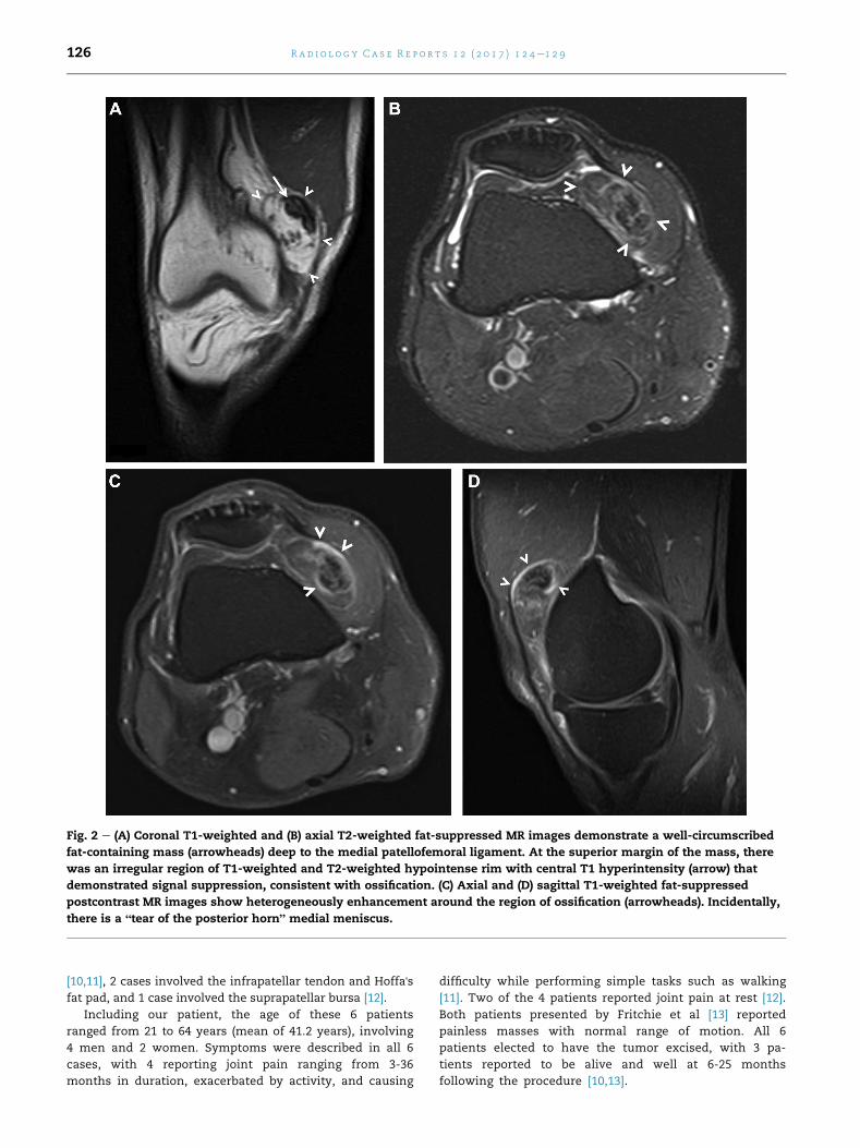
Fig. 2 e (A) Coronal T1-weighted and (B) axial T2-weighted fat-suppressed MR images demonstrate a well-circumscribed
fat-containing mass (arrowheads) deep to the medial patellofemoral ligament. At the superior margin of the mass, there
was an irregular region of T1-weighted and T2-weighted hypointense rim with central T1 hyperintensity (arrow) that
demonstrated signal suppression, consistent with ossification. (C) Axial and (D) sagittal T1-weighted fat-suppressed
postcontrast MR images show heterogeneously enhancement around the region of ossification (arrowheads). Incidentally,
there is a “tear of the posterior horn” medial meniscus.
R a d i o l o g y C a s e R e p o r t s 1 2 ( 2 0 1 7 ) 1 2 4e1 2 9126

MR Imaging characteristics were described in 5 of the 6
cases. All reports describe the mass as heterogeneous on T1
and/or T2 sequences, with areas of macroscopic fat signal and
ossification. Including our patient, gadolinium contrast was
administered in 3 cases, all of which demonstrated hetero-
geneous enhancement [13]. Two of the cases showed evidence
of underlying osseous involvement [10,11].
The radiographic differential diagnosis for an ossifiedmass
is dependent onmany factors, including location.When intra-
articular or juxta-articular in location, mechanical symptoms
may lead to earlier presentation, and therefore, smaller mass
sizes whichmay result in difficulty differentiating ossification
from calcification. The end result is a broad radiographic
differential diagnosis of a calcified and/or ossified intra-
articular mass including benign entities, such as a hemangi-
oma, synovial chondromatosis, calcified synovitis, myositis
ossificans, or a loose body. However, malignancies such as
conventional or surface-based osteosarcoma, or soft tissue
sarcomas such as a synovial sarcoma, may also enter the
differential [14]. This emphasizes the importance of MR eval-
uation of juxta and/or intra-articular masses to evaluate for
characteristics that can narrow the differential diagnosis, as
well as aid in presurgical planning or staging.
Demonstration of a fat-containing mass on MR directs the
differential to lipomatous tumors, primarily lipoma and lip-
osarcoma. In the extremities, well-defined lesions composed
of pure fat or containing thin septa (<2 mm) on MR are
considered lipomas, especially in the absence of contrast
enhancement [15]. However, the mature fat of a lipoma is
subject to the same secondary inflammatory processes of fat
anywhere in the body and can be complicated by osseous
metaplasia, fat necrosis, calcification, fibrosis, and myxoid
change, leading to prominent nonadipose areas mimicking
the findings commonly associated with liposarcoma. Imaging
features that suggest malignancy include the presence of
thickened septa (>2mm), large size, deep (subfascial) location,
presence of nodular nonadipose areas, contrast enhance-
ment, and decreased percentage of fat composition [16]. In
addition to the well-differentiated subtype, the World Health
Organization Classification of Soft Tissue Tumors identifies 3
other subtypes of liposarcoma: myxoid and/or round cell,
pleomorphic, and dedifferentiated [1]. On CT and MR images,
the appearance of liposarcoma reflects the degree of tumor
differentiation: the more differentiated the tumor, the more
closely it resembles adipose tissue [17]. Thus, a well-
differentiated liposarcoma will typically appear as a
Fig. 3 e Hematoxylin-eosin morphology of the osteolipoma. (A) Cortical-type bone with fatty marrow, associated with
mature adipose tissue. A separate island of hyaline cartilage undergoing ossification is noted. Original magnification 20£.
(B) Cartilage island undergoing ossification. Original magnification 40£. (C) Mature adipose tissue and scant fibrous tissue
within the osteolipoma. Original magnification 200£.
R a d i o l o g y C a s e R e p o r t s 1 2 ( 2 0 1 7 ) 1 2 4e1 2 9 127

predominantly fatty mass having irregularly thickened,
linear, swirled, and/or nodular septa. In general, the other 3
histologic subtypes of liposarcomas contain significantly less
fat radiologically (<25%). Notably, the dedifferentiated sub-
type has characteristics of a well-differentiated liposarcoma
with an associated nonlipomatous component, which might
show signs of hemorrhage and/or necrosis [18].
In our case, the mass was well circumscribed, predomi-
nantly fat-containing with no thickened septa, and with a
focus of ossification, favoring an osteolipoma. However, the
deep location and heterogeneous enhancement were findings
that prevented excluding well-differentiated liposarcoma
from the differential. This is in keeping with prior studies
demonstrating the difficulty distinguishing osteolipomas
from well-differentiated liposarcomas secondary to the het-
erogeneous appearance of osteolipomas on imaging studies
and nonspecific presentation [17]. In this setting, we felt a
wide local excision, preoperatively planned as if managing a
sarcoma was the most appropriate treatment.
In conclusion, osteolipomas are a rare occurrence. When
arising in a juxta and/or intra-articular location, they result in
a broad differential diagnosis that should be further evaluated
withMR. Because of the absence of specific radiologic findings,
the differential diagnosis for lesions with fatty and osseous
components should include not only malignant entities such
as liposarcoma but also heterologous differentiation of rather
benign lipomas such as an osteolipoma, especially in the
setting of internal mature bony formation. In our view,
multidisciplinary communication between the radiologist and
treating surgeon is essential to correlate symptomatology and
any prior treatment response that may raise a suspicion for
malignancy. Although extremely rare, definitive evaluation
and treatment of osteolipomas may require wide local exci-
sion, and ideally should be undertaken by an orthopedic
oncologist given the potential for a sarcoma diagnosis.
r e f e r e n c e s
[1] Fletcher CD. The evolving classification of soft tissuetumours - an update based on the new 2013 WHOclassification. Histopathology 2014;64(1):2e11.
[2] Fletcher CDM, World Health Organization, InternationalAgency for Research on Cancer. WHO classification oftumours of soft tissue and bone. 4th ed. Lyon: IARC Press;2013.
[3] Plaut GS, Salm R, Truscott DE. Three cases of ossifyinglipoma. J Pathol Bacteriol 1959;78:292e5.
[4] Saghafi S, Mellati E, Sohrabi M, Raahpeyma A, Salehinejad J,Zare-Mahmoodabadi R. Osteolipoma of the oral andpharyngeal region: report of a case and review of theliterature. Oral Surg Oral Med Oral Pathol Oral Radiol Endod2008;105(6):e30e4.
[5] Obermann EC, Bele S, Brawanski A, Knuechel R,Hofstaedter F. Ossifying lipoma. Virchows Arch1999;434(2):181e3.
[6] Adebiyi KE, Ugboko VI, Maaji SM, Ndubuizu G. Osteolipomaof the palate: report of a case and review of the literature.Niger J Clin Pract 2011;14(2):242e4.
[7] Durmaz A, Tosun F, Kurt B, Gerek M, Birkent H. Osteolipomaof the nasopharynx. J Craniofac Surg 2007;18(5):1176e9.
Table
1e
Sum
mary
ofosteolipom
asaboutth
eknee,clin
ical,andra
diologic
data.
Case
Reference
Age/sex
Loca
tion
Size,
cmX-ray
MRappearance
Symptoms/duration
Treatm
ent/
follow-u
p
1Chengetal
21/M
Distalfemur
12�
6�
2NA
T1:well-d
emarcated,hetero
geneous
T2:lobularco
ntour,
hetero
geneoussignal
intensity
Activity-relatedpain/36
mo
Excision/n
o
recu
rrence
at6mo
2Hash
mietal
45/F
Distalfemur
8�
6.5
�14
Ossifica
tionwithfine
trabecu
lation
T1/T
2:lobulatedandtrabecu
latedareas
Painless
mass/1.5
yBiopsy
/NA
3Fritchie
etal
51/M
Lateralanterior
Infra-p
atellarknee
4.2
�4�
2.8
Mass
withca
lcific
stippling
T1:hetero
geneouslow
T2:hetero
geneouslow
andhigh
C:intense
hetero
geneousenhance
ment
Painless
mass/3
mo
Excision/n
o
recu
rrence
at8mo
4Fritchie
etal
31/F
Infrapatellartendon
5.2
�4.3
�4.1
Retropatellarmass
T1:hetero
geneouslyisointense
andhypointense
tofat.
C:brisk
enhance
mentin
nonfattyco
mponents
Nontender,
irritating
mass/1
y
Excision/n
o
recu
rrence
at25mo
5Pudlowsk
ietal
53/M
Suprapatellarbursa
5.5
�4.5
�2.5
Ossifiedmass
NA
Pain,sw
elling,
instability/5
mo
Medial
menisce
ctomy/N
A
664/M
Anteriorkneejoint
8.1
�3.7
�1.1
Ossifiedmass
T1:well-circu
msc
ribed,hetero
geneouslylobular
C:intense
hetero
geneousenhance
ment
thro
ughout
Kneepain,exace
rbated
byactivity/12mo
Excision/N
A
M,male;F,female;NA,notavailable;C,co
ntrast-enhance
d.
R a d i o l o g y C a s e R e p o r t s 1 2 ( 2 0 1 7 ) 1 2 4e1 2 9128

[8] Kameyama K, Akasaka Y, Miyazaki H, Hata J. Ossifyinglipoma independent of bone tissue. ORL J OtorhinolaryngolRelat Spec 2000;62(3):170e2.
[9] Amaral MB, Borges CF, de Freitas JB, Capistrano HM,Mesquita RA. Osteolipoma of the oral cavity: a case report.J Maxillofac Oral Surg 2015;14(Suppl. 1):195e9.
[10] Cheng S, Lu SC, Zhang B, Xue Z, Wang HW. Rare massiveosteolipoma in the upper part of the knee in a young adult.Orthopedics 2012;35(9):e1434e7.
[11] Hashmi AA, Malik B, Edhi MM, Faridi N, Ashraful M. A largeparosteal ossifying lipoma of lower limb encircling thefemur. Int Arch Med 2014;7(1):5.
[12] Pudlowski RM, Gilula LA, Kyriakos M. Intraarticular lipomawith osseous metaplasia: radiographic-pathologiccorrelation. AJR Am J Roentgenol 1979;132(3):471e3.
[13] Fritchie KJ, Renner JB, Rao KW, Esther RJ. Osteolipoma:radiological, pathological, and cytogenetic analysis of threecases. Skeletal Radiol 2012;41(2):237e44.
[14] Friedman MV, Kyriakos M, Matava MJ, McDonald DJ,Jennings JW, Wessell DE. Intra-articular synovial sarcoma.Skeletal Radiol 2013;42(6):859e67.
[15] Murphey MD, Arcara LK, Fanburg-Smith J. From the archivesof the AFIP: imaging of musculoskeletal liposarcoma withradiologic-pathologic correlation. Radiographics2005;25(5):1371e95.
[16] Kransdorf MJ, Bancroft LW, Peterson JJ, Murphey MD,Foster WC, Temple HT. Imaging of fatty tumors: distinctionof lipoma and well-differentiated liposarcoma. Radiology2002;224(1):99e104.
[17] Peterson JJ, Kransdorf MJ, Bancroft LW, O'Connor MI.Malignant fatty tumors: classification, clinical course,imaging appearance and treatment. Skeletal Radiol2003;32(9):493e503.
[18] Kransdorf MJ, Meis JM, Jelinek JS. Dedifferentiatedliposarcoma of the extremities: imaging findings in fourpatients. AJR Am J Roentgenol 1993;161(1):127e30.
R a d i o l o g y C a s e R e p o r t s 1 2 ( 2 0 1 7 ) 1 2 4e1 2 9 129