Embed Size (px)
Citation preview
OSTEOMA OF THE FRONTAL SINUS
Report of Two Cases
PAUL M. MOORE, JR., M.D. Department of Otolaryngology
Although osteoma of the frontal sinus is not rare, the following 2 cases present some features of special interest. Osteomata are usually found on routine examination in the early stage, as in the first examina-tion of case 1, or not until they have produced symptoms, as in case 2. The opportunity seldom arises to observe their development from inno-cent, rather unimportant findings to the obstructing, dangerous stage as in case 1. Osteomata are slow growing and usually do not reach a size that requires operative interference. Although they are more prone to increase in size during youth, the growth in case 1 occurred during the period between the ages of 38 and 46.
Case Reports Case 1. A man, aged 36, was first seen on September 24, 1937, complaining of
nasal congestion and chronic purulent postnasal drip. He had had a submucous resection seventeen years previously with some relief of nasal obstruction but with subsequent nasal crusting. Family history revealed that both grandfathers had suffered from asthma, one having had hay fever as well. The patient had had eczema in infancy and had noted sneezing and nasal congestion on exposure to horse hair and dust.
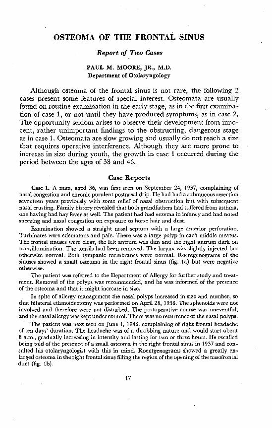
Examination showed a straight nasal septum with a large anterior perforation. Turbinates were edematous and pale. There was a large polyp in each middle meatus. The frontal sinuses were clear, the left antrum was dim and the right antrum dark on transillumination. The tonsils had been removed. The larynx was slightly injected but otherwise normal. Both tympanic membranes were normal. Roentgenograms of the sinuses showed a small osteoma in the right frontal sinus (fig. la) but were negative otherwise.
The patient was referred to the Department of Allergy for further study and treat-ment. Removal of the polyps was recommended, and he was informed of the presence of the osteoma and that it might increase in size.
In spite of allergy management the nasal polyps increased in size and number, so that bilateral ethmoidectomy was performed on April 28, 1938. The sphenoids were not involved and therefore were not disturbed. The postoperative course was uneventful, and the nasal allergy was kept under control. There was no recurrence of the nasal polyps.
The patient was next seen on June 1, 1946, complaining of right frontal headache of ten days' duration. The headache was of a throbbing nature and would start about 8 a.m., gradually increasing in intensity and lasting for two or three hours. He recalled being told of the presence of a small osteoma in the right frontal sinus in 1937 and con-sulted his otolaryngologist with this in mind. Roentgenograms showed a greatly en-larged osteoma in the right frontal sinus filling the region of the opening of the nasofrontal duct (fig. lb).
1 7
P A U L M . M O O R E , J R .
a b F i a 1. Case 1. (a) September, 1937 (b) June, 1946.
On June 12, 1946, an external frontal operation was performed on the right side, and the entire floor of the right frontal sinus was removed. Just enough of the anterior wall was removed to permit delivery of the osteoma, which was 3.5 x 2 cm. and rather firmly attached at its base. Thick mucopus was trapped above and laterally to this mass. Cultures of this material were sterile. The sinus was cleaned of foreign material. The lining mucosa appeared normal, and neither it nor the "nasofrontal duct were disturbed. A small rubber tissue drain was placed in the wound at the inner end of the eyebrow, and the wound was closed with interrupted dermal sutures. The drain was removed after twenty-four hours. The patient experienced diplopia for about two months, but there was no recurrence of the headache and no difficulty from collection of fluid in the frontal sinus.
Case 2. A man, aged 25, was first seen February 22, 1943, complaining of a swelling of his right eye of two weeks' duration. This had been preceded by an acute upper respira-tory infection with nasal congestion and discharge and sore throat. There had been little or no fever. Severe headache was present in the right frontal area, tending to in-crease in severity in the evening and at night. The patient had experienced an attack of acute right frontal sinusitis six years previously. The symptoms of this condition had responded to conservative treatment, and the patient had had no further trouble other than a chronic mucoid postnasal drip until the present illness.
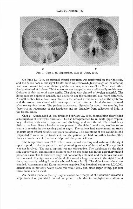
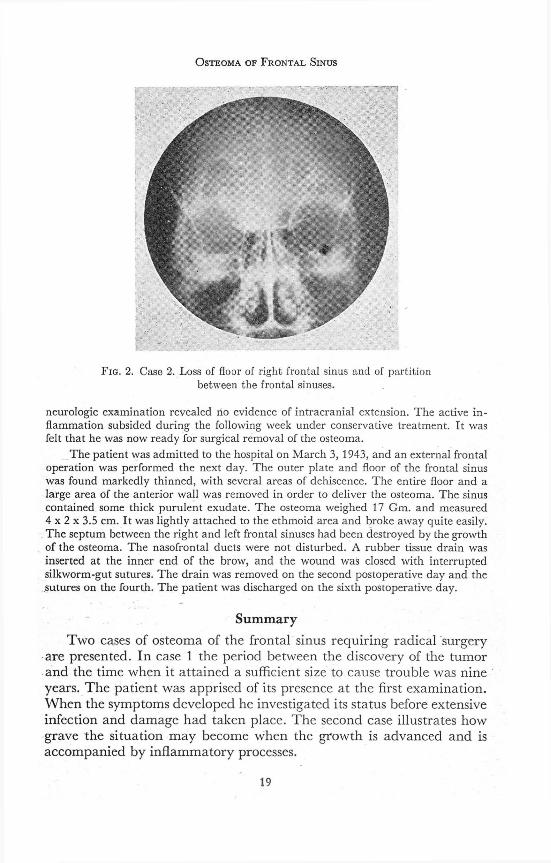
The temperature was 99.8°. There was marked swelling and redness of the right upper eyelid, tender to palpation and presenting an area of fluctuation. The eye itself was not involved. The nasal septum was not obstructive. The turbinates on the right side were swollen, and mucopus could be seen on this same side in the nasopharynx and posterior naris. The tonsils were large but not acutely inflamed, and the larynx and ears were normal. Roentgenograms of the skull showed a large osteoma in the right frontal sinus, apparently arising from the ethmoid bone (fig. 2). The right frontal sinus was clouded. Wassermann and Kahn tests were negative. Red blood cell count was 5,030,000, hemoglobin 78 per cent, white blood cell count 5600, and blood sugar 89 mg. per cent three hours after a meal.
An incision made in the right upper eyelid over the point of fluctuation released a large amount of pus which on culture proved to be due to Staphylococcus albus. A
1 8
OSTEOMA OF F R O N T A L SINUS
FIG. 2. Case 2. Loss of floor of right frontal sinus and of partition between the frontal sinuses.
neurologic examination revealed no evidence of intracranial extension. The active in-flammation subsided during the following week under conservative treatment. It was felt that he was now ready for surgical removal of the osteoma.
The patient was admitted to the hospital on March 3, 1943, and an external frontal operation was performed the next day. The outer plate and floor of the frontal sinus was found markedly thinned, with several areas of dehiscence. The entire floor and a large area of the anterior wall was removed in order to deliver the osteoma. The sinus contained some thick purulent exudate. The osteoma weighed 17 Gm. and measured 4 x 2 x 3.5 cm. It was lightly attached to the ethmoid area and broke away quite easily. The septum between the right and left frontal sinuses had been destroyed by the growth of the osteoma. The nasofrontal ducts were not disturbed. A rubber tissue drain was inserted at the inner end of the brow, and the wound was closed with interrupted silkworm-gut sutures. The drain was removed on the second postoperative day and the sutures on the fourth. The patient was discharged on the sixth postoperative day.
Summary Two cases of osteoma of the frontal sinus requiring radical surgery
are presented. In case 1 the period between the discovery of the tumor and the time when it attained a sufficient size to cause trouble was nine years. The patient was apprised of its presence at the first examination. When the symptoms developed he investigated its status before extensive infection and damage had taken place. The second case illustrates how grave the situation may become when the growth is advanced and is accompanied by inflammatory processes.
1 9