Embed Size (px)
Citation preview

OSTEOPOROSIS IN MENWHY CHANGE NEEDS TO HAPPEN
www.iofbonehealth.org

2
NORMAL BONE OSTEOPOROTIC BONE
Osteoporosis is a disease characterized by low bone mass and deterioration in the microarchitecture of bone tissue, leading to an increased risk of fracture. Osteoporosis occurs when the bone mass decreases more quickly than the body can replace it, leading to a net loss of bone strength. As a result the skeleton becomes fragile, so that even a slight bump or fall can lead to a broken bone, (referred to as a fragility fracture). Osteoporosis has no signs or symptoms until a fracture occurs – this is why it is often called a ‘silent disease’.
Osteoporosis affects all bones in the body; however, fractures occur most frequently in the vertebrae (spine), wrist and hip. Osteoporotic fractures of the pelvis, upper arm and lower leg
are also common. Osteoporosis itself is not painful but the broken bones can result in severe pain, significant disability and even mortality. Both hip and spine fractures are also associated with a higher risk of death - 20% of those who suffer a hip fracture die within 6 months after the fracture.
A COMMON DISEASE
It is estimated that worldwide an osteoporotic fracture occurs every three seconds. At 50 years of age, one in three women and one in five men will suffer a fracture in their remaining lifetime. For women, the risk of hip fracture is higher than the risk of breast, ovarian and uterine cancer combined. For men, the risk is higher than the risk for prostate cancer.
Approximately 50% of people with one osteoporotic fracture will have another, with the risk of new fractures rising exponentially with each fracture.
A GROWING PUBLIC HEALTH PROBLEM
The risk of sustaining a fracture increases exponentially with age due not only to the decrease in bone mineral density, but also due to the increased rate of falls among the elderly. The elderly represent the fastest growing segment of the population. Thus, as life expectancy increases for the majority of the world’s population, the financial and human costs associated with osteoporotic fractures will increase dramatically unless preventive action is taken.
WHAT IS OSTEOPOROSIS?
TABLE OF CONTENTS
§ Foreword 3
§ The burden of osteoporosis in men 4
§ Bone development and loss in men 8
§ Causes of osteoporosis in men 12
§ Challenges in diagnosis and treatment 14
§ Guidance for men, health-care professionals and policymakers 17
§ Osteoporosis in men – why change needs to happen 20
§ References 21

3
One-third of hip fractures worldwide occur in men and they are associated with greater mortality when compared with women.
This statistic is remarkable because hip fractures represent the most serious complication of osteoporosis, a disease that for far too long has been considered to be exclusively a problem for women. While improving management of osteoporosis for women is critical, the time has now come for a radical reappraisal of osteoporosis management in men.
The world’s men are ageing fast; by 2050 the number of men aged 60 years or over will increase 10-fold. As male baby boomers enter old age, the number of men living with osteoporosis and the associated suffering from consequent fragility fractures is set to escalate to an unprecedented level.
Although all of the world’s regions will be affected, Asia and Latin America will bear the brunt of increased demand for acute fracture care services because of the growth in their ageing populations over the next 30 years. Given that 3.5 million fragility fractures occurred in men in 2000, the costs that will result from the projected increases in male fracture incidence will place an unbearable strain on overstretched health-care budgets.
To avert this calamity, a concerted international effort is required to improve the awareness of osteoporosis in men amongst both doctors and the community, and to implement systems of care to prevent fragility fractures. In this regard, there is good news. There are a range of therapies now available that have proven effective in the treatment of osteoporosis in men. These treatments have been shown to work against the various types of osteoporosis which can affect men, including primary (or idiopathic) osteoporosis and when secondary causes are responsible for bone loss (e.g. glucocorticoids or low sex hormone levels).
FOREWORD
Peter EbelingHead, Department of Medicine, Monash University, Victoria, Australia
IOF Board member
The key challenge facing health-care professionals and policymakers is to ensure that men who are clearly at high risk of suffering fragility fractures get the care they need. First and foremost, this includes men who have already suffered a fragility fracture. A broken bone is a very clear signal of elevated future fracture risk – nevertheless osteoporosis assessment and treatment rates among these men are very low – being mostly under 20%. Studies from around the world, reviewed in this report, demonstrate a near universal absence of secondary fracture prevention systems for men who have already suffered fragility fractures. Similar poor attention to bone health is evident among men receiving androgen deprivation therapy for prostate cancer or glucocorticoid treatment for many other conditions, the most common causes of secondary osteoporosis in men.
A systematic approach to osteoporosis management in men is required on a global scale, including the implementation of awareness and educational programmes as well as Fracture Liaison Services (FLS), which are proven systems of care for patients who suffer fragility fractures. FLS place a fracture coordinator at its centre and can result in fewer fractures, cost savings for the health system and improvement in the quality of life of patients. FLS is the focus of the International Osteoporosis
Foundation’s (IOF) Capture the Fracture Campaign. A growing number of centres of excellence are sharing their experience with colleagues elsewhere to catalyse the establishment of FLS in many countries. Governments are recognizing the need to incorporate FLS into national policy. Closure of the evidence-treatment gap for men with fragility fractures or men who have been initiated on bone-thinning treatments for other diseases can be achieved so easily. Development of robust protocols and systems of care to deliver them – which ensure a bone-health assessment goes hand-in-hand with the presence of a fragility fracture or upon initiation of bone-thinning drugs – will transform osteoporosis care for men.
Policymakers must not discriminate against men by their omission from national clinical guidelines and reimbursement policies. Governments and health-care professionals the world over must ask themselves whether this is an issue inhibiting optimal osteoporosis care for men in their jurisdictions. Where change is needed, it must happen now.
The demographic tsunami of ageing is upon us. Elimination of the osteoporosis evidence-treatment gap for men is an essential component of our response to this unprecedented threat to the sustainability of our health-care systems.

4
In 1950, there were approximately 90 million men in the world aged 60 years or over. By the turn of the century, there were almost 275 million and by 2050 there will be more than 900 million men who
have lived into their seventh decade (Figure 1)1,2. This 10-fold increase in the older male population in just a century is a longevity miracle. However, a demographic shift on this scale creates challenges which – with
absolute certainty – will include an explosion in the incidence of chronic diseases afflicting older men. These diseases will not only impose a great burden upon men and their families but they will also test our health and social care systems to the limit. Osteoporosis will be at the vanguard of this battle set to rage between quantity and quality of life.
All too often, osteoporosis is perceived to be a ‘woman’s disease’ that is not preventable or an urgent health concern to men. The primary purpose of this report is to debunk these myths and raise awareness of the threat that osteoporosis poses to older men throughout the world. It is estimated that the residual lifetime risk of experiencing an osteoporotic fracture in men over the age of 50 is up to 27%3 higher than the lifetime risk of developing prostate cancer of 11.3%4.
And just as osteoporosis does not discriminate between the sexes – with
THE BURDEN OF OSTEOPOROSIS IN MEN
FIGURE 1 The ageing of the world’s male population 1950–20501,2
0
200
400
600
800
1000
≥80 years
≥60 years
2050202520001950
nu
mb
er o
f m
en (
mill
ion
s)

5
osteoporotic fractures affecting one in five men versus one in three women aged over 50 years – its impact will be felt in the coming decades in the majority of the world’s regions. As illustrated in Figure 2, the population of men aged over 60 years who are potentially at risk of suffering fragility fractures will continue to grow in Europe, Northern America and Oceania, whilst in Asia and Latin America the rate of growth of the male population aged 60 years or over will be exponential.
Osteoporosis causes fragility fractures, which are fractures that usually result from a fall from a standing height or less5. Arguably, the most serious fragility fracture is a hip fracture, and one-third of all hip fractures
worldwide occur in men6. Audits from several countries have shown that a significant proportion of men who suffer hip fractures have broken other bones before they broke their hip7-9. Furthermore, a study from Sweden, which followed a cohort of older men for 22 years, reported that 27% of men who had suffered a hip fracture sustained subsequent fractures in their remaining lifetime10. When men suffer fractures caused by osteoporosis – like women – too many become trapped in the fragility fracture cycle11.
For older working men, fragility fractures have been demonstrated to have a significant impact on productivity. In Denmark, a national evaluation of the impact of fragility fractures concluded that almost 5,000
working days would be lost on account of fractures in men aged 50–65 years12. A recently published burden of disease analysis from Osteoporosis Australia concluded that productivity losses among Australian men aged 50 years or over with fragility fractures cost more than 46 million AUD in 201213.
In terms of mortality related to fragility fractures, men fare particularly badly and are the ‘weaker sex’. A national registry study14 from Denmark published in 2010 echoed the findings of previous studies15-18: Hip fractures in men are associated with greater mortality compared with women, with rates as high as 37% in the first year following fracture. In addition, mortality is increased after most fragility fractures in men, not only following hip fractures19.
In recent years, substantial geographic variation in the incidence of hip and other fragility fractures has been observed20. In general, hip fracture rates appear to be increasing rapidly in the East whilst age-adjusted rates for women have stabilized, or declined, in the West11,21-33. This decline in age-adjusted rates of hip fractures in the West has been less marked amongst men. Notably, a growing number of studies have reported large increases in the absolute incidence of hip fracture in men during short intervals of time21,28,34,38. A recent study from the UK of more than 10,000 hip fracture admissions to a major trauma centre noted a substantial increase in the proportion of hip fractures occurring in men in a 12-year period39. In 2000, 23.5% of the hip fractures occurred in men, which increased to 30.7% by 2012.
FIGURE 2 Proportion of men aged ≥60 years by world region 1950–20501,2
0
5
10
15
20
25
30Oceania
North America
Latin America & Caribbean
Europe
Asia
Africa
20502025200019751950
per
cen
tag
e o
f m
ale
po
pu
lati
on
Hip fractures in men are associated with greater mortality
compared with women, with rates as high as 37% in
the first year following fracture. In addition, mortality is
increased after most fragility fractures in men, not only
following hip fractures.19
6
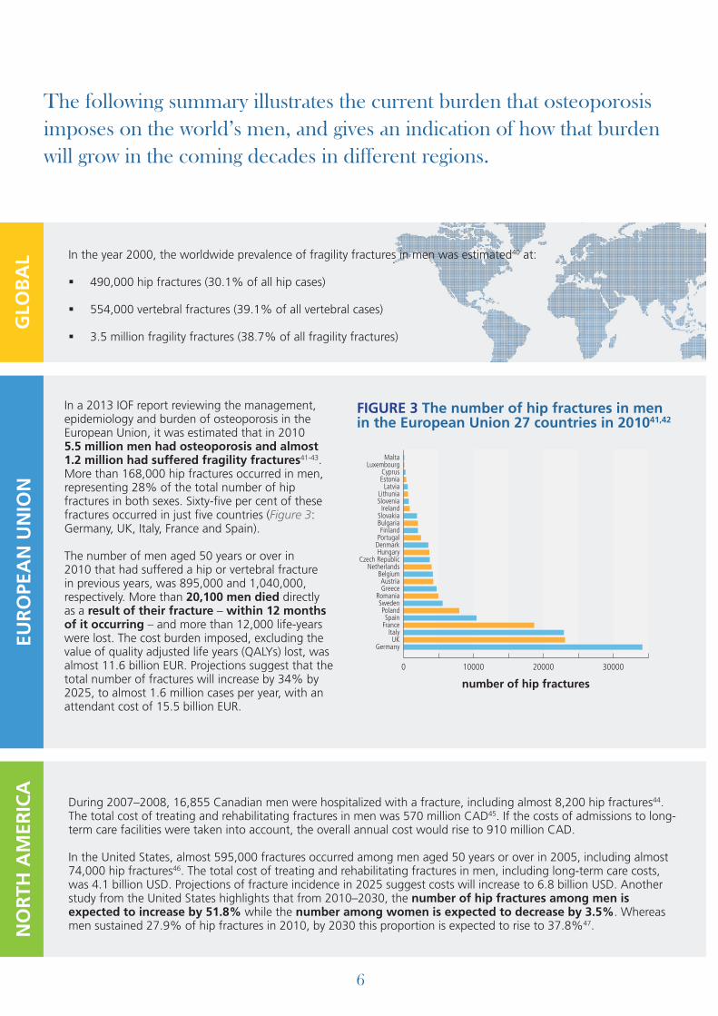
The following summary illustrates the current burden that osteoporosis imposes on the world’s men, and gives an indication of how that burden will grow in the coming decades in different regions.
In the year 2000, the worldwide prevalence of fragility fractures in men was estimated40 at:
§ 490,000 hip fractures (30.1% of all hip cases)
§ 554,000 vertebral fractures (39.1% of all vertebral cases)
§ 3.5 million fragility fractures (38.7% of all fragility fractures)
In a 2013 IOF report reviewing the management, epidemiology and burden of osteoporosis in the European Union, it was estimated that in 2010 5.5 million men had osteoporosis and almost 1.2 million had suffered fragility fractures41-43. More than 168,000 hip fractures occurred in men, representing 28% of the total number of hip fractures in both sexes. Sixty-five per cent of these fractures occurred in just five countries (Figure 3: Germany, UK, Italy, France and Spain).
The number of men aged 50 years or over in 2010 that had suffered a hip or vertebral fracture in previous years, was 895,000 and 1,040,000, respectively. More than 20,100 men died directly as a result of their fracture – within 12 months of it occurring – and more than 12,000 life-years were lost. The cost burden imposed, excluding the value of quality adjusted life years (QALYs) lost, was almost 11.6 billion EUR. Projections suggest that the total number of fractures will increase by 34% by 2025, to almost 1.6 million cases per year, with an attendant cost of 15.5 billion EUR.
During 2007–2008, 16,855 Canadian men were hospitalized with a fracture, including almost 8,200 hip fractures44. The total cost of treating and rehabilitating fractures in men was 570 million CAD45. If the costs of admissions to long-term care facilities were taken into account, the overall annual cost would rise to 910 million CAD.
In the United States, almost 595,000 fractures occurred among men aged 50 years or over in 2005, including almost 74,000 hip fractures46. The total cost of treating and rehabilitating fractures in men, including long-term care costs, was 4.1 billion USD. Projections of fracture incidence in 2025 suggest costs will increase to 6.8 billion USD. Another study from the United States highlights that from 2010–2030, the number of hip fractures among men is expected to increase by 51.8% while the number among women is expected to decrease by 3.5%. Whereas men sustained 27.9% of hip fractures in 2010, by 2030 this proportion is expected to rise to 37.8%47.
EUR
OPE
AN
UN
ION
NO
RTH
AM
ERIC
AG
LOB
AL
FIGURE 3 The number of hip fractures in men in the European Union 27 countries in 201041,42
0 10000 20000 30000
GermanyUK
ItalyFranceSpain
PolandSweden
RomaniaGreeceAustria
BelgiumNetherlands
Czech RepublicHungary
DenmarkPortugalFinland
BulgariaSlovakia
IrelandSloveniaLithunia
LatviaEstoniaCyprus
LuxembourgMalta
number of hip fractures

7
In Argentina, 9,444 hip fractures were estimated to have occurred in men in 2009 at a total cost of 35.9 million USD48. By 2050, projections suggest the incidence of hip fractures in men will increase to 13,000 cases per year.
In Brazil, the prevalence of osteoporosis at the femoral neck among men aged 50 years or over has been reported as 15.4%49. The Brazilian Osteoporosis Study (BRAZOS) found the prevalence of fragility fracture among men aged 40 years and over to be 12.8%50. The number of men suffering hip fractures every year is estimated to be 24,20051.
In Mexico its estimated that almost 7,800 hip fractures occurred in men in 2009, at a cost of 39 million USD52. Hip fracture incidence is projected to increase to 11,700 and 35,500 cases per year by 2020 and 2050, respectively. Among men aged 50 years and over, the prevalence of radiographically detected vertebral fractures is almost 10%53.
In 2011, IOF published the Eastern European and Central Asian Regional Audit54. This report identified a paucity of epidemiological data on osteoporosis and fragility fractures across the region. Another finding was surprisingly low levels of hospitalization and surgery for hip fracture sufferers. In the Russian Federation, between 33–40% of hip fracture sufferers were hospitalized and just 13% received surgical treatment. Consequently, mortality rates for hip fracture in some Russian cities are very high at 45–52%55.
In 2012, epidemiological modelling was published for the Russian Federation. More than 142,000 fragility fractures were estimated to have occurred in men in 2010, including more than 32,000 hip fractures. By 2035, projections suggest the number of fragility fractures and hip fractures will increase to more than 177,000 and almost 43,700, respectively55.
Osteoporosis Australia recently published a new burden of disease analysis for the period 2012–202213. It showed that in 2012, almost 202,000 Australian men aged 50 years or over had osteoporosis and more than 40,700 suffered a fragility fracture, including 6,670 hip fractures. Other key findings related to men included:
The total cost of hip fractures in men in 2012 was almost 188 million AUD (28,177 AUD per case) comprised of: § Total hospital costs: 144,634,902 AUD § Pre-hospital ambulance/paramedic costs: 4,592,466 AUD § Sub-acute care (i.e. rehabilitation) costs: 20,215,518 AUD § Community costs for fracture management: 773,009 AUD § Nursing home care costs: 17,724,884 AUD
The total cost of all fragility fractures in men in 2012 was almost 426 million AUD. By 2022, older men will suffer more than 55,300 fractures, including 10,000 hip fractures.
In China, as the enormous Chinese population simultaneously ages and urbanizes, fracture incidence is changing dramatically. In Beijing, from 2002–2006, hip fracture rates in men aged 50 years or over increased by 49%21. In Tangshan in Hebei province, from 1994–2010, hip fracture rates in men aged 70 years or over increased by 85%56.
In Japan, hip fracture incidence has been reported in a nationwide survey conducted every five years since 198757. The number of hip fractures occurring annually in men rose from 13,500 cases in 1987 to 31,300 in 2007.
In Saudi Arabia, estimates suggest that almost 8,800 hip fractures occurred in men and women combined during 200458. With a notably high observed male to female ratio of 1.2:1, approximately 4,800 hip fractures occur annually in Saudi men. The total cost of managing hip fractures in men was estimated at 622 million USD.
In Turkey, almost 6,500 men were estimated to have suffered a hip fracture in 201059. By 2035, projections suggest that each year 14,860 men will break their hip. The remaining lifetime risk of hip fracture for a 50 year old Turkish man is 3.5%.
In Iran, almost 22,000 hip fractures occurred in men in 2010, a figure which is expected to increase to almost 29,000 by 2020 and 43,500 by 2050 (B. Larijani, personal communication, July 21, 2014).
LATI
N A
MER
ICA
ASI
A-P
AC
IFIC
CEN
TRA
L A
SIA
MID
DLE
-EA
ST

8
BONE DEVELOPMENT AND LOSS IN MEN
CHILDHOOD THROUGH TO YOUNG ADULTHOOD
Many factors influence the growth of our skeleton and maintenance of its bone mass throughout life. As illustrated in Figure 4, both males and females attain peak bone mass between ages 20–30 years. Up to the age of 10–12 years, there are no significant differences in bone mass between boys and girls. However, at the onset of puberty, the bone mass increases more in males60.
Why does this occur? Accrual of bone mass during childhood and adolescence is controlled by sex steroids and the growth hormone/insulin-like growth factor 1 (IGF-I) axis of the endocrine system62. A study of young men from Gothenburg sought to establish whether androgens increase the size of cortical bone – the hard ‘outer casing’ of bones – and whether oestrogens have the opposite effect63. Levels of free testosterone and oestradiol were
measured and correlated with the size of cortical bone. The results supported the notion that androgens increase, whereas oestrogens reduce, cortical bone size. Consequently, during
puberty, boys develop larger bones than girls and so accrue greater bone mass. The size of bones and the thickness of their cortex are major determinants of bone strength, and thus men generally
FIGURE 4 Bone mass throughout the life cycle61
7050 604020 3010
puberty menopausemen
women
years
bone
mas
s
peak bone mass

9
have larger bone size and greater bone strength than women.
Achieving one’s genetic potential for peak bone mass during childhood and adolescence is the primary objective during this first stage of the skeleton’s life cycle. The consequence of not doing so has been illustrated by computer modelling developed to predict the relative influences of peak bone mineral density (BMD), menopause and age-related bone loss on the development of osteoporosis in women65. A 10% increase in peak BMD was predicted to delay the development of osteoporosis by 13 years. Important influences on peak bone mass for young males include:
Exercise Osteoporosis Australia’s Building healthy bones throughout life strategy66 published in 2013 stated ‘Childhood and adolescence may represent the optimal window of opportunity in which exercise can improve bone strength and protect against osteoporosis and associated fragility fractures in old age, assuming the gains achieved are maintained in later life.’ Systematic literature review has reported beneficial effects on BMD for children participating in moderate to high impact weight-bearing physical activities67. Long-term follow-up from the Australian Schools Health and Fitness Survey conducted in 1985 suggests that higher levels of fitness as a child are predictive of greater peak bone mass at age 30 years68,69.
Calcium intake approximately 40% of adult peak bone mass is acquired during the two years around puberty70. Accordingly, ensuring adequate dietary calcium intake during this period of growth is essential. In this regard, it is of great concern that a multinational study of calcium intakes in adolescent boys reported levels of only 60% of country-specific requirements71.
Vitamin D levels the association between vitamin D deficiency and
rickets is well documented and understood. However, the impact that vitamin D deficiency in childhood has on bone health at the population level is also likely to be significant72. Reports from Europe73-78, the Middle East79, North America80 and Oceania81-84 suggest that low levels of vitamin D in children are a cause for concern throughout the world. In 2011, the Institutes of Medicine report on dietary intakes of vitamin D and calcium defined the adequate intake of vitamin D of infants (0–12 months old) to be 400 IU and the recommended dietary allowance of vitamin D for children aged 1–18 years to be 600 IU/day85.
Protein intake proteins are building blocks and help to maintain strong bones, conversely low protein intake is associated with impaired skeletal growth thereby influencing peak bone mass86. Proteins may have a positive effect on bone and muscle through hepatic production of insulin-like growth factor I (IGF-I)87. Serum levels of IGF-I are closely related to growth, increasing from birth to puberty.
Furthermore IGF-I is considered as a major factor for bone longitudinal growth, stimulating chondrocyte from the growth plate and stimulating the production of active form of vitamin D (1,25 dihydroxyvitamin D) in the kidney. Dairy products, fish, meat, nuts and legumes are a good dietary source of proteins. Both animal and plant proteins sources appear to favour strong bones.
Other factors which can adversely affect peak bone mass and BMD in young males include delayed puberty88, smoking89-91, alcohol consumption89 and certain childhood diseases such as acute lymphoblastic leukaemia92 and medications such as glucocorticoids93 and anti-epileptic drugs94.
AGES 20–60 YEARS
During these decades of adulthood, the primary objective is to avoid premature bone loss and maintain a healthy skeleton. On account of the muscular system being the generator of the strongest mechanical forces
Osteoporosis has been described as a ‘paediatric
disease with geriatric consequences’.64
FIGURE 5 The structure of bone
Corticalbone
Lamellar
Haversian canal
Trabecularbone Trabecular
boneCortical
bone
Periosteum
Canaliculus
Nerve
Volkman’s canal
Artery
Endosteum
Venous sinus

10
applied to bones95, avoiding loss of muscle mass – known as sarcopenia – is also of paramount importance in this stage of life. Accordingly, as for younger males, regular exercise has an important role to play. Recommendations for building healthy bones in healthy adults from Osteoporosis Australia66 and others96,97 provide an illustration of the type and frequency of activities that current knowledge suggests will be of benefit:
Be habitually physically active and undertake regular weight-bearing and/or muscle strengthening exercises.
§ Encourage regular participation in moderate impact weight-bearing physical activity, high impact training (e.g. 50–100 jumps) or related impact loading sports for at least 30 minutes 3–5 days per week.
§ Include muscle-strengthening exercises on at least 2 days per week. For maximum benefits, the programme should be high intensity (60–80% of peak capacity), become progressively more challenging over time and target the major muscles around the hip and spine.
§ Where possible, encourage participation in a multi-modal exercise regimen (inclusive of weight bearing/high impact/high intensity resistance exercise) at least three times per week.
With regard to calcium intake and vitamin D levels, men should aim to comply with the relevant national recommendations from agencies within their respective countries.
As suggested in Figure 4, bone loss appears to commence soon after young men reach peak bone mass. A study from Sweden investigated changes in BMD in men aged between 17–26 years98. A significant year-on-year loss of BMD at the hip was observed from age 19 years, when peak bone mass had occurred. Analysis of bone density data from these young men’s fathers suggested that 25% of BMD at the hip may be lost by 50 years of age and that bone remodelling may be regulated differently at the hip than at other sites.
There are important differences between the ways in which bone loss occurs with ageing in men as compared with women. To appreciate these differences, the basics of bone biology must be firstly considered.
Bone is a living tissue able to impart tremendous strength to support our bodies, yet simultaneously must also have the capacity to be flexible to absorb shock without breaking. As illustrated in Figure 5, bone comes in two major forms, the cortical bone, which forms the casing or outer shell, and the trabecular bone – also known as spongy or cancellous bone – which forms a honeycomb-type mesh within the cortex. The trabecular bone provides structural support when loads are applied and enables the entire bone to be flexible.
Bone is in a perpetual state of remodelling throughout life, with the entire skeleton being replaced every 10 years99. One group of cells – osteoclasts – are drawn to sites of microdamage to remove old bone (bone resorption). Once the osteoclasts have completed their task, bone forming cells – osteoblasts – deposit new bone to fill the gap created. This process is known as the bone remodelling cycle and is represented in Figure 6 for a healthy young adult. For bone mass to remain constant, the amount of bone being resorbed by the osteoclasts needs to be equivalent to the amount of bone being formed by the osteoblasts.
As men age, the rate of bone resorption by osteoclasts on the inside surface of cortical bone increases (known as endocortical resorption). At the same time, new bone is being deposited on the outer surface of the cortex (known as periosteal apposition). These concurrent processes lead to an increase in the circumference of bones, which serves to increase the bone size and moves the cortex further away from the centre of the bone. From a biomechanical perspective, both of these changes result in greater bone strength. However, the cortex also becomes thinner which reduces bone strength. So, in men aged younger than 70 years, there is a degree of balance between these two competing processes.
In postmenopausal women, there is evidence to suggest that the rate of endocortical resorption is such that periosteal apposition cannot serve as a sufficient compensatory mechanism to prevent bone fragility100-103. The change
BoneRemodeling
Cycle
OsteoclastsBone ResorptionBone resorption begins when osteoclasts remove a portion of the bone to be replaced later by the action of osteoblasts. This is a vital step for signaling bone formation.
OsteoblastsBone Formation
Osteoblasts lay down collagen and mineral deposits over the area
previously remodeled by osteoclasts. Osteoblast activity is
vital for maintaining bone mineral density and bone strength.
Resorption
Reversal
Formation
FIGURE 6 Bone renewal through the remodelling cycle

11
in cross-sectional structure of bone for men and women with ageing is illustrated in Figure 7. These seemingly subtle differences in the way that our bones change with ageing contribute to our understanding of why fracture rates increase in women to a greater extent than in men.
Another aspect whereby men differ from women is in the mechanisms underlying age-related trabecular bone loss. In men trabecular thinning occurs and may be associated with decreases in IGF-1, whereas in women there is resorption and loss of trabeculae, particularly horizontal trabeculae, associated with oestrogen deficiency at the time of menopause104. This is another reason why skeletal fragility is higher in women.
AGE 70 YEARS ONWARDS
As men enter old age, the focus becomes prevention and treatment of osteoporosis with the objective of minimizing the risk of fragility fractures. Longitudinal studies suggest that the rate of bone loss accelerates after age 70 years in men109,110. As ageing progresses, bone loss in the marrow cavity is not compensated by bone deposition on the periosteum, which results in loss of cortical bone111. A systematic review established that men aged over 70 years were 50% more likely to suffer a fragility fracture than younger men112.
As indicated on the next page, secondary causes of osteoporosis are highly prevalent in men, the most common secondary causes being: § Hypogonadism § Glucocorticoid use § Excessive alcohol use § Smoking
Hypogonadism – as defined by a serum testosterone level less than 300 ng/dL – has been shown to be present in two-thirds of American male nursing home residents who have suffered hip fractures113 (see page 13).
Prostate cancer and fractures
Androgen deprivation therapy (ADT) is the mainstay of treatment for metastatic prostate cancer and a significant risk factor for osteoporosis in older men114. Bone loss is rapid in men treated with ADT, of the order 2–4% at the lumbar spine and hip during the first year of treatment115,116. A U.S. study of more than 50,000 men who had received a diagnosis of prostate cancer in the 1990s evaluated fracture incidence117; 19.4% of men who took ADT had a fracture, as compared with 12.6% of those not receiving ADT, a highly statistically significant difference (P<0.001). All-cause mortality has also been shown to be higher for men taking ADT for prostate cancer, as compared to men with prostate cancer who were not taking ADT or men without prostate cancer118.
Glucocorticoids (GC) are used to treat many conditions including chronic obstructive pulmonary disease, inflammatory bowel disease and rheumatological diseases119. In the United States, 0.2–0.5% of the general population take GC120. GC-induced osteoporosis is the second most common form of osteoporosis after postmenopausal osteoporosis, with up to half of long-term GC users suffering fragility fractures121,122.
Daily alcohol intake of two or fewer units are not associated with increased fracture risk123. However, above this threshold, alcohol intake is associated with a 38% increased risk of suffering any fragility fracture and a 68% increased risk of hip fracture. Accordingly, with respect to bone health, moderation is best.
Smoking has negative effects on bone health124. Compared with non-smokers, current smoking is associated with a 29% increased risk of suffering a fragility fracture and an 84% increased risk of hip fracture. As it is for the heart and the brain, smoking is bad for your bones and should be avoided.
FIGURE 7 The influence of bone geometry on bone strength105
LEFT For the same areal BMD, bone C has progressively greater bending strength and axial strength than bone B and bone A because the mass of bone C is distributed further away from the centre – adapted from Bouxsein106.
RIGHT Sex and ageing differences in periosteal apposition and endocortical resorption in tubular bones. Adapted from Seeman107.
NEUTRAL AXIS
BEFOREPUBERTY
DURINGPUBERTY
AGEING
MALE
FEMALE
A
B
C
NEUTRAL AXIS
Periostealsurface
Endocorticalsurface
Periostealsurface
Endocorticalsurface

CAUSES OF OSTEOPOROSIS IN MEN
Secondary causes of osteoporosis in men, both common and rare, include104:
Common § Cushing’s syndrome or chronic corticosteroid use (>5 mg per day for more than 3 months) § Excessive alcohol use (more than 2 units a day) § Primary or secondary hypogonadism (serum testosterone levels <300 ng/dL) § Inadequate calcium intake (<600 mg per day) § Vitamin D deficiency/insufficiency § Smoking § Family history (genetics)
Less common § Low body mass index (BMI <20) § Lack of exercise or excessive exercise that leads to a low BMI § Antiepileptic drugs (phenytoin, phenobarbitone, primidone, carbamazepine) § Thyrotoxicosis § Primary hyperparathyroidism § Type 1 and type 2 diabetes mellitus § Chronic liver or kidney disease § Malabsorption, including coeliac disease § Hypercalciuria § Rheumatoid arthritis or ankylosing spondylosis § Inflammatory bowel disease § Malignancy, for example prostate cancer
» Chemotherapy » Androgen deprivation therapy
§ Warfarin
Rare § Multiple myeloma § Human immunodeficiency virus infection or its treatment with protease inhibitors (tenofovir) § Mastocytosis § Immunosuppressive therapy (cyclosporin, tacrolimus) § Osteogenesis imperfecta
12

Hypogonadism – testosterone deficiency in men – occurs in up to 12.3% of men, and is a significant contributor to osteoporosis108. The causes of male hypogonadism may be usefully categorized as primary or secondary:
Primary hypogonadism defects of the testes § Genetic/chromosomal disorders (Klinefelter’s syndrome XXY) § Anorchia (congenital or postorchidectomy) § Cryptorchidism § Chemotherapy (alkylating agents), radiotherapy § Orchitis (mumps, HIV, autoimmune) § Testicular trauma or torsion § Medications (glucocorticoids, colchicine) § Alcohol § Chronic liver or kidney disease § Haemochromatosis
Secondary hypogonadism defects of the hypothalamus or pituitary gland § Idiopathic: Kallmann syndrome (anosmia and hypogonatrophic hypogonadism) § Functional
» Excessive exercise, weight change » Low BMI » Systemic or intercurrent illness
§ Structural » Pituitary or hypothalamic tumour, prolactinoma » Infiltration (sarcoidosis, haemochromatosis, histiocytosis X, lymphoma) » Cranial irradiation, surgery, head trauma
§ Medications/Iatrogenic » Androgen deprivation therapy for treatment of prostate cancer » Opioids, marijuana » Exogenous administration of androgens
13
14
CHALLENGES IN DIAGNOSIS AND TREATMENTWorldwide, a lack of awareness of the threat that osteoporosis poses to men is evident among men themselves, health-care professionals responsible for their care and the policymakers determining priorities within health systems. Three specific ‘gaps’ exist which will be considered in more detail: evidence-treatment gaps; gaps in clinical guidelines; and gaps in access to medicines.
EVIDENCE-TREATMENT GAPS
During the last decade, the observation that fracture begets fracture has underpinned major international125-127 and national initiatives128-139 intended to reduce the incidence of fragility fractures in men and women. The strategy shown in Figure 8, which was developed by the Department of Health in England in 2009140,141, serves to illustrate the systematic approach advocated by many of these leading initiatives.
Numerous audits conducted by IOF throughout the world have shown a pervasive and persistent osteoporosis care gap for patients who present with hip fractures or fragility fractures at other skeletal sites142-144. In the absence of a systematic approach the vast majority of fragility fracture sufferers do not receive the secondary preventive care that they need to prevent future fractures. Examples of this care gap for male fracture patients follow:
Australia: almost 38,000 patients (55% female, 45% male) aged 40 years or over were identified by 1,258 general practitioners in 2006–2007145. Among the 17,075 men, 6.8% had a prior fracture history. Overall, fewer than 30% of men and women with a prior fracture history received specific medication for osteoporosis. A recent analysis146 of the 45&Up study147 – a very large scale study of more than 213,000 older men and women in New South Wales – assessed rates of
bone density testing and osteoporosis treatment. Two and a half times as many women had undergone bone density testing compared with men (22.5% versus 9.0%), and almost three and a half times as many women had received osteoporosis treatment compared with men (26.8% versus 8.0%).
Canada: osteoporosis treatment rates were evaluated for male participants in the Canadian Multicentre Osteoporosis Study (CaMos) who had suffered fragility fractures148. At the beginning of the study, just over 20% of men had a prevalent clinical fragility fracture, of which just 2.3% reported a diagnosis of osteoporosis and fewer than 1% were taking a bisphosphonate medicine. By year five of the study, 10.3% of the men who had a fracture at baseline, or had suffered a new fracture in the intervening 5 years, reported a diagnosis of osteoporosis. Furthermore, fewer than 10% of men who had

15
a fracture history at year five were receiving treatment for osteoporosis.
Denmark: national registers were used to identify patients born in 1945 or earlier who sustained a fracture between 1997–2004149. Initiation of osteoporosis treatment in men with vertebral fractures increased from 8% in 1997 to 16.5% in 2004. For men with hip fractures, treatment rates increased from 0.7% in 1997 to 3.4% in 2004.
Switzerland: a nationwide survey of hospital Emergency Departments identified almost 5,000 consecutive patients who presented with one or more fractures between 2004–2006150. Of the 870 men in the study, 13.8% were adequately treated for osteoporosis.
The Netherlands: the PHARMO database in the Netherlands was analysed to establish what proportion of patients hospitalized with a fragility fracture were treated with osteoporosis medicines during the year after fracture151. Less than 5% of men with fractures were treated.
United Kingdom: in 2011, the Royal College of Physicians published
findings from the national audit of falls and bone health in older people152. Only 37% of local health services provided any kind of Fracture Liaison Service (FLS) and not all of these could demonstrate reliable assessment of all fracture patients. The proportion of men treated for osteoporosis after hip fracture was 47% for men aged less than 75 years and 55% for older men. The proportion of men treated for osteoporosis after a non-hip fragility fracture was 15% for men aged less than 75 years and 26% for older men.
United States of America: a nationally representative study of more than 51,000 patients admitted to one of 318 hospitals across the United States with a hip fracture between 2003–2005 assessed levels of secondary preventive care153. Among men, 2.2% received osteoporosis medication. A recent study has shown an alarming reduction in the proportion of hip fracture patients being treated for osteoporosis in U.S. hospitals154. For men and women combined, treatment rates have reduced from around 40% in 2002 to 20% in 2011. Men were 50% less likely to receive treatment than women. Another large-scale study of health insurance claims for fractures
occurring in men between 2000 and 2005 found that 8% of men with a fragility fracture at any skeletal site received bisphosphonate treatment155.
As highlighted previously in this report, both ADT and GC treatment are leading secondary causes of osteoporosis. Studies from several countries have evaluated osteoporosis assessment and treatment rates among men starting ADT:
Canada: among men treated with ADT at the Juravinksi Cancer Centre in Hamilton, Ontario in 2008 and 2009, 28% were appropriately screened and managed for osteoporosis156.
United States of America: a study of men treated with ADT in the Veterans Affairs health system in New Mexico evaluated osteoporosis care157. Just 13% of men underwent BMD testing and 21% received treatment with an intravenous or oral bisphosphonate drug.
Similar low levels of osteoporosis assessment and treatment have been reported for men receiving glucocorticoid therapy158-161. There are very few data on the use of glucocorticoids in men younger than
FIGURE 8 A systematic approach to fragility fracture care and prevention in England140,141
hip fracturepatients
non-hip fragility fracture patients
individuals at highrisk of 1st fragility fracture
or other injurious falls
older people
Step
wise im
plem
enta
tion
- bas
ed o
n siz
e of
impa
ct
Objective 1Improve outcomes and improve efficiency of care after hip fractures - by following the 6 “Blue Book” standards
Objective 2Respond to the first fracture, prevent the second - through Fracture Liaison Services in accute and primary care
Objective 3Early intervention to restore independence - through falls care pathway linking acute and urgent care services to secondary falls prevention
Objective 4Prevent frailty, preserve bone health, reduce accidents - through preserving physical activity, healthy lifestyles and reducing environmental hazards

16
50 years. The lack of prophylactic treatment for osteoporosis in men receiving GC is another cause of potentially avoidable fragility fractures.
United Kingdom: data from the General Practice Research Database (GPRD) have demonstrated that fracture risk is increased even with relatively low daily doses (2.5–7.5 mg) of prednisolone or its equivalent and rises further with increasing daily dose162.
United States of a America: a study reported BMD measurement being performed in less than 5% of men, as compared with 13% of women, and osteoporosis treatment being initiated for fewer than 9% of men, as compared with 57% of women158.
Canada: in the Canadian Osteoporosis Study (CaMos), the risk of incident fragility fractures over 10 years was significantly increased with prior use of glucocorticoids for a month or more163.
GAPS IN CLINICAL GUIDELINES
Given that one-third of hip fractures occur in men, assessment and treatment of osteoporosis in men has not featured adequately in national clinical guidance in many countries. A good example of this oversight relates to guidance issued by the National Institute for Health and Clinical Excellence (NICE) in the UK.
Over the last decade, NICE has published a comprehensive suite of guidelines relating to prevention of fragility fractures among postmenopausal women. The first secondary fracture prevention
treatment guideline was published in 2005164. In 2008, a revised treatment guideline for secondary fracture prevention and a new primary fracture prevention guideline were published for women, and subsequently updated in 2011165,166. In 2012, clinical management guidelines concerned with assessment of risk for fragility fracture did make mention of men167. However, in the absence of specific treatment guidance for men, a key component of mandatory prescribing recommendations for the UK National Health Service is missing.
As men continue to live longer lives and suffer increasing numbers of fragility fractures – and hip fractures in particular – policymakers in all countries should ensure that new national clinical guidelines on
osteoporosis management always include the care of men.
GAPS IN ACCESS TO MEDICINES
A consequence of the fact that the majority of the major phase III clinical trials conducted to fulfil drug registration requirements with the world’s regulatory authorities have been conducted in postmenopausal women is that osteoporosis medicines have been licensed to treat men, often, many years after they were first available for women. As considered in the next section of this report, the evidence-base for treatment of osteoporosis in men has grown substantially in the last decade and, as such, access to medicines to treat osteoporosis in men needs to keep pace with this progress.
Assessment and treatment of men has not
featured adequately in national clinical guidance
in many countries.

17
GUIDANCE FOR MEN, HEALTH-CARE PROFESSIONALS AND POLICYMAKERS
This report has summarized the burden osteoporosis imposes upon men throughout the world, how osteoporosis develops in men and the current gaps in treatment, clinical guidelines and access to medicines. The take home message is that the vast majority of men who are at high risk of suffering fractures caused by osteoporosis are unaware of their risk, as are those delivering their health care. This status quo must be challenged, and this challenge is the focus of the last section of this report.
GUIDANCE FOR MEN
Who should be tested?
Men who have suffered a fracture as a result of a fall from standing height or less since age 50 years should undergo assessment for osteoporosis and fracture risk125,168,169. In addition to those who have fractured, based on
the recommendations of the Endocrine Society in the United States170, men with the following common risk factors for osteoporosis should have BMD measured:
§ Causes related to modifiable lifestyle factors: » Excessive alcohol consumption » Smoking » Excessive exercise
§ Causes related to nutritional deficiencies: » Eating disorders and low BMI » Malabsorption » Vitamin D deficiency
§ Causes related to diseases and their treatments:
» Chronic kidney disease » Chronic obstructive pulmonary
disease » Delayed puberty
» Glucocorticoid excess (endogenous or exogenous)
» HIV and protease inhibitor therapy
» Hypercalciuria » Hypogonadism (including
Androgen Deprivation Therapy) » Inflammatory arthritis » Mastocytosis » Multiple myeloma » Osteogenesis imperfecta » Primary hyperparathyroidism » Thyrotoxicosis
Men with these risk factors should ask their doctor the following questions:
§ I have a common risk factor for osteoporosis, so do you agree that I should have a bone density test done? How often should it be repeated?
§ Can you calculate my risk of suffering future fractures?

18
§ What should I be doing with respect to calcium, vitamin D and exercise?
§ Can you advise me of specific lifestyle changes I can make to improve my bone health?
§ Do I need specific therapy to treat osteoporosis?
Lifestyle measures
Exercise has been shown to improve BMD in older men171 and decrease falls risk172. Accordingly, the U.S. Endocrine Society recommends that men at risk of developing osteoporosis should participate in weight-bearing activities – such as walking – for 30–40 minutes per session, 3–4 sessions per week170.
Men should maintain adequate dietary intake of calcium in accordance with the recommended national daily intake in their country. The Endocrine Society has specified 1,000–1,200 mg as an appropriate level for the United States, with the option of calcium supplementation if dietary intake does not achieve this level170. Vitamin D, the primary source of which is via sun exposure, plays a major role in bone health. Recommendations from Osteoporosis Australia highlight the need for regular and safe sunlight exposure, which aims to avoid skin redness and any attendant increased risk of developing skin cancer66. Clearly, safe levels of sunlight exposure
depend on latitude and season of the year, so men should consider appropriate guidance for their own country of residence. The Australian66, U.S.170 and IOF173 recommendations identify a serum 25-hydroxyvitamin D level of 75 nmol/L (30 ng/ml) as optimal for reducing risk of fractures.
GUIDANCE FOR HEALTH-CARE PROFESSIONALS
The assessment and treatment of osteoporosis in men has been the subject of several recent review articles111,174,175. A summary of the benefits of the various osteoporosis treatments provided in one review is illustrated in Table 1. A précis of the evidence-base for the individual treatments follows.
Bisphosphonates
Alendronate: many studies have evaluated the efficacy of alendronate in men with osteoporosis. The most recent confirmed findings of previous studies with regards to improved BMD and reduced bone turnover markers176. Fracture reduction was demonstrated in a study of men with hypogonadism or eugonadism (normal testosterone levels)177. The incidence of radiologically detected vertebral fractures was 0.8% in the patients taking alendronate as compared to 7.1% in the controls. A cost-effectiveness analysis supports the use of alendronate in men with primary
osteoporosis who are at high fracture risk178. Alendronate has also been shown to improve BMD for patients receiving ADT178 or GC179.
Risedronate: Risedronate has been shown to increase BMD180 and, in the context of a non-blinded study, reduce vertebral fracture incidence in primary osteoporosis in men181.
Intravenous bisphosphonates: monthly intravenous (i.v.) ibandronate therapy has been shown to improve BMD and bone turnover markers in men with osteoporosis182. In men receiving ADT, i.v. pamidronate has been shown to prevent bone loss183. The most well studied i.v. bisphosphonate in men is zoledronic acid, which has been shown to improve BMD176,184 and reduce the incidence of both vertebral184 and nonvertebral fractures185 in men with primary osteoporosis. Zoledronic acid has also improved BMD for men receiving ADT186 and GC187.
Alternative and adjunctive therapies
Denosumab: a fully human monoclonal antibody which is an alternative to bisphosphonate therapy. Denosumab has been shown to improve BMD in men with primary osteoporosis188, and improve BMD and reduce vertebral fracture incidence in men taking ADT189. In a study of Japanese men and women with
TABLE 1 Summary of benefits of osteoporosis therapy in men111
Treatment
Primary osteoporosis Androgen deprivation therapy Osteoporosis secondary to glucocorticoids
BMD Vertebral fracture
Non-vertebral fracture
BMD Vertebral fracture
Non-vertebral fracture
BMD Vertebral fracture
Non-vertebral fracture
Bisp
hosp
hona
tes Alendronate x x x x
Risedronate x x
Ibandronate x
Pamidronate x
Zoledronic acid x x x x x
Alte
rnat
ive
ther
apie
s Denosumab x x x
Strontium ranelate
x
Teriparatide x x x x
Modified from Sim l-W, Ebeling PR. Treatment of osteoporosis in men with bisphosphonates: rationale and the latest evidence. Ther Adv Musculoskel Dis 2013;5(5):259-267. Reproduced with kind permission.

19
osteoporosis, denosumab significantly reduced the incidence of new or worsening vertebral fracture by almost 66% in two years190.
Teriparatide: the primary anabolic agent for the treatment of osteoporosis, teriparatide has been shown to increase BMD191 in men with hypogonadism or eugonadism and osteoporosis, and reduce vertebral fracture incidence192. Teriparatide has also been shown to prevent bone loss193,194 in men and vertebral fractures in men and women with GC-induced osteoporosis195. Teriparatide treatment also showed larger improvements in spinal BMD, microstructure, and finite element-derived bone strength than risedronate in men with GC-induced osteoporosis194.
Testosterone: studies of testosterone as a treatment for osteoporosis are limited and no study has used fracture as a primary endpoint. Testosterone therapy has been shown to improve BMD and bone turnover markers in men with hypogonadism196,197. Whilst studies combining testosterone and bisphosphonates have not been conducted, a rationale exists for bisphosphonate use in men receiving sex steroids to restore eugonadism175.
Clinical guidelines for osteoporosis treatment in men
The following clinical guidelines provide clinicians with more detailed analysis and recommendations regarding osteoporosis treatment in men:
Australia: Clinical Guideline for the Prevention and Treatment of Osteoporosis in Postmenopausal Women and Older Men. 2010. The Royal Australian College of General Practitioners198.
Germany: 2006 DVO-guideline for prevention, diagnosis, and therapy of osteoporosis for women after menopause, for men after age 60 - executive summary guidelines199.
Japan: Japanese 2011 guidelines for prevention and treatment of osteoporosis - executive summary200.
United Kingdom: Diagnosis and management of osteoporosis in postmenopausal women and older men in the UK: National Osteoporosis Guideline Group (NOGG) update 2013201.
United States of America: Osteoporosis in Men: An Endocrine Society Clinical Practice Guideline170.
IOF Scientific Working Groups have published position papers relating to the prevention and treatment of osteoporosis in men receiving ADT and GCs:
§ Cancer-associated bone disease202.
§ A framework for the development of guidelines for the management of glucocorticoid-induced osteoporosis203.
GUIDANCE FOR POLICYMAKERS
Given that one-third of hip fractures occur in men6 and the number of older men throughout the world is increasing very rapidly1,2 combined with the fact that mortality after hip fracture is higher in men. Policymakers have a critical role to play in enabling health-care professionals to reduce the incidence of fragility fractures in men. This will also significantly reduce the financial burden that fractures caused by osteoporosis place on national health-care systems, now and in the future. The following issues should be prioritized by policymakers:
Fracture Liaison Services: individuals who have suffered a first fragility fracture are at considerably increased risk of suffering second and subsequent fractures204,205. In the absence of a systematic approach to delivery of secondary fracture prevention, the vast majority of fragility fracture patients do not receive the osteoporosis care that they need142,143.
Fracture Liaison Services (FLS) have been demonstrated to provide clinically effective care in a highly cost-effective manner in a growing number of countries throughout the world206,207. Governments in several countries have explicitly endorsed their implementation as a means to close the current global care gap132,133,140,141,208-210. The IOF Capture the Fracture Campaign125,126,168 serves as a global hub for resources developed to support policymakers and health-care professionals in the implementation of FLS. IOF has also developed globally endorsed standards for FLS168: www.capturethefracture.org
National clinical guidelines: national guidelines development groups and/or national health-care quality agencies have published guidelines on the treatment and clinical care of osteoporosis in women. However, a comparative vacuum exists regarding national guidance on the treatment of osteoporosis in men. Policymakers should ensure that national guidelines on osteoporosis developed by government agencies always address osteoporosis in both men and women.
Access to medicines: access to medicines for osteoporosis is highly variable throughout the world. Policymakers should ensure that access to osteoporosis treatments, and reimbursement mechanisms, do not discriminate against men.
Support national education and awareness campaigns: helping to raise public awareness of the preventive actions that can be taken to reduce risk of bone muscle and joint diseases will avoid escalating costs to health-care systems and the pain, death and suffering of millions of people.
Exercise has been shown to improve BMD in
older men and decrease falls risk.

20
It’s not just a woman’s disease
The common misconception is that osteoporosis affects only women, but it affects millions of men around the world too, with devastating consequences. The facts:
§ Osteoporosis affects men too
§ Fractures rates are increasing rapidly in men
§ Men more likely than women to be disabled or die from osteoporosis
§ Fractures in men are costly to health-care systems
§ Fractures cause loss of work days
§ Poor lifestyle in boys and men impact their future risk of osteoporosis
§ Men are not being diagnosed and treated for osteoporosis
§ Men can take steps to build strong bones and prevent fractures
Make change happen
Osteoporosis and related fractures pose a serious and growing threat to the health and well-being of men around the world. IOF joins national patient and medical societies worldwide in calling for concerted efforts on the part of governments and health professionals to reduce the burden of osteoporosis in the male population. Measures must be taken to:
§ Encourage and support efforts to increase awareness of osteoporosis risk among men
§ Improve knowledge within the health professional community
so that at-risk men are identified and treated
§ Support the development and dissemination of osteoporosis management guidelines targeted to men
§ Promote research into osteoporosis in men
§ Facilitate reimbursement of osteoporosis testing and treatment in men at risk
§ Implement systems of care to prevent secondary fragility fractures so that men who have suffered a fracture are identified and treated in a timely manner
OSTEOPOROSIS IN MEN – WHY CHANGE NEEDS TO HAPPEN

REFERENCES1. United Nations Department of Economic and Social
Affairs Population Division (2013) World Population Prospects: The 2012 Revision, Highlights and Advance Tables. Working Paper No. ESA/P/WP.228. New York
2. United Nations Department of Economic and Social Affairs Population Division (2013) World Population Prospects: The 2012 Revision Online database. http://esa.un.org/unpd/wpp/index.htm Accessed 14 August 2013
3. Cooley H, Jones G (2001) A population-based study of fracture incidence in southern Tasmania: lifetime fracture risk and evidence for geographic variations within the same country. Osteoporos Int 12:124-130
4. Merrill RM, Weed DL, Feuer EJ (1997) The lifetime risk of developing prostate cancer in white and black men. Cancer epidemiology, biomarkers & prevention : a publication of the American Association for Cancer Research, cosponsored by the American Society of Preventive Oncology 6:763-768
5. Eisman JA, Bogoch ER, Dell R, et al. (2012) Making the first fracture the last fracture: ASBMR task force report on secondary fracture prevention. Journal of bone and mineral research : the official journal of the American Society for Bone and Mineral Research 27:2039-2046
6. Gullberg B, Johnell O, Kanis JA (1997) World-wide projections for hip fracture. Osteoporos Int 7:407-413
7. Gallagher JC, Melton LJ, Riggs BL, Bergstrath E (1980) Epidemiology of fractures of the proximal femur in Rochester, Minnesota. Clin Orthop Relat Res 163-171
8. Port L, Center J, Briffa NK, Nguyen T, Cumming R, Eisman J (2003) Osteoporotic fracture: missed opportunity for intervention. Osteoporos Int 14:780-784
9. Edwards BJ, Bunta AD, Simonelli C, Bolander M, Fitzpatrick LA (2007) Prior fractures are common in patients with subsequent hip fractures. Clin Orthop Relat Res 461:226-230
10. von Friesendorff M, McGuigan FE, Besjakov J, Akesson K (2011) Hip fracture in men-survival and subsequent fractures: a cohort study with 22-year follow-up. Journal of the American Geriatrics Society 59:806-813
11. Cooper C, Mitchell P, Kanis JA (2011) Breaking the fragility fracture cycle. Osteoporos Int 22:2049-2050
12. Hansen L, Mathiesen AS, Vestergaard P, Ehlers LH, Petersen KD (2013) A health economic analysis of osteoporotic fractures: who carries the burden? Arch Osteoporos 8:126
13. Watts JJ, Abimanyi-Ochom J, Sanders KM (2013) Osteoporosis costing all Australians A new burden of disease analysis – 2012 to 2022. Osteoporosis Australia, Glebe, NSW
14. Kannegaard PN, van der Mark S, Eiken P, Abrahamsen B (2010) Excess mortality in men compared with women following a hip fracture. National analysis of comedications, comorbidity and survival. Age and ageing 39:203-209
15. Todd CJ, Freeman CJ, Camilleri-Ferrante C, Palmer CR, Hyder A, Laxton CE, Parker MJ, Payne BV, Rushton N (1995) Differences in mortality after fracture of hip: the east Anglian audit. Bmj 310:904-908
16. Pande I, Scott DL, O’Neill TW, Pritchard C, Woolf AD, Davis MJ (2006) Quality of life, morbidity, and mortality after low trauma hip fracture in men. Annals of the rheumatic diseases 65:87-92
17. Alegre-Lopez J, Cordero-Guevara J, Alonso-Valdivielso JL, Fernandez-Melon J (2005) Factors associated with mortality and functional disability after hip fracture: an inception cohort study. Osteoporos Int 16:729-736
18. Endo Y, Aharonoff GB, Zuckerman JD, Egol KA, Koval KJ (2005) Gender differences in patients with hip fracture: a greater risk of morbidity and mortality in men. Journal of orthopaedic trauma 19:29-35
19. Bliuc D, Nguyen ND, Milch VE, Nguyen TV, Eisman JA, Center JR (2009) Mortality risk associated with low-trauma osteoporotic fracture and subsequent fracture in men and women. JAMA : the journal of the American Medical Association 301:513-521
20. Cooper C, Cole ZA, Holroyd CR, Earl SC, Harvey NC, Dennison EM, Melton LJ, Cummings SR, Kanis JA, Epidemiology ICWGoF (2011) Secular trends in the incidence of hip and other osteoporotic fractures. Osteoporos Int 22:1277-1288
21. Xia WB, He SL, Xu L, Liu AM, Jiang Y, Li M, Wang O, Xing XP, Sun Y, Cummings SR (2012) Rapidly increasing rates of hip fracture in Beijing, China. Journal of bone and mineral research : the official journal of the American Society for Bone and Mineral Research 27:125-129
22. Langley J, Samaranayaka A, Davie G, Campbell AJ (2011) Age, cohort and period effects on hip fracture incidence: analysis and predictions from New Zealand data 1974-2007. Osteoporos Int 22:105-111
23. Fisher AA, O’Brien ED, Davis MW (2009) Trends in hip fracture epidemiology in Australia: possible impact of bisphosphonates and hormone replacement therapy. Bone 45:246-253
24. Pasco JA, Brennan SL, Henry MJ, Nicholson GC, Sanders KM, Zhang Y, Kotowicz MA (2011) Changes in hip fracture rates in southeastern Australia spanning the period 1994-2007. Journal of bone and mineral research : the official journal of the American Society for Bone and Mineral Research 26:1648-1654
25. Cassell E, Clapperton A (2013) A decreasing trend in fall-related hip fracture incidence in Victoria, Australia. Osteoporos Int 24:99-109
26. Kannus P, Niemi S, Parkkari J, Palvanen M, Vuori I, Jarvinen M (2006) Nationwide decline in incidence of hip fracture. Journal of bone and mineral research : the official journal of the American Society for Bone and Mineral Research 21:1836-1838
27. Nymark T, Lauritsen JM, Ovesen O, Rock ND, Jeune B (2006) Decreasing incidence of hip fracture in the Funen County, Denmark. Acta orthopaedica 77:109-113
28. Maravic M, Taupin P, Landais P, Roux C (2011) Change in hip fracture incidence over the last 6 years in France. Osteoporos Int 22:797-801
29. Holt G, Smith R, Duncan K, Hutchison JD, Reid D (2009) Changes in population demographics and the future incidence of hip fracture. Injury 40:722-726
30. Lofman O, Berglund K, Larsson L, Toss G (2002) Changes in hip fracture epidemiology: redistribution between ages, genders and fracture types. Osteoporos Int 13:18-25
31. Guilley E, Chevalley T, Herrmann F, Baccino D, Hoffmeyer P, Rapin CH, Rizzoli R (2008) Reversal of the hip fracture secular trend is related to a decrease in the incidence in institution-dwelling elderly women. Osteoporos Int 19:1741-1747
32. Brauer CA, Coca-Perraillon M, Cutler DM, Rosen AB (2009) Incidence and mortality of hip fractures in the United States. JAMA : the journal of the American Medical Association 302:1573-1579
33. Leslie WD, O’Donnell S, Jean S, Lagace C, Walsh P, Bancej C, Morin S, Hanley DA, Papaioannou A, Osteoporosis Surveillance Expert Working G (2009) Trends in hip fracture rates in Canada. JAMA : the journal of the American Medical Association 302:883-889
34. Gordon J, Pham CT, Karnon J, Crotty M (2012) Monitoring progress in the management of hip fractures in South Australia, Australia. Arch Osteoporos 7:267-273
35. Hiligsmann M, Bruyere O, Roberfroid D, Dubois C, Parmentier Y, Carton J, Detilleux J, Gillet P, Reginster JY (2012) Trends in hip fracture incidence and in the prescription of antiosteoporosis medications during the same time period in Belgium (2000-2007). Arthritis care & research 64:744-750
36. Icks A, Arend W, Becker C, Rapp K, Jungbluth P, Haastert B (2013) Incidence of hip fractures in Germany, 1995-2010. Arch Osteoporos 8:140
37. McGowan B, Casey MC, Silke C, Whelan B, Bennett K (2013) Hospitalisations for fracture and associated costs between 2000 and 2009 in Ireland: a trend analysis. Osteoporos Int 24:849-857
38. Wilk R, Skrzypek M, Kowalska M, Kusz D, Wielgorecki A, Horyniecki M, Sliwiak J, Piejczyk S, Pluskiewicz W (2014) Standardized incidence and trend of osteoporotic hip fracture in Polish women and men: a nine year observation. Maturitas 77:59-63
39. Baker PN, Salar O, Ollivere BJ, Forward DP, Weerasuriya N, Moppett IK, Moran CG (2014) Evolution of the hip fracture population: time to consider the future? A retrospective observational analysis. BMJ open 4:e004405
40. Johnell O, Kanis JA (2006) An estimate of the worldwide prevalence and disability associated with osteoporotic fractures. Osteoporos Int 17:1726-1733
41. Hernlund E, Svedbom A, Ivergard M, Compston J, Cooper C, Stenmark J, McCloskey EV, Jonsson B, Kanis JA (2013) Osteoporosis in the European Union: medical management, epidemiology and economic burden : A report prepared in collaboration with the International Osteoporosis Foundation (IOF) and the European Federation of Pharmaceutical Industry Associations (EFPIA). Arch Osteoporos 8:136
42. Svedbom A, Hernlund E, Ivergard M, Compston J, Cooper C, Stenmark J, McCloskey EV, Jonsson B, Kanis JA, IOF EUrpot (2013) Osteoporosis in the European Union: a compendium of country-specific reports. Arch Osteoporos 8:137
43. Kanis JA, Borgstrom F, Compston J, Dreinhofer K, Nolte E, Jonsson L, Lems WF, McCloskey EV, Rizzoli R, Stenmark J (2013) SCOPE: a scorecard for osteoporosis in Europe. Arch Osteoporos 8:144
44. Tarride JE, Hopkins RB, Leslie WD, Morin S, Adachi JD, Papaioannou A, Bessette L, Brown JP, Goeree R (2012) The burden of illness of osteoporosis in Canada. Osteoporos Int 23:2591-2600
45. Tarride JE, Guo N, Hopkins R, Leslie WD, Morin S, Adachi JD, Papaioannou A, Bessette L, Brown JP, Goeree R (2012) The burden of illness of osteoporosis in Canadian men. Journal of bone and mineral research : the official journal of the American Society for Bone and Mineral Research 27:1830-1838
46. Burge R, Dawson-Hughes B, Solomon DH, Wong JB, King A, Tosteson A (2007) Incidence and economic burden of osteoporosis-related fractures in the United States, 2005-2025. Journal of bone and mineral research : the official journal of the American Society for Bone and Mineral Research 22:465-475
47. Stevens JA, Rudd RA (2013) The impact of decreasing U.S. hip fracture rates on future hip fracture estimates. Osteoporos Int 24:2725-2728
48. Sanchez A, Spivacow FR (2010) Epidemiology, costs, and burden of osteoporosis in Argentina, 2009. Arch Osteoporos 5:1-6
49. Tanaka T, Latorre MR, Jaime PC, Florindo AA, Pippa MG, Zerbini CA (2001) Risk factors for proximal femur osteoporosis in men aged 50 years or older. Osteoporos Int 12:942-949
50. Pinheiro MM, Ciconelli RM, Martini LA, Ferraz MB (2009) Clinical risk factors for osteoporotic fractures in Brazilian women and men: the Brazilian Osteoporosis Study (BRAZOS). Osteoporos Int 20:399-408
51. International Osteoporosis Foundation (2012) The Latin America Regional Audit: Epidemiology, costs & burden of osteoporosis in 2012. Nyon, Switzerland
52. Clark P, Carlos F, Vazquez Martinez JL (2010) Epidemiology, costs and burden of osteoporosis in Mexico. Arch Osteoporos, 5:9-17
53. Clark P, Cons-Molina F, Deleze M, Talavera JO, Palermo L, Cummings SO (2010) The prevalence of radiographic vertebral fractures in Mexican men. Osteoporos Int 21:1523-1528
54. International Osteoporosis Foundation (2011) The Eastern European & Central Asian Regional Audit: Epidemiology, costs & burden of osteoporosis in 2010.
55. Men’shikova LV (2002) [Proximal hip fractures and their medico-social sequelae]. Klinicheskaia meditsina 80:39-41
56. Tian FM, Zhang L, Zhao HY, Liang CY, Zhang N, Song HP (2014) An increase in the incidence of hip fractures in Tangshan, China. Osteoporos Int 25:1321-1325
57. Orimo H, Yaegashi Y, Onoda T, Fukushima Y, Hosoi T, Sakata K (2009) Hip fracture incidence in Japan: estimates of new patients in 2007 and 20-year trends. Arch Osteoporos 4:71-77
58. Bubshait D, Sadat-Ali M (2007) Economic implications of osteoporosis-related femoral fractures in Saudi Arabian society. Calcified tissue international 81:455-458
59. Tuzun S, Eskiyurt N, Akarirmak U, Saridogan M, Senocak M, Johansson H, Kanis JA, Turkish Osteoporosis S (2012) Incidence of hip fracture and prevalence of osteoporosis in Turkey: the FRACTURK study. Osteoporos Int 23:949-955
60. Bonjour JP, Chevalley T, Ferrari S, Rizzoli R (2009) The importance and relevance of peak bone mass in the prevalence of osteoporosis. Salud publica de Mexico 51 Suppl 1:S5-17
61. Cooper C, Melton LJ, 3rd (1992) Epidemiology of osteoporosis. Trends in endocrinology and metabolism: TEM 3:224-229
62. Russell M, Breggia A, Mendes N, Klibanski A, Misra M (2011) Growth hormone is positively associated with surrogate markers of bone turnover during puberty. Clinical endocrinology 75:482-488
63. Lorentzon M, Swanson C, Andersson N, Mellstrom D, Ohlsson C (2005) Free testosterone is a positive, whereas free estradiol is a negative, predictor of cortical bone size in young Swedish men: the GOOD study. Journal of bone and mineral research : the official journal of the American Society for Bone and Mineral Research 20:1334-1341
64. Hightower L (2000) Osteoporosis: pediatric disease with geriatric consequences. Orthopaedic nursing / National Association of Orthopaedic Nurses 19:59-62
65. Hernandez CJ, Beaupre GS, Carter DR (2003) A theoretical analysis of the relative influences of peak BMD, age-related bone loss and menopause on the development of osteoporosis. Osteoporos Int 14:843-847
66. Ebeling PR, Daly RM, Kerr DA, Kimlin MG (2013) An evidence-informed strategy to prevent osteoporosis in Australia. The Medical journal of Australia 198:90-91
67. Hind K, Burrows M (2007) Weight-bearing exercise and bone mineral accrual in children and adolescents: a review of controlled trials. Bone 40:14-27
68. Foley S, Quinn S, Dwyer T, Venn A, Jones G (2008) Measures of childhood fitness and body mass index are associated with bone mass in adulthood: a 20-year prospective study. Journal of bone and mineral research : the official journal of the American Society for Bone and Mineral Research 23:994-1001
69. Bonaiuti D, Shea B, Iovine R, Negrini S, Robinson V, Kemper HC, Wells G, Tugwell P, Cranney A (2002) Exercise for preventing and treating osteoporosis in postmenopausal women. The Cochrane database of systematic reviews CD000333
21
70. National Health and Medical Research Council (2006) Nutrient Reference Values for Australia and New Zealand including Recommended Dietary Intakes.
71. Looker AC (2006) Dietary calcium intake. In Weaver CM, Heaney RP (eds) Calcium in Human Health. Humana Press, Towata, NJ, pp 105-127
72. Winzenberg T, Jones G (2013) Vitamin D and bone health in childhood and adolescence. Calcified tissue international 92:140-150
73. Zamora SA, Rizzoli R, Belli DC, Slosman DO, Bonjour JP (1999) Vitamin D supplementation during infancy is associated with higher bone mineral mass in prepubertal girls. The Journal of clinical endocrinology and metabolism 84:4541-4544
74. Outila TA, Karkkainen MU, Lamberg-Allardt CJ (2001) Vitamin D status affects serum parathyroid hormone concentrations during winter in female adolescents: associations with forearm bone mineral density. The American journal of clinical nutrition 74:206-210
75. Guillemant J, Le HT, Maria A, Allemandou A, Peres G, Guillemant S (2001) Wintertime vitamin D deficiency in male adolescents: effect on parathyroid function and response to vitamin D3 supplements. Osteoporos Int 12:875-879
76. Lehtonen-Veromaa MK, Mottonen TT, Nuotio IO, Irjala KM, Leino AE, Viikari JS (2002) Vitamin D and attainment of peak bone mass among peripubertal Finnish girls: a 3-y prospective study. The American journal of clinical nutrition 76:1446-1453
77. Cheng S, Tylavsky F, Kroger H, et al. (2003) Association of low 25-hydroxyvitamin D concentrations with elevated parathyroid hormone concentrations and low cortical bone density in early pubertal and prepubertal Finnish girls. The American journal of clinical nutrition 78:485-492
78. Tylavsky FA, Cheng S, Lyytikainen A, Viljakainen H, Lamberg-Allardt C (2006) Strategies to improve vitamin D status in northern European children: exploring the merits of vitamin D fortification and supplementation. The Journal of nutrition 136:1130-1134
79. El-Hajj Fuleihan G, Nabulsi M, Choucair M, Salamoun M, Hajj Shahine C, Kizirian A, Tannous R (2001) Hypovitaminosis D in healthy schoolchildren. Pediatrics 107:E53
80. Looker AC, Dawson-Hughes B, Calvo MS, Gunter EW, Sahyoun NR (2002) Serum 25-hydroxyvitamin D status of adolescents and adults in two seasonal subpopulations from NHANES III. Bone 30:771-777
81. Munns C, Zacharin MR, Rodda CP, et al. (2006) Prevention and treatment of infant and childhood vitamin D deficiency in Australia and New Zealand: a consensus statement. The Medical journal of Australia 185:268-272
82. Rockell JE, Skeaff CM, Williams SM, Green TJ (2006) Serum 25-hydroxyvitamin D concentrations of New Zealanders aged 15 years and older. Osteoporos Int 17:1382-1389
83. Rockell JE, Green TJ, Skeaff CM, et al. (2005) Season and ethnicity are determinants of serum 25-hydroxyvitamin D concentrations in New Zealand children aged 5-14 y. The Journal of nutrition 135:2602-2608
84. Jones G, Dwyer T, Hynes KL, Parameswaran V, Greenaway TM (2005) Vitamin D insufficiency in adolescent males in Southern Tasmania: prevalence, determinants, and relationship to bone turnover markers. Osteoporos Int 16:636-641
85. Institute of Medicine (2011) Dietary Reference Intakes for Calcium and Vitamin D.
86. Garn SM, Rohmann CG, Behar M, Viteri F, Guzman MA (1964) Compact Bone Deficiency in Protein-Calorie Malnutrition. Science 145:1444-1445
87. Thissen JP, Triest S, Maes M, Underwood LE, Ketelslegers JM (1990) The decreased plasma concentration of insulin-like growth factor-I in protein-restricted rats is not due to decreased numbers of growth hormone receptors on isolated hepatocytes. The Journal of endocrinology 124:159-165
88. Ambler GR (2009) Androgen therapy for delayed male puberty. Current opinion in endocrinology, diabetes, and obesity 16:232-239
89. Korkor AB, Eastwood D, Bretzmann C (2009) Effects of gender, alcohol, smoking, and dairy consumption on bone mass in Wisconsin adolescents. WMJ : official publication of the State Medical Society of Wisconsin 108:181-188
90. Taes Y, Lapauw B, Vanbillemont G, Bogaert V, De Bacquer D, Goemaere S, Zmierczak H, Kaufman JM (2010) Early smoking is associated with peak bone mass and prevalent fractures in young, healthy men. Journal of bone and mineral research : the official journal of the American Society for Bone and Mineral Research 25:379-387
91. Eleftheriou KI, Rawal JS, James LE, et al. (2013) Bone structure and geometry in young men: the influence of smoking, alcohol intake and physical activity. Bone 52:17-26
92. Thomas IH, Donohue JE, Ness KK, Dengel DR, Baker KS, Gurney JG (2008) Bone mineral density in young adult survivors of acute lymphoblastic leukemia. Cancer 113:3248-3256
93. Harris M, Hauser S, Nguyen TV, Kelly PJ, Rodda C, Morton J, Freezer N, Strauss BJ, Eisman JA, Walker JL (2001) Bone mineral density in prepubertal asthmatics receiving corticosteroid treatment. Journal of paediatrics and child health 37:67-71
94. Sheth RD, Binkley N, Hermann BP (2008) Gender differences in bone mineral density in epilepsy. Epilepsia 49:125-131
95. Cederholm T, Cruz-Jentoft AJ, Maggi S (2013) Sarcopenia and fragility fractures. European journal of physical and rehabilitation medicine 49:111-117
96. Bass SL, Saxon L, Daly RM, Turner CH, Robling AG, Seeman E, Stuckey S (2002) The effect of mechanical loading on the size and shape of bone in pre-, peri-, and postpubertal girls: a study in tennis players. Journal of bone and mineral research : the official journal of the American Society for Bone and Mineral Research 17:2274-2280
97. Bielemann RM, Martinez-Mesa J, Gigante DP (2013) Physical activity during life course and bone mass: a systematic review of methods and findings from cohort studies with young adults. BMC musculoskeletal disorders 14:77
98. Nordstrom P, Neovius M, Nordstrom A (2007) Early and rapid bone mineral density loss of the proximal femur in men. The Journal of clinical endocrinology and metabolism 92:1902-1908
99. Office of the Surgeon General (2004) Bone Health and Osteoporosis: A Report of the Surgeon General. In US Department of Health and Human Services (ed)Washington
100. Russo CR, Lauretani F, Seeman E, Bartali B, Bandinelli S, Di Iorio A, Guralnik J, Ferrucci L (2006) Structural adaptations to bone loss in aging men and women. Bone 38:112-118
101. Szulc P, Seeman E, Duboeuf F, Sornay-Rendu E, Delmas PD (2006) Bone fragility: failure of periosteal apposition to compensate for increased endocortical resorption in postmenopausal women. Journal of bone and mineral research : the official journal of the American Society for Bone and Mineral Research 21:1856-1863
102. Szulc P, Delmas PD (2007) Bone loss in elderly men: increased endosteal bone loss and stable periosteal apposition. The prospective MINOS study. Osteoporos Int 18:495-503
103. Lauretani F, Bandinelli S, Griswold ME, Maggio M, Semba R, Guralnik JM, Ferrucci L (2008) Longitudinal changes in BMD and bone geometry in a population-based study. Journal of bone and mineral research : the official journal of the American Society for Bone and Mineral Research 23:400-408
104. Ebeling PR (2008) Clinical practice. Osteoporosis in men. The New England journal of medicine 358:1474-1482
105. Martin RM, Correa PHS (2010) Bone quality and osteoporosis therapy. Arq Bras Endocrinol Metabol 54:185-199
106. Bouxsein ML (2005) Determinants of skeletal fragility. Best practice & research Clinical rheumatology 19:897-911
107. Seeman E (2008) Bone quality: the material and structural basis of bone strength. Journal of bone and mineral metabolism 26:1-8
108. Mohr BA, Guay AT, O’Donnell AB, McKinlay JB (2005) Normal, bound and nonbound testosterone levels in normally ageing men: results from the Massachusetts Male Ageing Study. Clinical endocrinology 62:64-73
109. Szulc P, Delmas PD (2001) Biochemical markers of bone turnover in men. Calcified tissue international 69:229-234
110. Jones G, Nguyen T, Sambrook P, Kelly PJ, Eisman JA (1994) Progressive loss of bone in the femoral neck in elderly people: longitudinal findings from the Dubbo osteoporosis epidemiology study. Bmj 309:691-695
111. Ebeling PR (2013) Osteoporosis in men. Current opinion in rheumatology 25:542-552
112. Drake MT, Murad MH, Mauck KF, et al. (2012) Clinical review. Risk factors for low bone mass-related fractures in men: a systematic review and meta-analysis. The Journal of clinical endocrinology and metabolism 97:1861-1870
113. Abbasi AA, Rudman D, Wilson CR, Drinka PJ, Basu SN, Mattson DE, Richardson TJ (1995) Observations on nursing home residents with a history of hip fracture. The American journal of the medical sciences 310:229-234
114. Helsen C, Van den Broeck T, Voet A, Prekovic S, Van Poppel H, Joniau S, Claessens F (2014) Androgen receptor antagonists for prostate cancer therapy. Endocrine-related cancer
115. Maillefert JF, Sibilia J, Michel F, Saussine C, Javier RM, Tavernier C (1999) Bone mineral density in men treated with synthetic gonadotropin-releasing hormone agonists for prostatic carcinoma. The Journal of urology 161:1219-1222
116. Mittan D, Lee S, Miller E, Perez RC, Basler JW, Bruder JM (2002) Bone loss following hypogonadism in men with prostate cancer treated with GnRH analogs. The Journal of clinical endocrinology and metabolism 87:3656-3661
117. Shahinian VB, Kuo YF, Freeman JL, Goodwin JS (2005) Risk of fracture after androgen deprivation for prostate cancer. The New England journal of medicine 352:154-164
118. Van Hemelrijck M, Garmo H, Michaelsson K, Thorstenson A, Akre O, Stattin P, Holmberg L, Adolfsson J (2013) Mortality following hip fracture in men with prostate cancer. PloS one 8:e74492
119. Adler RA (2011) Osteoporosis in men: insights for the clinician. Therapeutic advances in musculoskeletal disease 3:191-200
120. Newman ED, Matzko CK, Olenginski TP, Perruquet JL, Harrington TM, Maloney-Saxon G, Culp T, Wood GC (2006) Glucocorticoid-Induced Osteoporosis Program (GIOP): a novel, comprehensive, and highly successful care program with improved outcomes at 1 year. Osteoporos Int 17:1428-1434
121. Saag KG (2003) Glucocorticoid-induced osteoporosis. Endocrinology and metabolism clinics of North America 32:135-157, vii
122. Cohen D, Adachi JD (2004) The treatment of glucocorticoid-induced osteoporosis. The Journal of steroid biochemistry and molecular biology 88:337-349
123. Kanis JA, Johansson H, Johnell O, Oden A, De Laet C, Eisman JA, Pols H, Tenenhouse A (2005) Alcohol intake as a risk factor for fracture. Osteoporos Int 16:737-742
124. Kanis JA, Johnell O, Oden A, et al. (2005) Smoking and fracture risk: a meta-analysis. Osteoporos Int 16:155-162
125. International Osteoporosis Foundation (2012) Capture the Fracture: A global campaign to break the fragility fracture cycle. Nyon
126. International Osteoporosis Foundation (2013) Capture the Fracture: Break the worldwide fragility fracture cycle. http://www.capturethefracture.org/ Accessed 21 June 2013
127. International Osteoporosis Foundation (2013) Capture the Fracture: Best Practice Framework. http://www.capturethefracture.org/best-practice-framework Accessed 27 June 2013
128. Osteoporosis Canada (2011) Osteoporosis: Towards a fracture free future. Toronto
129. Osteoporosis Canada (2011) Osteoporosis patient bill of rights. http://www.osteoporosis.ca/osteoporosis-and-you/copn/patient-bill-of-rights/
130. Osteoporosis Canada (2013) Make the FIRST break the LAST with Fracture Liaison Services.
131. Osteoporosis New Zealand (2012) Bone Care 2020: A systematic approach to hip fracture care and prevention for New Zealand. Wellington
132. New Zealand National Party (2013) Opening of Waikato DHB Older Persons and Rehabilitation Building: Speech delivered by Hon Todd McClay, Associate Minister of Health, on behalf of Hon Jo Goodhew, Minister of Health. http://www.national.org.nz/Article.aspx?ArticleId=41324 Accessed 7 August 2013
133. Chandran M, Tan MZ, Cheen M, Tan SB, Leong M, Lau TC (2013) Secondary prevention of osteoporotic fractures-an “OPTIMAL” model of care from Singapore. Osteoporos Int
134. National Osteoporosis Society (2009) Protecting fragile bones: A strategy to reduce the impact of osteoporosis and fragility fractures in England/Scotland/Wales/Northern Ireland.
135. National Osteoporosis Society (2012) The Falls and Fractures Declaration. http://www.nos.org.uk/page.aspx?pid=1248 Accessed 1 November 2012
136. National Osteoporosis Society (2012) The Falls and Fractures Alliance. http://www.nos.org.uk/page.aspx?pid=1247 Accessed 18 July 2013
137. National Bone Health Alliance (2013) NBHA Secondary Fracture Prevention Initiative. http://www.nbha.org/projects/secondary-fracture-prevention-initiative Accessed 4 February 2013
138. National Bone Health Alliance (2013) Fracture Prevention CENTRAL. http://www.nbha.org/fpc Accessed 27 June 2013
139. National Bone Health Alliance (2013) 2Million2Many. http://www.2million2many.org/ Accessed 7 August 2013
140. Department of Health (2009) Falls and fractures: Effective interventions in health and social care. In Department of Health (ed)
141. Department of Health (2009) Fracture prevention services: an economic evaluation.
142. Elliot-Gibson V, Bogoch ER, Jamal SA, Beaton DE (2004) Practice patterns in the diagnosis and treatment of osteoporosis after a fragility fracture: a systematic review. Osteoporos Int 15:767-778
143. Giangregorio L, Papaioannou A, Cranney A, Zytaruk N, Adachi JD (2006) Fragility fractures and the osteoporosis care gap: an international phenomenon. Seminars in arthritis and rheumatism 35:293-305
144. Greenspan SL, Wyman A, Hooven FH, et al. (2012) Predictors of treatment with osteoporosis medications after recent fragility fractures in a multinational cohort of postmenopausal women. Journal of the American Geriatrics Society 60:455-461
145. Chen JS, Hogan C, Lyubomirsky G, Sambrook PN (2009) Management of osteoporosis in primary care in Australia. Osteoporos Int 20:491-496
146. Chen JS, Simpson JM, Blyth FM, March LM (2014) Managing osteoporosis with FRAX® in Australia: proposed new treatment thresholds from the 45&Up Study cohort. Osteoporos Int In press:
147. Up Study C, Banks E, Redman S, et al. (2008) Cohort profile: the 45 and up study. International journal of epidemiology 37:941-947
148. Papaioannou A, Kennedy CC, Ioannidis G, et al. (2008) The osteoporosis care gap in men with fragility fractures: the Canadian Multicentre Osteoporosis Study. Osteoporos Int 19:581-587
22

149. Roerholt C, Eiken P, Abrahamsen B (2009) Initiation of anti-osteoporotic therapy in patients with recent fractures: a nationwide analysis of prescription rates and persistence. Osteoporos Int 20:299-307
150. Suhm N, Lamy O, Lippuner K, OsteoCare study g (2008) Management of fragility fractures in Switzerland: results of a nationwide survey. Swiss medical weekly 138:674-683
151. Panneman MJ, Lips P, Sen SS, Herings RM (2004) Undertreatment with anti-osteoporotic drugs after hospitalization for fracture. Osteoporos Int 15:120-124
152. Royal College of Physicians’ Clinical Effectiveness and Evaluation Unit (2011) Falling standards, broken promises: Report of the national audit of falls and bone health in older people 2010.
153. Jennings LA, Auerbach AD, Maselli J, Pekow PS, Lindenauer PK, Lee SJ (2010) Missed opportunities for osteoporosis treatment in patients hospitalized for hip fracture. Journal of the American Geriatrics Society 58:650-657
154. Solomon DH, Johnston SS, Boytsov NN, McMorrow D, Lane JM, Krohn KD (2014) Osteoporosis Medication Use after Hip Fracture in U.S. Patients between 2002 and 2011. Journal of bone and mineral research : the official journal of the American Society for Bone and Mineral Research
155. Shepherd AJ, Cass AR, Ray LA, Tan A, Wilkinson GS (2012) Treatment for older men with fractures. Osteoporos Int 23:1041-1051
156. Al-Shamsi HO, Lau AN, Malik K, Alamri A, Ioannidis G, Corbett T, Adachi JD, Papaioannou A (2012) The current practice of screening, prevention, and treatment of androgen-deprivation-therapy induced osteoporosis in patients with prostate cancer. Journal of oncology 2012:958596
157. Yee EF, White RE, Murata GH, Handanos C, Hoffman RM (2007) Osteoporosis management in prostate cancer patients treated with androgen deprivation therapy. Journal of general internal medicine 22:1305-1310
158. Feldstein AC, Elmer PJ, Nichols GA, Herson M (2005) Practice patterns in patients at risk for glucocorticoid-induced osteoporosis. Osteoporos Int 16:2168-2174
159. Guzman-Clark JR, Fang MA, Sehl ME, Traylor L, Hahn TJ (2007) Barriers in the management of glucocorticoid-induced osteoporosis. Arthritis Rheum 57:140-146
160. Feng Z, Zeng S, Wang Y, Zheng Z, Chen Z (2013) Bisphosphonates for the prevention and treatment of osteoporosis in patients with rheumatic diseases: a systematic review and meta-analysis. PloS one 8:e80890
161. Overman RA, Toliver JC, Yeh JY, Gourlay ML, Deal CL (2014) U.S. adults meeting 2010 American College of Rheumatology criteria for treatment and prevention of glucocorticoid-induced osteoporosis. Arthritis care & research
162. van Staa TP, Leufkens HG, Cooper C (2002) The epidemiology of corticosteroid-induced osteoporosis: a meta-analysis. Osteoporos Int 13:777-787
163. Ioannidis G, Pallan S, Papaioannou A, et al. (2014) Glucocorticoids predict 10-year fragility fracture risk in a population-based ambulatory cohort of men and women: Canadian Multicentre Osteoporosis Study (CaMos). Arch Osteoporos 9:169
164. National Institute for Health and Clinical Excellence (2005) Bisphosphonates (alendronate, etidronate, risedronate), selective oestrogen receptor modulators (raloxifene) and parathyroid hormone (teriparatide) for the secondary prevention of osteoporotic fragility fractures in postmenopausal women. Technology appraisal guidance 87.
165. National Institute for Health and Clinical Excellence (2011) Alendronate, etidronate, risedronate, raloxifene, strontium ranelate and teriparatide for the secondary prevention of osteoporotic fragility fractures in postmenopausal women. NICE technology appraisal guidance 161 (amended).
166. National Institute for Health and Clinical Excellence (2011) Alendronate, etidronate, risedronate, raloxifene and strontium ranelate for the primary prevention of osteoporotic fragility fractures in postmenopausal women. NICE technology appraisal guidance 160 (amended).
167. National Institute for Health and Clinical Excellence (2012) Osteoporosis: assessing the risk of fragility fracture. NICE clinical guideline 146. London
168. Akesson K, Marsh D, Mitchell PJ, McLellan AR, Stenmark J, Pierroz DD, Kyer C, Cooper C, Group IOFFW (2013) Capture the Fracture: a Best Practice Framework and global campaign to break the fragility fracture cycle. Osteoporos Int
169. Marsh D, Akesson K, Beaton DE, et al. (2011) Coordinator-based systems for secondary prevention in fragility fracture patients. Osteoporos Int 22:2051-2065
170. Watts NB, Adler RA, Bilezikian JP, Drake MT, Eastell R, Orwoll ES, Finkelstein JS, Endocrine S (2012) Osteoporosis in men: an Endocrine Society clinical practice guideline. The Journal of clinical endocrinology and metabolism 97:1802-1822
171. Kukuljan S, Nowson CA, Sanders KM, Nicholson GC, Seibel MJ, Salmon J, Daly RM (2011) Independent and combined effects of calcium-vitamin D3 and exercise on bone structure and strength in older men: an 18-month factorial design randomized controlled trial. The Journal of clinical endocrinology and metabolism 96:955-963
172. Smulders E, Weerdesteyn V, Groen BE, Duysens J, Eijsbouts A, Laan R, van Lankveld W (2010) Efficacy of a short multidisciplinary falls prevention program for elderly persons with osteoporosis and a fall history: a randomized controlled trial. Archives of physical medicine and rehabilitation 91:1705-1711
173. International Osteoporosis Foundation (2011) Three Steps to Unbreakable Bones: Vitamin D, calcium and exercise. Nyon, Switzerland
174. Walsh JS, Eastell R (2013) Osteoporosis in men. Nature reviews Endocrinology 9:637-645
175. Sim Ie W, Ebeling PR (2013) Treatment of osteoporosis in men with bisphosphonates: rationale and latest evidence. Therapeutic advances in musculoskeletal disease 5:259-267
176. Orwoll ES, Miller PD, Adachi JD, Brown J, Adler RA, Kendler D, Bucci-Rechtweg C, Readie A, Mesenbrink P, Weinstein RS (2010) Efficacy and safety of a once-yearly i.v. Infusion of zoledronic acid 5 mg versus a once-weekly 70-mg oral alendronate in the treatment of male osteoporosis: a randomized, multicenter, double-blind, active-controlled study. Journal of bone and mineral research : the official journal of the American Society for Bone and Mineral Research 25:2239-2250
177. Orwoll E, Ettinger M, Weiss S, et al. (2000) Alendronate for the treatment of osteoporosis in men. The New England journal of medicine 343:604-610
178. Borgstrom F, Johnell O, Jonsson B, Zethraeus N, Sen SS (2004) Cost effectiveness of alendronate for the treatment of male osteoporosis in Sweden. Bone 34:1064-1071
179. Stoch SA, Saag KG, Greenwald M, Sebba AI, Cohen S, Verbruggen N, Giezek H, West J, Schnitzer TJ (2009) Once-weekly oral alendronate 70 mg in patients with glucocorticoid-induced bone loss: a 12-month randomized, placebo-controlled clinical trial. The Journal of rheumatology 36:1705-1714
180. Boonen S, Orwoll ES, Wenderoth D, Stoner KJ, Eusebio R, Delmas PD (2009) Once-weekly risedronate in men with osteoporosis: results of a 2-year, placebo-controlled, double-blind, multicenter study. Journal of bone and mineral research : the official journal of the American Society for Bone and Mineral Research 24:719-725
181. Ringe JD, Faber H, Farahmand P, Dorst A (2006) Efficacy of risedronate in men with primary and secondary osteoporosis: results of a 1-year study. Rheumatol Int 26:427-431
182. Orwoll ES, Binkley NC, Lewiecki EM, Gruntmanis U, Fries MA, Dasic G (2010) Efficacy and safety of monthly ibandronate in men with low bone density. Bone 46:970-976
183. Smith MR, McGovern FJ, Zietman AL, Fallon MA, Hayden DL, Schoenfeld DA, Kantoff PW, Finkelstein JS (2001) Pamidronate to prevent bone loss during androgen-deprivation therapy for prostate cancer. The New England journal of medicine 345:948-955
184. Boonen S, Reginster JY, Kaufman JM, et al. (2012) Fracture risk and zoledronic acid therapy in men with osteoporosis. The New England journal of medicine 367:1714-1723
185. Lyles KW, Colon-Emeric CS, Magaziner JS, et al. (2007) Zoledronic acid and clinical fractures and mortality after hip fracture. The New England journal of medicine 357:1799-1809
186. Smith MR, Eastham J, Gleason DM, Shasha D, Tchekmedyian S, Zinner N (2003) Randomized controlled trial of zoledronic acid to prevent bone loss in men receiving androgen deprivation therapy for nonmetastatic prostate cancer. The Journal of urology 169:2008-2012
187. Sambrook PN, Roux C, Devogelaer JP, Saag K, Lau CS, Reginster JY, Bucci-Rechtweg C, Su G, Reid DM (2012) Bisphosphonates and glucocorticoid osteoporosis in men: results of a randomized controlled trial comparing zoledronic acid with risedronate. Bone 50:289-295
188. Orwoll E, Teglbjaerg CS, Langdahl BL, et al. (2012) A randomized, placebo-controlled study of the effects of denosumab for the treatment of men with low bone mineral density. The Journal of clinical endocrinology and metabolism 97:3161-3169
189. Smith MR, Egerdie B, Hernandez Toriz N, et al. (2009) Denosumab in men receiving androgen-deprivation therapy for prostate cancer. The New England journal of medicine 361:745-755
190. Nakamura T, Matsumoto T, Sugimoto T, et al. (2014) Fracture Risk Reduction with Denosumab in Japanese Postmenopausal Women and Men with Osteoporosis: Denosumab fracture Intervention RandomizEd placebo Controlled Trial (DIRECT). The Journal of clinical endocrinology and metabolism jc20134175
191. Orwoll ES, Scheele WH, Paul S, Adami S, Syversen U, Diez-Perez A, Kaufman JM, Clancy AD, Gaich GA (2003) The effect of teriparatide [human parathyroid hormone (1-34)] therapy on bone density in men with osteoporosis. Journal of bone and mineral research : the official journal of the American Society for Bone and Mineral Research 18:9-17
192. Kaufman JM, Orwoll E, Goemaere S, San Martin J, Hossain A, Dalsky GP, Lindsay R, Mitlak BH (2005) Teriparatide effects on vertebral fractures and bone mineral density in men with osteoporosis: treatment and discontinuation of therapy. Osteoporos Int 16:510-516
193. Langdahl BL, Marin F, Shane E, Dobnig H, Zanchetta JR, Maricic M, Krohn K, See K, Warner MR (2009) Teriparatide versus alendronate for treating glucocorticoid-induced osteoporosis: an analysis by gender and menopausal status. Osteoporos Int 20:2095-2104
194. Gluer CC, Marin F, Ringe JD, et al. (2013) Comparative effects of teriparatide and risedronate in glucocorticoid-induced osteoporosis in men: 18-month results of the EuroGIOPs trial. Journal of bone and mineral research : the official journal of the American Society for Bone and Mineral Research 28:1355-1368
195. Saag KG, Shane E, Boonen S, Marin F, Donley DW, Taylor KA, Dalsky GP, Marcus R (2007) Teriparatide or alendronate in glucocorticoid-induced osteoporosis. The New England journal of medicine 357:2028-2039
196. Katznelson L, Finkelstein JS, Schoenfeld DA, Rosenthal DI, Anderson EJ, Klibanski A (1996) Increase in bone density and lean body mass during testosterone administration in men with acquired hypogonadism. The Journal of clinical endocrinology and metabolism 81:4358-4365
197. van der Werff ten Bosch JJ, Bot A (1992) Some skeletal dimensions of males with isolated gonadotrophin deficiency. The Netherlands journal of medicine 41:259-263
198. The Royal Australian College of General Practitioners (2010) Clinical Guideline for the Prevention and Treatment of Osteoporosis in Postmenopausal Women and Older Men. South Melborune, Australia
199. Pfeilschifter J, German Specialist Organisation for O (2006) 2006 DVO-guideline for prevention, diagnosis, and therapy of osteoporosis for women after menopause, for men after age 60 executive summary guidelines. Experimental and clinical endocrinology & diabetes : official journal, German Society of Endocrinology [and] German Diabetes Association 114:611-622
200. Orimo H, Nakamura T, Hosoi T, et al. (2012) Japanese 2011 guidelines for prevention and treatment of osteoporosis--executive summary. Arch Osteoporos 7:3-20
201. Compston J, Bowring C, Cooper A, et al. (2013) Diagnosis and management of osteoporosis in postmenopausal women and older men in the UK: National Osteoporosis Guideline Group (NOGG) update 2013. Maturitas 75:392-396
202. Rizzoli R, Body JJ, Brandi ML, et al. (2013) Cancer-associated bone disease. Osteoporos Int 24:2929-2953
203. Lekamwasam S, Adachi JD, Agnusdei D, et al. (2012) A framework for the development of guidelines for the management of glucocorticoid-induced osteoporosis. Osteoporos Int 23:2257-2276
204. Kanis JA, Johnell O, De Laet C, et al. (2004) A meta-analysis of previous fracture and subsequent fracture risk. Bone 35:375-382
205. Klotzbuecher CM, Ross PD, Landsman PB, Abbott TA, 3rd, Berger M (2000) Patients with prior fractures have an increased risk of future fractures: a summary of the literature and statistical synthesis. Journal of bone and mineral research : the official journal of the American Society for Bone and Mineral Research 15:721-739
206. Mitchell PJ (2011) Fracture Liaison Services: the UK experience. Osteoporos Int 22 Suppl 3:487-494
207. Mitchell PJ (2013) Best Practices in Secondary Fracture Prevention: Fracture Liaison Services. Current osteoporosis reports
208. New South Wales Agency for Clinical Innovation Musculoskeletal Network (2011) NSW Model of Care for Osteoporotic Refracture Prevention. Chatswood, NSW
209. Statewide Orthopaedic Clinical Network and Rehabilitation Clinical Network (2011) Models of Care for Orthopaedic Rehabilitation - Fragility Fractures General Orthopaedic Trauma and Arthroplasty. In Government of South Australia, SA Health (eds)Adelaide
210. Government of Western Australia (2011) Osteoporosis Model of Care. In Department of Health Musculoskeletal Diabetes & Endocrine Falls Prevention and Aged Care Health Networks (WA) (ed)Perth
23

24
AUTHOR Peter Ebeling Department of Medicine, Monash University, AustraliaEDITORS Paul Mitchell Synthesis Medical Limited and University of Derby, UKREVIEWERS Prof Cyrus Cooper, Dr Mark Edwards, Dr Nick Harvey MRC Lifecourse Epidemiology Unit, University of Southampton, UKDESIGN Gilberto D Lontro IOF
International Osteoporosis Foundationrue Juste-Olivier, 9 • CH-1260 NyonSwitzerlandT +41 22 994 01 00 F +41 22 994 01 [email protected]
COVER PHOTO GILBERTO LONTRO
©2014 International Osteoporosis Foundation D140728 P3000
World Osteoporosis Day 2014 Sponsors
Osteoporosis poses a serious and growing threat to the health
and well-being of millions of men worldwide. Unfortunately,
the disease is often under-diagnosed and under-treated which is
leading to early death and disability. Often mistakenly considered
a woman’s disease, osteoporotic fractures affect one in five men
globally. In fact, one-third of all hip fractures worldwide occur in
men and they are twice as likely as women to die from them. This
report highlights the cost-effective evidence-based solutions that
governments, health authorities and medical professionals must
implement to help prevent and control osteoporosis in men.
PROF CYRUS COOPER Chair of the Committee of Scientific Advisors, IOF