Embed Size (px)
Citation preview
Rev Bras Ter Intensiva. 2009; 21(3):231-236
Outcome of influenza A (H1N1) patients admitted to intensive care units in the Paraná state, Brazil
Pacientes com infecção por vírus A (H1N1) admitidos em unidades de terapia intensiva do Estado do Paraná, Brasil
ORIGINAL ARTICLE
INTRODUCTION
In March-April 2009,cases of acute respiratory syndrome associated to in-fluenza were described in Mexico and the United States and identified as a new swine influenza A virus.(1) This was a new virus, genotipically different from other A viruses: avian H1N1, swine or human (responsible for the Spanish In-fluenza in 1918). It quickly spread through the North Hemisphere within the next weeks, reaching Europe in May.
On June 11, 2009 the World Health Organization (WHO) raised the in-
Péricles Almeida Delfino Duarte1, Alisson Venazzi2, Nazah Cherif Mohamad Youssef3, Mirella Cristine de Oliveira4, Luana Alves Tannous5, César Barros Duarte6, Cíntia Magalhães Carvalho Grion7, Almir Germano8, Paulo Marcelo Schiavetto9, Alexandre Luiz de Gonzaga Pinho Lins10, Marcos Menezes Freitas Campos11, Cecília Keiko Miúra12, Carla Sakuma de Oliveira Bredt13, Luiz Carlos Toso14, Álvaro Réa-Neto15
1. Physician General Intensive Care, Unit Hospital São Lucas - FAG and Professor of Medicine at Universidade Estadual do Oeste do Paraná - UNIOESTE - Cascavel (PR), Brazil.2. Medical Graduation Student at Universidade Estadual do Oeste do Paraná - UNIOESTE - Cascavel (PR), Brazil. 3. Physician of the Adult Intensive Care Unit, Hospital das Nações - Curitiba (PR), Brazil. 4. Physician of the Adult Intensive Care Unit, Hospital do Trabalhador - Curitiba (PR), Brazil.5. Physician of the Adult Intensive Care Unit Hospital Cajuru - Curitiba (PR), Brazil.6. Physician of the Adult Intensive Care Unit - Hospital São Lucas - Curitiba (PR), Brazil.7. Physician of the Adult Intensive Care Unit Hospital Universitário Regional - Assistant Professor of the Intensive Care Discipline, Universidade Estadual de Londrina - UEL - Londrina (PR), Brazil.8. Physician of the Adult Intensive Care Unit, Hospital Universitário - Maringá (PR), Brazil.9. Physician of the Adult Intensive Care Unit, Santa Casa - Campo Mourão (PR), Brazil.10. Physician of the Adult Intensive Care Unit, Hospital Policlínica - Physician and Professor of Infectology at Universidade Estadual do Oeste do Paraná - UNIOESTE – Cascavel (PR), Brazil.11. Physician, of the Adult Intensive Care Unit, Hospital Universitário do Oeste do Paraná’s - Cascavel (PR), Brazil.12. Physician of the Adult Intensive Care Unit, Hospital Costa Cavalcanti’s - Foz do Iguaçu (PR), Brazil.13. Physician Residency Coordinator, Hospital Universitário do Oeste do Paraná’s - Cascavel (PR), Brazil.14. Physician, of the Adult Intensive Care Unit, Hospital São Lucas - Faculdade Assis Gurgacz (FAG)–Cascavel (PR), Brazil.15. Physician of the Intensive Care Study and Research Center – CEPETI and Medicine Professor at Universidade Federal do Paraná - Curitiba (PR), Brazil.
ABSTRACT
Objective: This study aimed to analyze outcome, clinical and epide-miological characteristics and sever-ity factors in adult patients admitted with a diagnosis of infection by virus A (H1N1) to public and private inten-sive care units, in Paraná, Brazil.
Methods: Cohort study of medi-cal charts of patients older than 12 years admitted to 11 intensive care units in 6 cities in the state of Parana, Brazil, during a period of 45 days, with diagnosis of swine influenza. The diagnosis of infection with A (H1N1) was made by real time poly-merase chain reaction (RT-PCR) of nasopharyngeal secretion, or strong clinical suspicion when other causes had been ruled out (even with nega-tive RT-PCR). Descriptive statistics were performed, analysis by the Chi square test was used to compare per-centages and the Student’s t test for continuous variables with univariate analysis, assuming a significance level of p <0.05.
Results: There were 63 adult pa-
tients admitted with a diagnosis of H1N1, 37 (58.7%) being RT-PCR positive. Most patients were young adults (65% under 40 years of age) with no gender predominance and high incidence of obesity (27.0% with Body Mass Index > 30). Mean of the Acute Physiologic Chronic Health Evaluation II (APACHE II) score was 15.0 + 8.1. Mortality in the intensive care unit was 39.7%. The main fac-tors associated with mortality were: positive RT-PCR, low levels of initial PaO2/FiO2, high initial levels of urea and lactate dehydrogenase, required level of positive end expiratory pres-sure, need for the prone position and vasopressors.
Conclusions: Adult patients with A (H1N1) virus infection admitted to intensive care units had a high risk of death, particularly due to respiratory impairment. Positive RT-PCR, urea and lactic dehydrogenase, low initial PaO2/FiO2 and high levels of PEEP were correlated with higher mortality.
Keywords: Influenza A virus; Inten-sive care units; Respiration, artificial.
Received from the Hospital São Lucas - Cascavel (PR), Brazil.
Submitted September 15, 2009Accepted September 22, 2009
Potential conflicts of interest:The authors reported no potential conflicts of interest.
Author for Correspondence:Péricles Almeida Delfino DuarteHospital São Lucas/FAG’s General ICURua Engenheiro Rebouças, 2219 CEP: 85840.000 - Cascavel (PR), Brazil.E-mail: [email protected]
232 Duarte PAD, Venazzi A, Youssef NCM, Oliveira MC, Tannous LA, Duarte CB et al.
Rev Bras Ter Intensiva. 2009; 21(3):231-236
fluenza A: H1N1 epidemic level to the maximum alert level of 6, officially declaring the world in a new influenza pandemic, which was considered “uncontrollable”.(2)
The H1N1 pandemic reached Parana, Brazil early in June 2009. Shortly after, Parana became one of the states with the highest incidence and mortality rates of the disease in Brazil, apparently because of its frontier with Argentina, a country that along with Chile had the first South American H1N1 cases in May (end of fall in the Southern hemisphere).
This study aimed to observe and analyze Influenza A: H1N1 cases in patients aged above 12 years admitted to 11 Intensive Care Units (ICU) in 6 different cities in the Parana State, Brazil, for 45 days.
METHODS
This was a cohort observational study with all patients above 12 years of age diagnosed with acute A: H1N1 virus infection admitted to an adult ICU in 11 hospitals of 6 cities in the state of Parana (Southern Brazil) evaluated from July 18, 2009 to August 21, 2009.
Patients’ medical records and clinical epidemiologic information, plus ICU admission laboratory tests, oxygen therapy and mechanic ventilation data, along with ICU and hospital course were evaluated.
The Influenza A:H1N1 diagnostic testing was per-formed by the Real Time Polymerase Chain Reaction (RT-
PCR) using oropharyngeal secretion - “Kit Superscript III Platinum One-Step Quantitative RT-PCR System”® (In-vitrogen, Carlsbad, USA).
This test was performed at the Parana State Health Secretariat Central Laboratory. The inclusion criteria were: (1) patients with RT-PCR virological diagnosis or (2) patients clinically diagnosed (based on clinical-epide-miologic data), however with a negative RT-PCR test. In RT-PCR negative cases, clinical diagnosis was reached by excluding other conditions such as seasonal influenza and other viruses, or bacterial pneumonia.
Descriptive statistics and Chi square percentual com-parisons were performed, and the quantitative variables were compared using the Student’s t test, with a p<0.05 significance level. Analysis was univariated. The study was approved by the Universidade Estadual do Oeste do Paraná Ethics Committee.
RESULTS
During the study period there were 574 admissions in the 11 studied ICUs (55.7% in private hospitals and 44.3% in public or philanthropic hospitals), of which 63 patients (11.0%) with influenza H1N1 infection were in-cluded. The patients’ epidemiological data are shown on table 1. The mean age was 35.0 years, and 46.1% were male. Of the H1N1 patients, 42.9% were treated in pri-
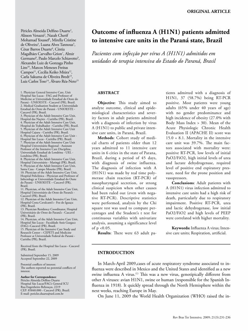
Table 1- Clinical-epidemiologic data of patients with H1N1 diagnosis admitted to the intensive care unit (n=63)Variable Total RT-PCR pos RT-PCR neg P value
(N=63) (N=37) (N=26)Male 46.0 48.6 47.8 0.847Age (years)
13 to 25 23.8 24.3 17.4 0.73026 to 40 41.3 45.9 34.8 0.53541 to 55 20.6 16.3 30.4 0.308≥ 56 14.3 13.5 17.4 0.946
Time (days) from the onset of symptoms to ICU admission 6.7± 3.70 7.27 ± 3.58 6.22 ± 3.82 0.269Comorbidities 42.8 48.6 39.1 0.625
Obesity (BMI >30) 27.0 32.4 21.7 0.518COPD 9.5 10.8 8.7 0.878DM 3.2 2.7 4.3 0.717CHF functional class III or IV 4.8 5.4 4.3 0.695CRF plus dialysis 0 0 0Neoplasms 1.6 2.7 0 0.857HIV 1.6 2.7 0 0.857SLE 1.6 2.7 0 0.857
Current pregnancy 12.7 10.8 17.4 0.704APACHE II first ICU 24h 15.0 ± 8.11 16.2 ± 7.84 15.4 ± 8.64 0.704
ICU - intensive care unit; COPD - chronic obstructive pulmonary disease; BMI - body mass index; DM - diabetes mellitus; CHF - congestive heart failure; CRF – chronic renal failure; HIV - human immunodeficiency virus; SLE- systemic lupus erythematosus; APACHE - Acute Physiology and Chronic Health Evaluation. Results expressed as mean ± standard deviation or %.
Virus A (H1N1) infection in intensive care unit 233
Rev Bras Ter Intensiva. 2009; 21(3):231-236
vate ICUs, and 57.1% in public ICUs. The mean time from the onset of symptoms to ICU admission was 6.0 days. All patients underwent RT-PCR nasopharyngeal se-cretion testing, which was positive in 37 (58.7%) patients. Invasive mechanic ventilation was required for 71.4%.
The admission laboratory tests and clinical features are shown in table 2.
The ICU mortality was 39.7%, while of those under mechanic ventilation (MV) it was 53.3%. There was no
difference in mortality between private and public ICUs (36.1% vs 44.4%, p=0.685).
Eight of the patients were pregnant (12.7%), includ-ing one in the 3rd post-partum day. Maternal mortality in the ICU was of 25%.
There was no age-related difference in mortality (x2=5.09; p=0.16).
The main ICU mortality related factors (Table 3) were positive RT-PCR test, initially low arterial oxygen pres-
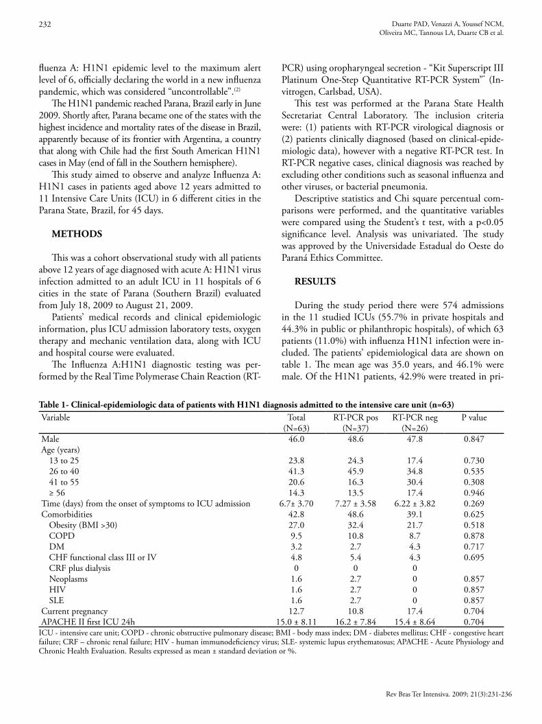
Table 2 –Clinical-epidemiological and laboratory characteristics at intensive care unit admission (n=63) Variable Total RT-PCR pos RT-PCR neg P value
N=63 N=37 N=26Leucocytes (Cells x 103/mm3) 9.89 ± 7.57 7.28 ± 3.34 14.22 ± 10.59 <0.005Platelets (Cells/mm3) 172.0 ± 98.92 162.9 ± 79.58 196.6 ± 123.16 0.192LDH(UI/ml) 504.7 ± 591.4 1067.2 ± 668.9 504.7 ± 270.3 <0.001CPK (UI/ml) 694.6 ± 1324.1 856.9 ± 1335 694.6 + 252 0.544Baseline Creatinine (mg/dl) 1.26 ± 1.25 1.28 ± 1.17 1.27 ± 1.46 0.976Creatinine > 1.5 mg/dl 17.4 18.9 17.4 0.858Arterial Lactate 2.44 ± 2.34 2.32 ± 4.72 2.28 ± 6.81 0.978Baseline PaO2/FiO2 150.0 ± 91.23 119.4 ± 79.36 189.4 ± 102.6 0.003Baseline PaCO2 (mmHg) 40.7 ± 20.4 38.5 ± 13.2 46.7 ± 27.94 0.124Higher PEEP / first 12 h 15.1 ± 7.29 16.9 ± 7.37 12.7 ± 6.29 0.021VD use in the first 04h 42.8 43.2 47.8 0.917
RT-PCR - Real time - Polymerase Chain Reaction; LDH – Lactic dehydrogenase; CPK - Creatino-phosfokynase; PEEP – Positive end expiratory pressure; VD - Vasopressor drugs. Results expressed as mean ± standard deviation or %. Table 3 – Mortality risk factors
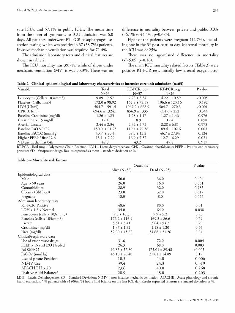
Outcome P valueAlive (N=38) Dead (N=25)
Epidemiological data Male 50.0 36.0 0.404Age > 50 years 26.0 16.0 0.531Comorbidities 28.9 32.0 0.985Obesity (BMI>30) 23.0 32.0 0.617Pregnant 18.0 8.0 0.455
Admission laboratory testsRT-PCR Positive 48.6 80.0 0.01LDH > 1.5 x Normal 34.0 64.0 0.038Leucocytes (cells x 103/mm3) 9.8 ± 10.3 9.9 ± 5.2 0.95 Platelets (cells x 103/mm3) 176.2 ± 116.9 169.3 ± 86.6 0.79Lactate 5.51 ± 5.41 3.84 ± 5.67 0.29Creatinine (mg/dl) 1.37 ± 1.32 1.18 ± 1.20 0.56Urea (mg/dl) 52.90 ± 45.87 34.68 ± 21.26 0.04
Clinical/respiratory dataUse of vasopressor drugs 31.6 72.0 0.004PEEP > 15 cmH2O Needed 26.3 68.0 0.003PaO2/FiO2 96.83 ± 57.80 175.01 ± 89.48 <0.005PaCO2 (mmHg) 45.10 ± 26.40 37.81 ± 14.89 0.17Use of prone Position 10.5 44.0 0.006NIMV Use 39.4 24.3 0.319APACHE II > 20 23.6 40.0 0.268Positive fluid balance* 28.9 48.0 0.203
LDH – Lactic Dehydrogenase; SD – Standard Deviation; NIMV – non-invasive mechanic ventilation; APACHE - Acute physiology and chronic health evaluation. * % patients with >1800ml/24 hours fluid balance on the first ICU day. Results expressed as mean ± standard deviation or %.
234 Duarte PAD, Venazzi A, Youssef NCM, Oliveira MC, Tannous LA, Duarte CB et al.
Rev Bras Ter Intensiva. 2009; 21(3):231-236
sure/inspired oxygen fraction rate (PaO2/FiO2), initially increased urea and lactic dehydrogenase (LDH), positive end-expiratory pressure (PEEP) required , need for prone position and use of vasopressor drugs.
Table 4 displays the outcomes of positive and negative RT-PCR results.
DISCUSSION
In March 1918 (during World War I) a severe influ-enza epidemic spread throughout the world, beginning simultaneously in the U.S. and Europe and killing (par-ticularly in the “second wave”, that started 5 months later) about 40 million people, becoming the most catastrophic medical event ever in human history.(3) The strain involved was Influenza A (H1N1) from birds. Since then, the influ-enza virus surveillance systems had identified changes in the A virus epidemic strains every 1 to 2 years (by surface glycoprotein mutations, hemagglutinin and neuropepti-dase). In the last 91 years, two A virus pandemics were identified: in 1957 (H2N2) and 1968 (H3N2), although they were much less lethal.(4-6) During the last years, with epidemics observed such as severe acute respiratory syn-drome (SARS), the imminence of an influenza pandemic has been alerted by the health authorities.(3,7,8)
In this trial, it was seen that 11.0% of ICU admis-sions were due to H1N1 cases. An important bias is that these hospitals were mostly considered as reference for treatment of H1N1 patients, which could increase disease incidence. Another important factor to bear in mind is that in, at least one hospital, a special unit was created for H1N1 patients. Thus, in practice the number of ICU beds increased during this period (as the original ICU continued to take care of non-H1N1 patients). Finally, there were instructions to reduce the elective surgeries in public hospitals as a health system strategy, which could temporarily reduce some indica-tions for ICU admissions.
The ICU mortality in this study was high, particularly among invasive MV patients. This finding is also found in literature. In 30 of the admitted cases (either with viro-logical or clinical diagnosis) in California hospitals (USA),
six patients were admitted to the ICU (4 under mechanic ventilation), with no death (although three were still hospi-talized at the reporting time).(9)According to Perez-Padilha et al.,(10) of the 18 H1N1 cases confirmed in patients with respiratory failure, 10 under mechanical ventilation were reported; of the total of patients under mechanical ventila-tion (MV), 70% died.
The main mortality risk factors identified were related to clinical severity, particularly respiratory impairment. Another trial found these main factors associated to higher risk of mortality: hypotension requiring vasoactive drugs, acute renal failure, metabolic acidosis, APACHE II, PaO2/FiO2 and Sequential Organ Failure Assessment (SOFA) admission score.(10) The severe respiratory impairment is similar to severe influenza cases, just as SARS.(11)
As previously described in literature,(10) ICU mortal-ity was higher among positive RT-PCR test patients than among those with a negative test. This could reflect a high-er viral load and morbidity in positive RT-PCR patients, although as mentioned below, this could also mean that among the RT-PCR negative patients other diagnoses, with milder courses could be included. Another possibili-ty is inappropriate material collection, which could reflect in increased false-negatives rates.(12)
Pregnancy is acknowledged as a risk factor for respira-tory complications in infections by influenza.(13,14) Among the factors justifying the high incidence in this group, physiological changes of pregnancy, including reduced pulmonary functional residual capacity and cell-mediated immunity impairment, are emphasized.(15) However, in patients with severe infection requiring ICU stay, the mortality rate is similar to that of the general popula-tion.(9) In our study the maternal mortality among preg-nant women was similar to the overall group (25.0% vs 41.8%; p=0.602).
Obesity has clearly been incriminated as a severity and mortality risk factor among swine influenza patients.(16) It is believed that this is due both to the disease-related re-spiratory effects (reduced pulmonary functional residual capacity) and presence of typically associated comorbidi-ties (such as diabetes, asthma, cardiovascular diseases, etc).(9) Obesity was very frequent among this group of patients,
Table 4 – Clinical outcomes
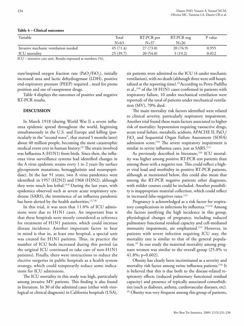
Variable Total RT-PCR pos RT-PCR neg P valueN=63 N=37 N=26
Invasive mechanic ventilation needed 45 (71.4) 27 (73.0) 20 (76.9) 0.955ICU mortality 25 (39.7) 20 (54.0) 5 (19.2) 0.012
ICU – intensive care unit. Results expressed as numbers (%).
Virus A (H1N1) infection in intensive care unit 235
Rev Bras Ter Intensiva. 2009; 21(3):231-236
similar to literature: 27.0% had a body mass index (BMI) above 30.
The time from onset of the symptoms and ICU admis-sion was relatively long (6 days). In the Spanish trial,(16) time from onset of symptoms and beginning of treatment was 4 days.
This study has limitations. The total number of cases may have been overestimated. In our study, were consid-ered as H1N1 cases, not only patients with confirmed laboratory diagnosis, but also those with strong clinical-epidemiological suspicion, although with negative RT-PCR. According to the Health Ministry of Brazil,(17) “in-fluenza A:H1N1 will be discarded if influenza A virus is not detected by RT-PCR or culture techniques.” How-ever, although RT-PCR is the World Health Organiza-tion (WHO) and Center for Disease Control (CDC) recommended method,(12,18) false-negative results reach 10%.(19,20) Thus, the investigators decided to assess the strongly suspected cases (clinical diagnosis), even though with a negative influenza A test (since other causes were discarded). This was the same methodology as that used in previous reports,(9,10) although in the Spanish trial(16) only positive cases were accepted for diagnosis. Howev-er, the authors recognize that this approach constitutes a significant limitation for this study regarding overall data interpretation.
The univariate analysis may have been a restricted tool to define mortality-associated risk factors and therefore a limitation for interpretation of this study data.
CONCLUSIONS
Adult patients with an influenza A(H1N1) diagnosis admitted to an ICU have an increased mortality rate. The main mortality predictive factors found in this trial were: severe respiratory impairment, positive RT-PCR test, increased baseline urea and LDH, and need for va-sopressor drugs. However, diagnostic methods (includ-ing clinical diagnosis) and the reduced number of pa-tients may have contributed to jeopardize interpretation of this study data.
RESUMO
Objetivo: Analisar a evolução, características clínico-epidemiológicas e fatores de gravidade em pacientes adultos admitidos com diagnóstico de infecção por vírus A(H1N1) em unidades de terapia intensiva públicas e privadas no es-tado do Paraná, sul do Brasil.
Métodos: Estudo coorte de análise de prontuários de pacientes com idade superior a 12 anos admitidos em 11 unidades de terapia intensiva de 6 cidades no estado do Paraná (Brasil), durante um período de 45 dias, com diag-nóstico de gripe suína. O diagnóstico de infecção por vírus A(H1N1) foi feito através de real time -polimerase chain re-action (RT-PCR) da secreção nasofaríngea, ou de forte sus-peita clínica quando descartadas outras causas (mesmo com RT-PCR negativo). Foi feita estatística descritiva e análise com teste chi quadrado, para comparação entre porcenta-gens e teste t de student para variáveis continuas, com análi-se univariada, admitindo-se como significante um p<0,05.
Resultados: Foram admitidos 63 pacientes adultos com diagnóstico de H1N1, sendo 37 (58,7%) RT-PCR positi-vos. A maioria dos pacientes era de adultos jovens (65% com idade inferior a 40 anos), sem predominância de sexo e alta incidência de obesidade (27,0% com índice de massa corpórea>30). A média do escore Acute Physiologic Chronic Heatlh Evaluation II (APACHE II) foi de 15,0 ± 8,1. A mortalidade na unidade de terapia intensiva foi de 39,7%. Os principais fatores associados a essa mortalidade foram exame positivo no teste RT-PCR, níveis baixos de relação PaO2/FiO2 inicial, níveis elevados de uréia e desidrogena-se lática iniciais, nível de pressão expiratória final positiva necessária, necessidade de posição prona e de drogas vaso-pressoras.
Conclusões: Pacientes admitidos em unidades de terapia intensiva com infecção por vírus A(H1N1) apresentaram alto risco de óbito, particularmente devidos ao comprome-timento respiratório. O exame RT-PCR positivo, níveis de uréia e de desidrogenase láctica, além baixa PaO2/FiO2 e necessidades de PEEP alta, foram relacionados com uma maior mortalidade.
Descritores: Vírus da influenza A; Unidade de terapia intensiva; Ventilação mecânica
REFERENCES
1. Novel Swine-Origin Influenza A (H1N1) Virus Investiga-tion Team, Dawood FS, Jain S, Finelli L, Shaw MW, Lin-dstrom S, Garten RJ, et al. Emergence of a novel swine-ori-gin influenza A (H1N1) virus in humans. N Engl J Med. 2009;360(25):2605-15.
2. World Health Organization. Global Alert and Response (GAR). DG Statement following the meeting of the Emer-gency Committee [Internet]. [cited 2009 Sep 29]. Available from: http://www.who.int/csr/disease/swineflu/4th_mee-ting_ihr/en/index.html .
3. World Health Organization. Global Alert and Response (GAR). Avian influenza: assessing the pandemic threat [In-
236 Duarte PAD, Venazzi A, Youssef NCM, Oliveira MC, Tannous LA, Duarte CB et al.
Rev Bras Ter Intensiva. 2009; 21(3):231-236
ternet]. [cited 2009 Sep 29]. Available from: www.who.int/csr/disease/influenza/WHO_CDS_2005_29/en/ .
4. Kamps BS, Hoffmann C, Preiser W, editors. Influenza re-port 2006 [Internet]. Paris: Flying Publisher; 2006. 225 p. [cited 2009 Sep 29]. Available from: http://www.influenza-report.com/influenzareport.pdf
5. Morens DM, Taubenberger JK, Fauci AS. The per-sistent legacy of the 1918 influenza virus. N Engl J Med. 2009;361(3):225-9. Erratum in: N Engl J Med. 2009;361(11):1123.
6. Zimmer SM, Burke DS. Historical perspective - Emer-gence of influenza A (H1N1) viruses. N Engl J Med. 2009;361(3):279-85
7. World Health Organization. Director-General. Pandemics: working together for an effective and equitable response. [Internet]. [cited 2009 Sep 13]. Disponível em: http://www.who.int/dg/speeches/2007/20070613_seattle/en/in-dex.html.
8. Lazzari S, Stöhr K. Avian influenza and influenza pande-mics. Bull World Health Organ. 2004; 82(4):242.
9. Centers for Disease Control and Prevention (CDC). Hos-pitalized patients with novel influenza A (H1N1) virus infection - California, April-May, 2009. MMWR Morb Mortal Wkly Rep. 2009;58(19):536-41.
10. Perez-Padilla R, de la Rosa-Zamboni D, Ponce de Leon S, Hernandez M, Quiñones-Falconi F, Bautista E, Rami-rez-Venegas A, Rojas-Serrano J, Ormsby CE, Corrales A, Higuera A, Mondragon E, Cordova-Villalobos JA; INER Working Group on Influenza . Pneumonia and respiratory failure from swine-origin influenza A (H1N1) in Mexico. N Engl J Med. 2009;361(7):680-9.
11. Phua GC, Govert J. Mechanical ventilation in an airborne epidemic. Clin Chest Med. 2008;29(2):323-8, vii.
12. World Health Organization. CDC protocol of realtime RTPCR for influenza A (H1N1). [Internet]. [citado 2009 Set 13]. Disponível em: http://www.who.int/csr/resour-ces/publications/swineflu/CDCRealtimeRTPCR_Swi-neH1Assay-2009_20090430.pdf.
13. Fiore AE, Shay DK, Broder K, Iskander JK, Uyeki TM, Mootrey G, Bresee JS, Cox NS; Centers for Disease Con-trol and Prevention (CDC); Advisory Committee on Im-
munization Practices (ACIP). Prevention and control of in-fluenza: recommendations of the Advisory Committee on Immunization Practices (ACIP), 2008. MMWR Recomm Rep. 2008;57(RR-7):1-60.
14. Centers for Disease Control and Prevention (CDC). No-vel influenza A (H1N1) virus infections in three pregnant women - United States, April-May 2009. MMWR Morb Mortal Wkly Rep. 2009;58(18):497-500. Erratum in: MMWR Morb Mortal Wkly Rep. 2009;58(19):541.
15. Rasmussen SA, Jamieson DJ, Bresee JS. Pandemic influen-za and pregnant women. Emerg Infect Dis. 2008;14(1):95-100.
16. Rello J, Rodriguez A, Ibanez P, Socias L, Cebrian J, Mar-ques A, Guerrero J, Ruiz-Santana S, Marquez E, Del No-gal-Saez F, Alvarez-Lerma F, Martinez S, Ferrer M, Avella-nas M, Granada R, Maravi-Poma E, Albert P, Sierra R, Vi-daur L, Ortiz P, Prieto Del Portillo I, Galvan B, Leon-Gil C, H1N1 Semicyuc Working Group I. Intensive care adult patients with severe respiratory failure caused by Influenza A (H1N1)v in Spain. Crit Care. 2009;13(5):R148.
17. Brasil. Ministério da Saúde. Secretaria de Vigilância em Saúde. Gabinete Permanente de Emergências em Saúde Pública. Emergência de Saúde Pública de Importância In-ternacional - ESPII. Protocolo de manejo clínico e vigilân-cia epidemiológica da influenza. Portal da Saúde [Internet]. [citado2009 Set 13]. Disponível em: http://portal.saude.gov.br/portal/arquivos/pdf/protocolo_de_manejo_clini-co_05_08_2009.pdf.
18. Centers for Disease Control and Prevention. Interim gui-dance on case definitions to be used for investigations of novel influenza A (H1N1) cases. [Internet]. [cited 2009 Sep 13]. Disponível em: http://www.cdc.gov/h1n1flu/ca-sedef.htm.
19. Ellis J, Iturriza M, Allen R, Bermingham A, Brown K, Gray J, Brown D. Evaluation of four real-time PCR assays for detection of influenza A (H1N1)v viruses. Euro Surveill. 2009;14(22):pii:19230.
20. Ginocchio CC, Zhang F, Manji R, Arora S, Bornfreund M, Falk L, et al. Evaluation of multiple test methods for the de-tection of the novel 2009 influenza A (H1N1) during the New York City outbreak. J Clin Virol. 2009;45(3):191-5.