Embed Size (px)
Citation preview
Listen to this manuscript’s
audio summary by
JACC Editor-in-Chief
Dr. Valentin Fuster.
J O U R N A L O F T H E A M E R I C A N C O L L E G E O F C A R D I O L O G Y V O L . 7 0 , N O . 6 , 2 0 1 7
ª 2 0 1 7 T H E A U T H O R S . P U B L I S H E D B Y E L S E V I E R O N B E H A L F O F T H E AM E R I C A N
C O L L E G E O F C A R D I O L O G Y F O U N DA T I O N . T H I S I S A N O P E N A C C E S S A R T I C L E U N D E R
T H E C C B Y - N C - N D L I C E N S E ( h t t p : / / c r e a t i v e c o mm o n s . o r g / l i c e n s e s / b y - n c - n d / 4 . 0 / ) .
I S S N 0 7 3 5 - 1 0 9 7
h t t p : / / d x . d o i . o r g / 1 0 . 1 0 1 6 / j . j a c c . 2 0 1 7 . 0 6 . 0 1 2
REVIEW TOPIC OF THE WEEK
Overcoming the Roadblocks to CardiacCell Therapy Using Tissue Engineering
Mounica Yanamandala, MD,a Wuqiang Zhu, MD, PHD,b Daniel J. Garry, MD, PHD,c Timothy J. Kamp, MD, PHD,dJoshua M. Hare, MD,e Ho-wook Jun, PHD,b Young-sup Yoon, MD, PHD,f Nenad Bursac, PHD,g
Sumanth D. Prabhu, MD,h Gerald W. Dorn II, MD,i Roberto Bolli, MD,j Richard N. Kitsis, MD,a Jianyi Zhang, MD, PHDb
ABSTRACT
FrobD
Ins
Un
Mi
CohDiCejIn
No
co
co
Ma
Transplantations of various stem cells or their progeny have repeatedly improved cardiac performance in animal models
of myocardial injury; however, the benefits observed in clinical trials have been generally less consistent. Some of the
recognized challenges are poor engraftment of implanted cells and, in the case of human cardiomyocytes, functional
immaturity and lack of electrical integration, leading to limited contribution to the heart’s contractile activity and
increased arrhythmogenic risks. Advances in tissue and genetic engineering techniques are expected to improve the
survival and integration of transplanted cells, and to support structural, functional, and bioenergetic recovery of
the recipient hearts. Specifically, application of a prefabricated cardiac tissue patch to prevent dilation and to
improve pumping efficiency of the infarcted heart offers a promising strategy for making stem cell therapy a
clinical reality. (J Am Coll Cardiol 2017;70:766–75) © 2017 The Authors. Published by Elsevier on behalf of the
American College of Cardiology Foundation. This is an open access article under the CC BY-NC-ND license
(http://creativecommons.org/licenses/by-nc-nd/4.0/).
A lthough transplanted cells and engineeredtissues result in improved cardiac perfor-mance when tested in animal models of
myocardial injury, the benefits observed in clinicaltrials have generally been modest at best. Opinionsregarding the optimal cell type or combination ofcell types have yet to reach consensus, and only avery small proportion of the administered cells areengrafted by the native myocardium. Cellular attri-tion is often attributed to a lack of perfusion in theinfarcted region, but the recipient’s immune systemmay also play a role, particularly in preclinical studies
m the aDepartment of Medicine, Montefiore Medical Center and Albe
epartment of Biomedical Engineering, The University of Alabama at B
titute, University of Minnesota, Minneapolis, Minnesota; dDepartment o
iversity of Wisconsin-Madison, Madison, Wisconsin; eDepartment of Med
ami, Florida; fDepartment of Medicine, Emory University, and Severan
llege of Medicine, Atlanta, Georgia; gDepartment of Biomedical Engin
epartment of Medicine, Division of Cardiovascular Disease, University o
nter for Pharmacogenomics, Department of Medicine, Washington Univer
stitute of Molecular Cardiology, University of Louisville, Louisville, Kentuc
rthStar Genomics. Dr. Kamp is a consultant for Cellular Dynamics Internati
nsultant and board member for Longeveron LLC. All other authors have rep
ntents of this paper to disclose.
nuscript received May 24, 2017; revised manuscript received June 5, 2017
with human-derived tissues or other xenogeneictransplantation experiments. Furthermore, thesurviving cells rarely produce grafts of substantialsize and may remain electrically isolated from thenative myocardium, which would prevent the graftfrom contributing to the contractile activity of theheart and, more importantly, could lead to arrhyth-mogenic complications, which may be the primarysafety concern associated with transplanted myocar-dial cells and tissues. Tissue engineering strategiesare expected to improve engraftment of transplantedcells, as well as structural, functional, and
rt Einstein College of Medicine, Bronx, New York;
irmingham, Birmingham, Alabama; cLillehei Heart
f Medicine, School of Medicine and Public Health,
icine, University of Miami Miller School of Medicine,
ce Biomedical Science Institute, Yonsei University
eering, Duke University, Durham, North Carolina;
f Alabama at Birmingham, Birmingham, Alabama;
sity School of Medicine, St. Louis, Missouri; and the
ky. Dr. Garry is a consultant for Boston Scientific and
onal. Dr. Hare is a board member for Vestion; and is a
orted that they have no relationships relevant to the
, accepted June 5, 2017.
AB BR E V I A T I O N S
AND ACRONYM S
CM = cardiomyocyte
CPC = cardiac progenitor cell
Cx43 = connexin 43
EC = endothelial cell
hiPSC = human induced
pluripotent stem cell
HLA = human leukocyte
antigen
hPSC = human pluripotent
stem cell
MHC = major
histocompatibility complex
MI = myocardial infarction
PSC = pluripotent stem cell
TNF = tumor necrosis factor
J A C C V O L . 7 0 , N O . 6 , 2 0 1 7 Yanamandala et al.A U G U S T 8 , 2 0 1 7 : 7 6 6 – 7 5 Cardiac Cell Therapy: Roadblocks to Overcome
767
bioenergetic recovery of the infarcted heart. Theseand many other topics were discussed by attendeesof the National Institutes of Health 2016 ProgenitorCell Biology Consortium and Cardiovascular TissueEngineering Symposium at the University of Ala-bama, Birmingham, on March 28, 2016. Here, we pre-sent some of the more provocative ideas andadvances that were discussed at the meeting andthat may facilitate the translation of cardiac cell-and tissue-engineering therapies from the laboratoryto the clinic.
CELL TYPES FOR USE IN CARDIAC THERAPY
A wide variety of cell sources have been evaluated forrepair of the ischemic myocardium in animal models,and a subset of these have undergone testing inclinical trials. A recent review by Nguyen et al. (1)summarized the clinical trials on stem cell therapyfor ischemic heart diseases and heart failure fromJanuary 1, 2000, to July 2016. Table 1 adds the newclinical trials for ischemic heart diseases and heartfailure published between July 27, 2016, and May 18,2017, based on our PubMed search results, as well asclinical trials for stem cell therapy for congenitalheart diseases.
Human pluripotent stem cells (hPSCs) and newercell types, such as induced cardiac progenitor cells(CPCs) (2,3), are especially promising for cardiac celland tissue engineering therapy (4–6) because theycan be efficiently differentiated into functional car-diomyocytes (CMs), endothelial cells (ECs), andsmooth muscle cells (SMCs) (7–11). However, theoptimal proportions of each cell type have yet to beidentified, and a variety of other cell lineages (e.g.,resident CPCs) (12) may be needed to maximizetherapeutic effectiveness. Furthermore, mitochon-dria have important functions in cardiac metabolism(13) and programmed CM death (14), and emergingroles in cardiac differentiation were recentlysuggested (15,16). Specifically, CM differentiationrequires the membranes of adjacent mitochondria tofuse, and fusion of the outer membranes is regulatedby mitofusins 1 and 2, which direct ESC differentia-tion into CMs via regulation of calcineurin and Notchsignaling (16).
The importance of restoring functional CMs forpost-infarction repair is self-evident because theyprovide the mechanical force needed for contraction.In a rat model of myocardial infarction (MI),measurements of cardiac function and remodelingwere significantly better when implanted fibrin gel-based tissue-engineered patches were created fromthe complete population of neonatal rat cardiac cells
than when the CMs were omitted (17).Furthermore, extracellular matrix productionin tissue-engineered patches appears to in-crease in response to production of trans-forming growth factor b1 by CMs (18,19), andCMs are an important source of vascularendothelial growth factor in cell sheet–basedengineered tissues (20). Thus, CMs and cy-tokines that mediate CM–non-CM communi-cation are crucial components of thebeneficial activity induced by transplantedengineered cardiac tissues.
Other cell sources of potential utility forcardiac repair include mesenchymal stemcells (prototypically derived from bonebarrow), cardiac stem cells, cardiospheresisolated from endocardial biopsies (in miceand humans), which are composed of a
heterogeneous cell population, but are exceptionallyproliferative, and Abcg2-expressing progenitor cells(21). When suspended in saline and transplanted intothe hearts of mice after an acute infarction, Sca-1þ/CD31� cells appear to attenuate decline in cardiacfunction, increase myocardial neovascularization,and modestly promote CM differentiation from graftcells, as well as host cell proliferation (22).Variation of interspecies responses to cell therapyis another critical issue. Commonly used preclinicalmodels include nonhuman primates, large mammals(swine, dog, and others), and rodents. Although itmight be necessary, there is neither guidance onselection of preclinical animal models nor consensuscriteria on experimental design for preclinicalstudies thus far. Genetic background may affect theinterpretation of experimental results. Therefore,comparison and validation of data collected fromdifferent species are important when translatingpreclinical research to clinical trials.
Although transplantation of various cell types hasbeen reported to improve left ventricular functionand structure after MI, overwhelming evidence hasdemonstrated that there is no significant long-termengraftment of adoptively transferred cells into thehost myocardium. The persistence of beneficialeffects, despite the disappearance of transplantedcells, indicates that cell therapy may act via paracrinemechanisms. In addition, the rapid clearance of cellsfrom the host myocardium suggests that the benefitsof cell therapy are limited by the poor engraftment ofthe cells, implying that 1 dose does not adequatelytest the efficacy of that cell product (23). However,almost all preclinical and clinical studies of celltherapy performed heretofore have based theirassessment of efficacy on the outcome of

TABLE 1 Published Stem/Progenitor Cell Clinical Trials for Heart Diseases
Diseases Trial Design Sample Size Cell Type Cell Source Delivery Route
HLHS Nonrandomized (phase 1) 7 Autologous CDC Cardiosphere IC
HLHS Randomized (phase 2) 34 Autologous CDC Cardiosphere IC
Ischemic heart disease Randomized control (phase 2) 90 Autologous BMCs Bone marrow IC vs. IM
Ischemic heart disease Randomized control (phase 3) 271 Autologous MSCs Bone marrow IM
Nonischemic cardiomyopathy Randomized control (phase 2) 22 Allogeneic MSCs Bone marrow IV
Refractory angina Randomized control (phase 2) 31 Autologous CD133þ cells Bone marrow TEP
Ischemic vs. nonischemicdilated cardiomyopathy
Nonrandomized (phase 1) 27 Autologous skeletalstem-cell sheets
Skeletal muscle(vastus medialis)
Sutured toheart surface
Nonischemic dilatedcardiomyopathy
Randomized (phase 2) 37 Allogeneic MSC (19 patients) vs.autologous MSC (18 patients)
Bone marrow TEP
STEMI Randomized control (phase 2) 188 Autologous BMCs Bone marrow IC
BMC ¼ bone marrow–derived cell; BNP ¼ B-type natriuretic peptide; CDC ¼ cardiosphere-derived cells; HLHS ¼ hypoplastic left heart syndrome; hMSC ¼ human mesenchymal stem cell; IC ¼ intracoronaryinfusion; IM ¼ intramyocardial injection; IV ¼ intravenous; LVEF ¼ left ventricular ejection fraction; MSC ¼ mesenchymal stem cell; NT-proBNP ¼ N-terminal pro-B-type natriuretic peptide; RVEF ¼ rightventricular ejection fraction; STEMI ¼ ST-segment elevation myocardial infarction; TEP ¼ transendocardial injection; TNF ¼ tumor necrosis factor.
Yanamandala et al. J A C C V O L . 7 0 , N O . 6 , 2 0 1 7
Cardiac Cell Therapy: Roadblocks to Overcome A U G U S T 8 , 2 0 1 7 : 7 6 6 – 7 5
768
1 administration of a cell product. The problem ofmodest or no beneficial effects might be overcome byrepeated cell doses. The rationale is that just as mostpharmacological agents are ineffective when givenonce, but can be highly effective when given repeat-edly, a cell product might be ineffective or modestlyeffective as a single treatment, but might be quiteefficacious if given repeatedly.
On the basis of preclinical data performed in largeanimal models, human testing of various adult cellsources has progressed from phase I to phase III trials(24,25). From a mechanistic standpoint, cell therapyin the iterations described earlier reduces tissuefibrosis, restores tissue perfusion, has a powerfulanti-inflammatory effect, and stimulates myogenesis,largely by promoting endogenous myogenesis (24).Moreover, clinical trial activity is extending intonumerous cardiomyopathic disease states, includinghypoplastic left heart syndrome, adriamycin-inducedcardiomyopathy, and idiopathic dilated cardiomyop-athy. Future efforts to apply tissue-engineeringstrategies in the clinical setting will emergefollowing appropriate preclinical testing.
CELL ENGRAFTMENT
As many as 1 billion CMs are lost during MI, andalthough the typical dose of transplanted cells mayapproach or exceed this number, just 0.1% to 10% ofthe cells are engrafted into the myocardium andcontinue to survive for more than a few weeks aftertransplantation (26,27). Much of this attrition can beattributed to the harsh environment in and near theregion of the infarct; thus, one strategy for improving
engraftment and survival of transplanted cells is touse natural or artificial biomaterials that provide aprotective environment for the transplanted cells(28–30). Overall, published reports indicate that thefunctional benefits of the cardiac cell patch therapycritically depend upon the longer-term structuralintegrity of the cell patches. Maintenance of longer-term graft size remains a major challenge in preclin-ical trials of cell therapy.
IMMUNOGENICITY
One of the chief benefits of human induced pluripotentstem cells (hiPSCs) is that they can be generated from apatient’s own somatic cells, and consequently are notexpected to provoke an immune response after trans-plantation. Although this approach holds appeal foravoiding pharmacological immunosuppression andassociated complications, it requires a substantial timewindow (months) for hiPSC generation, followed bydifferentiation and graft formation. Thus, this autol-ogous approach is better suited to treatment of morechronic heart failure with existing technology. How-ever, some studies suggest that the immune toleranceof patient-specific induced PSC–derived cells may varydepending on the cells’ lineage (31–34). Questionsregarding immunogenicity are often addressed byperforming experiments in mice with humanized im-mune systems, but the currently available modelshave a limited lifespan and inconsistent immuneresponse, perhaps because the animals’ endogenousimmune system is eliminated with sublethal doses ofradiation. Collectively, these observations suggestthat translation of hiPSC-derived cell technology to

TABLE 1 Continued
Follow-Up
Summary/Observation
Trial Name/IdentifierHeart Function Others
18–36 months [ RVEF Improved somatic growth TICAP/NCT01273857 (78,79)
12 months [ RVEF Reduced fibrosis, improved somatic growth PERSEUS/NCT01829750 (80)
6–12 months [ LVEF in IM group, but not in IC group YNT-proBNP in IM group, but not IC group REGENERATE-IHD/NCT00747708 (81)
39 weeks No change LVEF Y incidence of sudden or aborted sudden deaths CHART-1/NCT01768702 (82)
90 days No change LVEF [ health status and Y circulating inflammatory cells NCT02467387 (83)
1, 4, 6, and 12 months No change LVEF Y angina REGENT-VSEL/NCT01660581 (84)
12 months [ LVEF in patients with ischemic heartdiseases, but not in patients withnonischemic heart diseases
Y BNP in patients with ischemic heart diseases, butnot in patients with nonischemic heart diseases
UMIN000003273 (85)
12 months [ LVEF in allo-MSC group, but not in theauto-MSC group
Y TNF-a, to a greater extent with allo-hMSCs vs.auto-hMSCs at 6 months
POSEIDON-DCM/NCT01392625 (24)
6 months [ LVEF in BMC group No treatment effect in irradiated BMC group BOOST-2/ISRCTN17457407 (86)
J A C C V O L . 7 0 , N O . 6 , 2 0 1 7 Yanamandala et al.A U G U S T 8 , 2 0 1 7 : 7 6 6 – 7 5 Cardiac Cell Therapy: Roadblocks to Overcome
769
clinical applications will likely require testing of theimmunogenicity of each hiPSC-derived cell lineage in anew generation of models that can provide moreconsistent and reliable results.
Allograft rejection is primarily mediated byhost-derived reactive T lymphocytes that recognizenon-self human leukocyte antigens (HLAs) on thesurface of transplanted donor cells. Humans have 2main categories of HLAs: major histocompatibilitycomplex (MHC) classes I and II. The Townes laboratoryrecently showed that surface expression of HLA class Imolecules can be largely eliminated in a line of humanembryonic stem cells (H9) using the clustered regu-larly interspaced short palindromic repeats (CRISPR)/CRISPR-associated protein (Cas) gene-editing systemto knock out both alleles of the gene for b2 micro-globulin, which is essential for cell-surface expressionof HLA class I and stability of the peptide-bindinggroove. CRISPR/Cas gene editing has also been usedto knock out the Class II MHC transactivator (CIITA) inhuman ECs, and the CIITA-knockout ECs could betransplanted into mice without producing an immuneresponse (35). The degree of differentiation of thehiPSCs derivatives engrafted may also influence theimmunogenicity of the tissue grafts. This is particu-larly relevant for tissue-engineering applications,because even when professional antigen-presentingcells are depleted, cell-mediated allograft rejectioncan still occur (36). This is perhaps because human ECsactivate alloantigen-reactive memory CD4þ T cells viaa mechanism requiring expression of class II MHCs.Thus, gene-editing technologies may enableresearchers to create “universal donor” hiPSC lines, aswell as hiPSC-derived cells and engineered tissueswith substantially higher rates of engraftment that can
be used to treat a wider variety of patients anddiseases.
IMMUNOMODULATION
As described earlier, stem cell engraftment rates andsurvival following transplantation are disappoint-ingly low. Moreover, among surviving transplantedprogenitor cells, the demonstrable magnitude of dif-ferentiation into functional CMs has been variable,ranging from no evidence of CM differentiation togeneration of small, apparently integrated CMs(22,26,37–39). Most studies of cell therapy seek tointervene therapeutically either after MI or duringchronic heart failure. The proinflammatory environ-ment of the failing heart may also be responsible forthe reported functional benefits of cardiac cell ther-apies. As both of these pathological scenarios exhibitheightened inflammatory activation and innate andadaptive immune cell infiltration in the myocardium(40–43), these microenvironmental factors may beimportant contributors to the suboptimal responsesto cell therapy. For example, tumor necrosis factor(TNF), a proinflammatory cytokine elaborated byboth immune cells and failing CMs, restrains CM dif-ferentiation of resident cardiac stem cells, and canchannel an alternate neuroadrenergic-like fatein vitro (44); these effects would be expected todiminish the reparative effects of stem cell therapy.Such findings suggest that immunomodulation of theproinflammatory microenvironment in recipients ofprogenitor cell therapy may be a high-yield strategyto enhance cell engraftment and CM differentiation.To date, such approaches have been relatively unex-plored, but could include targeting specific innate
Yanamandala et al. J A C C V O L . 7 0 , N O . 6 , 2 0 1 7
Cardiac Cell Therapy: Roadblocks to Overcome A U G U S T 8 , 2 0 1 7 : 7 6 6 – 7 5
770
immune cell populations (e.g., infiltrating andproinflammatory macrophages), specific cytokines(e.g., TNF), and/or antigen-independent T-cellresponses. These methodologies would be comple-mentary to the suppression of antigen-dependentMHC responses described earlier, and would ideallycomprise circumscribed interventions designed toimprove cell engraftment at the time of delivery andsubsequent CM differentiation during initial repair.Interestingly, mesenchymal stem cells suppress TNFlevels substantially, and this effect may thereforecontribute to some of the positive effects of thesecells in clinical trials (24).
ARRHYTHMOGENESIS
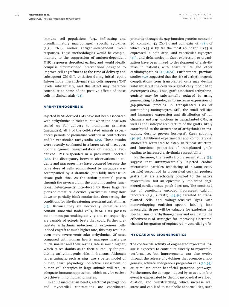
Injected hPSC-derived CMs have not been associatedwith arrhythmias in rodents, but when the dose wasscaled up for delivery to nonhuman primates(macaques), all 4 of the cell-treated animals experi-enced periods of premature ventricular contractionsand/or ventricular tachycardia (45). These resultswere recently confirmed in a larger set of macaquesupon allogeneic transplantation of macaque PSC-derived CMs suspended in a prosurvival cocktail(46). The discrepancy between observations in ro-dents and macaques may have occurred because thelarge dose of cells administered to macaques wasaccompanied by a dramatic ($10-fold) increase intissue graft size. As the action potential passesthrough the myocardium, the anatomic and/or func-tional heterogeneity introduced by these large re-gions of immature, electrically active tissue may slowdown or partially block conduction, thus setting theconditions for life-threatening re-entrant arrhythmias(47). Because they are electrically immature andcontain sinoatrial nodal cells, hPSC CMs possessautonomous pacemaking activity and consequently,are capable of ectopic beats that could further pre-cipitate arrhythmia induction. If exogenous cellsindeed engraft at much higher rate, this may result ineven more severe ventricular arrhythmias. Of note,compared with human hearts, macaque hearts aremuch smaller and their resting rate is much higher,which raises doubts as to their suitability for pre-dicting arrhythmogenic risks in humans. Althoughlarger animals, such as pigs, are a better model ofhuman heart physiology, objective assessment ofhuman cell therapies in large animals will requireadequate immunosuppression, which may be easiestto achieve in nonhuman primates.
In adult mammalian hearts, electrical propagationand myocardial contractions are coordinated
primarily through the gap-junction proteins connexin40, connexin 43 (Cx43), and connexin 45 (48), ofwhich Cx43 is by far the most abundant. Cx43 isexpressed in both atrial and ventricular myocytes(49), and deficiencies in Cx43 expression or organi-zation have been linked to development of arrhyth-mias in patients with heart failure and othercardiomyopathies (48,50,51). Furthermore, previousstudies (52) suggested that the risk of arrhythmogeniccomplications from transplanted cells may declinesubstantially if the cells were genetically modified tooverexpress Cx43. Thus, graft-associated arrhythmo-genicity may be substantially reduced by usinggene-editing technologies to increase expression ofgap-junction proteins in transplanted CMs orsurrounding nonmyocytes. Still, the small cell sizeand immature expression and distribution of ionchannels and gap junctions in transplanted CMs, aswell as the isotropic architecture of the grafts, likelycontributed to the occurrence of arrhythmias in ma-caques, despite proven host–graft Cx43 coupling(30,46). Additional experimental and computationalstudies are warranted to establish critical structuraland functional properties of transplanted graftsleading to increased arrhythmia susceptibility.
Furthermore, the results from a recent study (53)suggest that intramyocardially injected cardiacmicrotissue particles (consisting of w1,000 cells/particle) suspended in prosurvival cocktail producegrafts that are electrically coupled to the nativemyocardium, but an epicardially implanted engi-neered cardiac tissue patch does not. The combineduse of genetically encoded fluorescent calciumreporters (e.g., GCaMP) (45,46) targeted to trans-planted cells and voltage-sensitive dyes withnonoverlapping emission spectra labeling hostmyocardial tissue will be valuable for exploring themechanisms of arrhythmogenesis and evaluating theeffectiveness of strategies for improving electrome-chanical integration of engineered myocardial grafts.
MYOCARDIAL BIOENERGETICS
The contractile activity of engineered myocardial tis-sue is expected to contribute directly to myocardialperformance, but improvements can also evolvethrough the release of cytokines that promote angio-genesis, activate endogenous progenitor cells (20,54),or stimulate other beneficial paracrine pathways.Furthermore, the damage induced by an acute infarctevent is exacerbated by chronic myocardial overload,dilation, and overstretching, which increase wallstress and can lead to metabolic abnormalities, such

J A C C V O L . 7 0 , N O . 6 , 2 0 1 7 Yanamandala et al.A U G U S T 8 , 2 0 1 7 : 7 6 6 – 7 5 Cardiac Cell Therapy: Roadblocks to Overcome
771
as declines in the rate of adenosine triphosphate(ATP) use or in the ratio of phosphocreatine to ATPin surrounding CMs (55–58). These bioenergetic ab-normalities were largely corrected when hiPSCs weredifferentiated into hiPSC-ECs and hiPSC-SMCs, andthen suspended in a fibrin scaffold positioned over thesite of infarction in swine hearts (55). Thus, a consid-erable amount of the benefit associated withengineered tissue transplantation may stem from thestructural support of the graft or its cytokine produc-tion, in addition to direct remuscularization of theinjured region.
ENGINEERED ORGANS
Current limitations of human organ transplants haveprompted researchers to consider use of xenogeneicorgans as an alternative strategy. For example, func-tional pancreatata composed of rat cells have beengenerated in mice by injecting murine blastocystswith rat PSCs (59), and then transplanting the blas-tocysts into surrogate mouse dams. Importantly, theblastocysts could not generate the target organbecause they expressed a mutated form of Pdx1, themaster regulatory gene for pancreatic development,and consequently provided a niche for the develop-ment of the wild-type rat organ. This “blastocystcomplementation” strategy has also been used toproduce livers and kidneys in rodents and pan-creatata in pigs (60–62), whereas members of theGarry laboratory used an analogous approach thatcombined gene editing with somatic cell nucleartransfer to engineer pig embryos that lacked cardio-vascular cells, and then rescued this deficiency withwild-type, green fluorescent protein–labeled pigblastomeres. Although these studies support thefeasibility of generating patient-specific organs thatcan be used as models for preclinical work or,perhaps, as organs for transplantation therapy, theutility of this technology will remain limited untilmethods for generating organs from human stem cellsbecome more efficient.
STIMULATING ENDOGENOUS
REGENERATION AND REPAIR
In addition to transplantation of exogenous stem cellsand engineered tissues or organs, the ability tostimulate endogenous cardiac repair could eventuallylead to development of effective cell-free therapiesfor MI. Lower organisms, such as the newt andzebrafish, as well as neonatal mice, have a tremen-dous ability to regenerate from severe myocardial
injury (22,63), but the regenerative capacity of adultmouse and human hearts is much more limited.Nevertheless, studies that map cell fate or useradiocarbon dating indicate that both murine andhuman CMs are continually replaced, albeit at a verylow rate: an average of w1% of human CMs are newlyformed each year, with roughly one-half of the cellsreplaced over a lifetime (59,64). A number of studiesindicate that adult hearts can be remuscularizedthrough the proliferation and differentiation of c-Kitþ
CPCs, and endogenous CPCs may also release exo-somes or paracrine factors that modulate the repairprocess and promote neovascularization. Neverthe-less, genetic fate-mapping assessments by van Berloet al. (65,66) indicated that although c-Kitþ CPCs cangive rise to CMs, they do so in an extremely limitedfashion. Newly emerging studies are showing thatendogenous repair mechanisms can be dramaticallyup-regulated (67).
Furthermore, although the fibroproliferativeresponse (i.e., scar formation) is beneficial for short-term stability at the injury site, it interferes withsubsequent repair processes, such as vascular growthand potentially remuscularization. Thus, researchershave also begun to investigate methods for control-ling or reverting fibrosis by reprogramming fibro-blasts into CMs or ECs (68–70), and by identifying thecellular source(s) contributing to scar formation(71,72). The use of tissue-engineered systems mayincrease in vitro efficacy and improve understandingof the direct cardiac reprogramming processes(73,74), as well as permit well-controlled mechanisticstudies of CM/nonmyocyte interactions (75).
DISPARITY BETWEEN PRECLINICAL
AND CLINICAL STUDY RESULTS
The positive results from studies of cell therapy forthe treatment of MI in small-animal models havegenerally not been observed in clinical trials. Forexample, results from a phase I clinical trial indicatedthat although intracoronary administration of autolo-gous cardiac sphere-derived stem cells associatedwitha significantly decreased infarction size in patientswith acute MI, left ventricular chamber function didnot improve (76). Many of the factors that determinethe effectiveness of cell- or engineered-tissue–basedtherapies likely depend both on the unique charac-teristics of each specific disease state and on complexinteractions among numerous mechanisms of action,but these variables cannot be adequately or safelyexplored in clinical investigations. Future tissue-engineering therapies for MI are expected to face the
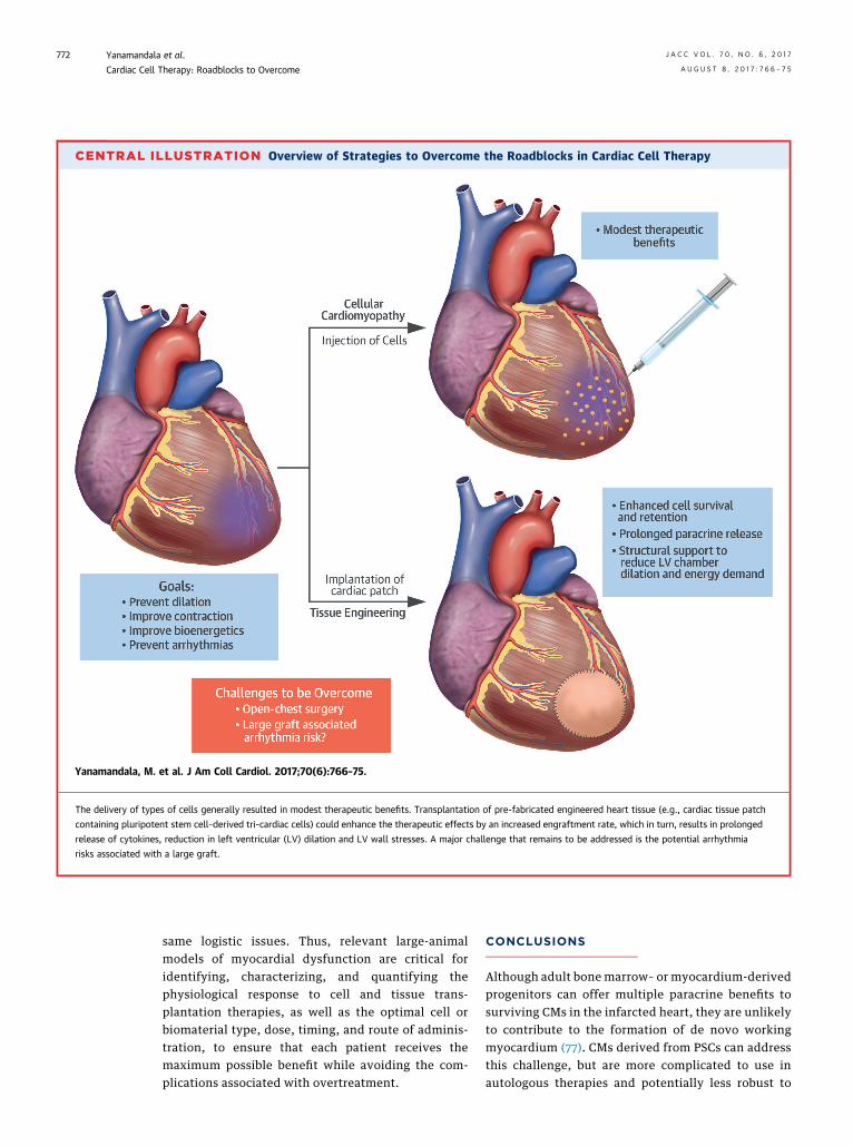
CENTRAL ILLUSTRATION Overview of Strategies to Overcome the Roadblocks in Cardiac Cell Therapy
Yanamandala, M. et al. J Am Coll Cardiol. 2017;70(6):766–75.
The delivery of types of cells generally resulted in modest therapeutic benefits. Transplantation of pre-fabricated engineered heart tissue (e.g., cardiac tissue patch
containing pluripotent stem cell–derived tri-cardiac cells) could enhance the therapeutic effects by an increased engraftment rate, which in turn, results in prolonged
release of cytokines, reduction in left ventricular (LV) dilation and LV wall stresses. A major challenge that remains to be addressed is the potential arrhythmia
risks associated with a large graft.
Yanamandala et al. J A C C V O L . 7 0 , N O . 6 , 2 0 1 7
Cardiac Cell Therapy: Roadblocks to Overcome A U G U S T 8 , 2 0 1 7 : 7 6 6 – 7 5
772
same logistic issues. Thus, relevant large-animalmodels of myocardial dysfunction are critical foridentifying, characterizing, and quantifying thephysiological response to cell and tissue trans-plantation therapies, as well as the optimal cell orbiomaterial type, dose, timing, and route of adminis-tration, to ensure that each patient receives themaximum possible benefit while avoiding the com-plications associated with overtreatment.
CONCLUSIONS
Although adult bone marrow– or myocardium-derivedprogenitors can offer multiple paracrine benefits tosurviving CMs in the infarcted heart, they are unlikelyto contribute to the formation of de novo workingmyocardium (77). CMs derived from PSCs can addressthis challenge, but are more complicated to use inautologous therapies and potentially less robust to

J A C C V O L . 7 0 , N O . 6 , 2 0 1 7 Yanamandala et al.A U G U S T 8 , 2 0 1 7 : 7 6 6 – 7 5 Cardiac Cell Therapy: Roadblocks to Overcome
773
surviving transplantation. Although the jury for theoptimal cell source is still out, it is possible thatdifferent disease applications will require differentcell types, and that a mixture of immune-matched orimmune-engineered PSC-derived CMs and host-derived stromal progenitors will prove optimal ininducing heart remuscularization while supportingcell survival and engraftment. Notably, applications ofpre-formed engineered cardiac tissue patches withspecifically tailored cell compositions could signifi-cantly increase both the survival and the beneficialeffects of transplanted cells. Furthermore, becauseparacrine factors, including extracellular vesicles, areresponsible for much of the observed beneficial effectsof cardiac cell therapy, amaintained tissue patch couldserve as a continued source of such beneficial para-crine signaling to the native heart tissue (CentralIllustration). As our understanding of exosomalbiology advances, patches can be engineered to opti-mize this signaling for cardiac regeneration. Use ofgenome-editing technologies may further enhance thepotency and functional integration of delivered cells.
Major challenges will be to address the potentialarrhythmogenicity risks associated with a large graft.With exciting translational prospects ahead, futurestudies to optimize engineered cardiac tissue thera-pies in large animals and decipher the mechanisms ofaction are fully warranted.
ACKNOWLEDGMENTS The authors gratefully acknowl-edge the National Institutes of Health (NIH) supportof their research, NIH UO1 HL134764, the ProgenitorCell Biology Consortium (grant HL099997), andthe Symposium held at University of Alabama-Birmingham in March 2016. This work was a productof discussions at the NIH Progenitor Cell BiologyConsortium Cardiovascular Tissue Engineering Sym-posium, March 2016.
ADDRESS FOR CORRESPONDENCE: Dr. Jianyi (Jay)Zhang, Department of Biomedical Engineering, Uni-versity of Alabama at Birmingham, 1670 UniversityBoulevard, Volker Hall, Birmingham, Alabama 35294.E-mail: [email protected].
RE F E RENCE S
1. Nguyen PK, Rhee JW, Wu JC. Adult stem celltherapy and heart failure, 2000 to 2016: a sys-tematic review. JAMA Cardiol 2016;1:831–41.
2. Lalit PA, Salick MR, Nelson DO, et al. Lineagereprogramming of fibroblasts into proliferativeinduced cardiac progenitor cells by definedfactors. Cell Stem Cell 2016;18:354–67.
3. Zhang Y, Cao N, Huang Y, et al. Expandablecardiovascular progenitor cells reprogrammedfrom fibroblasts. Cell Stem Cell 2016;18:368–81.
4. Jackman CP, Carlson AL, Bursac N. Dynamicculture yields engineered myocardium with near-adult functional output. Biomaterials 2016;111:66–79.
5. Jackman CP, Shadrin IY, Carlson AL, Bursac N.Human cardiac tissue engineering: from pluripo-tent stem cells to heart repair. Curr Opin ChemEng 2015;7:57–64.
6. Zhang D, Shadrin IY, Lam J, Xian HQ,Snodgrass HR, Bursac N. Tissue-engineered car-diac patch for advanced functional maturation ofhuman ESC-derived cardiomyocytes. Biomaterials2013;34:5813–20.
7. Zhang J, Klos M, Wilson GF, et al. Extracellularmatrix promotes highly efficient cardiac differen-tiation of human pluripotent stem cells: the matrixsandwich method. Circ Res 2012;111:1125–36.
8. Lian X, Hsiao C, Wilson G, et al. Robustcardiomyocyte differentiation from humanpluripotent stem cells via temporal modulation ofcanonical Wnt signaling. Proc Natl Acad Sci U S A2012;109:E1848–57.
9. Yang L, Geng Z, Nickel T, et al. Differentiationof human induced-pluripotent stem cells into
smooth-muscle cells: two novel protocols. PLoSOne 2016;11:e0147155.
10. Ye L, Zhang S, Greder L, et al. Effective cardiacmyocyte differentiation of human induced plurip-otent stem cells requires VEGF. PLoS One 2013;8:e53764.
11. Zhang S, Dutton JR, Su L, Zhang J, Ye L. Theinfluence of a spatiotemporal 3D environment onendothelial cell differentiation of human inducedpluripotent stem cells. Biomaterials 2014;35:3786–93.
12. Campagnolo P, Tsai TN, Hong X, et al. c-Kitþprogenitors generate vascular cells for tissue-engineered grafts through modulation of theWnt/Klf4 pathway. Biomaterials 2015;60:53–61.
13. Dorn GW II, Vega RB, Kelly DP. Mitochondrialbiogenesis and dynamics in the developing anddiseased heart. Genes Dev 2015;29:1981–91.
14. Dorn GW II, Kitsis RN. The mitochondrialdynamism-mitophagy-cell death interactome:multiple roles performed by members of a mito-chondrial molecular ensemble. Circ Res 2015;116:167–82.
15. Chung S, Dzeja PP, Faustino RS, Perez-Terzic C,Behfar A, Terzic A. Mitochondrial oxidative meta-bolism is required for the cardiac differentiation ofstem cells. Nat Clin Pract Cardiovasc Med 2007;4Suppl 1:S60–7.
16. Kasahara A, Cipolat S, Chen Y, Dorn GW II,Scorrano L. Mitochondrial fusion directs car-diomyocyte differentiation via calcineurin andNotch signaling. Science 2013;342:734–7.
17. Wendel JS, Ye L, Zhang P, Tranquillo RT,Zhang JJ. Functional consequences of a tissue-engineered myocardial patch for cardiac repair in
a rat infarct model. Tissue Eng Part A 2014;20:1325–35.
18. Ruwhof C, van Wamel AE, Egas JM, van derLaarse A. Cyclic stretch induces the release ofgrowth promoting factors from cultured neonatalcardiomyocytes and cardiac fibroblasts. Mol CellBiochem 2000;208:89–98.
19. van Wamel AJ, Ruwhof C, van der Valk-Kokshoorn LJ, Schrier PI, van der Laarse A.Stretch-induced paracrine hypertrophic stimuliincrease TGF-beta1 expression in cardiomyocytes.Mol Cell Biochem 2002;236:147–53.
20. Masumoto H, Matsuo T, Yamamizu K, et al.Pluripotent stem cell-engineered cell sheetsreassembled with defined cardiovascular pop-ulations ameliorate reduction in infarct heartfunction through cardiomyocyte-mediated neo-vascularization. Stem Cells 2012;30:1196–205.
21. Martin CM, Meeson AP, Robertson SM, et al.Persistent expression of the ATP-binding cassettetransporter, Abcg2, identifies cardiac SP cells inthe developing and adult heart. Dev Biol 2004;265:262–75.
22. Wang X, Hu Q, Nakamura Y, et al. The role ofthe sca-1þ/CD31� cardiac progenitor cell popula-tion in postinfarction left ventricular remodeling.Stem Cells 2006;24:1779–88.
23. Tokita Y, Tang XL, Li Q, et al. Repeatedadministrations of cardiac progenitor cells aremarkedly more effective than a single adminis-tration: a new paradigm in cell therapy. Circ Res2016;119:635–51.
24. Hare JM, DiFede DL, Castellanos AM, et al.Randomized comparison of allogeneic versusautologous mesenchymal stem cells for
Yanamandala et al. J A C C V O L . 7 0 , N O . 6 , 2 0 1 7
Cardiac Cell Therapy: Roadblocks to Overcome A U G U S T 8 , 2 0 1 7 : 7 6 6 – 7 5
774
nonischemic dilated cardiomyopathy: POSEIDON-DCM Trial. J Am Coll Cardiol 2017;69:526–37.
25. Karantalis V, Suncion-Loescher VY, Bagno L,et al. Synergistic effects of combined cell therapyfor chronic ischemic cardiomyopathy. J Am CollCardiol 2015;66:1990–9.
26. Zeng L, Hu Q, Wang X, et al. Bioenergetic andfunctional consequences of bone marrow-derivedmultipotent progenitor cell transplantation inhearts with postinfarction left ventricular remod-eling. Circulation 2007;115:1866–75.
27. Guo Y, Wysoczynski M, Nong Y, et al. Repeateddoses of cardiac mesenchymal cells are thera-peutically superior to a single dose in mice withold myocardial infarction. Basic Res Cardiol 2017;112:18.
28. Ban K, Park HJ, Kim S, et al. Cell therapy withembryonic stem cell-derived cardiomyocytesencapsulated in injectable nanomatrix gelenhances cell engraftment and promotes cardiacrepair. ACS Nano 2014;8:10815–25.
29. Segers VF, Lee RT. Biomaterials to enhancestem cell function in the heart. Circ Res 2011;109:910–22.
30. Vunjak-Novakovic G, Lui KO, Tandon N,Chien KR. Bioengineering heart muscle: a para-digm for regenerative medicine. Annu Rev BiomedEng 2011;13:245–67.
31. Zhao T, Zhang ZN, Rong Z, Xu Y. Immunoge-nicity of induced pluripotent stem cells. Nature2011;474:212–5.
32. Zhao T, Zhang ZN, Westenskow PD, et al.Humanized mice reveal differential immunoge-nicity of cells derived from autologous inducedpluripotent stem cells. Cell Stem Cell 2015;17:353–9.
33. Araki R, Uda M, Hoki Y, et al. Negligibleimmunogenicity of terminally differentiated cellsderived from induced pluripotent or embryonicstem cells. Nature 2013;494:100–4.
34. Guha P, Morgan JW, Mostoslavsky G,Rodrigues NP, Boyd AS. Lack of immune responseto differentiated cells derived from syngeneicinduced pluripotent stem cells. Cell Stem Cell2013;12:407–12.
35. Abrahimi P, Chang WG, Kluger MS, et al. Effi-cient gene disruption in cultured primary humanendothelial cells by CRISPR/Cas9. Circ Res 2015;117:121–8.
36. Shiao SL, Kirkiles-Smith NC, Shepherd BR,McNiff JM, Carr EJ, Pober JS. Human effectormemory CD4þ T cells directly recognize alloge-neic endothelial cells in vitro and in vivo.J Immunol 2007;179:4397–404.
37. Hong KU, Guo Y, Li QH, et al. c-kitþ cardiacstem cells alleviate post-myocardial infarction leftventricular dysfunction despite poor engraftmentand negligible retention in the recipient heart.PLoS One 2014;9:e96725.
38. Malliaras K, Li TS, Luthringer D, et al. Safetyand efficacy of allogeneic cell therapy in infarctedrats transplanted with mismatched cardiosphere-derived cells. Circulation 2012;125:100–12.
39. Sanganalmath SK, Bolli R. Cell therapy forheart failure: a comprehensive overview ofexperimental and clinical studies, current
challenges, and future directions. Circ Res 2013;113:810–34.
40. Ismahil MA, Hamid T, Bansal SS, Patel B,Kingery JR, Prabhu SD. Remodeling of the mono-nuclear phagocyte network underlies chronicinflammation and disease progression in heartfailure: critical importance of the cardiosplenicaxis. Circ Res 2014;114:266–82.
41. Nevers T, Salvador AM, Grodecki-Pena A, et al.Left ventricular T-cell recruitment contributes tothe pathogenesis of heart failure. Circ Heart Fail2015;8:776–87.
42. Prabhu SD, Frangogiannis NG. The biologicalbasis for cardiac repair after myocardial infarction:from inflammation to fibrosis. Circ Res 2016;119:91–112.
43. Sager HB, Hulsmans M, Lavine KJ, et al.Proliferation and recruitment contribute tomyocardial macrophage expansion in chronic heartfailure. Circ Res 2016;119:853–64.
44. Hamid T, Xu Y, Ismahil MA, et al. TNF receptorsignaling inhibits cardiomyogenic differentiationof cardiac stem cells and promotes aneuroadrenergic-like fate. Am J Physiol Heart CircPhysiol 2016;311:H1189–201.
45. Chong JJ, Yang X, Don CW, et al. Humanembryonic-stem-cell-derived cardiomyocytesregenerate non-human primate hearts. Nature2014;510:273–7.
46. Shiba Y, Gomibuchi T, Seto T, et al. Allogeneictransplantation of iPS cell-derived cardiomyocytesregenerates primate hearts. Nature 2016;538:388–91.
47. Weiss JN, Qu Z, Chen PS, et al. The dynamicsof cardiac fibrillation. Circulation 2005;112:1232–40.
48. Fontes MS, van Veen TA, de Bakker JM, vanRijen HV. Functional consequences of abnormalCx43 expression in the heart. Biochim BiophysActa 2012;1818:2020–9.
49. Davis LM, Rodefeld ME, Green K, Beyer EC,Saffitz JE. Gap junction protein phenotypes of thehuman heart and conduction system. J CardiovascElectrophysiol 1995;6:813–22.
50. Dupont E, Matsushita T, Kaba RA, et al.Altered connexin expression in human congestiveheart failure. J Mol Cell Cardiol 2001;33:359–71.
51. Kostin S, Rieger M, Dammer S, et al. Gapjunction remodeling and altered connexin43expression in the failing human heart. Mol CellBiochem 2003;242:135–44.
52. Roell W, Lewalter T, Sasse P, et al. Engraft-ment of connexin 43-expressing cells preventspost-infarct arrhythmia. Nature 2007;450:819–24.
53. Gerbin KA, Yang X, Murry CE, Coulombe KL.Enhanced electrical integration of engineeredhuman myocardium via intramyocardial versusepicardial delivery in infarcted rat hearts. PLoSOne 2015;10:e0131446.
54. Xiong Q, Ye L, Zhang P, et al. Bioenergetic andfunctional consequences of cellular therapy: acti-vation of endogenous cardiovascular progenitorcells. Circ Res 2012;111:455–68.
55. Xiong Q, Ye L, Zhang P, et al. Functionalconsequences of human induced pluripotent stemcell therapy: myocardial ATP turnover rate in thein vivo swine heart with postinfarction remodel-ing. Circulation 2013;127:997–1008.
56. Bolognese L, Neskovic AN, Parodi G, et al. Leftventricular remodeling after primary coronaryangioplasty: patterns of left ventricular dilationand long-term prognostic implications. Circulation2002;106:2351–7.
57. HuQ,WangX, Lee J, et al. Profoundbioenergeticabnormalities in peri-infarct myocardial regions. AmJ Physiol Heart Circ Physiol 2006;291:H648–57.
58. Feygin J, Mansoor A, Eckman P, Swingen C,Zhang J. Functional and bioenergetic modulationsin the infarct border zone following autologousmesenchymal stem cell transplantation. Am JPhysiol Heart Circ Physiol 2007;293:H1772–80.
59. Kobayashi T, Yamaguchi T, Hamanaka S, et al.Generation of rat pancreas in mouse by interspe-cific blastocyst injection of pluripotent stem cells.Cell 2010;142:787–99.
60. Usui J, Kobayashi T, Yamaguchi T, Knisely AS,Nishinakamura R, Nakauchi H. Generation of kid-ney from pluripotent stem cells via blastocystcomplementation. Am J Pathol 2012;180:2417–26.
61. Bort R, Signore M, Tremblay K, MartinezBarbera JP, Zaret KS. Hex homeobox gene controlsthe transition of the endoderm to a pseudos-tratified, cell emergent epithelium for liver buddevelopment. Dev Biol 2006;290:44–56.
62. Matsunari H, Nagashima H, Watanabe M, et al.Blastocyst complementation generates exogenicpancreas in vivo in apancreatic cloned pigs. ProcNatl Acad Sci U S A 2013;110:4557–62.
63. Porrello E, Mahmoud A, Simpson E, et al.Transient regenerative potential of the neonatalmouse heart. Science 2011;331:1078–80.
64. Bergmann O, Bhardwaj R, Bernard S, et al.Evidence for cardiomyocyte renewal in humans.Science 2009;324:98–102.
65. van Berlo JH, Kanisicak O, Maillet M, et al.c-kitþ cells minimally contribute cardiomyocytesto the heart. Nature 2014;509:337–41.
66. van Berlo JH, Molkentin JD. Most of the dusthas settled: cKitþ progenitor cells are an irrele-vant source of cardiac myocytes in vivo. Circ Res2016;118:17–9.
67. Hatzistergos KE, Takeuchi LM, Saur D, et al.cKitþ cardiac progenitors of neural crest origin.Proc Natl Acad Sci U S A 2015;112:13051–6.
68. Mohamed TM, Stone NR, Berry EC, et al. Chem-ical enhancement of in vitro and in vivo direct cardiacreprogramming. Circulation 2017;135:978–95.
69. Nam YJ, Song K, Olson EN. Heart repair bycardiac reprogramming. Nat Med 2013;19:413–5.
70. Srivastava D, Yu P. Recent advances in directcardiac reprogramming. Curr Opin Genet Dev2015;34:77–81.
71. Kanisicak O, Khalil H, Ivey MJ, et al. Genetic line-age tracing definesmyofibroblast origin and functionin the injured heart. Nat Commun 2016;7:12260.
72. Moore-Morris T, Cattaneo P, Puceat M,Evans SM. Origins of cardiac fibroblasts. J Mol CellCardiol 2016;91:1–5.

J A C C V O L . 7 0 , N O . 6 , 2 0 1 7 Yanamandala et al.A U G U S T 8 , 2 0 1 7 : 7 6 6 – 7 5 Cardiac Cell Therapy: Roadblocks to Overcome
775
73. Li Y, Dal-Pra S, Mirotsou M, et al. Tissue-engineered 3-dimensional (3D) microenvironmentenhances the direct reprogramming of fibroblastsinto cardiomyocytes by microRNAs. Sci Rep 2016;6:38815.
74. Sia J, Yu P, Srivastava D, Li S. Effect ofbiophysical cues on reprogramming to car-diomyocytes. Biomaterials 2016;103:1–11.
75. Bursac N, Kirkton RD, McSpadden LC, Liau B.Characterizing functional stem cell-cardiomyocyteinteractions. Regen Med 2010;5:87–105.
76. Makkar RR, Smith RR, Cheng K, et al.Intracoronary cardiosphere-derived cells forheart regeneration after myocardial infarction(CADUCEUS): a prospective, randomised phase 1trial. Lancet 2012;379:895–904.
77. A futile cycle in cell therapy. Nat Biotechnol2017;35:291.
78. Ishigami S, Ohtsuki S, Tarui S, et al. Intra-coronary autologous cardiac progenitor celltransfer in patients with hypoplastic left heartsyndrome: the TICAP prospective phase 1controlled trial. Circ Res 2015;116:653–64.
79. Tarui S, Ishigami S, Ousaka D, et al. Trans-coronary infusion of cardiac progenitor cells in
hypoplastic left heart syndrome: three-yearfollow-up of the Transcoronary Infusion ofCardiac Progenitor Cells in Patients WithSingle-Ventricle Physiology (TICAP) trial. J ThoracCardiovasc Surg 2015;150:1198–207, 1208.e1–2.
80. Ishigami S, Ohtsuki S, Eitoku T, et al. Intra-coronary cardiac progenitor cells in singleventricle physiology: the PERSEUS (Cardiac Pro-genitor Cell Infusion to Treat Univentricular HeartDisease) Randomized Phase 2 Trial. Circ Res 2017;120:1162–73.
81. Choudhury T, Mozid A, Hamshere S, et al. Anexploratory randomized control study of combi-nation cytokine and adult autologous bone marrowprogenitor cell administration in patients withischaemic cardiomyopathy: the REGENERATE-IHDclinical trial. Eur J Heart Fail 2017;19:138–47.
82. Bartunek J, Terzic A, Davison BA, et al. Car-diopoietic cell therapy for advanced ischaemicheart failure: results at 39 weeks of the prospec-tive, randomized, double blind, sham-controlledCHART-1 clinical trial. Eur Heart J 2017;38:648–60.
83. Butler J, Epstein SE, Greene SJ, et al. Intra-venous allogeneic mesenchymal stem cells for
nonischemic cardiomyopathy: safety and efficacyresults of a phase II-A randomized trial. Circ Res2017;120:332–40.
84. Wojakowski W, Jadczyk T, Michalewska-Wludarczyk A, et al. Effects of transendocardialdelivery of bone marrow-derived CD133þ cellson left ventricle perfusion and function in pa-tients with refractory angina: final results ofrandomized, double-blinded, placebo-controlled REGENT-VSEL Trial. Circ Res 2017;120:670–80.
85. Miyagawa S, Domae K, Yoshikawa Y, et al.Phase I clinical trial of autologous stem cell-sheet transplantation therapy for treatingcardiomyopathy. J Am Heart Assoc 2017;6:e003918.
86. Wollert KC, Meyer GP, Müller-Ehmsen J, et al.Intracoronary autologous bone marrow celltransfer after myocardial infarction: the BOOST-2randomised placebo-controlled clinical trial. EurHeart J 2017 Apr 19 [E-pub ahead of print].
KEY WORDS biocompatible materials,heart failure, myocardial infarction,myocardium, stem cells