Embed Size (px)
Citation preview

Liu and Makaroff BMC Plant Biology (2015) 15:74 DOI 10.1186/s12870-015-0452-2
RESEARCH ARTICLE Open Access
Overexpression of a truncated CTF7 constructleads to pleiotropic defects in reproductionand vegetative growth in ArabidopsisDesheng Liu and Christopher A Makaroff*
Abstract
Background: Eco1/Ctf7 is essential for the establishment of sister chromatid cohesion during S phase of the cellcycle. Inactivation of Ctf7/Eco1 leads to a lethal phenotype in most organisms. Altering Eco1/Ctf7 levels or pointmutations in the gene can lead to alterations in nuclear division as well as a wide range of developmental defects.Inactivation of Arabidopsis CTF7 (AtCTF7) results in severe defects in reproduction and vegetative growth.
Results: To further investigate the function(s) of AtCTF7, a tagged version of AtCTF7 and several AtCTF7 deletionconstructs were created and transformed into wild type or ctf7+/–plants. Transgenic plants expressing 35S:NTAP:AtCTF7Δ299–345 (AtCTF7ΔB) displayed a wide range of phenotypic alterations in reproduction and vegetative growth.Male meiocytes exhibited chromosome fragmentation and uneven chromosome segregation. Mutant ovulescontained abnormal megasporocyte-like cells during pre-meiosis, megaspores experienced elongated meiosis andmegagametogenesis, and defective megaspores/embryo sacs were produced at various stages. The transgenicplants also exhibited a broad range of vegetative defects, including meristem disruption and dwarfism that wereinherited in a non-Mendelian fashion. Transcripts for epigenetically regulated transposable elements (TEs) wereelevated in transgenic plants. Transgenic plants expressing 35S:AtCTF7ΔB displayed similar vegetative defects,suggesting the defects in 35S:NTAP:AtCTF7ΔB plants are caused by high-level expression of AtCTF7ΔB.
Conclusions: High level expression of AtCTF7ΔB disrupts megasporogenesis, megagametogenesis and malemeiosis, as well as causing a broad range of vegetative defects, including dwarfism that are inherited in anon-Mendelian fashion.
Keywords: Meiosis, Sister chromatid cohesion, Megasporogenesis, Megagametogenesis, Megaspore mother cell,Functional megaspore-like, Epigenetic
BackgroundThe precise establishment and release of sister chromatidcohesion are vital for cell division during mitosis and meiosis[1-3]. The cohesin complex, which forms a ring to entrapsister chromatids, is composed of four subunits: a heterodi-mer of Structural Maintenance of Chromosome (SMC) pro-teins, SMC1 and SMC3, an α-kleisin, Sister ChromatidCohesion1 (SCC1) in somatic cells or REC8 in meiotic cellsand SCC3 [2,4,5]. The core cohesin complex associates witha number of factors during the cohesion cycle [1-3,6-8] andits association with the chromosomes is controlled by
* Correspondence: [email protected] of Chemistry and Biochemistry, Miami University, Oxford, OH45056, USA
© 2015 Liu and Makaroff; licensee BioMed CenCommons Attribution License (http://creativecreproduction in any medium, provided the orDedication waiver (http://creativecommons.orunless otherwise stated.
modifications, such as acetylation, phosphorylation, sumoy-lation and proteolysis [9-13].Sister chromatid cohesion is established during S phase by
the Establishment of Cohesion protein, ECO1/CTF7 [14-18].In budding yeast, ECO1/CTF7 acetylates lysine residuesK112, K113 of SMC3 and K84, K210 of SCC1 to stabilizethe ring and keep it closed [10,19,20]. ECO1/CTF1 is an es-sential gene in many organisms with the complete inactiva-tion of CTF7 resulting in lethality [1-3,14-16,21]. Inactivationor mutations in ECO1/CTF1 leads to a broad range of de-fects, including chromosome mis-segregation, defects inDNA double strand break repair, defects in homologous re-combination and transcriptional alterations [14,15,17,21-25].Humans contain two ECO1/CTF7 orthologs, ESCO1 andESCO2 [24]. Mutations in ESCO2 lead to Roberts syndrome
tral. This is an Open Access article distributed under the terms of the Creativeommons.org/licenses/by/4.0), which permits unrestricted use, distribution, andiginal work is properly credited. The Creative Commons Public Domaing/publicdomain/zero/1.0/) applies to the data made available in this article,

Liu and Makaroff BMC Plant Biology (2015) 15:74 Page 2 of 16
(RBS), which is associated with a variety of defects includinggrowth retardation, limb reduction/asymmetric limb growth,cleft lip/palate and missing fingers/toes [26,27].Arabidopsis contains a single CTF7 ortholog (AtCTF7),
which can complement the temperature-sensitive yeast ctf7-203 mutant [16]. AtCTF7 encodes a 345 amino acid protein,containing a PIP box at residues 82 to 86, a C2H2 zinc fingermotif at residues 92–130 and an acetyltransferase domainfrom residues 184 to 335. The acetyltransferase domain canbe further separated into three motifs: D (amino acids 184–204), A (amino acids 266–301) and B (amino acids 311–335). Motif D provides the framework of the acetyltransfer-ase domain. Motif A participates in acetyl-CoA binding andis critical for catalytic activity, while motif B is the most C-terminal region and participates in substrate recognition andcatalytic activity regulation [28,29]. Variations in AtCTF7 ex-pression have been shown to result in a wide range of de-fects. Plants heterozygous for a T-DNA insertion in AtCTF7grow normally but their siliques contain approximately 25%arrested seeds, consistent with the belief that CTF7 is an es-sential protein [16]. However, Atctf7 homozygous plants canbe detected at very low frequencies [17]. Atctf7 plants exhibita wide range of developmental defects, including extremedwarfism and sterility with Atctf7 meiocytes exhibiting ab-normal chromosome segregation and defective sister chro-matid cohesion. Knockdown of AtCTF7 mRNA levels usingRNAi leads to growth retardation and defective sister chro-matid cohesion [18]. Finally, overexpression of full lengthAtCTF7 with the CaMV 35S promoter leads to ovule abor-tion, but plant growth is not affected [16].To further investigate the roles of CTF7 in Arabidop-
sis and to identify AtCTF7 interacting proteins, trans-genic plants expressing AtCTF7 constructs that encodeeither TAP-tagged [30] or non-tagged versions of fulllength or truncated AtCTF7 were generated and ana-lyzed. Transgenic plants that express a truncated versionof AtCTF7, missing motif B, from the 35S promoter ex-hibited impaired reproduction and vegetative growth.Ovules in 35S:NTAP:AtCTF7ΔB plants displayed alter-ations in cell identity and the timing of megasporogene-sis and megagametogenesis, while male meiocytesexhibited alterations during meiosis II. The transgenicplants also displayed alterations in vegetative growth thatwere not inherited in a Mendelian fashion.
ResultsArabidopsis plants expressing high levels of NTAP:AtCTF7ΔB display reduced fertilityAn AtCTF7 construct missing the C-terminal 46 aminoacids (Δ299-345) was generated, fused with NTAP [30]and expressed from the CaMV 35S promoter in wild-type Columbia plants (35S:NTAP:AtCTF7ΔB; Additionalfile 1: Figure S1). Twenty out of the 36 independent linesexamined exhibited reduced fertility, with fertility levels
varying significantly between the lines. Plants exhibitinga weak phenotype, which accounted for two of the 20 re-duced fertility lines, produced shorter siliques with re-duced numbers of seeds, but the seeds appeared normal(Figure 1Aii). For example Line 19 produced 39.2 ± 3.9seeds per silique (n = 35) compared to wild type plantsthat produce 54.2 ± 4.1 seeds per silique (n = 35). An-thers from Line 19 plants were smaller and contained re-duced numbers of pollen (574 vs 1175 in wild type), butthe pollen appeared viable (Figure 1Bii). The other 18 re-duced fertility lines exhibited more severe defects. Theseplants produced siliques containing large numbers ofunfertilized ovules and aborted seeds (Figure 1Aiii).Unfertilized ovules appeared as white dots, resemblingthe situation in atctf7-1 plants [17]. Aborted seeds ap-peared white and plump, similar to seeds containingarrested embryos in Atctf7-1+/− plants [16]. Seed set var-ied considerably between the lines, ranging from 13.3 ±5.6 seeds per silique to 34.7 ± 8.4 seeds per silique. An-thers from these plants typically contained reducednumbers of pollen, much of which was not viable. Forexample, anthers from Line 11 produced on average ap-proximately 210 pollen, of which only 20% was viable(Figure 1Biii). Given that most lines exhibited severe fer-tility defects, one representative line (#11) was chosenand characterized in detail.The reduced fertility phenotype of 35S:NTAP:AtCTF7ΔB
plants suggested that co-suppression may be loweringAtCTF7 transcript levels in the lines. Therefore, quantitativereverse transcription polymerase chain reaction (qRT-PCR)was conducted to examine transcript levels of nativeAtCTF7 as well as the CTF7 transgene. Surprisingly, totalAtCTF7 (35S:NTAP:AtCTF7ΔB and native AtCTF7) tran-script levels were approximately seven-fold higher in 35S:NTAP:AtCTF7ΔB plants than CTF7 transcript levels in wildtype plants, while native AtCTF7 transcript levels werealmost three-fold higher in the transgenic plants (Fig-ure 1C). Therefore, the transgenic lines contained highlevels of AtCTF7ΔB transcripts along with elevatedlevels of native AtCTF7 mRNA, indicating that the ob-served phenotypes are not the result of reduced AtCTF7expression. Instead these results suggest that the 35S:NTAP:AtCTF7ΔB construct may be exerting a domin-ant negative effect.Reciprocal crossing experiments were carried out to
determine the effect of the 35S:NTAP:AtCTF7ΔB con-struct on male and female gametes. The 35S:NTAP:AtCTF7ΔB construct was transmitted at reduced levelsthrough both male and female gametes (Additional file1: Table S1), consistent with our preliminary resultsthat both male and female fertility were affected in35S:NTAP:AtCTF7ΔB plants. Nonviable seeds wereobtained when 35S:NTAP:AtCTF7ΔB plants were usedas either the male or female parent; however the

Figure 1 35S:NTAP:AtCTF7ΔB plants exhibit reduced fertility. (A) Open siliques from wild type (Ai) and 35S:NTAP:AtCTF7ΔB (Aii,Aiii) plants.i, Wild type silique with full seed set. ii, Silique from a 35S:NTAP:AtCTF7ΔB plant (Line 19) exhibiting a weak phenotype. iii, Silique from a 35S:NTAP:AtCTF7ΔB plant (Line 11) exhibiting a strong phenotype. Arrows indicate shriveled, unfertilized ovules. White stars show white, plump seeds,which aborted after fertilization. Scale bar = 0.5 cm. (B) Alexander staining of mature anthers from wild type (i) and 35S:NTAP:AtCTF7ΔB (ii, iii)plants. i, Wild type anther. ii, Anther from Line 19. The anther is smaller and contains less pollen; all the pollen is viable. iii, Anther from Line 11.The anther is smaller and contains low numbers of viable pollen. Scale bar = 50 μm. (C). Expression analysis of AtCTF7 in wild type and 35S:NTAP:AtCTF7ΔB plants. Transcript levels of total AtCTF7 (35S:NTAP:AtCTF7ΔB and native AtCTF7) and native AtCTF7 are increased in 35S:NTAP:AtCTF7ΔBplants with Line 11 plants exhibiting the highest levels. Buds of wild type, non-dwarf, 4th generation Line 11 plants and 4th generation Line19plants were used for this experiment. Data are shown as means ± SD (n = 3).
Liu and Makaroff BMC Plant Biology (2015) 15:74 Page 3 of 16
relative proportion of nonviable seed was three timesgreater (9.3% verses 31.3%) when 35S:NTAP:AtCTF7ΔBplants served as the female. The number of nonviableseeds was greatest (65%) in self-pollenated plants. There-fore, while both male and female gametes are affected, the35S:NTAP:AtCTF7ΔB construct has a greater effect on fe-male reproduction. Inheritance of 35S:NTAP:AtCTF7ΔB-associated defects from both parents has a compoundingeffect on seed production.
Male meiotic chromosome segregation is altered in 35S:NTAP:AtCTF7ΔB plantsPrevious studies showed that inactivation of AtCTF7 via T-DNA insertion or reduction in AtCTF7 mRNA levels viaRNAi disrupts sister chromatid cohesion, resulting in un-even chromosome segregation during meiosis and ultim-ately reduced pollen viability [17,18]. The reduced fertilityobserved in 35S:NTAP:AtCTF7ΔB plants suggested thatmeiosis might also be affected in these lines. Therefore,

Liu and Makaroff BMC Plant Biology (2015) 15:74 Page 4 of 16
chromosome spreading experiments were carried out toexamine the effect of 35S:NTAP:AtCTF7ΔB expression onmale meiosis.Male meiocytes in 35S:NTAP:AtCTF7ΔB plants resem-
bled wild type during early stages of meiosis, with normalchromosome morphology during pachytene (Figure 2A,E),diakinesis (Figure 2B,F) and metaphase I (Figure 2C,G).The first noticeable defect was observed at telophase Iwhen lagging chromosomes were observed (Figure 2D,H),followed by mis-segregated chromosomes at prophase II(Figure 2M). More than twenty individual chromosomeswere typically observed in meiocytes beginning at meta-phase II (Figure 2N), indicating that sister chromatid co-hesion was prematurely lost. Chromosomes did notsegregate evenly at anaphase II (Figure 2O) resulting inthe production of polyads with varying DNA contents attetrad stage (Figure 2P). Similar to atctf7 plants, a smallnumber of relatively normal meiocytes were also observedthroughout meiosis with fewer normal-appearing meio-cytes in later stages of meiosis. For example, the percent-ages of defective meiocytes observed at various stages ofmeiosis in Line 11 were: metaphase I: 0% (0/41), telophase
Figure 2 35S:NTAP:AtCTF7ΔB male meiocytes exhibit defective meiot(E-H) and (M-P) 35S:NTAP:AtCTF7ΔB meiocytes. A, E pachytene; B, F diakinmetaphase II; K, O anaphase II; L, P telophase II. Lagging chromosomes anchromosomes are stained by 4’, 6-diamidino-2-phenylindole (DAPI). Scale b
I: 6.8% (7/87), prophase II: 23.5% (24/102), metaphase II:40.7% (24/59), anaphase II: 56.8% (25/44) and telophase II:64.0% (114/178).Immunolocalization experiments were then carried
out to examine the distribution of the meiotic cohesinprotein SYN1 [31] in 35S:NTAP:AtCTF7ΔB male meio-cytes. Consistent with meiotic chromosome spreadingexperiments the overall distribution of SYN1 was notdramatically affected in meiocytes of 35S:NTAP:AtCTF7ΔB plants (Additional file 1: Figure S2). SYN1exhibited diffuse nuclear labeling during interphase withstrong signal observed on the developing chromosomalaxes from early leptotene into zygotene (Additional file1: Figures S2A,D). During late zygotene and pachytenethe protein lined the chromosomes (Additional file 1:Figures S2B,E,C and F). SYN1 was released normallyfrom the condensing chromosomes during diploteneand diakinesis and similar to the situation in wild type,SYN1 was barely detectable on chromosomes by prome-taphase I. Therefore, cohesin appears to load and be re-moved normally from meiotic chromosomes in 35S:NTAP:AtCTF7ΔB plants.
ic chromosome segregation. (A-D) and (I-L) Wild type meiocytes.esis; C, G metaphase I; D, H telophase I; I, M prophase II; J, Nd/or chromosome fragments are denoted with arrows. Meioticar = 10 μm.
Liu and Makaroff BMC Plant Biology (2015) 15:74 Page 5 of 16
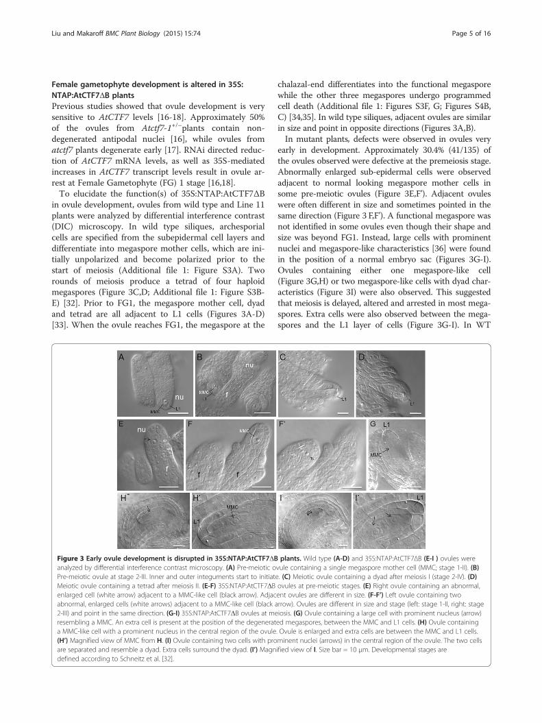
Female gametophyte development is altered in 35S:NTAP:AtCTF7ΔB plantsPrevious studies showed that ovule development is verysensitive to AtCTF7 levels [16-18]. Approximately 50%of the ovules from Atctf7-1+/–plants contain non-degenerated antipodal nuclei [16], while ovules fromatctf7 plants degenerate early [17]. RNAi directed reduc-tion of AtCTF7 mRNA levels, as well as 35S-mediatedincreases in AtCTF7 transcript levels result in ovule ar-rest at Female Gametophyte (FG) 1 stage [16,18].To elucidate the function(s) of 35S:NTAP:AtCTF7ΔB
in ovule development, ovules from wild type and Line 11plants were analyzed by differential interference contrast(DIC) microscopy. In wild type siliques, archesporialcells are specified from the subepidermal cell layers anddifferentiate into megaspore mother cells, which are ini-tially unpolarized and become polarized prior to thestart of meiosis (Additional file 1: Figure S3A). Tworounds of meiosis produce a tetrad of four haploidmegaspores (Figure 3C,D; Additional file 1: Figure S3B-E) [32]. Prior to FG1, the megaspore mother cell, dyadand tetrad are all adjacent to L1 cells (Figures 3A-D)[33]. When the ovule reaches FG1, the megaspore at the
Figure 3 Early ovule development is disrupted in 35S:NTAP:AtCTF7ΔBanalyzed by differential interference contrast microscopy. (A) Pre-meiotic oPre-meiotic ovule at stage 2-III. Inner and outer integuments start to initiateMeiotic ovule containing a tetrad after meiosis II. (E-F) 35S:NTAP:AtCTF7ΔBenlarged cell (white arrow) adjacent to a MMC-like cell (black arrow). Adjacabnormal, enlarged cells (white arrows) adjacent to a MMC-like cell (black a2-III) and point in the same direction. (G-I) 35S:NTAP:AtCTF7ΔB ovules at meresembling a MMC. An extra cell is present at the position of the degenerata MMC-like cell with a prominent nucleus in the central region of the ovule(H’) Magnified view of MMC from H. (I) Ovule containing two cells with proare separated and resemble a dyad. Extra cells surround the dyad. (I’) Magndefined according to Schneitz et al. [32].
chalazal-end differentiates into the functional megasporewhile the other three megaspores undergo programmedcell death (Additional file 1: Figures S3F, G; Figures S4B,C) [34,35]. In wild type siliques, adjacent ovules are similarin size and point in opposite directions (Figures 3A,B).In mutant plants, defects were observed in ovules very
early in development. Approximately 30.4% (41/135) ofthe ovules observed were defective at the premeiosis stage.Abnormally enlarged sub-epidermal cells were observedadjacent to normal looking megaspore mother cells insome pre-meiotic ovules (Figure 3E,F’). Adjacent ovuleswere often different in size and sometimes pointed in thesame direction (Figure 3 F,F’). A functional megaspore wasnot identified in some ovules even though their shape andsize was beyond FG1. Instead, large cells with prominentnuclei and megaspore-like characteristics [36] were foundin the position of a normal embryo sac (Figures 3G-I).Ovules containing either one megaspore-like cell(Figure 3G,H) or two megaspore-like cells with dyad char-acteristics (Figure 3I) were also observed. This suggestedthat meiosis is delayed, altered and arrested in most mega-spores. Extra cells were also observed between the mega-spores and the L1 layer of cells (Figure 3G-I). In WT
plants. Wild type (A-D) and 35S:NTAP:AtCTF7ΔB (E-I ) ovules werevule containing a single megaspore mother cell (MMC; stage 1-II). (B). (C) Meiotic ovule containing a dyad after meiosis I (stage 2-IV). (D)ovules at pre-meiotic stages. (E) Right ovule containing an abnormal,ent ovules are different in size. (F-F’) Left ovule containing tworrow). Ovules are different in size and stage (left: stage 1-II, right: stageiosis. (G) Ovule containing a large cell with prominent nucleus (arrow)ed megaspores, between the MMC and L1 cells. (H) Ovule containing. Ovule is enlarged and extra cells are between the MMC and L1 cells.minent nuclei (arrows) in the central region of the ovule. The two cellsified view of I. Size bar = 10 μm. Developmental stages are

Liu and Makaroff BMC Plant Biology (2015) 15:74 Page 6 of 16
plants, a small number (6.7%, 8/120) of ovules were foundto contain twin megaspore mother cells; however no de-fects were observed during or after meiosis. In Line 11plants, 14.1% (19/135) of the ovules contained multiplemegaspore mother-like cells at pre-meiosis. Furthermore,72.5% (50/69) of ovules with dyads contained extra cellsbetween the dyad and L1 layer, and 27.5% (19/69) thedyads were in elongated shape. Ultimately 50.4% (64/127)of the ovules observed were defective at FG1 stage. There-fore, alterations in archesporial cell differentiation, the on-set and progression of meiosis and possibly somatic cellidentity are observed in ovules of 35S:NTAP:AtCTF7ΔBplants.The effects of 35S:NTAP:AtCTF7ΔB on megagameto-
genesis were investigated by confocal laser scanning mi-croscopy (CLSM) and DIC microscopy. During wild typemegagametogenesis, the functional megaspore undergoesthree rounds of mitosis accompanied by nuclear migration,fusion, degeneration and cellularization to form the finalembryo sac (Additional file 1: Figures S3H-L; S4D-J). AtFG1, the functional megaspore undergoes mitosis to pro-duce a two-nucleate embryo sac (FG2; Additional file 1:Figure S3H). Formation of a vacuole between the two nu-clei marks stage FG3 (Figure 4,D and L; Additional file 1:Figures S3I, S4D). During FG3 the ovule becomes curvedand the inner integument embraces the nucellus. A secondround of mitosis produces a four-nucleate embryo sac(FG4; Figure 4E,M; Additional file 1: Figures S3J, S4E). Thisis followed by migration of the two chalazal nuclei from anorthogonal orientation to a chalazal-micropylar orientation.After nuclear migration, a third round of mitosis gives riseto eight nuclei in a 4n + 4n configuration (FG5; Additionalfile 1: Figure S3K). The two polar nuclei, one from eachside, meet at the embryo sac’s micropylar half and fuse toform the central cell, while the antipodal nuclei start to de-generate (Additional file 1: Figure S4H). The central cellhas formed and the antipodal nuclei are completed degen-erated by FG7 (Additional file 1: Figures S3K, S4I). Prior tofertilization, one synergid nucleus degenerates, such thatthe embryo sac consists of one egg cell, one central cell andone synergid nucleus (FG8; Additional file 1: Figures S3L,S4J).Ovule development is typically synchronous in wild type
sliques with predominately one or two developmentalstages present in a given pistil (Additional file 1: Table S2).In contrast, 35S:NTAP:AtCTF7ΔB ovule development ap-peared slowed and asynchronous (Additional file 1: TableS3). Female gametophytes in the same pistil were often atseveral different stages, indicating that the synchrony ofgametophyte development was disturbed and embryo sacmaturation was delayed. Ovules containing a megaspore(Figures 3G,H’), FG1 embryo sacs with a functionalmegaspore-like cell [37] and degrading megaspores(Figures 4G,H,N), and abnormal FG2 and FG3 embryo
sacs were commonly observed in the same slique(Figure 4I,O-P). Ovules containing degraded/degradingmegasporocytes, and degrading FG1 embryo sacs were alsoobserved (Additional file 1: Figures S5A-D). Alterations innuclear division appeared to precede arrest in some mega-spores (Additional file 1: Figure S5E-H). Most embryo sacsarrest at FG2/FG3; although some terminal ovules appearedto progress beyond FG3 (Additional file 1: Table S3).Common phenotypes included degraded/degrading nuclei(Additional file 1: Figure S5J-K), degenerated embryo sacs(Additional file 1: Figure S5M), polar nuclei fusion defects(Additional file 1: Figure S5N) and vacuole developmentdefects (Additional file 1: Figure S5N-P).To determine if the alterations observed in Line 11 are
representative of 35S:NTAP:AtCTF7ΔB plants in general,ovules from other lines exhibiting severely reduced fertility(#13 and #15) were examined. Similar to the situation inLine11, pre-meiotic ovules from Line15 contained abnor-mally enlarged sub-epidermal cells adjacent to megasporemother-like cells (Figures 5A-C). During meiosis, ovulescontained abnormal cells adjacent to degenerated mega-spores (Figures 5D-Q). Some ovules contained functionalmegaspore-like cells (Figure 5F-H,Q). Mature ovulescontained differentiated functional megaspore-like cells(Figures 5R,S) and female gametophytes with various de-fects (Figures 5U,V). Similar to Line 11, female gameto-phytes from Lines 13 and 15 developed slowly andasynchronously; embryo sacs arrested at FG1 and FG3(Additional file 1: Figure S6A,C). Additional alterationsnot observed in Line 11 were also identified, includingsome ovules that appeared to contain two functionalmegaspore-like cells (Additional file 1: Figure S6B). In someovules the middle megaspores appeared to differentiate intofunctional megaspore-like cells while the megaspores at thechalazal-end degenerated (Additional file 1: Figure S6D),suggesting that ovule polarity was disrupted. All together,30.4% (48/158) ovules examined in Line 15 plants displayedalterations at pre-meiosis; 66.0% (68/103) of the ovuleswere defective at meiosis and 79.3% (73/92) of the ovuleswere defective at FG1. Therefore, common defects associ-ated with NTAP:AtCTF7ΔB include a delay and alterationsin both megasporogenesis and megagametogenesis withembryo sacs arresting at various stages of development.Because the ovules of 35S:NTAP:AtCTF7ΔB plants ap-
peared to exhibit a delay in the onset of meiosis, qRT-PCR was carried out to examine transcript levels forseveral genes important for meiosis and ovule develop-ment (Additional file 1: Figure S7A,B). Transcripts forWUS1 [38], MMD1 [39], SPO11-1 [40] and ZYP1a [41]were elevated between two and three fold in 35S:NTAP:AtCTF7ΔB plants relative to wild type. Transcript levelsof DMC1 [42], SYN3 [43,44] and OSD1 [45] showedmodest increases, while the transcript levels of othergenes were unchanged (Additional file 1: Figures S7A,B).

Figure 4 Embryo sac development is delayed and arrests early in 35S:NTAP:AtCTF7ΔB ovules. (A-E) and (J-M) Wild type ovules. (F-I) and(N-P) 35S:NTAP:AtCTF7ΔB ovules. (A) FG0 ovule. No FM is identified. (B) Early FG1 ovule showing the FM and DM. The nucellus is not surroundedby the integument. (C) FG1 ovule. The nucellus is surrounded by the outer integument but not the inner integument. (D) FG3 ovule containing atwo-nucleate embryo sac. The nucellus is enclosed by the inner integument. (E) FG4 ovule containing a four-nucleate embryo sac. (F-I) Embryosac development in 35S:NTAP:AtCTF7ΔB ovules observed by CLSM. (F) Ovule containing megaspore(s). (G) Ovule containing a FG1 embryo sac.Chalazal end megaspore becomes functional megaspore like (FML) and the other megaspores are degrading. (H) FG1 embryo sac. FML locates ata more chalazal position. (I) FG3 embryo sac containing two nuclei with a vacuole between them. (J-M) Embryo sac development in WT ovulesvisualized by DIC (also see Additional file 1: Figure S3). (J) Meiotic ovule containing a dyad (stage 2-IV). (K) FG1 ovule. FM (arrow) is uni-nucleate.(L) FG3 ovule, containing an embryo sac with two nuclei and a vacuole. (M) FG4 ovule. (N-P) Embryo sac development in 35S:NTAP:AtCTF7ΔBovules observed by DIC. (N) FG1 embryo sac. The nucellus is surrounded by the outer integument and the inner integument. Extra cells arepresent between the FM (arrow) and L1 cells. (O-O’) Ovule containing a two-nucleate embryo sac. Non-degenerated L1 cells are present. (P)Ovule with a FG3 embryo sac. FMLs are identified as having distinctly bright nuclear autofluorescence and DMs contain a diffuse signalthroughout the cells, but no clearly defined nucleus, defined according to Barrell and Grossniklaus [37]. Size bar = 10 μm. Developmental stagesin CLSM and DIC are defined according to Christensen et al. [34,35] and Schneitz et al. [32], respectively.
Liu and Makaroff BMC Plant Biology (2015) 15:74 Page 7 of 16
Expression of 35S:NTAP:AtCTF7ΔB causes pleiotropicgrowth defectsDuring the analysis of 35S:NTAP:AtCTF7ΔB reduced fer-tility lines, plants displaying vegetative defects began toappear in the T2 or T3 generations. Specifically, later gen-erations grew progressively worse in 12 of the 18 inde-pendent severely reduced fertility lines examined. Theremaining six lines continued to display reduced fertility,but did not exhibit vegetative defects through the seventhgeneration. A wide range of morphological defects was ob-served in the 12 lines (Figure 6). The defects varied be-tween lines and between progeny of the same line. Theobserved vegetative abnormalities included dwarf plants,fused stems and disruption of phyllotaxis (Figures 6C-E).The proportion of plants exhibiting vegetative alterations
increased in successive generations. Likewise, the severityof the vegetative alterations also became successively worsein subsequent generations. For example, the frequency of
dwarf plants increased from approximately 18% in thethird generation to 82% by generation six (Figure 6;Table 1). The dwarf phenotype also became more severewith later generations containing smaller, more defectiveplants. Dwarf plants varied in morphology and exhibited arange of alterations, including acaulescent plants, floralabnormalities, homeotic changes and irregular leaves(Figure 6F-I). While some of the most severe 35S:NTAP:AtCTF7ΔB dwarf plants resembled atctf7-1 plants(Figure 6J), in general the phenotype was less severe thanatctf7-1 plants. Furthermore, while most atctf7-1 plantsexhibit early senescence, 35S:NTAP:AtCTF7ΔB plants typ-ically did not. In order to determine if the phenotypic al-terations were due to increased 35S:NTAP:AtCTF7ΔBexpression, native CTF7 and total CTF7 levels were mea-sured in both dwarf and non-dwarf plants of three differ-ent generations of 35S:NTAP:AtCTF7ΔB plants, whichshowed progressively more severe phenotypes. Native

Figure 5 Phenotypes of 35S:NTAP:AtCTF7ΔB ovules from Line 15. (A-C) Pre-meiotic 35S:NTAP:AtCTF7ΔB ovules containing abnormalenlarged cells (white arrows) adjacent to megaspore mother cell-like cells (MMC like; black arrows). (D) Pre-meiotic ovule containing a MMCwith degenerating nucleus. (E-Q) Meiotic ovules containing abnormal enlarged cells (white arrows) adjacent to DMs (stars). Cells with functionalmegaspore-like (FML) characteristics are indicated by black arrows in F, G, H and Q. The abnormal cell in Q contains two nuclei (white arrows).(R-S) Post-meiotic ovules containing FMLs. (R) The FML is associated with abnormal cells, which are not degenerating (white arrows). (S) Extracells/nuclei are present between the DM (star) and FML. (T) Ovule containing two abnormal cells (white arrows) adjacent to the DM (star). (U-V)Post-meiotic ovules containing female gametophytes with one nucleus. DM, degenerated megaspore are denoted by stars; FML, functionalmegaspore like; MMC, megaspore mother cell. Size bars, 10 μm.
Liu and Makaroff BMC Plant Biology (2015) 15:74 Page 8 of 16
CTF7 levels ranged from 2.6-3.7 fold above wild type inthe different plants, while total CTF7 transcript levels wereelevated 5.4-7.6 fold relative to wild type. However, no con-sistent difference was observed between dwarf and non-dwarf plants or between one generation and the other.
Figure 6 35S:NTAP:AtCTF7ΔB plant morphology changes in later genget progressively worse in 35S:NTAP:AtCTF7ΔB plants through self-pollinatiBoth dwarf and non-dwarf, reduced fertile plants are observed in third genshowing a higher frequency of dwarf plants. Defects such as reduced apicaobserved. (F-I) Representative 35S:NTAP:AtCTF7ΔB dwarf plants. Inflorescenthe first node (H) and no inflorescence (I). Leave defects include aberrant rsenescence phenotype (arrow). Plants B to I are from Line 11. All plants areapproximately 30 days old; plant in I is 40 days old. Scale bar = 5 cm in A-
Therefore, the phenotypic differences observed are not dueto dramatic changes in AtCTF7ΔB transcript levels.Rather, and most surprisingly, the dwarf phenotype was
not inherited in a Mendelian fashion (Table 1). When dwarfplants were selfed they produced a mixture of dwarf and
erations. (A) Wild type Columbia plant. (B-E) Morphological alterationson. (B) Second generation plants are normal, but reduced fertile. (C)eration plants. (D) Fourth generation plants. (E) Sixth generation plants,l dominance (arrow) and phyllotaxis disturbances (asterisks) arece defects include acaulescent (G), multiple inflorescence branches atosette size and shape (F, H and I). (J) atctf7-1 plant showing an earlygrown under the same conditions. Plants in A-H and J are
E and 2 cm in F-J.

Table 1 Non-Mendelian inheritance in 35S:NTAP:AtCTF7ΔB plants
Generation Dwarfplants (%)
Reduced fertile,non-dwarf plants (%)
2nd 125 (100%)
3rd 24 (18.5%) 106 (81.5%)
4th
Seeds from dwarf Parent 16 (42.1% ) 22 (57.9%)
Seeds from non-dwarf Parent 25 (45.5%) 30 (54.5%)
5th
Seeds from dwarf plants 55 (78.6%) 15 (21.4%)
Seeds from non-dwarf plants 81 (67.5%) 39 (32.5%)
6th
Seeds from dwarf plants 55 (81.4%) 13 (18.6%)
Seeds from non-dwarf plants 66 (82.5%) 14 (17.5%)
Plants were from Line 11. Seeds from reduced fertile, non-dwarf plants andfrom dwarf plants were collected and sown separately.
Liu and Makaroff BMC Plant Biology (2015) 15:74 Page 9 of 16
non-dwarf, reduced fertile plants. The frequency of dwarfplants produced from selfed dwarf plants was similar to thefrequency of dwarf plants resulting from selfing a non-dwarf plant. The stochastic appearance of the dwarf pheno-type and variation in phenotypes suggested that the alter-ations could be the result of epigenetic changes. In order toinvestigate this possibility, qRT-PCR was carried out tomeasure the expression levels of several epigenetically regu-lated transposable elements (TEs), including MU1, COPIA28 and solo LTR [46], as well as several genes associatedwith epigenetic events [47-50]. Expression levels of MU1,COPIA 28 and solo LTR were increased between five(MU1) and 24 fold (COPIA 28) in 35S:NTAP:AtCTF7ΔBplants (Figure 7A). Subtle changes were also observed inthe transcript levels of several siRNA associated genes(Figure 7B). ARGONAUTE1 (AGO1), RDR2 and mir156transcript levels were reduced approximately 40-60% whileAGO4 transcripts were elevated slightly. Transcript levelsof HDA19 and RDM4 were also decreased approximately50% (Figure 7B), while transcript levels of the canonicalDNA methylation genes, MET1 and DMT7, did not varysignificantly (Figure 7B).Transcript levels of several cell cycle (RBR, CYCB1.1
and CYCA1.1) and DNA repair (BRCA1 and BRCA2B)genes have previously been shown to be elevated in atctf7plants [17]. These genes were tested and found to also beelevated in 35S:NTAP:AtCTF7ΔB plants (Figure 7C). Thesimilarities in morphological defects and expressionpatterns observed between 35S:NTAP:AtCTF7ΔB andatctf7-1 plants [17] are consistent with the hypothesis thatthe 35S:NTAP:AtCTF7ΔB construct is exerting a domin-ant negative effect.Finally, experiments were carried out to determine if the
35S:NTAP:AtCTF7ΔB-associated defects are due to thepresence of the N-terminal tag or the absence of the
acetyltransferase B motif. A 35S:AtCTF7ΔB construct wasgenerated and transformed into wild type Columbia plants(Additional file 1: Figure S1). Eight out of the 13 35S:AtCTF7ΔB transgenic lines examined exhibited reducedfertility, with the 35S:AtCTF7ΔB defects typically appearingmore severe than those in 35S:NTAP:AtCTF7ΔB plants(Additional file 1: Figure S8). Like 35S:NTAP:AtCTF7ΔBplants, dwarf plants were not observed until the third gen-eration in 35S: AtCTF7ΔB plants. However, dwarf plantsappeared at higher frequencies and their phenotypes weremore varied than 35S:NTAP:AtCTF7ΔB plants (Additionalfile 1: Figure S8B). For example, the viability of Line 21 de-creased with successive generations, such that seeds fromthis line were not viable by the 4th generation. Likewise,while abnormalities associated with 35S:AtCTF7ΔB plantswere similar to those observed in 35S:NTAP:AtCTF7ΔBplants, additional alterations were also present. For ex-ample, 35S:AtCTF7ΔB Line 24 segregated for two types ofplants, those without an inflorescence and “normal” re-duced fertility plants (Additional file 1: Figure S8C). 35S:AtCTF7ΔB Line 29 plants produced siliques that pointeddownward and contained fewer seeds (46.4 ± 2.5 versus54.2 ± 4.1 per silique in WT; n = 35) (Additional file 1:Figure S8D). This phenotype is similar to bp/knat1mutations [51].Finally, in order to investigate which aspect(s) of the 35S:
NTAP:AtCTF7ΔB construct was causing the fertility andgrowth defects, several additional constructs were gener-ated and introduced into wild type or Atctf7-1+/− plants.Wild type plants transformed with a 35S:NTAP:AtCTF7construct (Additional file 1: Figure S1) resembled 35S:AtCTF7 plants [16]. Specifically, the plants grew normallyand produced pollen, but exhibited reduced female fertility.A CTF7:AtCTF7ΔB construct (Additional file 1: Figure S1)was also created and transformed into Atctf7-1+/–plants.The native CTF7 promoter is expressed at low levelsthroughout the plant [17]. Wild type plants containing theCTF7:AtCTF7ΔB construct exhibited normal growth anddevelopment and normal fertility levels (52.0 ± 2.2 seed/si-lique, n = 33 versus 54.2 ± 4.1 seed/silique in wild type, n =35). No alterations were observed in six different transgeniclines over six generations. atctf7-1 plants containing theCTF7:AtCTF7ΔB construct were obtained in T2 popula-tions, but at frequencies (7%, 6/86) much lower than ex-pected (25%) if the construct complemented the atctf7-1mutation. While these plants were dwarf, they grew betterthan atctf7-1 plants (Additional file 1: Figure S8E). Some-what similar to 35S:AtCTF7ΔB plants, plant morphologyvaried between plants with some plants appearing acaules-cence or producing fewer rosette leaves. The plants pro-duced approximately 40 ovules per silique; however theyfailed to set seed; siliques contained aborted ovules resem-bling atctf7-1 plants (Additional file 1: Figure S8G). There-fore, the defects observed in 35S:NTAP:AtCTF7ΔB plants

Figure 7 Transcript levels of epigenetically regulated transposable elements and other select genes in 35S:NTAP:AtCTF7ΔB plants. (A)Transcript levels of MU1, COPIA28 and soloLTR, are increased dramatically in 35S:NTAP:AtCTF7ΔB plants. (B) Transcript levels of genes associatedwith epigenetic events are differently affected. HDA19 and RDM4 transcript levels are decreased, while MET1 and DMT7 transcripts are not altered.(C) Transcript levels of cell cycle genes, CYCB1.1, CYCA1.1 and RBR, and DNA repair genes, BRCA1 and BRCA2B, are increased. Buds of wild type andnon-dwarf, reduced fertile 4th generation Line 11 plants were used. Data are shown as means ± SD (n = 3).
Liu and Makaroff BMC Plant Biology (2015) 15:74 Page 10 of 16
are caused by high-level expression of AtCTF7ΔB and notthe presence of the NTAP tag. Rather, the presence of theNTAP appears to reduce the severity of the alterations, ei-ther by reducing the stability or activity of the protein, pos-sibly by affecting its interaction with other proteins. Further,expression of a truncated version of CTF7, missing the Bmotif, can restore some vegetative growth to atctf7-1 plants;however the plants are still completely sterile. Therefore, anintact acetyltransferase domain is required for full CTF7activity.
DiscussionThe acetylation of cohesin complexes at conserved lysineresidues by CTF7/Eco1 plays an essential role in the es-tablishment of cohesion during S phase and thereforenuclear division. Consistent with this, CTF7/Eco1 nullmutations are typically lethal. Organisms expressing al-tered CTF7/Eco1 levels or point mutations in the pro-tein typically display relatively normal levels of cohesinduring nuclear division, but exhibit a range of devel-opmental alterations. For example, mutations in the

Liu and Makaroff BMC Plant Biology (2015) 15:74 Page 11 of 16
N-terminus of the protein typically lead to defects incohesion and often chromosome loss during mitosis[52]. In contrast certain mutations in the C-terminalacetyltransferase domain of yeast CTF7/ECO1 havelittle effect on S-phase cohesion and chromosomesegregation, but cause an increased sensitivity toDNA-damaging agents. Likewise, Roberts Syndromein humans has been linked to point mutations inESCO2. Cells from patients with Roberts Syndromeare typically hypersensitive to DNA-damaging agentsand show premature centromere separation; however,only 10–20% of cells show abnormal mitosis [27,53].Finally, numerous studies have shown that cohesinmutations or reductions in cohesin levels result intranscriptional alterations that can have far-rangingdevelopmental consequences [54].Generally similar results have been obtained from
studies on CTF7 in plants. Arabidopsis plants hetero-zygous for a T-DNA insertion in AtCTF7 grow nor-mally but produce approximately 25% aborted seeds,consistent with the conclusion that CTF7 is an essen-tial protein [16]. Likewise knockdown of AtCTF7mRNA levels leads to growth retardation and defectivesister chromatid cohesion [18]. However, unlike otherorganisms, homozygous Atctf7 plants have been de-tected at very low frequencies [17]. Atctf7 plants ex-hibit a wide range of developmental defects, includingextreme dwarfism and sterility.In our current study we show that high-level ex-
pression of a truncated form of AtCTF7 results in re-duced fertility and dramatic alterations in vegetativegrowth. Specifically, plants that express a 35S:NTAP:AtCTF7Δ299–345 construct exhibit defects in male andfemale meiocytes, with female reproduction being af-fected more dramatically. Male meiocytes exhibitedchromosome fragmentation and uneven chromosomesegregation during meiosis II that resulted in abnormalpollen development and ultimately pollen abortion.Ovules contained abnormal megasporocyte-like cells dur-ing pre-meiosis, megaspores that experienced elongatedand aborted meiosis and defective megaspores and em-bryo sacs that arrested at various stages. A broad range ofvegetative defects was also observed beginning in T2 gen-erations of AtCTF7ΔB transgenic plants. The appearanceof these defects was stochastic and inherited in a non-Mendelian fashion. Comparison of AtCTF7ΔB transgenicplants with AtCTF7 RNAi and atctf7 plants and CTF7/Eco1 mutants in other organisms suggests that CTF7 mayhave multiple roles in the cell.
Reproductive defects in 35S:NTAP:AtCTF7ΔB plants differfrom those in AtCTF7RNAi and atctf7 plantsInactivation of AtCTF7 by T-DNA insertion or a reductionin AtCTF7 levels by AtCTF7-RNAi lead to alterations in
chromosome condensation and sister chromatid cohesionduring early meiotic prophase followed by defects in hom-ologous chromosome pairing and segregation later in mei-osis [17,18]. The effect of 35S:NTAP:AtCTF7ΔB on malemeiocytes was less severe and occurred later in meiosis.The first noticeable defect was the appearance of laggingand broken chromosomes during telophase I (Figure 2H).Twenty or more individual chromosomes were often ob-served beginning at metaphase II, suggesting that cohesionmight be prematurely lost. Overexpression of AtCTF7ΔBdid not have a noticeable effect on the initial establishmentof cohesion, as the distribution of SYN1 on meiotic chro-mosomes was normal throughout prophase (Additionalfile 1: Figure S2). Likewise, chromosome condensation, sis-ter chromatid cohesion and homologous chromosomepairing were normal during male meiotic prophase in 35S:NTAP:AtCTF7ΔB plants. This suggests that AtCTF7ΔBdoes not affect the bulk of meiotic cohesin complexes to asignificant extent, but rather may alter centromeric cohesinlevels, or possibly cohesin interactions with SGO1 orPATRONUS [55].In contrast to male reproduction, the effect(s) of 35S:
NTAP:AtCTF7ΔB on female reproduction are observedearlier and are more variable than those in atctf7 andAtCTF7 RNAi plants. Over expression of AtCTF7 fromthe 35S promoter or knockdown of AtCTF7 using RNAiblocks early ovule development, typically at FG1 or FG2[16,18]. In contrast, atctf7 ovules in AtCTF7+/–plants de-velop normally, but arrest soon after fertilization [16]. Inall three situations the alterations are relatively uniformwith arrest occurring at a specific developmental stage.In contrast, 35S:NTAP:AtCTF7ΔB causes pleiotropicovule/seed defects. NTAP:AtCTF7ΔB lines displayed awide range of defects, including additional abnormalcells adjacent to gametic cells, delayed/arrested meiosis,the production of functional megaspore-like cells ofwhich some are mis-positioned, and delayed and alteredembryo sac development. Although alterations werecommonly first observed prior to and during meiosis,most megaspores progressed to FG2 or FG3 before ar-resting (Additional file 1: Table S3).
35S:NTAP:AtCTF7ΔB leads to defects consistentwith epigenetic alterations35S:NTAP:AtCTF7ΔB lines exhibited relatively normalvegetative growth and development for the first two gen-erations. However, severe vegetative abnormalities beganto appear starting in the T2 or T3 generations of differ-ent lines. The defects, which included dwarf plants,fused stems and disrupted phyllotaxis (Figure 6C-E), var-ied between lines and between progeny of the same line.The proportion of plants exhibiting vegetative alterationsas well as the severity of the vegetative alterations in-creased in successive generations. It is interesting to
Liu and Makaroff BMC Plant Biology (2015) 15:74 Page 12 of 16
note that phenotypic variability is very common in RBSpatients [26,27].The delayed appearance of vegetative defects and the in-
creased frequency of defects in subsequent generationscould result from the accumulation of defects in 35S:NTAP:AtCTF7ΔB plants. Consistent with this possibility isthe observation that ctf7/eco1 mutations are commonly as-sociated with sensitivity to DNA damaging agents [56]. Asexpected, both 35S:NTAP:AtCTF7ΔB and atctf7 plantscontain elevated transcript levels for DNA repair and re-combination genes (Figure 7; Additional file 1:Figure S7)[17]. While the vegetative defects in 35S:NTAP:AtCTF7plants may result from spontaneous mutations, the situ-ation is clearly more complex as the dwarf phenotype isnot inherited in a Mendelian fashion (Table 1). When dwarfplants were selfed they produced a mixture of dwarf andnon-dwarf, reduced fertile plants. Further, the frequency ofdwarfs in the progeny of selfed dwarf plants was similar tothe frequency of dwarf plants resulting from selfing of non-dwarf plants. This raised the possibility that epigenetic al-terations may be present in 35S:NTAP:AtCTF7ΔB plants.Consistent with this possibility is our observation that tran-script levels of MU1, COPIA 28 and solo LTR were in-creased between five (MU1) and 24 fold (COPIA 28) in35S:NTAP:AtCTF7ΔB plants (Figure 7A). Subtle changeswere observed in the transcript levels of several siRNA as-sociated genes. AGO1, RDR2 and mir156 transcript levelswere reduced approximately 40-60% while AGO4 was ele-vated slightly. HDA19 and RDM4 transcripts were also de-creased approximately 50% (Figure 7B).Our observation that epigenetic alterations may be
present in 35S:NTAP:AtCTF7ΔB plants is consistent withthe alterations we observe in female reproduction. Recentstudies have shown that embryo sacs are enriched fortranscripts of proteins involved in RNA metabolism andtranscriptional regulation, and that they display distinctepigenetic regulatory mechanisms [57-59]. Disruption ofgenes in small RNA regulatory pathways, such as AGO1,AGO9, DICER-LIKE1 (DCL1) and MEIOSIS ARRESTEDAT LEPTOTENE1 (MEL1), leads to multiple gametic cellsat premeiosis, abnormal meiotic divisions, gametic cell fatealterations and twin female gametophytes [33,59,60]. Forexample, mutations in AGO9, which participates in smallRNA silencing by cleaving endogenous mRNAs, results inadditional gametic cells in pre-meiotic ovules, which mayskip meiosis and twin female gametophytes in post-meiotic ovules [33]. Several of these defects are observedin 35S:NTAP:AtCTF7ΔB ovules before and during mei-osis. Moreover, AGO9 participates in the epigeneticallyregulated silencing of TEs [33]. At this time it is not clearif the apparent epigenetic alterations we observed are thedirect result of high-level NTAP:AtCTF7ΔB expression ora secondary effect. For example, it is possible that NTAP:AtCTF7ΔB expression directly affects the expression of
genes involved in epigenetic regulation. The involvementof cohesin complexes in transcriptional regulation is welldocumented in other organisms [54]. It is also possiblethat the changes we observe are an indirect effect ofNTAP:AtCTF7ΔB expression. For example, previous stud-ies have shown that eco1/ctf7 mutations result in defects innucleolar integrity, rRNA production, ribosome biogenesisand protein biosynthesis in Saccharomyces cerevisiae andhuman [25,61]. In Arabidopsis, mutations in genes partici-pating in mRNA production and rRNA/ribosome biogen-esis slow mitotic progression in female gametophytes andresult in pleiotropic defects in embryo sacs [58,59,62-66].For example, mutations in SLOW-WALKER1 (SWA1),which participates in 18S pre-rRNA processing, results inasynchronous megagametophyte development, and embryosac arrest over a wide range of stages [62]. Likewise, muta-tions in ribosomal protein genes lead to defects in inflores-cence, leaf and plant stature in Arabidopsis, similar tothose observed in 35S:NTAP:AtCTF7ΔB plants [66-69].Therefore, many of the alterations we observe in 35S:NTAP:AtCTF7ΔB plants could be the result of alterationsin rRNA biogenesis or ribosome biogenesis, which in turncould indirectly impact epigenetic pathways.
AtCTF7ΔB likely acts on several levelsThe alterations observed in 35S:NTAP:AtCTF7ΔB plantsappear to result from the presence of high levels ofAtCTF7ΔB and not a reduction of native AtCTF7 levelsor the presence of the NTAP. 35S:AtCTF7ΔB plants ex-hibit similar, if not more dramatic phenotypes than 35S:NTAP:AtCTF7ΔB plants, indicating that the NTAP is notresponsible for the observed phenotypes. Likewise, expres-sion studies show that in addition to high AtCTF7ΔB tran-script levels, transgenic plants also contain elevated levelsof native AtCTF7 transcripts (Figure 1C). Therefore, the35S:AtCTF7ΔB construct does not cause co-suppression.Consistent with this are the apparently normal SYN1cohesin patterns observed in male meiocytes (Additionalfile 1: Figure S2). Interestingly, the elevated levels of nativeAtCTF7 transcript suggest that the cellular cohesion statusis altered to some extent in AtCTF7ΔB plants and that afeedback loop exists to monitor and maintain cohesionlevels in plants.High-level expression of AtCTF7ΔB appears to exert a
dominant negative effect, resulting in a relatively widerange of alterations. Less clear is how high level expressionof AtCTF7ΔB exerts its effect or if the alterations we ob-serve are all related. Deletion of the last 46 amino acids ofthe acetyltransferase domain is expected to eliminate mostof the actyltransferase activity. High-level expression of theprotein may directly compete with native AtCTF7 forcohesin substrates resulting in an overall reduction or re-distribution of cohesin levels throughout the genome.These changes could in turn result in a wide range of

Liu and Makaroff BMC Plant Biology (2015) 15:74 Page 13 of 16
transcriptional alterations, similar to the situation observedin other organisms [54]. While this is the most-likely effect,it may not explain all of the observed alterations. For ex-ample, to our knowledge apparent epigenetic alterationshave not been observed in either AtCTF7 RNAi or atctf7plants [17,18]. Therefore, it is also possible that the 46amino acid deletion alters acetyltransferase specificity suchthat the protein acts on off targets. For example, CTF7 hasbeen shown to not acetylate histones; however if alteredsubstrate specificity resulted in the acetylation of histones,then changes in chromatin structure could produce someof the alterations we observe. Finally, the possibility also ex-ists that the deletion may alter the interaction of AtCTF7with other proteins, either directly or indirectly involved inmaintaining chromatin structure. Further experiments arerequired to determine how specifically AtCTF7ΔB is acting,why male and female reproduction respond differently toalterations in AtCTF7 levels and what role, if any AtCTF7plays in epigenetic regulation.
ConclusionsProper levels of AtCTF7 are critical for proper plantgrowth and development with female gametophytes be-ing most sensitive to changes in AtCTF7 activity. Highlevel expression of NTAP:AtCTF7ΔB results in pleio-tropic defects in reproduction and vegetative growth.High levels of AtCTF7ΔB may affect small RNA pro-cessing, which in turn appears to result in epigenetic al-terations. These results indicate that CTF7 may playmultiple roles in plant cells.
MethodsPlant material and growth conditionsWild type Arabidopsis thaliana plants (ecotype Columbia),the SALK_059500 (ctf7-1) insertion line and all transgenicplants described in this report were grown in Metro-Mix200 soil (Scotts-Sierra Horticultural Products; http://www.scotts.com) in a growth chamber at 22°C with a 16-h-light/8-h-dark cycle as described [16]. T-DNA insertionand transgenic plants were genotyped by PCR with primerpairs specific for the T-DNA and wild-type loci.
Cloning procedures for construction of the transgenicplantsThe AtCTF7ΔB cDNA fragment (1–894 bp nucleotides,AtCTF7Δ299–345) was digested with NdeI/HindIII and clonedinto pIADL14 as described [16]. The 35S:NTAP:AtCTF7ΔBconstruct was generated using Gateway-compatible binaryvectors containing the NTAP tag ((NTAPi) [30]; a gift fromDr. Qinn Li lab, Miami Univeristy). AtCTF7ΔB, 1–894 bpcDNA nucleotides, was first PCR-amplified with primers(1111/1201) and cloned into the pENTR vector, which wasthen fused with the binary vector by LR recombination reac-tions (Invitrogen) to make the final 35S:NTAP:AtCTF7ΔB
construct. 35S:AtCTF7ΔB was generated by cloning theAtCTF7 cDNA (1–894 bp) as a NcoI/SpeI fragment into thebinary vector pFGC5941. The 35S:NTAP:AtCTF7 constructwas generated by the Gateway method with the correspond-ing primers. The AtCTF7 promoter (1.3 kb upstream of theATG) was amplified, digested and cloned into the 35S:AtCTF7ΔB construct. All the constructs were confirmed byDNA sequencing. The primers used are listed in Additionalfile 1: Table S4.Each construct was mobilized into Agrobacterium
tumefaciens strain AGL-1 and transformed into Arabi-dopsis thaliana using the floral dip method [70]. Trans-genic plants were screened by BASTA and furtherconfirmed by PCR.
Quantitative real-time RT–PCR (qRT-PCR)Buds of wild type and 35S:NTAP:AtCTF7ΔB Line 11 4thgeneration plants were harvested and pooled separately.For 35S:NTAP:AtCTF7ΔB samples, buds were only har-vested from reduced fertile, non-dwarf plants or dwarfplanys separately. Total RNA was extracted with the PlantRNeasy Mini kit (Qiagen, Hilden, Germany), and 10 μg ofRNA was treated with Turbo DNase I (Ambion, http://www.invitrogen.com/site/us/en/home/brands/ambion.html)and used for cDNA synthesis with an oligo(dT) primer anda First Strand cDNA Synthesis Kit (Roche, http://www.roche.com). PCR was performed with the SYBR-GreenPCR Mastermix (Bio-Rad, Hercules, CA, USA) and amplifi-cation was monitored on a MJR Opticon ContinuousFluorescence Detection System (Bio-Rad). Expression wasnormalized against β-tubulin-2. At least three biologicalreplicates were performed, with two technical replicates foreach sample. Student’s t-test was conducted to identifytranscripts that exhibit statistically significant variation atthe 95% confidence level. Sequences of primers used inthese studies are presented in Additional file 1: Table S4.
Chromosome analysisPollen morphology and viability were compared inflowers of 35S:NTAP:AtCTF7ΔB plants and wild-typeplants using Alexander staining [71]. Male meioticchromosome spreads were carried out on floral budsfixed in Carnoy’s fixative (ethanol:chloroform:aceticacid: 6:3:1) and prepared as described previously [72].Chromosomes were stained with 4,6-diamino-2-phe-nylindole dihydrochloride (DAPI, 1.5 μg ml−1; VectorLaboratories, Inc. Burlingame, CA, USA) and ob-served with an Olympus BX51 epifluorescence micro-scope system. Images were captured with a Spotcamera system (Diagnostic Instruments Inc., http://www.spotimaging.com) and processed. Meiotic stageswere assigned based on chromosome structure andmorphology [72].
Liu and Makaroff BMC Plant Biology (2015) 15:74 Page 14 of 16
ImmunolocalizationSYN1 immunolocalization studies were carried out as pre-viously described [31]. Primary antibodies for SYN1 wereraised from rabbit and diluted 1:500. The slides were de-tected with Alexa Fluor 488 labeled goat anti-rabbit sec-ondary antibody (1:2000; Molecular Probes, http://zt.invitrogen.com/). Slides were stained with DAPI and ob-served under an epifluorescence microscope.
Ovule analysis of 35S:NTAP:AtCTF7ΔB plants and wildtype plantsInflorescences from 35S:NTAP:AtCTF7ΔB plants andwild type plants were collected and fixed in 4% glutaral-dehyde under vacuum for 2 hrs, dehydrated in a gradedethanol series (40%, 60%, 80%, 100% steps for 1 h each),and cleared in a 2:1 mixture of benzyl benzoate:benzylalcohol. Ovules were dissected under a stereo dissectingmicroscope, mounted and sealed with coverslips. Ovuleswere observed on a Zeiss Axioskop microscope underdifferential interference contrast microscopy optics usinga 40 objective as described [32]. Images were collectedand processed. Ovules were also observed by confocallaser scanning microscopy [34,35]. Images were collectedand projected with Olympus Flouview 2.0 software(http://www.olympus-global.com/) and analyzed withImage Pro Plus (Media Cybernetics; http://www.medi-acy.com). All pistils from individual inflorescences weredissected and the ovules stages were recorded [34,35].
Availability of supporting dataThe supporting data of this article are included with thearticle and its additional files.
Additional file
Additional file 1: Figure S1. Schematic diagrams of wild type AtCTF7and AtCTF7 constructs used in this study. Table S1. Transfer efficiency of35S:NTAP:AtCTF7ΔB mutants. Figure S2. SYN1 distribution pattern is notaltered in 35S:NTAP:AtCTF7ΔB male meiocytes. Figure S3. Femalegametophyte development in wild type plants, revealed by differentialinterference contrast microscopy. Figure S4. Female gametophytedevelopment in wild type plants, revealed by confocal laser scanningmicroscopy. Table S2. Female gametophyte development in wild typeplants. Table S3. Female gametophyte development in 35S:NTAP:AtCTF7ΔB plants. Figure S5. 35S:NTAP:AtCTF7ΔB ovules exhibit variousdefects during meiosis and mitosis. Figure S6. Female gametophytesfrom Line 13 and Line 15 resemble those from Line 11. Figure S7.Transcript levels of select genes in female gametophyte development areincreased in 35S:NTAP:AtCTF7ΔB plants. Figure S8. Morphologicalalterations are associated with 35S:AtCTF7ΔB and atctf7-1 plantstransformed with CTF7pro:AtCTF7ΔB. Table S4. Primers used.
AbbreviationsAt: Arabidopsis thaliana; ΔB: Δ299-345; SMC: Structural Maintenance ofChromosome; SCC: Sister Chromatid Cohesion; RBS: Roberts syndrome;qRT-PCR: Quantitative reverse transcription polymerase chain reaction;FG: Female Gametophyte; DIC: Differential interference contrast microscopy;
MMC: Megaspore mother cell; FM: Functional megaspore; CLSM: Confocallaser scanning microscopy; EC: Egg cell; CC: Central cell; SN: Synergidnucleus; FML: Functional megaspore-like; DM: Degenerated megaspore;TEs: Transposable elements; AGO: ARGONAUTE; DAPI:4,6-diamino-2-phenylindole dihydrochloride.
Competing interestsThe authors declare that they have no competing interests.
Authors’ contributionsDL and CM designed the experiments. DL performed the experiments. DLand CM prepared the manuscript. Both authors read and approved the finalmanuscript.
AcknowledgementsThe authors thank Richard Edelmann and Matthew L. Duley for theirassistance in the confocal laser scanning microscopy (CLSM) and theMakaroff lab for technical support and helpful discussions. This work wassupported by a grant (MCB0718191) from the National Science Foundationto CAM.
Received: 23 November 2014 Accepted: 12 February 2015
References1. Nasmyth K, Haering CH. Cohesin: its roles and mechanisms. Ann Rev Genet.
2009;43:525–58.2. Brooker AS, Berkowitz KM. The roles of cohesins in mitosis, meiosis, and
human health and disease. Methods Mol Biol. 2014;1170:229–66.3. Zamariola L, Tiang CL, De Storme N, Pawlowski W, Geelen D. Chromosome
segregation in plant meiosis. Front Plant Sci. 2014;5:279.4. Watanabe Y, Nurse P. Cohesin Rec8 is required for reductional chromosome
segregation at meiosis. Nature. 1999;400:461–4.5. Haering CH, Lowe J, Hochwagen A, Nasmyth K. Molecular architecture of
SMC proteins and the yeast cohesin complex. Mol Cell. 2002;9:773–88.6. Bernard P, Schmidt CK, Vaur S, Dheur S, Drogat J, Genier S, et al. Cell-cycle
regulation of cohesin stability along fission yeast chromosomes. EMBO J.2008;27:111–21.
7. Díaz-Martínez LA, Giménez-Abián JF, Clarke DJ. Chromosomecohesion-rings, knots, orcs and fellowship. J Cell Sci. 2008;121(13):2107–14.
8. Yuan L, Yang X, Makaroff CA. Plant cohesins, common themes and uniqueroles. Cur Protein and Peptide Sci. 2011;12:93–104.
9. Tomonaga T, Nagao K, Kawasaki Y, Furuya K, Murakami A, Morishita J, et al.Characterization of fission yeast cohesin: essential anaphase proteolysis ofRad21 phosphorylated in the S phase. Genes Dev. 2000;14:2757–70.
10. Ben-Shahar TR, Heeger S, Lehane C, East P, Flynn H, Skehel M, et al.Eco1-dependent cohesin acetylation during establishment of sisterchromatid cohesion. Science. 2008;321:563–6.
11. Huang X, Andreu-Vieyra CV, Wang M, Cooney AJ, Matzuk MM, Zhang P.Preimplantation mouse embryos depend on inhibitory phosphorylation ofseparase to prevent chromosome missegregation. Mol Cell Biol.2009;29:1498–505.
12. Almedawar S, Colomina N, Bermúdez-López M, Pociño-Merino I,Torres-Rosell J. A SUMO-dependent step during establishment of sisterchromatid cohesion. Curr Biol. 2012;22(17):1576–81.
13. Wu N, Kong X, Ji Z, Zeng W, Potts PR, Yokomori K, et al. Scc1 sumoylationby Mms21 promotes sister chromatid recombination through counteractingWapl. Genes Dev. 2012;26(13):1473–85.
14. Skibbens RV, Corson LB, Koshland D, Hieter P. Ctf7p is essential for sisterchromatid cohesion and links mitotic chromosome structure to the DNAreplication machinery. Genes Dev. 1999;13:307–19.
15. Toth A, Ciosk R, Uhlmann F, Galova M, Schleifer A, Nasmyth K. Yeast cohesincomplex requires a conserved protein, Eco1p (Ctf7), to establish cohesionbetween sister chromatids during DNA replication. Genes Dev.1999;13:320–33.
16. Jiang L, Yuan L, Xia M, Makaroff CA. Proper levels of the Arabidopsiscohesion establishment factor CTF7 are essential for embryo andmegagametophyte, but not endosperm, development. Plant Physiol.2010;154(2):820–32.
Liu and Makaroff BMC Plant Biology (2015) 15:74 Page 15 of 16
17. Bolanos-Villegas P, Yang X, Wang H, Juan C, Chuang M, Makaroff CA, et al.Arabidopsis CHROMOSOME TRANSMISSION FIDELITY 7 (AtCTF7/ECO1) isrequired for DNA repair, mitosis and meiosis. Plant J. 2013;75:927–40.
18. Singh DK, Andreuzza S, Panoli AP, Siddiqi I. AtCTF7 is required forestablishment of sister chromatid cohesion and association of cohesin withchromatin during meiosis in Arabidopsis. BMC Plant Biol. 2013;13:117–24.
19. Heidinger-Pauli JM, Unal E, Guacci V, Koshland D. The kleisin subunit ofcohesin dictates damage-induced cohesion. Mol Cell. 2008;31:47–56.
20. Unal E, Heidinger-Pauli JM, Kim W, Guacci V, Onn I, Gygi SP, et al. Amolecular determinant for the establishment of sister chromatid cohesion.Science. 2008;321:566–9.
21. Whelan G, Kreidl E, Wutz G, Egner A, Peters JM, Eichele G. Cohesinacetyltransferase Esco2 is a cell viability factor and is required for cohesionin pericentric heterochromatin. EMBO J. 2012;31:71–82.
22. Tanaka K, Yonekawa T, Kawasaki Y, Kai M, Furuya K, Iwasaki M, et al. Fissionyeast eso1p is required for establishing sister chromatid cohesion during Sphase. Mol Cell Biol. 2000;20:3459–69.
23. Williams BC, Garrett-Engele CM, Li Z, Williams EV, Rosenman ED, Goldberg ML.Two putative acetyltransferases, San and Deco, are required forestablishing sister chromatid cohesion in Drosophila. Curr Biol. 2003;13:2025–36.
24. Hou F, Zou H. Two human orthologues of Eco1/Ctf7 acetyltransferases areboth required for proper sister-chromatid cohesion. Mol Biol Cell.2005;16(8):3908–18.
25. Gard S, Light W, Xiong B, Bose T, McNairn AJ, Harris B, et al. Cohesinopathymutations disrupt the subnuclear organization of chromatin. J Cell Biol.2009;187:455–62.
26. Vega H, Waisfisz Q, Gordillo M, Sakai N, Yanagihara I, Yamada M, et al.Roberts syndrome is caused by mutations in Esco2, a human homolog ofyeast Eco1 that is essential for the establishment of sister chromatidcohesion. Nat Genet. 2005;37:468–70.
27. Vega H, Trainer AH, Gordillo M, Crosier M, Kayserili H, Skovby F, et al.Phenotypic variability in 49 cases of ESCO2 mutations, including novelmissense and codon deletion in the acetyltransferase domain, correlateswith ESCO2 expression and establishes the clinical criteria for Robertssyndrome. J Med Genet. 2010;47(1):30–7.
28. Dyda F, Klein DC, Hickman AB. GCN5-related N-acetyltransferases: astructural overview. Annu Rev Biophys Biomol Struct. 2000;29:81–103.
29. Ivanov D, Schleiffer A, Eisenhaber F, Mechtler K, Christian H, Nasmyth K.Eco1 is a novel acetyltransferase that can acetylate proteins involved incohesion. Curr Biol. 2002;12:323–8.
30. Rohila JS, Chen M, Cerny R, Fromm ME. Improved tandem affinitypurification tag and methods for isolation of protein heterocomplexes fromplants. Plant J. 2004;38:172–81.
31. Cai X, Dong FG, Edelmann RE, Makaroff CA. The Arabidopsis SYN1 cohesinprotein is required for sister chromatid arm cohesion and homologouschromosome pairing. J Cell Sci. 2003;116:2999–3007.
32. Schneitz K, Hulskamp M, Pruitt RE. Wild-type ovule development inArabidopsis thaliana-a light microscope study of cleared whole-mounttissue. Plant J. 1995;7:731–49.
33. Olmedo-Monfil V, Durán-Figueroa N, Arteaga-Vázquez M, Demesa-Arévalo E,Autran D, Grimanelli D, et al. Control of female gamete formation by a smallRNA pathway in Arabidopsis. Nature. 2010;464:628–32.
34. Christensen CA, King EJ, Jordan JR, Drews GN. Megagametogenesis inArabidopsis wild type and the Gf mutant. Sex Plant Reprod. 1997;10:49–64.
35. Christensen CA, Subramanian S, Drews GN. Identification of gametophyticmutations affecting female gametophyte development in Arabidopsis. DevBiol. 1998;202:136–51.
36. Siddiqi I, Ganesh G, Grossniklaus U, Subbiah V. The dyad gene is requiredfor progression through female meiosis in Arabidopsis. Development.2000;127:197–207.
37. Barrell PJ, Grossniklaus U. Confocal microscopy of whole ovules for analysisof reproductive development: the elongate1 mutant affects meiosis II. PlantJ. 2005;43:309–20.
38. Gross-Hardt R, Lenhard M, Laux T. WUSCHEL signaling functions ininterregional communication during Arabidopsis ovule development. GenesDev. 2002;16:1129–38.
39. Yang X, Makaroff CA, Ma H. The Arabidopsis MALE MEIOCYTE DEATH1 geneencodes a PHD-finger protein that is required for male meiosis. Plant Cell.2003;15(6):1281–95.
40. Grelon M, Vezon D, Gendrot G, Pelletier G. AtSPO11-1 is necessary forefficient meiotic recombination in plants. EMBO J. 2001;20(3):589–600.
41. Higgins JD, Sanchez-Moran E, Armstrong SJ, Jones GH, Franklin FCH. TheArabidopsis synaptonemal complex protein ZYP1 is required forchromosome synapsis and normal fidelity of crossing over. Genes Dev.2005;19:2488–500.
42. Couteau F, Belzile F, Horlow C, Grandjean O, Vezon D. Randomchromosome segregation without meiotic arrest in both male andfemale meiocytes of a dmc1 mutant of Arabidopsis. Plant Cell.1999;11:1623–34.
43. Jiang L, Xia M, Strittmatter LI, Makaroff CA. The Arabidopsis cohesin proteinSYN3 localizes to the nucleolus and is essential for gametogenesis. Plant J.2007;50:1020–34.
44. Yuan L, Yang X, Ellis JL, Fisher NM, Makaroff CA. The Arabidopsis SYN3 cohesinprotein is important for early meiotic events. Plant J. 2012;71:147–60.
45. d’Erfurth I, Jolivet S, Froger N, Catrice O, Novatchkova M. Turning meiosisinto mitosis. PLoS Biol. 2009;7:e1000124.
46. Moissiard G, Cokus S, Cary J, Feng S, Billi AC, Stroud H, et al. MORC FamilyATPases Required for Heterochromatin Condensation and Gene Silencing.Science. 2012;336(6087):1448–51.
47. Tian L, Chen ZJ. Blocking histone deacetylation in Arabidopsis inducespleiotropic effects on plant gene regulation and development. Proc NatlAcad Sci U S A. 2001;98:200–5.
48. Chen X. Small RNAs and their roles in plant development. Annu Rev CellDev Biol. 2009;35:21–44.
49. Matzke M, Kanno T, Daxinger L, Huettel B, Matzke AJ. RNA-mediatedchromatin-based silencing in plants. Curr Opin Cell Biol. 2009;21:367–76.
50. Law JA, Jacobsen SE. Establishing, maintaining and modifying DNAmethylation patterns in plants and animals. Nat Rev Genet.2010;11:204–20.
51. Shi CL, Stenvik GE, Vie AK, Bones AM, Pautot V, Proveniers M, et al.Arabidopsis class I KNOTTED-like homeobox proteins act downstream inthe IDA-HAE/HSL2 floral abscission signaling pathway. Plant Cell.2011;23:2553–67.
52. Brands A, Skibbens RV. Ctf7p/Eco1p exhibits acetyltransferase activity–butdoes it matter? Curr Biol. 2005;15(2):R50–51.
53. Van Den Berg DJ, Francke U. Roberts syndrome, a review of 100 cases and anew rating system for severity. Am J Med Genet. 1993;47:1104–23.
54. Dorsett D, Merkenschlager M. Cohesin at active genes: a unifying theme forcohesin and gene expression from model organisms to humans. Curr OpinCell Bio. 2013;25(3):327–33.
55. Cromer L, Jolivet S, Horlow C, Chelysheva L, Heyman J, De Jaeger G,et al. Centromeric cohesion is protected twice at meiosis, bySHUGOSHINs at anaphase I and by PATRONUS at interkinesis. Curr Biol.2013;23(21):2090–99.
56. Lu S, Goering M, Gard S, Xiong B, McNairn AJ, Jaspersen SL, et al. Eco1 isimportant for DNA damage repair in S. cerevisiae. Cell Cycle. 2010;9:3315–27.
57. Wuest SE, Vijverberg K, Schmidt A, Weiss M, Gheyselinck J, Lohr M, et al.Arabidopsis female gametophyte gene expression map reveals similaritiesbetween plant and animal gametes. Curr Biol. 2010;20:506–12.
58. Schmidt A, Wuest SE, Vijverberg K, Baroux C, Kleen D, Grossniklaus U.Transcriptome analysis of the Arabidopsis megaspore mother cell uncoversthe importance of RNA helicases for plant germline development. PLoS Biol.2011;9(9):e1001155.
59. Shi DQ, Yang WC. Ovule development in Arabidopsis: progress andchallenge. Curr Opin Plant Biol. 2011;14(1):74–80.
60. Nonomura K, Morohoshi A, Nakano M, Eiguchi M, Miyao A, Hirochik H, et al.A germ cell–specific gene of the ARGONAUTE family is essential for theprogression of premeiotic mitosis and meiosis during sporogenesis in rice.Plant Cell. 2007;19:2583–94.
61. Bose T, Lee KK, Lu S, Xu B, Harris B. Cohesin proteins promote ribosomalRNA production and protein translation in yeast and human cells. PLoSGenet. 2012;8:e1002749.
62. Shi DQ, Liu J, Xiang YH, Ye D, Sundaresan V, Yang WC. SLOW WALKER1,essential for gametogenesis in Arabidopsis, encodes a WD40 proteininvolved in 18S ribosomal RNA biogenesis. Plant Cell. 2005;17:2340–54.
63. Coury D, Zhang C, Ko A, Skaggs M, Christensen C, Drews GN, et al.Segregation distortion in Arabidopsis gametophytic factor 1 (gfa1) mutantsis caused by a deficiency of an essential RNA splicing factor. Sex PlantReprod. 2007;20:87–97.
64. Groß-Hardt R, Kagi C, Baumann N, Moore JM, Baskar R, Gagliano WB, et al.LACHESIS restricts gametic cell fate in the female gametophyte ofArabidopsis. PLoS Biol. 2007;5:494–500.

Liu and Makaroff BMC Plant Biology (2015) 15:74 Page 16 of 16
65. Huang CK, Huang LF, Huang JJ, Wu SJ, Yeh CH, Lu CA. A DEAD-Box protein,AtRH36, is essential for female gametophyte development and is involvedin rRNA biogenesis in Arabidopsis. Plant Cell Physiol. 2010;51:694–706.
66. Zsögön A, Szakonyi D, Shi X, Byrne ME. Ribosomal protein RPL27a promotesfemale gametophyte development in a dose-dependent manner. PlantPhysio. 2014;165:1133–43.
67. Van Lijsebettens M, Vanderhaeghen R, De Block M, Bauw G, Villarroel R, VanMontagu M. An S18 ribosomal protein gene copy at the Arabidopsis PFLlocus affects plant development by its specific expression in meristems.EMBO J. 1994;13:3378–88.
68. Byrne ME, Simorowski J, Martienssen RA. ASYMMETRIC LEAVES1 reveals knoxgene redundancy in Arabidopsis. Development. 2002;129:1957–65.
69. Stirnberg P, Liu JP, Ward S, Kendall SL, Leyser O. Mutation of the cytosolicribosomal protein-encoding RPS10B gene affects shoot meristematicfunction in Arabidopsis. BMC Plant Biol. 2012;12:160.
70. Clough SJ, Bent AF. Floral dip: a simplified method for Agrobacterium-mediatedtransformation of Arabidopsis thaliana. Plant J. 1998;16:735–43.
71. Alexander P. Differential staining of aborted and nonaborted pollen. StainTechnol. 1969;44:117–22.
72. Ross KJ, Fransz P, Jones GH. A light microscopic atlas of meiosis inArabidopsis thaliana. Chromosome Res. 1996;4:507–16.
Submit your next manuscript to BioMed Centraland take full advantage of:
• Convenient online submission
• Thorough peer review
• No space constraints or color figure charges
• Immediate publication on acceptance
• Inclusion in PubMed, CAS, Scopus and Google Scholar
• Research which is freely available for redistribution
Submit your manuscript at www.biomedcentral.com/submit