Embed Size (px)
Citation preview
3
1Overview of Dental Materials
K E Y T E R M SAuxiliary dental material Substance used in the construction
of a dental prosthesis that does not become a part of the prosthesis.
Preventive dental material Cement, coating, or restorative mate-rial that either seals pits and fissures or releases a therapeutic agent, such as fluoride and/or mineralizing ions, to prevent or arrest the demineralization of the tooth structure.
Restorative dental material Metallic, ceramic, metal-ceramic, or resin-based substance used to replace, repair, or rebuild teeth
and/or enhance esthetics. A direct restorative material is placed in the tooth preparation and is transformed to be a restora-tion. An indirect restorative material is fabricated extraorally to produce prostheses.
Temporary restorative material Cement or resin-based com-posite used for a period ranging from a few days to several months to temporarily restore or replace missing teeth or tooth structure until a definitive, lasting prosthesis or restoration can be placed.
Dentists and engineers have similar long-range objectives in their professions—that is, to design, construct, apply, and evaluate devices or structures of materials that can be subjected to a wide range of environmental conditions. They must have a thorough knowledge of the properties and behavioral characteristics of the materials they intend to use. However, dentists need to make proper diagnoses, prevent dental caries, and surgically treat the affected tooth struc-tures. Subsequently, they must select a material, or materials, for either direct or indirect applications aiming to restore the patient’s intraoral functions. The science of dental materials covers a broad range of terminology, composition, microstructure, and properties used to describe or predict the performance of materials developed for dental applications. Previous courses in mathematics, chemistry, and physics should have prepared you to read this book and under-stand the terms and principles involved in describing the behavior of these materials as they are used clinically and in the testing labo-ratories of academia, governmental facilities, and industry.
Properties of materials can be categorized into chemical and physical properties. Chemical properties are generally composed of the composition and behavior of materials in a chemical environ-ment, regardless of any interaction with other external influences. These properties will be presented in chapters where specific materi-als are discussed. Physical properties (Chapter 3, Introduction) are measurable variables that describe how an object looks, feels, or acts when the object is probed by external agents, such as heat, light, moisture, or force. Mechanical properties are an aspect of physical properties, primarily related to the behavior of materials in response
to externally applied forces or pressures (Chapter 4, What Are Mechanical Properties?). In a clinical environment, the behavior of dental materials may depend on several variables simultaneously, but our ability to differentiate primary from secondary factors or properties will allow us to easily understand or predict a material’s performance. Furthermore, this potential to predict clinical per-formance will allow us to analyze the causes of structural degrada-tion and failure of these materials when they no longer serve their intended functions in the oral cavity.
In this chapter, we will describe the function of the oral cavity, the structure of the tooth, potential issues involving teeth that require intervention, categories of materials by application, chal-lenges to these materials in restoring the function of the teeth, safety issues of dental materials, the future need for dental bioma-terials, and the organization of the book.
The Oral CavityAs an anatomical space and part of the head and neck, the oral cavity consists of the lips, cheeks, minor salivary glands, gingiva, tongue, hard palate, and teeth. As part of human evolution, the oral cavity developed to allow humans to ingest food, chew, swal-low, breathe, and speak.
In addition, the oral cavity is a food processor for the body. The presence and colonization of bacteria, along with distinct teeth anatomy, saliva, and chewing (motion), begin the breakdown of food and initiate the digestive process. Therefore humans are able
O U T L I N EThe Oral Cavity
Structure of Teeth
Potential Issues and Treatments Associated With Teeth
Categories of Dental Materials
Challenges of Dental Materials in the Oral Cavity
The Future Need for Dental Biomaterials
Organization of the Book
4 PART I General Classes and Properties of Dental Materials
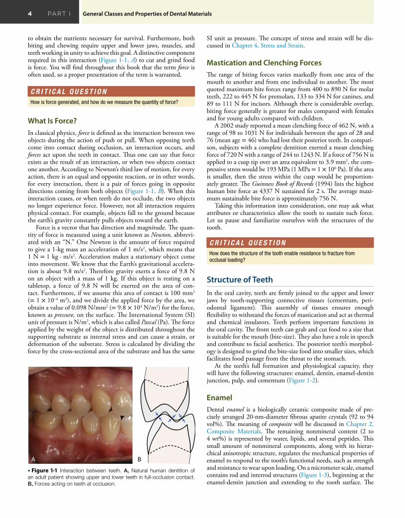
to obtain the nutrients necessary for survival. Furthermore, both biting and chewing require upper and lower jaws, muscles, and teeth working in unity to achieve this goal. A distinctive component required in this interaction (Figure 1-1, A) to cut and grind food is force. You will find throughout this book that the term force is often used, so a proper presentation of the term is warranted.
A B
• Figure 1-1 Interaction between teeth. A, Natural human dentition of an adult patient showing upper and lower teeth in full-occlusion contact. B, Forces acting on teeth at occlusion.
C R I T I C A L Q U E S T I O NHow is force generated, and how do we measure the quantity of force?
What Is Force?In classical physics, force is defined as the interaction between two objects during the action of push or pull. When opposing teeth come into contact during occlusion, an interaction occurs, and forces act upon the teeth in contact. Thus one can say that force exists as the result of an interaction, or when two objects contact one another. According to Newton’s third law of motion, for every action, there is an equal and opposite reaction, or in other words, for every interaction, there is a pair of forces going in opposite directions coming from both objects (Figure 1-1, B). When this interaction ceases, or when teeth do not occlude, the two objects no longer experience force. However, not all interaction requires physical contact. For example, objects fall to the ground because the earth’s gravity constantly pulls objects toward the earth.
Force is a vector that has direction and magnitude. The quan-tity of force is measured using a unit known as Newton, abbrevi-ated with an “N.” One Newton is the amount of force required to give a 1-kg mass an acceleration of 1 m/s2, which means that 1 N = 1 kg · m/s2. Acceleration makes a stationary object come into movement. We know that the Earth’s gravitational accelera-tion is about 9.8 m/s2. Therefore gravity exerts a force of 9.8 N on an object with a mass of 1 kg. If this object is resting on a tabletop, a force of 9.8 N will be exerted on the area of con-tact. Furthermore, if we assume this area of contact is 100 mm2 (= 1 × 10⁻⁴ m2), and we divide the applied force by the area, we obtain a value of 0.098 N/mm2 (= 9.8 × 10⁴ N/m2) for the force, known as pressure, on the surface. The International System (SI) unit of pressure is N/m2, which is also called Pascal (Pa). The force applied by the weight of the object is distributed throughout the supporting substrate as internal stress and can cause a strain, or deformation of the substrate. Stress is calculated by dividing the force by the cross-sectional area of the substrate and has the same
SI unit as pressure. The concept of stress and strain will be dis-cussed in Chapter 4, Stress and Strain.
Mastication and Clenching ForcesThe range of biting forces varies markedly from one area of the mouth to another and from one individual to another. The most quoted maximum bite forces range from 400 to 890 N for molar teeth, 222 to 445 N for premolars, 133 to 334 N for canines, and 89 to 111 N for incisors. Although there is considerable overlap, biting force generally is greater for males compared with females and for young adults compared with children.
A 2002 study reported a mean clenching force of 462 N, with a range of 98 to 1031 N for individuals between the ages of 28 and 76 (mean age = 46) who had lost their posterior teeth. In compari-son, subjects with a complete dentition exerted a mean clenching force of 720 N with a range of 244 to 1243 N. If a force of 756 N is applied to a cusp tip over an area equivalent to 3.9 mm2, the com-pressive stress would be 193 MPa (1 MPa = 1 × 10⁶ Pa). If the area is smaller, then the stress within the cusp would be proportion-ately greater. The Guinness Book of Records (1994) lists the highest human bite force as 4337 N sustained for 2 s. The average maxi-mum sustainable bite force is approximately 756 N.
Taking this information into consideration, one may ask what attributes or characteristics allow the tooth to sustain such force. Let us pause and familiarize ourselves with the structures of the tooth.
Structure of TeethIn the oral cavity, teeth are firmly joined to the upper and lower jaws by tooth-supporting connective tissues (cementum, peri-odontal ligament). This assembly of tissues ensures enough flexibility to withstand the forces of mastication and act as thermal and chemical insulators. Teeth perform important functions in the oral cavity. The front teeth can grab and cut food to a size that is suitable for the mouth (bite-size). They also have a role in speech and contribute to facial aesthetics. The posterior teeth’s morphol-ogy is designed to grind the bite-size food into smaller sizes, which facilitates food passage from the throat to the stomach.
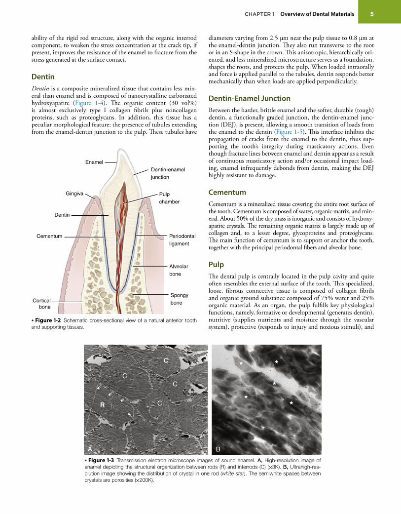
At the teeth’s full formation and physiological capacity, they will have the following structures: enamel, dentin, enamel-dentin junction, pulp, and cementum (Figure 1-2).
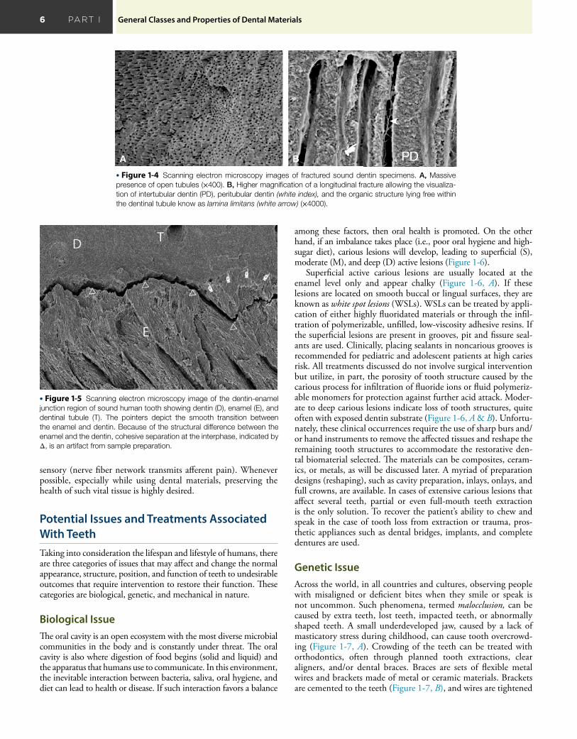
EnamelDental enamel is a biologically ceramic composite made of pre-cisely arranged 20-nm-diameter fibrous apatite crystals (92 to 94 vol%). The meaning of composite will be discussed in Chapter 2, Composite Materials. The remaining nonmineral content (2 to 4 wt%) is represented by water, lipids, and several peptides. This small amount of nonmineral components, along with its hierar-chical anisotropic structure, regulates the mechanical properties of enamel to respond to the tooth’s functional needs, such as strength and resistance to wear upon loading. On a micrometer scale, enamel contains rod and interrod structures (Figure 1-3), beginning at the enamel-dentin junction and extending to the tooth surface. The
C R I T I C A L Q U E S T I O NHow does the structure of the tooth enable resistance to fracture from occlusal loading?
5 CHAPTER 1 Overview of Dental Materials
ability of the rigid rod structure, along with the organic interrod component, to weaken the stress concentration at the crack tip, if present, improves the resistance of the enamel to fracture from the stress generated at the surface contact.
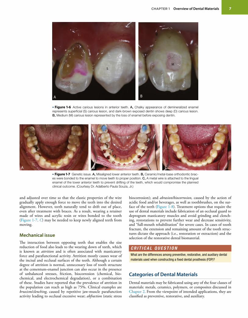
DentinDentin is a composite mineralized tissue that contains less min-eral than enamel and is composed of nanocrystalline carbonated hydroxyapatite (Figure 1-4). The organic content (30 vol%) is almost exclusively type I collagen fibrils plus noncollagen proteins, such as proteoglycans. In addition, this tissue has a peculiar morphological feature: the presence of tubules extending from the enamel-dentin junction to the pulp. These tubules have
B
R
A
C
C
C
C
• Figure 1-3 Transmission electron microscope images of sound enamel. A, High-resolution image of enamel depicting the structural organization between rods (R) and interrods (C) (×3K). B, Ultrahigh-res-olution image showing the distribution of crystal in one rod (white star). The semiwhite spaces between crystals are porosities (×200K).
diameters varying from 2.5 μm near the pulp tissue to 0.8 μm at the enamel-dentin junction. They also run transverse to the root or in an S-shape in the crown. This anisotropic, hierarchically ori-ented, and less mineralized microstructure serves as a foundation, shapes the roots, and protects the pulp. When loaded intraorally and force is applied parallel to the tubules, dentin responds better mechanically than when loads are applied perpendicularly.
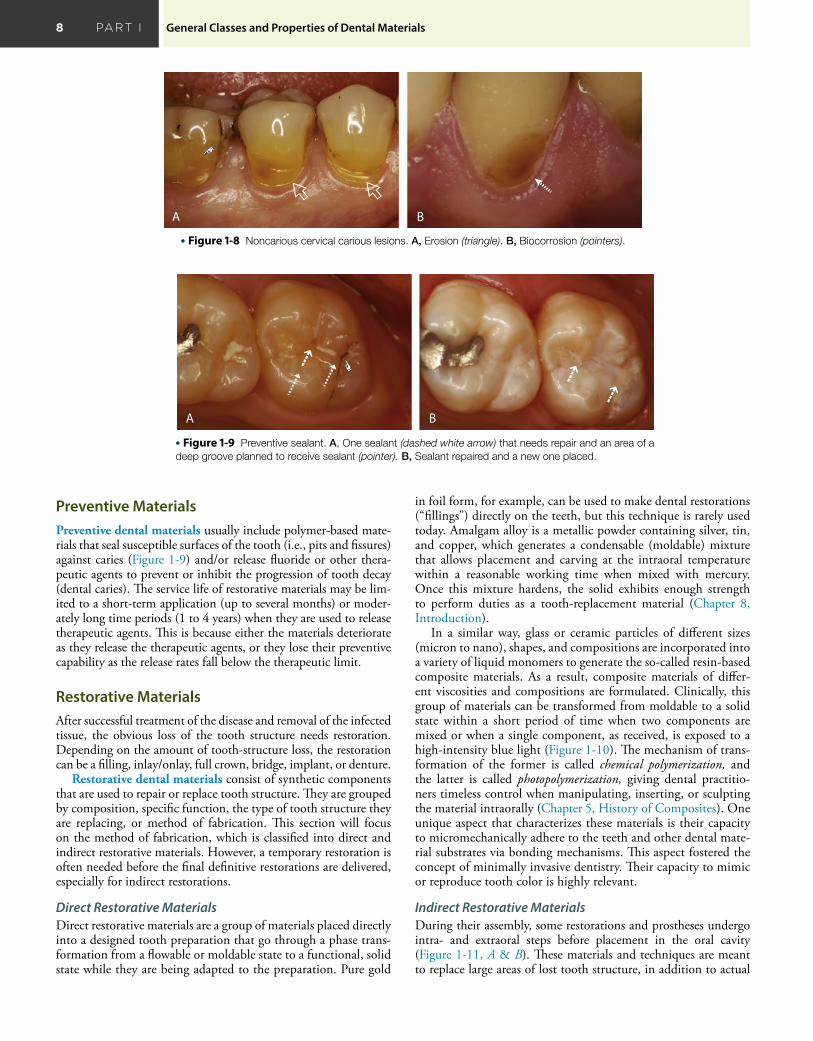
Dentin-Enamel JunctionBetween the harder, brittle enamel and the softer, durable (tough) dentin, a functionally graded junction, the dentin-enamel junc-tion (DEJ), is present, allowing a smooth transition of loads from the enamel to the dentin (Figure 1-5). This interface inhibits the propagation of cracks from the enamel to the dentin, thus sup-porting the tooth’s integrity during masticatory actions. Even though fracture lines between enamel and dentin appear as a result of continuous masticatory action and/or occasional impact load-ing, enamel infrequently debonds from dentin, making the DEJ highly resistant to damage.
CementumCementum is a mineralized tissue covering the entire root surface of the tooth. Cementum is composed of water, organic matrix, and min-eral. About 50% of the dry mass is inorganic and consists of hydroxy-apatite crystals. The remaining organic matrix is largely made up of collagen and, to a lesser degree, glycoproteins and proteoglycans. The main function of cementum is to support or anchor the tooth, together with the principal periodontal fibers and alveolar bone.
PulpThe dental pulp is centrally located in the pulp cavity and quite often resembles the external surface of the tooth. This specialized, loose, fibrous connective tissue is composed of collagen fibrils and organic ground substance composed of 75% water and 25% organic material. As an organ, the pulp fulfills key physiological functions, namely, formative or developmental (generates dentin), nutritive (supplies nutrients and moisture through the vascular system), protective (responds to injury and noxious stimuli), and
Enamel
Gingiva
Dentin
Cementum
Corticalbone
Pulpchamber
Periodontalligament
Alveolarbone
Spongybone
Dentin-enameljunction
• Figure 1-2 Schematic cross-sectional view of a natural anterior tooth and supporting tissues.
6 PART I General Classes and Properties of Dental Materials
sensory (nerve fiber network transmits afferent pain). Whenever possible, especially while using dental materials, preserving the health of such vital tissue is highly desired.
Potential Issues and Treatments Associated With TeethTaking into consideration the lifespan and lifestyle of humans, there are three categories of issues that may affect and change the normal appearance, structure, position, and function of teeth to undesirable outcomes that require intervention to restore their function. These categories are biological, genetic, and mechanical in nature.
Biological IssueThe oral cavity is an open ecosystem with the most diverse microbial communities in the body and is constantly under threat. The oral cavity is also where digestion of food begins (solid and liquid) and the apparatus that humans use to communicate. In this environment, the inevitable interaction between bacteria, saliva, oral hygiene, and diet can lead to health or disease. If such interaction favors a balance
among these factors, then oral health is promoted. On the other hand, if an imbalance takes place (i.e., poor oral hygiene and high-sugar diet), carious lesions will develop, leading to superficial (S), moderate (M), and deep (D) active lesions (Figure 1-6).
Superficial active carious lesions are usually located at the enamel level only and appear chalky (Figure 1-6, A). If these lesions are located on smooth buccal or lingual surfaces, they are known as white spot lesions (WSLs). WSLs can be treated by appli-cation of either highly fluoridated materials or through the infil-tration of polymerizable, unfilled, low-viscosity adhesive resins. If the superficial lesions are present in grooves, pit and fissure seal-ants are used. Clinically, placing sealants in noncarious grooves is recommended for pediatric and adolescent patients at high caries risk. All treatments discussed do not involve surgical intervention but utilize, in part, the porosity of tooth structure caused by the carious process for infiltration of fluoride ions or fluid polymeriz-able monomers for protection against further acid attack. Moder-ate to deep carious lesions indicate loss of tooth structures, quite often with exposed dentin substrate (Figure 1-6, A & B). Unfortu-nately, these clinical occurrences require the use of sharp burs and/or hand instruments to remove the affected tissues and reshape the remaining tooth structures to accommodate the restorative den-tal biomaterial selected. The materials can be composites, ceram-ics, or metals, as will be discussed later. A myriad of preparation designs (reshaping), such as cavity preparation, inlays, onlays, and full crowns, are available. In cases of extensive carious lesions that affect several teeth, partial or even full-mouth teeth extraction is the only solution. To recover the patient’s ability to chew and speak in the case of tooth loss from extraction or trauma, pros-thetic appliances such as dental bridges, implants, and complete dentures are used.
Genetic IssueAcross the world, in all countries and cultures, observing people with misaligned or deficient bites when they smile or speak is not uncommon. Such phenomena, termed malocclusion, can be caused by extra teeth, lost teeth, impacted teeth, or abnormally shaped teeth. A small underdeveloped jaw, caused by a lack of masticatory stress during childhood, can cause tooth overcrowd-ing (Figure 1-7, A). Crowding of the teeth can be treated with orthodontics, often through planned tooth extractions, clear aligners, and/or dental braces. Braces are sets of flexible metal wires and brackets made of metal or ceramic materials. Brackets are cemented to the teeth (Figure 1-7, B), and wires are tightened
• Figure 1-5 Scanning electron microscopy image of the dentin-enamel junction region of sound human tooth showing dentin (D), enamel (E), and dentinal tubule (T). The pointers depict the smooth transition between the enamel and dentin. Because of the structural difference between the enamel and the dentin, cohesive separation at the interphase, indicated by Δ, is an artifact from sample preparation.
A B
• Figure 1-4 Scanning electron microscopy images of fractured sound dentin specimens. A, Massive presence of open tubules (×400). B, Higher magnification of a longitudinal fracture allowing the visualiza-tion of intertubular dentin (PD), peritubular dentin (white index), and the organic structure lying free within the dentinal tubule know as lamina limitans (white arrow) (×4000).
7 CHAPTER 1 Overview of Dental Materials
and adjusted over time so that the elastic properties of the wire gradually apply enough force to move the teeth into the desired alignment. However, teeth naturally tend to drift out of place, even after treatment with braces. As a result, wearing a retainer made of wires and acrylic resin or wires bonded to the tooth (Figure 1-7, C) may be needed to keep newly aligned teeth from moving.
Mechanical issueThe interaction between opposing teeth that enables the size reduction of food also leads to the wearing down of teeth, which is known as attrition and is often associated with masticatory force and parafunctional activity. Attrition mostly causes wear of the incisal and occlusal surfaces of the teeth. Although a certain degree of attrition is normal, unnecessary loss of tooth structure at the cementum-enamel junction can also occur in the presence of unbalanced stresses, friction, biocorrosion (chemical, bio-chemical, and electrochemical degradation), or a combination of these. Studies have reported that the prevalence of attrition in the population can reach as high as 75%. Clinical examples are bruxism/clenching, caused by repetitive jaw-muscle parafunction activity leading to occlusal excessive wear; abfraction (static stress
biocorrosion); and abrasion/biocorrosion, caused by the action of acidic food and/or beverages, as well as toothbrushes, on the sur-face of the teeth (Figure 1-8). Treatment options that require the use of dental materials include fabrication of an occlusal guard to deprogram masticatory muscles and avoid grinding and clench-ing, restorations to prevent further wear and decrease sensitivity, and “full-mouth rehabilitation” for severe cases. In cases of tooth fracture, the extension and remaining amount of the tooth struc-tures dictate the approach (i.e., restoration or extraction) and the selection of the restorative dental biomaterial.
Categories of Dental MaterialsDental materials may be fabricated using any of the four classes of materials: metals, ceramics, polymers, or composites discussed in Chapter 2. From the viewpoint of intended applications, they are classified as preventive, restorative, and auxiliary.
M
BA
SD
• Figure 1-6 Active carious lesions in anterior teeth. A, Chalky appearance of demineralized enamel represents superficial (S) carious lesion, and dark-brown exposed dentin shows deep (D) carious lesion. B, Medium (M) carious lesion represented by the loss of enamel before exposing dentin.
A B C
• Figure 1-7 Genetic issue. A, Misaligned lower anterior teeth. B, Ceramic/metal-base orthodontic brac-es were bonded to the enamel to move teeth to proper position. C, A metal wire is attached to the lingual enamel of the lower anterior teeth to prevent drifting of the teeth, which would compromise the planned clinical outcome. (Courtesy Dr. Adalberto Paula Souza, Jr.)
C R I T I C A L Q U E S T I O NWhat are the differences among preventive, restorative, and auxiliary dental materials used when constructing a fixed dental prosthesis (FDP)?
8 PART I General Classes and Properties of Dental Materials
Preventive MaterialsPreventive dental materials usually include polymer-based mate-rials that seal susceptible surfaces of the tooth (i.e., pits and fissures) against caries (Figure 1-9) and/or release fluoride or other thera-peutic agents to prevent or inhibit the progression of tooth decay (dental caries). The service life of restorative materials may be lim-ited to a short-term application (up to several months) or moder-ately long time periods (1 to 4 years) when they are used to release therapeutic agents. This is because either the materials deteriorate as they release the therapeutic agents, or they lose their preventive capability as the release rates fall below the therapeutic limit.
Restorative MaterialsAfter successful treatment of the disease and removal of the infected tissue, the obvious loss of the tooth structure needs restoration. Depending on the amount of tooth-structure loss, the restoration can be a filling, inlay/onlay, full crown, bridge, implant, or denture.
Restorative dental materials consist of synthetic components that are used to repair or replace tooth structure. They are grouped by composition, specific function, the type of tooth structure they are replacing, or method of fabrication. This section will focus on the method of fabrication, which is classified into direct and indirect restorative materials. However, a temporary restoration is often needed before the final definitive restorations are delivered, especially for indirect restorations.
Direct Restorative MaterialsDirect restorative materials are a group of materials placed directly into a designed tooth preparation that go through a phase trans-formation from a flowable or moldable state to a functional, solid state while they are being adapted to the preparation. Pure gold
A B
• Figure 1-9 Preventive sealant. A, One sealant (dashed white arrow) that needs repair and an area of a deep groove planned to receive sealant (pointer). B, Sealant repaired and a new one placed.
A B
• Figure 1-8 Noncarious cervical carious lesions. A, Erosion (triangle). B, Biocorrosion (pointers).
in foil form, for example, can be used to make dental restorations (“fillings”) directly on the teeth, but this technique is rarely used today. Amalgam alloy is a metallic powder containing silver, tin, and copper, which generates a condensable (moldable) mixture that allows placement and carving at the intraoral temperature within a reasonable working time when mixed with mercury. Once this mixture hardens, the solid exhibits enough strength to perform duties as a tooth-replacement material (Chapter 8, Introduction).
In a similar way, glass or ceramic particles of different sizes (micron to nano), shapes, and compositions are incorporated into a variety of liquid monomers to generate the so-called resin-based composite materials. As a result, composite materials of differ-ent viscosities and compositions are formulated. Clinically, this group of materials can be transformed from moldable to a solid state within a short period of time when two components are mixed or when a single component, as received, is exposed to a high-intensity blue light (Figure 1-10). The mechanism of trans-formation of the former is called chemical polymerization, and the latter is called photopolymerization, giving dental practitio-ners timeless control when manipulating, inserting, or sculpting the material intraorally (Chapter 5, History of Composites). One unique aspect that characterizes these materials is their capacity to micromechanically adhere to the teeth and other dental mate-rial substrates via bonding mechanisms. This aspect fostered the concept of minimally invasive dentistry. Their capacity to mimic or reproduce tooth color is highly relevant.
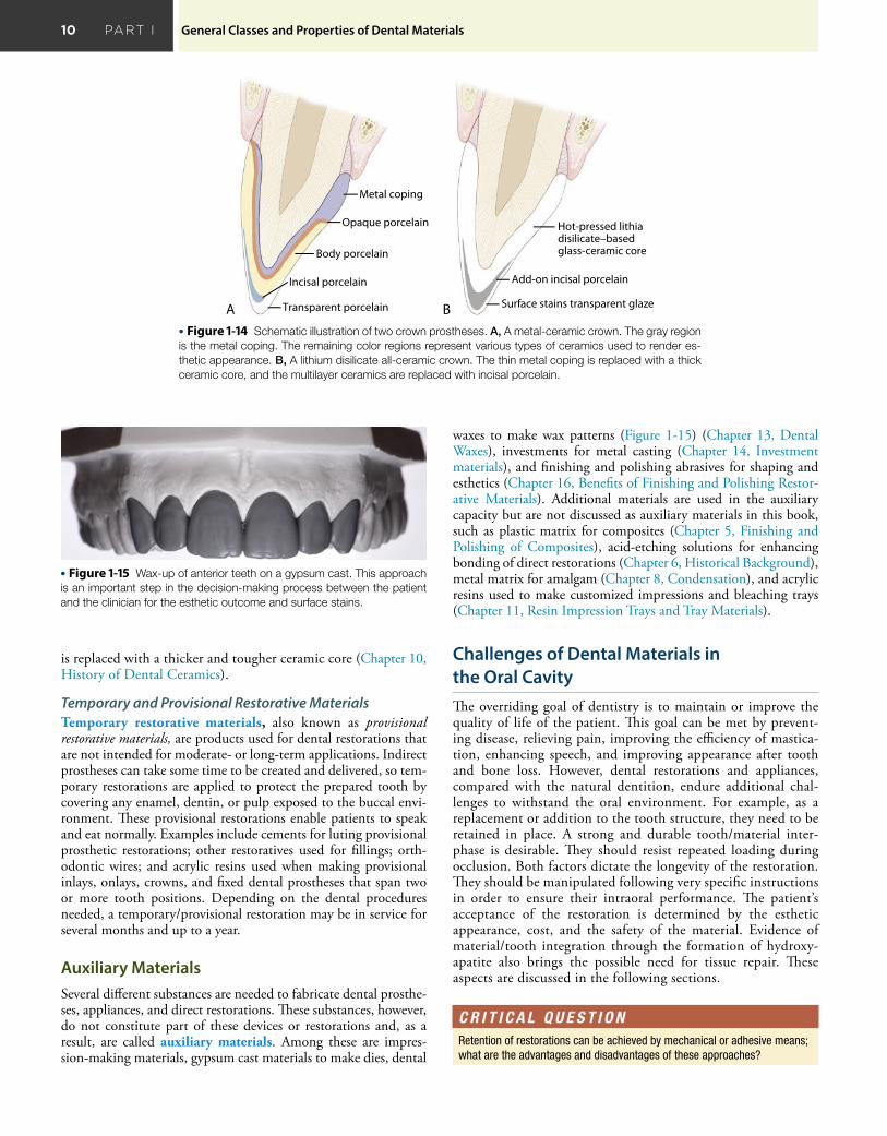
Indirect Restorative MaterialsDuring their assembly, some restorations and prostheses undergo intra- and extraoral steps before placement in the oral cavity (Figure 1-11, A & B). These materials and techniques are meant to replace large areas of lost tooth structure, in addition to actual
9 CHAPTER 1 Overview of Dental Materials
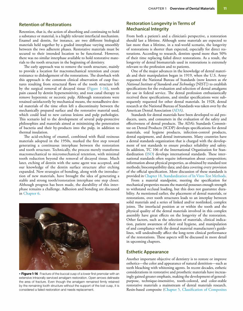
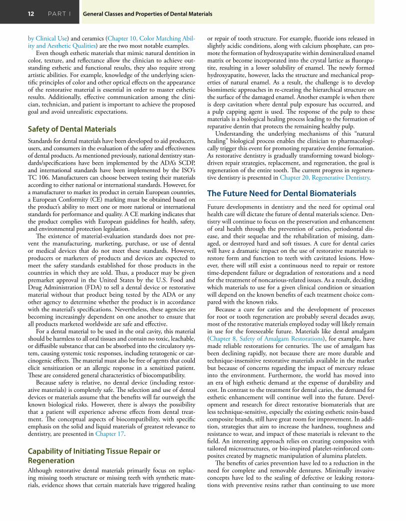
teeth. They are generally called indirect restorative materials and can be fixed by cementation (Figure 1-11, C) or remain removable, like a partial denture held by friction (Figure 1-12), or a complete denture (Figure 1-13), which is held to soft tissue by wetting. The issue of wetting will be discussed in Chapter 2, Wetting.
Metals and alloys, because of their mechanical properties, have been used for making indirect dental prosthetic restorations that include inlays/onlays, crowns and bridges, removable partial frameworks, and implants. A metal-ceramic or porcelain fused to metal (PFM) system is one in which esthetic ceramics are fused with metal substrates, where the metal functions as a coping,
• Figure 1-12 A removable partial denture framework in place. The point-ers indicate the retentive arms, and the arrows indicate the reciprocal arms. The wire mesh area between teeth provide sites for retaining artificial teeth with tissue-colored denture resin.
A B C
• Figure 1-11 Indirect ceramic restoration. A, Tooth preparation isolated with rubber dam. B, Indirect ce-ramic restoration made and adapted to a stone cast. C, Clinical view of the tooth/ceramic assembly after finishing the adhesive luting steps. (Courtesy Dr. Karine Barizon.)
• Figure 1-13 A set of complete removable dentures. (Courtesy Dr. Maged Abdelaal.)
A B
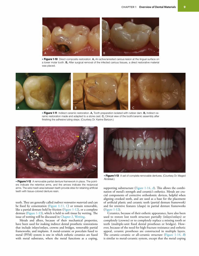
• Figure 1-10 Direct composite restoration. A, An active/arrested carious lesion at the lingual surface on a lower molar tooth. B, After surgical removal of the infected carious tissues, a direct restorative material was placed.
supporting substructure (Figure 1-14, A). This allows the combi-nation of metal’s strength and ceramic’s esthetics. Metals are cru-cial components of corrective orthodontic devices, helpful when aligning crooked teeth, and are used as a base for the placement of artificial plastic and ceramic teeth (partial denture framework) and for retentive features (clasps) in partial denture frameworks (Figure 1-12).
Ceramics, because of their esthetic appearance, have also been used to restore lost tooth structure partially (inlays/onlays) or completely (crowns) or to completely replace a missing tooth or teeth (multiple-unit fixed dental prostheses or bridges). How-ever, because of the need for high fracture resistance and esthetic appeal, ceramic prostheses are constructed in multiple layers. The ceramic-ceramic or all-ceramic structure (Figure 1-14, B) is similar to metal-ceramic system, except that the metal coping
10 PART I General Classes and Properties of Dental Materials
• Figure 1-15 Wax-up of anterior teeth on a gypsum cast. This approach is an important step in the decision-making process between the patient and the clinician for the esthetic outcome and surface stains.
is replaced with a thicker and tougher ceramic core (Chapter 10, History of Dental Ceramics).
Temporary and Provisional Restorative MaterialsTemporary restorative materials, also known as provisional re storative materials, are products used for dental restorations that are not intended for moderate- or long-term applications. Indirect prostheses can take some time to be created and delivered, so tem-porary restorations are applied to protect the prepared tooth by covering any enamel, dentin, or pulp exposed to the buccal envi-ronment. These provisional restorations enable patients to speak and eat normally. Examples include cements for luting provisional prosthetic restorations; other restoratives used for fillings; orth-odontic wires; and acrylic resins used when making provisional inlays, onlays, crowns, and fixed dental prostheses that span two or more tooth positions. Depending on the dental procedures needed, a temporary/provisional restoration may be in service for several months and up to a year.
Auxiliary MaterialsSeveral different substances are needed to fabricate dental prosthe-ses, appliances, and direct restorations. These substances, however, do not constitute part of these devices or restorations and, as a result, are called auxiliary materials. Among these are impres-sion-making materials, gypsum cast materials to make dies, dental
Surface stains transparent glaze
Hot-pressed lithia disilicate–basedglass-ceramic core
Add-on incisal porcelain
B
Body porcelain
Incisal porcelain
Metal coping
Opaque porcelain
Transparent porcelainA
• Figure 1-14 Schematic illustration of two crown prostheses. A, A metal-ceramic crown. The gray region is the metal coping. The remaining color regions represent various types of ceramics used to render es-thetic appearance. B, A lithium disilicate all-ceramic crown. The thin metal coping is replaced with a thick ceramic core, and the multilayer ceramics are replaced with incisal porcelain.
C R I T I C A L Q U E S T I O NRetention of restorations can be achieved by mechanical or adhesive means; what are the advantages and disadvantages of these approaches?
waxes to make wax patterns (Figure 1-15) (Chapter 13, Dental Waxes), investments for metal casting (Chapter 14, Investment materials), and finishing and polishing abrasives for shaping and esthetics (Chapter 16, Benefits of Finishing and Polishing Restor-ative Materials). Additional materials are used in the auxiliary capacity but are not discussed as auxiliary materials in this book, such as plastic matrix for composites (Chapter 5, Finishing and Polishing of Composites), acid-etching solutions for enhancing bonding of direct restorations (Chapter 6, Historical Background), metal matrix for amalgam (Chapter 8, Condensation), and acrylic resins used to make customized impressions and bleaching trays (Chapter 11, Resin Impression Trays and Tray Materials).
Challenges of Dental Materials in the Oral CavityThe overriding goal of dentistry is to maintain or improve the quality of life of the patient. This goal can be met by prevent-ing disease, relieving pain, improving the efficiency of mastica-tion, enhancing speech, and improving appearance after tooth and bone loss. However, dental restorations and appliances, compared with the natural dentition, endure additional chal-lenges to withstand the oral environment. For example, as a replacement or addition to the tooth structure, they need to be retained in place. A strong and durable tooth/material inter-phase is desirable. They should resist repeated loading during occlusion. Both factors dictate the longevity of the restoration. They should be manipulated following very specific instructions in order to ensure their intraoral performance. The patient’s acceptance of the restoration is determined by the esthetic appearance, cost, and the safety of the material. Evidence of material/tooth integration through the formation of hydroxy-apatite also brings the possible need for tissue repair. These aspects are discussed in the following sections.
11 CHAPTER 1 Overview of Dental Materials
Retention of RestorationsRetention, that is, the action of absorbing and continuing to hold a substance or material, is a highly relevant interfacial mechanism. Enamel and dentin, for instance, are two different biological materials held together by a graded interphase varying smoothly between the two adherent phases. Restorative materials must be secured to their intended location to be functional. However, there was no similar interphase available to hold restorative mate-rials to the tooth structure in the beginning of dentistry.
The early approach was to remove the tooth structure, mainly to provide a location for macromechanical retention and create resistance to dislodgement of the restorations. The drawback with this approach is the common clinical observation of cusp frac-tures resulting from structural flaws of the tooth structure left by the surgical removal of decayed tissue (Figure 1-16), tooth pain caused by dentin hypersensitivity, and root canal therapy to remove hyperemic or necrotic pulp. Although restorations were retained satisfactorily by mechanical means, the nonadhesive den-tal materials of the time often left a discontinuity between the mechanically prepared surface and the restorative material used, which could lead to new carious lesions and pulp pathologies. This scenario led to the development of several pulp-protective philosophies and materials aimed at minimizing the penetration of bacteria and their by-products into the pulp, in addition to thermal insulation.
The acid-etching of enamel, combined with fluid resinous materials adopted in the 1950s, marked the first step toward generating a continuous interphase between the restoration and tooth structure. Technically, the process merely transforms macromechanical to micromechanical retention, with minimal tooth reduction beyond the removal of decayed tissue. Much later, etching of dentin with the same agent was accepted, and our knowledge of the dentin surface structure after etching expanded. New strategies of bonding, along with the introduc-tion of new materials, have brought the idea of generating a stable and strong tooth/restoration interphase one step closer. Although progress has been made, the durability of this inter-phase remains a challenge. Adhesion and bonding are discussed in Chapter 6.
• Figure 1-16 Fracture of the buccal cusp of a lower first premolar with an extensive intraorally serviced amalgam restoration. Open arrows delineate the area of fracture. Even though the amalgam remained firmly retained by the remaining tooth structure without the support of the lost cusp, it is considered a failed restoration and needs replacement.
Restoration Longevity in Terms of Mechanical IntegrityFrom both a patient’s and a clinician’s perspective, a restoration should last a lifetime. Although some materials are expected to last more than a lifetime, in a real-world scenario, the longevity of restorations is shorter than expected, especially for direct res-torations. According to research, dentists spend more than 50% of their time replacing failed direct restorations. As a result, the longevity of dental biomaterials used in restorations is extremely relevant to the profession and to patients.
One of the major advances in the knowledge of dental materi-als and their manipulation began in 1919, when the U.S. Army requested the National Bureau of Standards (now known as the National Institute of Standards and Technology [NIST]) to establish specifications for the evaluation and selection of dental amalgams for use in federal service. The dental profession enthusiastically received these specifications, and similar specifications were sub-sequently requested for other dental materials. In 1928, dental research at the National Bureau of Standards was taken over by the American Dental Association (ADA).
Standards for dental materials have been developed to aid pro-ducers, users, and consumers in the evaluation of the safety and effectiveness of dental products. The ADA’s Standards Commit-tee on Dental Products (SCDP) develops specifications for dental materials, oral hygiene products, infection-control products, dental equipment, and dental instruments. Many countries have a dental standards organization that is charged with the develop-ment of test standards to ensure product reliability and safety. In addition, TC 106 of the International Organization for Stan-dardization (ISO) develops international standards. These inter-national standards often require information about composition; information about physical properties, as obtained by standard test methods; biocompatibility data; and data covering every provision of the official specification. More discussion of these standards is provided in Chapter 18, Standardization of In Vitro Test Methods.
From a material standpoint, meeting the specification for mechanical properties means the material possesses enough strength to withstand occlusal loading, but this does not guarantee dura-bility. As mentioned earlier, the placement of dental materials, or restorations, over tooth structures leads to an interplay between solid materials and a series of linked and/or nonlinked, complex joints. The interfacial position at or within the tooth and the physical quality of the dental materials involved in this complex assembly have great effects on the longevity of the restoration. Other factors, such as the selection of materials, clinical indica-tion, patient awareness of their oral hygiene, and manipulation of and compliance with the dental material manufacturer’s guide-lines, will undoubtedly affect the long-term clinical performance of the restorations. These aspects will be discussed in more detail in upcoming chapters.
Esthetic AppearanceAnother important objective of dentistry is to restore or improve esthetics—the color and appearance of natural dentition—such as teeth bleaching with whitening agents. In recent decades, esthetic considerations in restorative and prosthetic materials have increas-ingly gained greater emphasis, making the development of general-purpose, technique-insensitive, tooth-colored, and color-stable restorative materials a mainstream of dental materials research. Resin-based composite (Chapter 5, Classification of Composites
12 PART I General Classes and Properties of Dental Materials
by Clinical Use) and ceramics (Chapter 10, Color Matching Abil-ity and Aesthetic Qualities) are the two most notable examples.
Even though esthetic materials that mimic natural dentition in color, texture, and reflectance allow the clinician to achieve out-standing esthetic and functional results, they also require strong artistic abilities. For example, knowledge of the underlying scien-tific principles of color and other optical effects on the appearance of the restorative material is essential in order to master esthetic results. Additionally, effective communication among the clini-cian, technician, and patient is important to achieve the proposed goal and avoid unrealistic expectations.
Safety of Dental MaterialsStandards for dental materials have been developed to aid producers, users, and consumers in the evaluation of the safety and effectiveness of dental products. As mentioned previously, national dentistry stan-dards/specifications have been implemented by the ADA’s SCDP, and international standards have been implemented by the ISO’s TC 106. Manufacturers can choose between testing their materials according to either national or international standards. However, for a manufacturer to market its product in certain European countries, a European Conformity (CE) marking must be obtained based on the product’s ability to meet one or more national or international standards for performance and quality. A CE marking indicates that the product complies with European guidelines for health, safety, and environmental protection legislation.
The existence of material-evaluation standards does not pre-vent the manufacturing, marketing, purchase, or use of dental or medical devices that do not meet these standards. However, producers or marketers of products and devices are expected to meet the safety standards established for those products in the countries in which they are sold. Thus, a producer may be given premarket approval in the United States by the U.S. Food and Drug Administration (FDA) to sell a dental device or restorative material without that product being tested by the ADA or any other agency to determine whether the product is in accordance with the material’s specifications. Nevertheless, these agencies are becoming increasingly dependent on one another to ensure that all products marketed worldwide are safe and effective.
For a dental material to be used in the oral cavity, this material should be harmless to all oral tissues and contain no toxic, leachable, or diffusible substance that can be absorbed into the circulatory sys-tem, causing systemic toxic responses, including teratogenic or car-cinogenic effects. The material must also be free of agents that could elicit sensitization or an allergic response in a sensitized patient. These are considered general characteristics of biocompatibility.
Because safety is relative, no dental device (including restor-ative materials) is completely safe. The selection and use of dental devices or materials assume that the benefits will far outweigh the known biological risks. However, there is always the possibility that a patient will experience adverse effects from dental treat-ment. The conceptual aspects of biocompatibility, with specific emphasis on the solid and liquid materials of greatest relevance to dentistry, are presented in Chapter 17.
Capability of Initiating Tissue Repair or RegenerationAlthough restorative dental materials primarily focus on replac-ing missing tooth structure or missing teeth with synthetic mate-rials, evidence shows that certain materials have triggered healing
or repair of tooth structure. For example, fluoride ions released in slightly acidic conditions, along with calcium phosphate, can pro-mote the formation of hydroxyapatite within demineralized enamel matrix or become incorporated into the crystal lattice as fluorapa-tite, resulting in a lower solubility of enamel. The newly formed hydroxyapatite, however, lacks the structure and mechanical prop-erties of natural enamel. As a result, the challenge is to develop biomimetic approaches in re-creating the hierarchical structure on the surface of the damaged enamel. Another example is when there is deep cavitation where dental pulp exposure has occurred, and a pulp capping agent is used. The response of the pulp to these materials is a biological healing process leading to the formation of reparative dentin that protects the remaining healthy pulp.
Understanding the underlying mechanisms of this “natural healing” biological process enables the clinician to pharmacologi-cally trigger this event for promoting reparative dentine formation. As restorative dentistry is gradually transforming toward biology-driven repair strategies, replacement, and regeneration, the goal is regeneration of the entire tooth. The current progress in regenera-tive dentistry is presented in Chapter 20, Regenerative Dentistry.
The Future Need for Dental BiomaterialsFuture developments in dentistry and the need for optimal oral health care will dictate the future of dental materials science. Den-tistry will continue to focus on the preservation and enhancement of oral health through the prevention of caries, periodontal dis-ease, and their sequelae and the rehabilitation of missing, dam-aged, or destroyed hard and soft tissues. A cure for dental caries will have a dramatic impact on the use of restorative materials to restore form and function to teeth with cavitated lesions. How-ever, there will still exist a continuous need to repair or restore time-dependent failure or degradation of restorations and a need for the treatment of noncarious-related issues. As a result, deciding which materials to use for a given clinical condition or situation will depend on the known benefits of each treatment choice com-pared with the known risks.
Because a cure for caries and the development of processes for root or tooth regeneration are probably several decades away, most of the restorative materials employed today will likely remain in use for the foreseeable future. Materials like dental amalgam (Chapter 8, Safety of Amalgam Restorations), for example, have made reliable restorations for centuries. The use of amalgam has been declining rapidly, not because there are more durable and technique-insensitive restorative materials available in the market but because of concerns regarding the impact of mercury release into the environment. Furthermore, the world has moved into an era of high esthetic demand at the expense of durability and cost. In contrast to the treatment for dental caries, the demand for esthetic enhancement will continue well into the future. Devel-opment and research for direct restorative biomaterials that are less technique-sensitive, especially the existing esthetic resin-based composite brands, still have great room for improvement. In addi-tion, strategies that aim to increase the hardness, toughness and resistance to wear, and impact of these materials is relevant to the field. An interesting approach relies on creating composites with tailored microstructures, or bio-inspired platelet-reinforced com-posites created by magnetic manipulation of alumina platelets.
The benefits of caries prevention have led to a reduction in the need for complete and removable dentures. Minimally invasive concepts have led to the sealing of defective or leaking restora-tions with preventive resins rather than continuing to use more
13 CHAPTER 1 Overview of Dental Materials
destructive replacement procedures. Thus, the development of effective bio-inspired remineralizing agents for acid-damaged enamel and dentin repair and the development of smart materials that lead to self-repair of defective joints or of a material’s inter-nal flaws, as well as dental pulp regeneration, will be critical chal-lenges to the future of dental materials. In addition, the need for replacement of restorations should decrease over the next several decades, in lieu of the minimally invasive philosophy. However, this reduction will be offset by the increased demand for esthetic restorations (discussed in Chapter 20, Biomaterials).
Technology has advanced tremendously over the past 40 years, resulting in innovations such as laser applications, imaging pro-cedures, three-dimensional (3-D) printing technology, low-shrinkage composites, smart ceramics, and minimally invasive dental procedures, which have generated benefits for several fields. Computer-aided design/computer-aided manufacturing (CAD-CAM) technology for making indirect restorations, for example, has reduced the demand for both impression materi-als and some indirect auxiliary materials that were used by labo-ratory technicians to fabricate indirect prostheses (discussed in Chapter 15, Digital Impressions and Prototyping Tools).
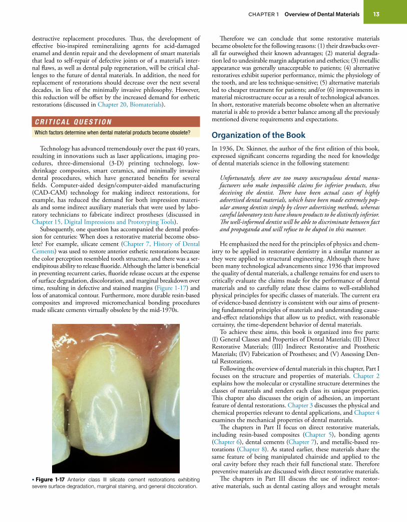
Subsequently, one question has accompanied the dental profes-sion for centuries: When does a restorative material become obso-lete? For example, silicate cement (Chapter 7, History of Dental Cements) was used to restore anterior esthetic restorations because the color perception resembled tooth structure, and there was a ser-endipitous ability to release fluoride. Although the latter is beneficial in preventing recurrent caries, fluoride release occurs at the expense of surface degradation, discoloration, and marginal breakdown over time, resulting in defective and stained margins (Figure 1-17) and loss of anatomical contour. Furthermore, more durable resin-based composites and improved micromechanical bonding procedures made silicate cements virtually obsolete by the mid-1970s.
Therefore we can conclude that some restorative materials became obsolete for the following reasons: (1) their drawbacks over-all far outweighed their known advantages; (2) material degrada-tion led to undesirable margin adaptation and esthetics; (3) metallic appearance was generally unacceptable to patients; (4) alternative restoratives exhibit superior performance, mimic the physiology of the tooth, and are less technique-sensitive; (5) alternative materials led to cheaper treatment for patients; and/or (6) improvements in material microstructure occur as a result of technological advances. In short, restorative materials become obsolete when an alternative material is able to provide a better balance among all the previously mentioned diverse requirements and expectations.
Organization of the BookIn 1936, Dr. Skinner, the author of the first edition of this book, expressed significant concerns regarding the need for knowledge of dental materials science in the following statement:
Unfortunately, there are too many unscrupulous dental manu-facturers who make impossible claims for inferior products, thus deceiving the dentist. There have been actual cases of highly advertised dental materials, which have been made extremely pop-ular among dentists simply by clever advertising methods, whereas careful laboratory tests have shown products to be distinctly inferior. The well-informed dentist will be able to discriminate between fact and propaganda and will refuse to be duped in this manner.
He emphasized the need for the principles of physics and chem-istry to be applied in restorative dentistry in a similar manner as they were applied to structural engineering. Although there have been many technological advancements since 1936 that improved the quality of dental materials, a challenge remains for end users to critically evaluate the claims made for the performance of dental materials and to carefully relate these claims to well-established physical principles for specific classes of materials. The current era of evidence-based dentistry is consistent with our aims of present-ing fundamental principles of materials and understanding cause-and-effect relationships that allow us to predict, with reasonable certainty, the time-dependent behavior of dental materials.
To achieve these aims, this book is organized into five parts: (I) General Classes and Properties of Dental Materials; (II) Direct Restorative Materials; (III) Indirect Restorative and Prosthetic Materials; (IV) Fabrication of Prostheses; and (V) Assessing Den-tal Restorations.
Following the overview of dental materials in this chapter, Part I focuses on the structure and properties of materials. Chapter 2 explains how the molecular or crystalline structure determines the classes of materials and renders each class its unique properties. This chapter also discusses the origin of adhesion, an important feature of dental restorations. Chapter 3 discusses the physical and chemical properties relevant to dental applications, and Chapter 4 examines the mechanical properties of dental materials.
The chapters in Part II focus on direct restorative materials, including resin-based composites (Chapter 5), bonding agents (Chapter 6), dental cements (Chapter 7), and metallic-based res-torations (Chapter 8). As stated earlier, these materials share the same feature of being manipulated chairside and applied to the oral cavity before they reach their full functional state. Therefore preventive materials are discussed with direct restorative materials.
The chapters in Part III discuss the use of indirect restor-ative materials, such as dental casting alloys and wrought metals
• Figure 1-17 Anterior class III silicate cement restorations exhibiting severe surface degradation, marginal staining, and general discoloration.
C R I T I C A L Q U E S T I O NWhich factors determine when dental material products become obsolete?
14 PART I General Classes and Properties of Dental Materials
(Chapter 9), ceramic-based materials (Chapter 10), denture-base resins (Chapter 11), and dental implants (Chapter 12).
Chapter 13 through 16 in Part IV describe the materials and technologies needed to fabricate indirect prostheses. Chapter 13 discusses auxiliary materials, which include impression materi-als, gypsum products, and dental waxes, which are used to fabri-cate the wax patterns needed for final prostheses. The process of making metal castings using the lost wax method is discussed in Chapter 14, and Chapter 15 discusses the use of digital technology for making dental prostheses without using traditional auxiliary materials but instead relying on digital imaging and CAD-CAM technology. Finally, all restorations, direct or indirect, require cer-tain degrees of surface modification to improve their clinical per-formance. The procedures and the essential instruments used for finishing and polishing restorations are discussed in Chapter 16.
The goal of dentistry is to maintain or improve the quality of life of the patient. The restorations involved must prevent disease, relieve pain, improve the efficiency of mastication, enhance speech, improve appearance, and most importantly, last for a long time. Part V aims to address the question of how we assess restorations. As stated, all materials undergo some sort of interaction with the
oral tissue. The biological considerations, known as biocompatibil-ity, are covered in Chapter 17. In vitro testing is conducted by subjecting a material made in a certain configuration to a known external factor (or factors) in a well-defined condition and record-ing the material’s response to such factors. The significance of in vitro testing is discussed in Chapter 18. Clinical evaluations of a dental restoration represent a different level of assessment for restorations. Chapter 19 discusses the components of clinical stud-ies, using one to illustrate how variables can be collected from the study. Finally, although new materials that address the particular needs of restorative dentistry are emerging and alternative treat-ment philosophies are being adopted, the advancement of new technologies in other disciplines has benefited dental materials science in a variety of ways. Chapter 20 analyzes potential future technologies in dentistry and describes both recently emerged technologies and those anticipated in the coming decades.
AcknowledgmentThe authors wish to acknowledge Dr. Kenneth J. Anusavice for his contribution to the earlier editions of this chapter.
Selected ReadingsBorcic J, Anic I, Urek MM, et al: The prevalence of non-carious cervical
lesions in permanent dentition, J Oral Rehabil 31:117–123, 2004.Erb RM, Libanori R, Rothfuchs N, et al: Composites reinforced in three
dimensions by using low magnetic fields, Science 335:199–204, 2012.He LH, Swain MV: Understanding the mechanical behaviour of human
enamel from its structural and compositional characteristics, J Mech Behav Biomed Mater 1:18–29, 2008.
Hensten-Pettersen A, Jacobsen N: Perceived side effects of biomaterials in prosthetic dentistry, J Prosthet Dent 65:138–144, 1991.
Jäger I, Fratzl P: Mineralized collagen fibrils: A mechanical model with a stag-gered arrangement of mineral particles, Biophys J 79:1737–1746, 2000.
Lobbezoo F, Ahlberg J, Raphael KG, et al: International consensus on the assessment of bruxism: Report of a work in progress, J Oral Rehabil 45:837–844, 2018.
Munksgaard EC: Toxicology versus allergy in restorative dentistry, Adv Dent Res 6:17–21, 1992.
Nanci A: Ten Cate’s Oral Histology: Development, Structure, and Function, 9th ed, St. Louis, 2018, Elsevier.
Stock SR, Vieira AE, Delbem AC, et al: Synchrotron micro computed tomography of the mature bovine dentinoenamel junction, J Struct Biol 161:162–171, 2008.
Thompson VP, Watson TF, Marshall GW Jr, et al: Outside-the-(cavity-prep)-box thinking, Adv Dent Res 25:24–32, 2013.
Volponi1 AA, Zaugg LK, Neves V, et al: Tooth repair and regeneration, Curr Oral Health Rep 5:295–303, 2018.
Full Selected Readings for this chapter can be found on www.expertcon-sult.com.
Selected Readings (Web Version)ADA Standards Committee on Dental Products (ADA SCDP) ADA
website: https://www.ada.org/en/science-research/dental-standards/dental -products. This website provides details on ANSI/ADA specifications for dental materials, instruments, and equipment and the working groups of the ADA Standards Committee on Dental Products.
Borcic J, Anic I, Urek MM, et al: The prevalence of non-carious cervical lesions in permanent dentition, J Oral Rehabil 31:117–123, 2004.
Coleman RL: Physical Properties of Dental Materials (Gold alloys and accessory materials), Research Paper No. 32, Washington, DC, 1928, US Govern-ment Printing Office.
Cramer NB, Stansbury JW, Bowman CN: Recent advances and develop-ments in composite dental restorative materials, J Dent Res 90:402–416, 2011. This publication represents the first major effort to relate the physical properties of dental materials to the clinical situation. The American Dental Association specification program was established based on this historical review of the philosophy and the content of the facility created at the National Bureau of Standards (now National Institute of Stan-dards and Technology).
Erb RM, Libanori R, Rothfuchs N, et al: Composites reinforced in three dimensions by using low magnetic fields, Science 335:199–204, 2012.
Gibbs CH, Anusavice KJ, Young HM, et al: Maximum clenching force of patients with moderate loss of posterior tooth support: A pilot study, J Prosthet Dent 88:498–502, 2002. The ability to sustain a maximum clenching force for a period of 2 s was reported in this study.
He LH, Swain MV: Understanding the mechanical behaviour of human enamel from its structural and compositional characteristics, J Mech Behav Biomed Mater 1:18–29, 2008.
Hensten-Pettersen A, Jacobsen N: Perceived side effects of biomaterials in prosthetic dentistry, J Prosthet Dent 65:138–144, 1991.
International Organization for Standardization (ISO) website: https://www.iso.org/home.html.
International Organization for Standardization (ISO) TC 106—Dentistry website. https://www.iso.org/committee/51218.html.
Jäger I, Fratzl P: Mineralized collagen fibrils: A mechanical model with a staggered arrangement of mineral particles, Biophys J 79:1737–1746, 2000.
Kallus T, Mjör IA: Incidence of adverse effects of dental materials, Eur J Oral Sci 99:236–240, 1991.
Lobbezoo F, Ahlberg J, Raphael KG, et al: International consensus on the assessment of bruxism: Report of a work in progress, J Oral Rehabil 45:837–844, 2018.
Munksgaard EC: Toxicology versus allergy in restorative dentistry, Adv Dent Res 6:17–21, 1992.
Nanci A: Ten Cate’s oral histology: development, structure, and function, 9th ed, St. Louis, 2018, Elsevier.
Phillips RW: Changing trends of dental restorative materials, Dent Clin North Am 33:285–291, 1989.
Stock SR, Vieira AE, Delbem AC, et al: Synchrotron micro computed tomography of the mature bovine dentinoenamel junction, J Struct Biol 161:162–171, 2008.
Thompson VP, Watson TF, Marshall GW Jr, et al: Outside-the-(cavity-prep)-box thinking, Adv Dent Res 25:24–32, 2013.
Volponi1 AA, Zaugg LK, Neves V, et al: Tooth repair and regeneration, Curr Oral Health Rep 5:295–303, 2018.
14.e1