Embed Size (px)
Citation preview
Oxidative phosphorylation
Lecture 11 2007 (J.S.)Biochemistry I

2
ATP is a high-energy compound that serves as the "universal currency" of free energy in biological systems through coupling with endergonic reactions.
The reaction, which is used to drive endergonic events, is very oft the hydrolysis of ATP.
Living organisms require a continual input of free energy for three major purposes:– the performance of mechanical work in cellular movements,– the active transport of molecules and ions across membranes
(osmotic work),– the synthesis of macromolecules and other biomolecules from simple
precursors (chemical work).
3
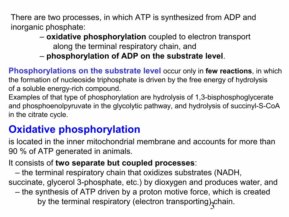
There are two processes, in which ATP is synthesized from ADP and inorganic phosphate:
– oxidative phosphorylation coupled to electron transport along the terminal respiratory chain, and– phosphorylation of ADP on the substrate level.
Phosphorylations on the substrate level occur only in few reactions, in which the formation of nucleoside triphosphate is driven by the free energy of hydrolysis of a soluble energy-rich compound.Examples of that type of phosphorylation are hydrolysis of 1,3-bisphosphoglycerateand phosphoenolpyruvate in the glycolytic pathway, and hydrolysis of succinyl-S-CoAin the citrate cycle.
Oxidative phosphorylationis located in the inner mitochondrial membrane and accounts for more than 90 % of ATP generated in animals.It consists of two separate but coupled processes: – the terminal respiratory chain that oxidizes substrates (NADH, succinate, glycerol 3-phosphate, etc.) by dioxygen and produces water, and – the synthesis of ATP driven by a proton motive force, which is created
by the terminal respiratory (electron transporting) chain.
4
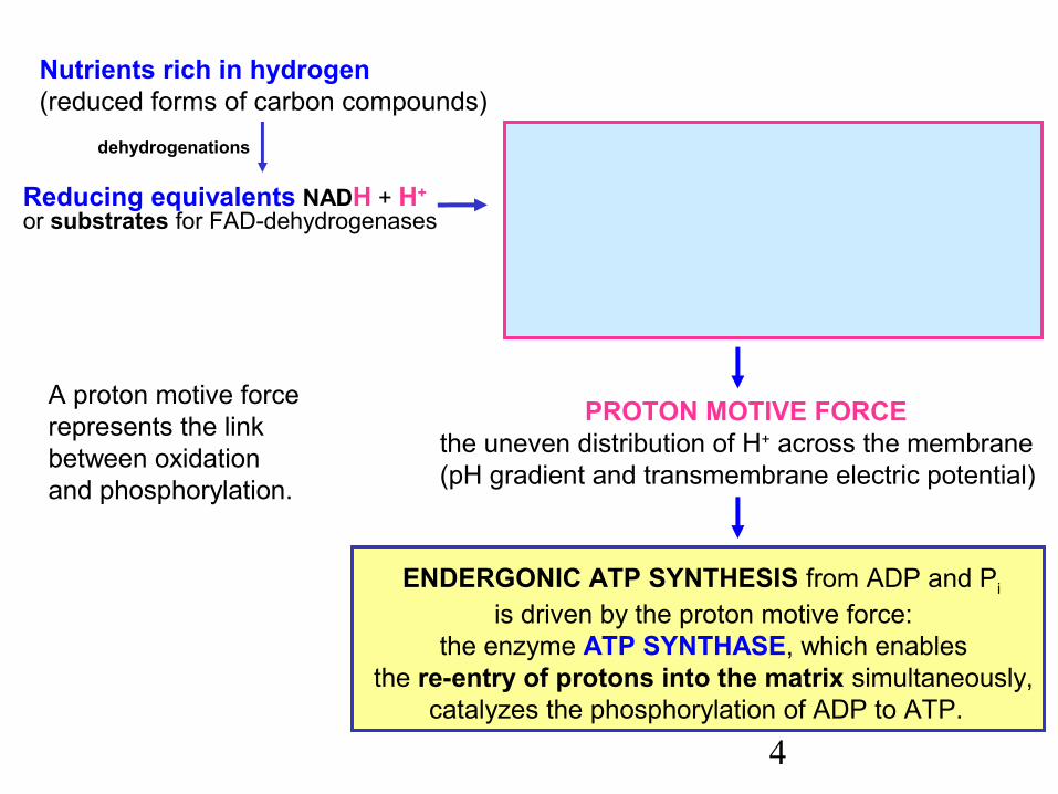
Reducing equivalents NADH + H+ or substrates for FAD-dehydrogenases
dehydrogenations
Nutrients rich in hydrogen (reduced forms of carbon compounds)
PROTON MOTIVE FORCEthe uneven distribution of H+ across the membrane (pH gradient and transmembrane electric potential)
TERMINAL RESPIRATORY CHAINtransfers electrons to O2 (dioxygen)
that is reduced to two O2– (oxide anions)accepting H+ and giving water. This
process results in the pumping of protonsout of the mitochondrial matrix
ENDERGONIC ATP SYNTHESIS from ADP and Pi
is driven by the proton motive force:the enzyme ATP SYNTHASE, which enables
the re-entry of protons into the matrix simultaneously,catalyzes the phosphorylation of ADP to ATP.
A proton motive forcerepresents the linkbetween oxidationand phosphorylation.
5
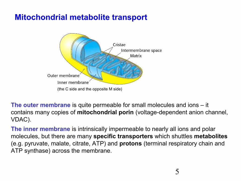
The outer membrane is quite permeable for small molecules and ions – it contains many copies of mitochondrial porin (voltage-dependent anion channel,VDAC).The inner membrane is intrinsically impermeable to nearly all ions and polarmolecules, but there are many specific transporters which shuttles metabolites(e.g. pyruvate, malate, citrate, ATP) and protons (terminal respiratory chain andATP synthase) across the membrane.
(the C side and the opposite M side)
Mitochondrial metabolite transport
6
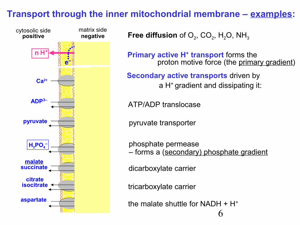
Transport through the inner mitochondrial membrane – examples:
Primary active H+ transport forms theproton motive force (the primary gradient)
Free diffusion of O2, CO2, H2O, NH3cytosolic side
positivematrix sidenegative
Ca2+
pyruvateOH–
ADP3–
ATP4–
OH–
malatesuccinate
citrateisocitrate
aspartate
H2PO4–
HPO42–
malate
malate
Secondary active transports driven by a H+ gradient and dissipating it:
ATP/ADP translocase
pyruvate transporter
phosphate permease– forms a (secondary) phosphate gradient
dicarboxylate carrier
tricarboxylate carrier
the malate shuttle for NADH + H+
7
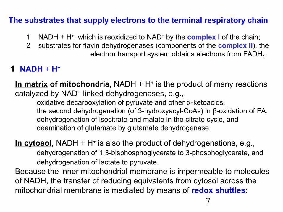
The substrates that supply electrons to the terminal respiratory chain
1 NADH + H+, which is reoxidized to NAD+ by the complex I of the chain;2 substrates for flavin dehydrogenases (components of the complex II), the
electron transport system obtains electrons from FADH2.
1 NADH + H+ In matrix of mitochondria, NADH + H+ is the product of many reactions catalyzed by NAD+-linked dehydrogenases, e.g., oxidative decarboxylation of pyruvate and other α-ketoacids, the second dehydrogenation (of 3-hydroxyacyl-CoAs) in β-oxidation of FA, dehydrogenation of isocitrate and malate in the citrate cycle, and deamination of glutamate by glutamate dehydrogenase.
In cytosol, NADH + H+ is also the product of dehydrogenations, e.g., dehydrogenation of 1,3-bisphosphoglycerate to 3-phosphoglycerate, and dehydrogenation of lactate to pyruvate.Because the inner mitochondrial membrane is impermeable to molecules of NADH, the transfer of reducing equivalents from cytosol across the mitochondrial membrane is mediated by means of redox shuttles:

8
Transport of reducing equivalents from cytoplasm into mitochondriaby redox shuttles:
The malate shuttle(is universal)
FADH2FAD UQ
glycerol-3-P
dihydroxyacetone-P
NADH+H+NAD+
mitochondrialglycerol phosphate
dehydrogenase(prosthetic group FAD)
cytosolic (NAD-linked)glycerol phosphate
dehydrogenase
The glycerophosphate shuttle(of minor importance in most human tissues)
Without entering the matrix, reducing equivalentsin the form of FADH2 supply electrons to theterminal respiratory chain (acceptor ubiquinone).
intermembranespace
matrix
NADH+H+NAD+
NADH+H+NAD+
oxaloacetate
oxaloacetate
aspartate
Glu
aspartatemalate
Pi
malateGlu
Glu
malatedehydrogenase
malatedehydrogenase
Glu
Pi
9
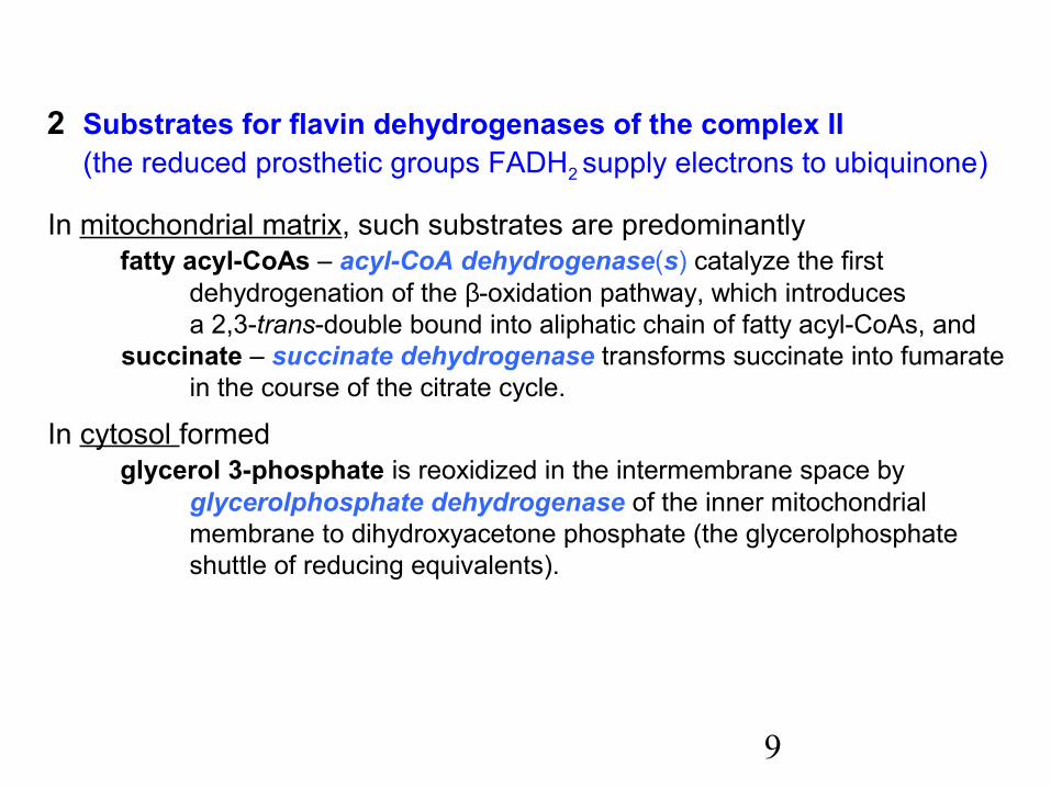
2 Substrates for flavin dehydrogenases of the complex II (the reduced prosthetic groups FADH2 supply electrons to ubiquinone)
In mitochondrial matrix, such substrates are predominantly fatty acyl-CoAs – acyl-CoA dehydrogenase(s) catalyze the first
dehydrogenation of the β-oxidation pathway, which introduces a 2,3-trans-double bound into aliphatic chain of fatty acyl-CoAs, and
succinate – succinate dehydrogenase transforms succinate into fumarate in the course of the citrate cycle.
In cytosol formed glycerol 3-phosphate is reoxidized in the intermembrane space by
glycerolphosphate dehydrogenase of the inner mitochondrial membrane to dihydroxyacetone phosphate (the glycerolphosphate shuttle of reducing equivalents).

10
The mitochondrial terminal respiratory chain
The chain reoxidizes NADH+H+ or FADH2 by transporting electrons to the terminal acceptor O2, which is reduced to form water.The free energy of oxidation of NADH or FADH2 is utilized for pumping protons to the outside of the inner mitochondrial membrane.The proton gradient across the inner mitochondrial membrane represents the proton motive force.
The proton motive force couples the terminal respiratory chain with the phosphorylation of ADP. It is a source of free energy for ATP synthesis that is driven by re-entry of protons into the matrix through an ATP synthase complex.
(The chemiosmotic hypothesis was mentioned primarily by Peter Mitchell in 1961.)
11
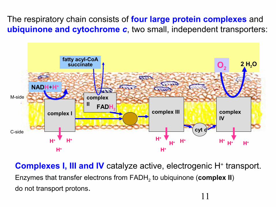
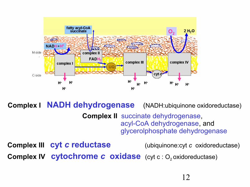
The respiratory chain consists of four large protein complexes and ubiquinone and cytochrome c, two small, independent transporters:
Complexes I, III and IV catalyze active, electrogenic H+ transport.Enzymes that transfer electrons from FADH2 to ubiquinone (complex II)do not transport protons.
complex I complex III complex IVUQ
cyt c
complex II FADH2
NADH+H+
fatty acyl-CoAsuccinate O2
2 H2O
M-side
C-side
H+H+H+H+
H+H+
H+H+ H+
H+
12
Complex I NADH dehydrogenase (NADH:ubiquinone oxidoreductase)
Complex II succinate dehydrogenase, acyl-CoA dehydrogenase, and
glycerolphosphate dehydrogenase
Complex III cyt c reductase (ubiquinone:cyt c oxidoreductase)
Complex IV cytochrome c oxidase (cyt c : O2 oxidoreductase)
13
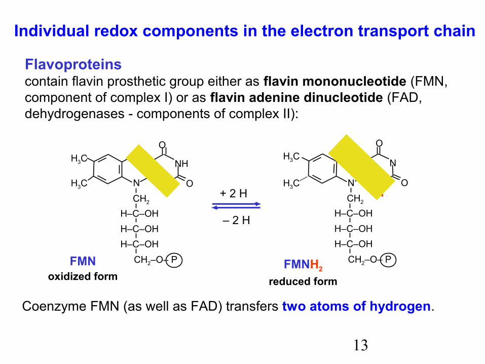
Coenzyme FMN (as well as FAD) transfers two atoms of hydrogen.
N
N
N
NH
O
O
H3C
N
N
N
N
O
O
H
H+ 2 H
– 2 H
FMNoxidized form
FMNH2
reduced form
H3C
H3C
H3C
CH2–O– P
CH2
H–C–OHH–C–OHH–C–OH
CH2–O– P
CH2
H–C–OHH–C–OHH–C–OH
Individual redox components in the electron transport chain
Flavoproteinscontain flavin prosthetic group either as flavin mononucleotide (FMN,component of complex I) or as flavin adenine dinucleotide (FAD, dehydrogenases - components of complex II):
14
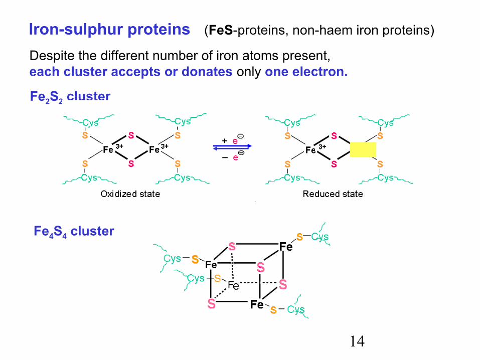
Iron-sulphur proteins (FeS-proteins, non-haem iron proteins)
Despite the different number of iron atoms present,each cluster accepts or donates only one electron.
Fe2S2 cluster
Fe4S4 cluster
15
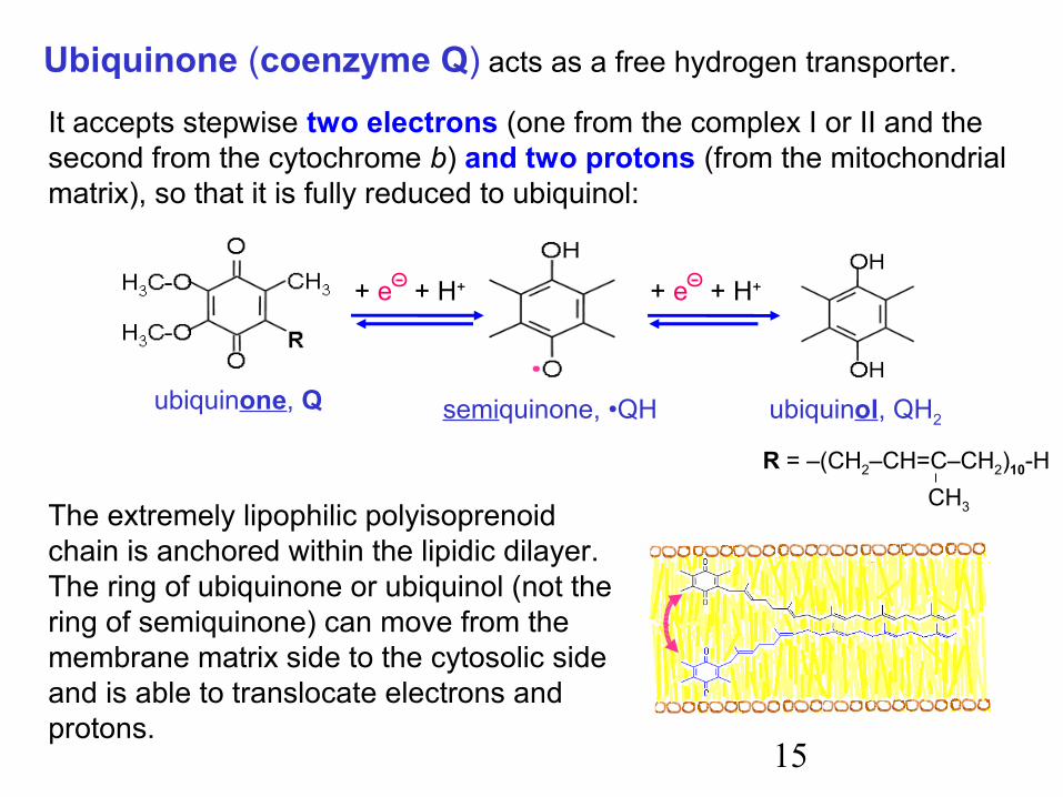
R = –(CH2–CH=C–CH2)10-HCH3
It accepts stepwise two electrons (one from the complex I or II and the second from the cytochrome b) and two protons (from the mitochondrial matrix), so that it is fully reduced to ubiquinol:
+ e + H+
ubiquinone, Q semiquinone, •QH ubiquinol, QH2
+ e + H+
Ubiquinone (coenzyme Q) acts as a free hydrogen transporter.
The extremely lipophilic polyisoprenoid chain is anchored within the lipidic dilayer. The ring of ubiquinone or ubiquinol (not the ring of semiquinone) can move from the membrane matrix side to the cytosolic side and is able to translocate electrons and protons.
16
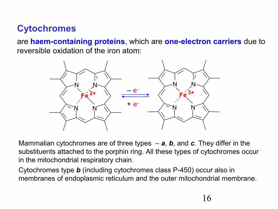
Cytochromesare haem-containing proteins, which are one-electron carriers due to reversible oxidation of the iron atom:
Mammalian cytochromes are of three types – a, b, and c. They differ in the substituents attached to the porphin ring. All these types of cytochromes occur in the mitochondrial respiratory chain.Cytochromes type b (including cytochromes class P-450) occur also in membranes of endoplasmic reticulum and the outer mitochondrial membrane.
N N
NN
Fe 2+
N N
NN
Fe 3+
+ e–
– e–
17
Haem a of cytochrome aa3
Haem of cytochrome c
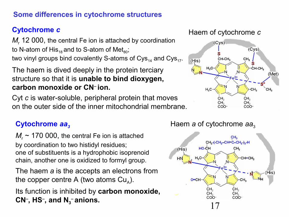
Some differences in cytochrome structures
Cytochrome cMr 12 000, the central Fe ion is attached by coordinationto N-atom of His18 and to S-atom of Met80;two vinyl groups bind covalently S-atoms of Cys14 and Cys17.
The haem is dived deeply in the protein terciarystructure so that it is unable to bind dioxygen,carbon monoxide or CN– ion.Cyt c is water-soluble, peripheral protein that moveson the outer side of the inner mitochondrial membrane.
Cytochrome aa3
Mr ~ 170 000, the central Fe ion is attachedby coordination to two histidyl residues;one of substituents is a hydrophobic isoprenoidchain, another one is oxidized to formyl group.
The haem a is the accepts an electrons fromthe copper centre A (two atoms CuA).Its function is inhibited by carbon monoxide,CN–, HS–, and N3
– anions.
18
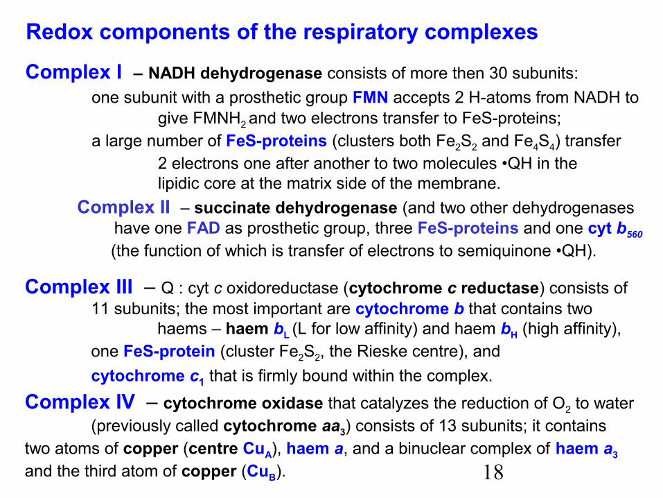
Redox components of the respiratory complexes
Complex I – NADH dehydrogenase consists of more then 30 subunits:one subunit with a prosthetic group FMN accepts 2 H-atoms from NADH to
give FMNH2 and two electrons transfer to FeS-proteins; a large number of FeS-proteins (clusters both Fe2S2 and Fe4S4) transfer
2 electrons one after another to two molecules •QH in thelipidic core at the matrix side of the membrane.
Complex II – succinate dehydrogenase (and two other dehydrogenases have one FAD as prosthetic group, three FeS-proteins and one cyt b560
(the function of which is transfer of electrons to semiquinone •QH).
Complex III – Q : cyt c oxidoreductase (cytochrome c reductase) consists of 11 subunits; the most important are cytochrome b that contains two
haems – haem bL (L for low affinity) and haem bH (high affinity),one FeS-protein (cluster Fe2S2, the Rieske centre), andcytochrome c1 that is firmly bound within the complex.
Complex IV – cytochrome oxidase that catalyzes the reduction of O2 to water(previously called cytochrome aa3) consists of 13 subunits; it contains
two atoms of copper (centre CuA), haem a, and a binuclear complex of haem a3 and the third atom of copper (CuB).
19
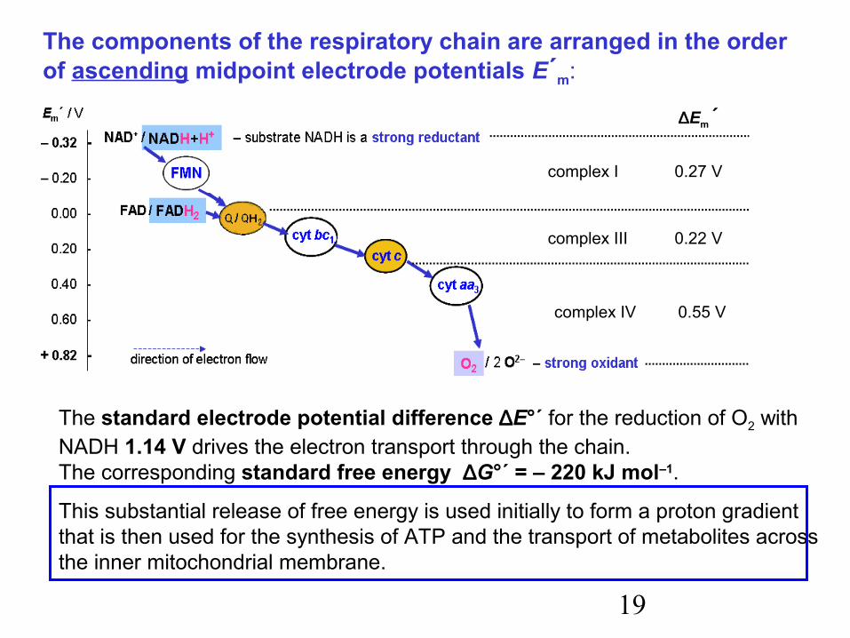
The components of the respiratory chain are arranged in the orderof ascending midpoint electrode potentials E´m:
∆Em´
complex I 0.27 V
complex III 0.22 V
complex IV 0.55 V
The standard electrode potential difference ∆E°´ for the reduction of O2 with NADH 1.14 V drives the electron transport through the chain.The corresponding standard free energy ∆G°´ = – 220 kJ mol–1.
This substantial release of free energy is used initially to form a proton gradientthat is then used for the synthesis of ATP and the transport of metabolites across the inner mitochondrial membrane.
20
H+
+2 H+ + 2 H+
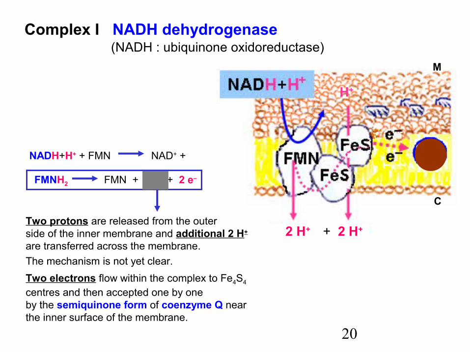
Complex I NADH dehydrogenase (NADH : ubiquinone oxidoreductase)
NADH+H+ + FMN NAD+ +
FMNH2 FMN + 2 H+ + 2 e–
Two protons are released from the outerside of the inner membrane and additional 2 H+
are transferred across the membrane.The mechanism is not yet clear.Two electrons flow within the complex to Fe4S4
centres and then accepted one by oneby the semiquinone form of coenzyme Q nearthe inner surface of the membrane.
M
C

21
Complex II Three independent flavin dehydrogenases that act in a similar way. Only one of them, succinate dehydrogenase, is mentioned here.
The enzymes that transfer electrons from FADH2 to Q (complex II), in contrast to complex I,do not transport protons across the inner membrane. Consequently, less proton gradient (and less ATP) is formed from the oxidation of FADH2 by dioxygen than from NADH.
Succinate.DH is an enzyme that catalyzes one of the reactions of the citrate cycle.However, it is the only integral membrane protein – the other enzymes of the citrate cycle are soluble, dispersed in the matrix..
M
C
22
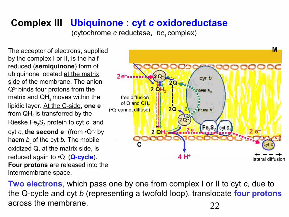
The acceptor of electrons, suppliedby the complex I or II, is the half-reduced (semiquinone) form ofubiquinone located at the matrixside of the membrane. The anionQ2– binds four protons from thematrix and QH2 moves within thelipidic layer. At the C-side, one e–
from QH2 is transferred by theRieske Fe2S2 protein to cyt c1 andcyt c, the second e– (from •Q–) byhaem bL
of the cyt b. The mobile oxidized Q, at the matrix side, isreduced again to •Q– (Q-cycle).Four protons are released into theintermembrane space.
Two electrons, which pass one by one from complex I or II to cyt c, due tothe Q-cycle and cyt b (representing a twofold loop), translocate four protonsacross the membrane.
Complex III Ubiquinone : cyt c oxidoreductase (cytochrome c reductase, bc1 complex)
4 H+
lateral diffusion
free diffusion of Q and QH2
(•Q– cannot diffuse)
M
C
23
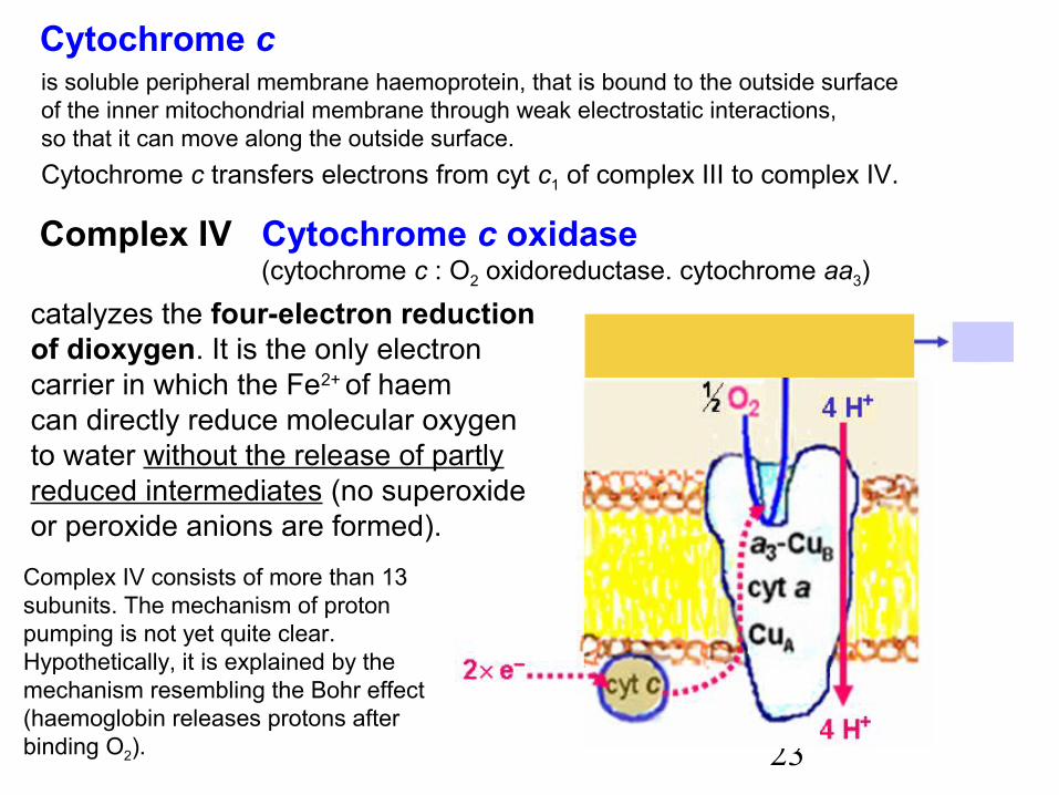
Cytochrome c is soluble peripheral membrane haemoprotein, that is bound to the outside surfaceof the inner mitochondrial membrane through weak electrostatic interactions,so that it can move along the outside surface.Cytochrome c transfers electrons from cyt c1 of complex III to complex IV.
Complex IV Cytochrome c oxidase (cytochrome c : O2 oxidoreductase. cytochrome aa3)
catalyzes the four-electron reductionof dioxygen. It is the only electroncarrier in which the Fe2+ of haemcan directly reduce molecular oxygento water without the release of partlyreduced intermediates (no superoxideor peroxide anions are formed).
Complex IV consists of more than 13subunits. The mechanism of protonpumping is not yet quite clear.Hypothetically, it is explained by themechanism resembling the Bohr effect(haemoglobin releases protons afterbinding O2).
24H+H+
H+H+ H+
H+ H+H+
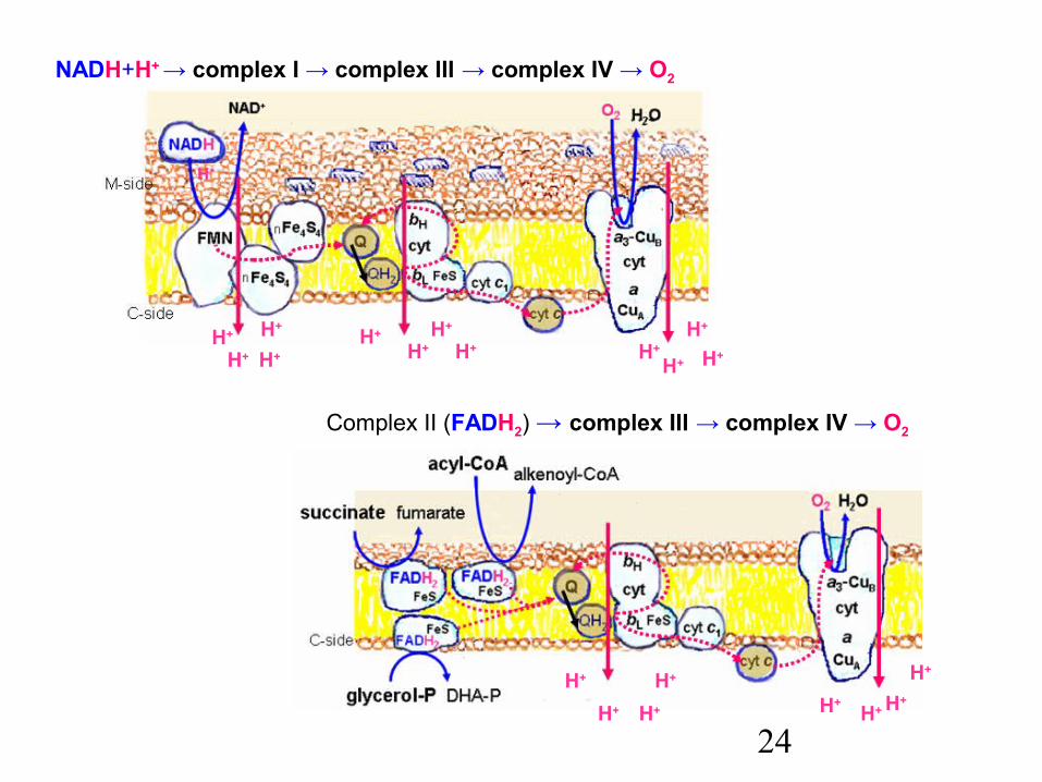
Complex II (FADH2) → complex III → complex IV → O2
NADH+H+ → complex I → complex III → complex IV → O2
H+H+H+H+
H+H+
H+
H+H+H+
H+H+
25
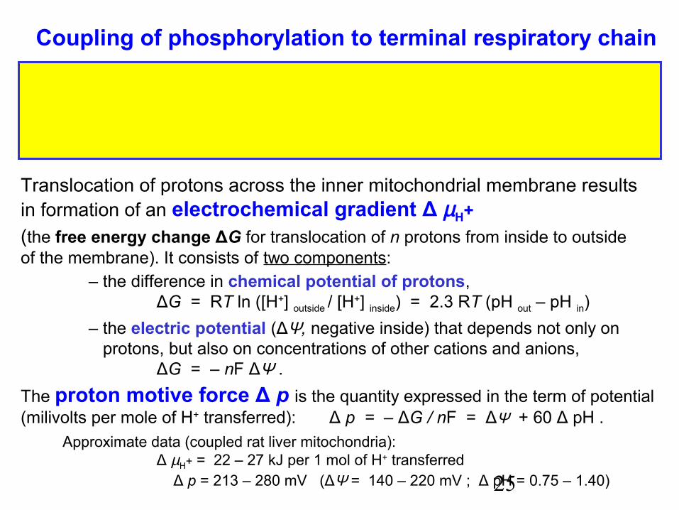
Coupling of phosphorylation to terminal respiratory chain
Oxidative phosphorylation of ADP to ATP consists of two separateprocesses, which are efficiently coupled only in intact mitochondria. Thelink between oxidation and phosphorylation is a proton motive force.
Translocation of protons across the inner mitochondrial membrane resultsin formation of an electrochemical gradient Δ µ H+(the free energy change ΔG for translocation of n protons from inside to outsideof the membrane). It consists of two components:
– the difference in chemical potential of protons,ΔG = RT ln ([H+] outside / [H+] inside) = 2.3 RT (pH out – pH in)
– the electric potential (ΔΨ, negative inside) that depends not only on protons, but also on concentrations of other cations and anions,
ΔG = – nF ΔΨ .The proton motive force Δ p is the quantity expressed in the term of potential(milivolts per mole of H+ transferred): Δ p = – ΔG / nF = ΔΨ + 60 Δ pH . Approximate data (coupled rat liver mitochondria):
Δ µH+ = 22 – 27 kJ per 1 mol of H+ transferred Δ p = 213 – 280 mV (ΔΨ = 140 – 220 mV ; Δ pH = 0.75 – 1.40)
26
H+
H+H+
H+– – –
+ + +
ADP + Pi
ATP
NADH+H+ O2 2H2ONAD+
H+
H+ H+H+
H+H+H+
H+ H+H+
H+H+H+H+
H+H+
H+H+
H+
e–
Terminal respiratory chain
Electrochemical gradient
M-side (negative)
C-side (positive)
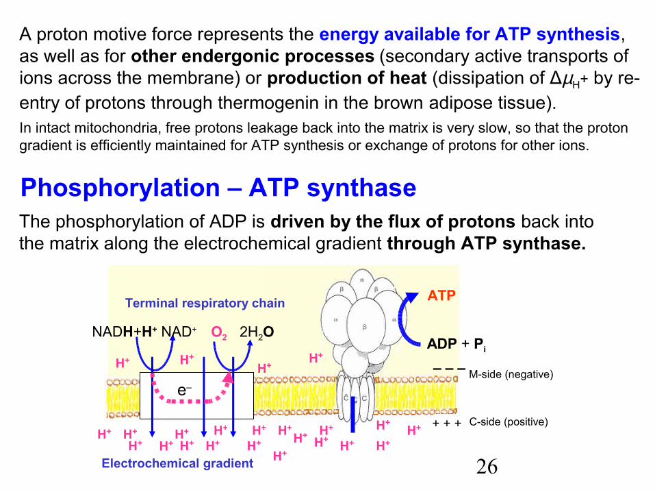
A proton motive force represents the energy available for ATP synthesis,as well as for other endergonic processes (secondary active transports of ions across the membrane) or production of heat (dissipation of ΔµH+ by re-entry of protons through thermogenin in the brown adipose tissue).In intact mitochondria, free protons leakage back into the matrix is very slow, so that the proton gradient is efficiently maintained for ATP synthesis or exchange of protons for other ions.
Phosphorylation – ATP synthaseThe phosphorylation of ADP is driven by the flux of protons back intothe matrix along the electrochemical gradient through ATP synthase.
27
H+
H+H+
H+H+ H+
H+H+
ADP + Pi
ATP
matrix half channel
cytosolic half-channel
subunit a10 subunits c
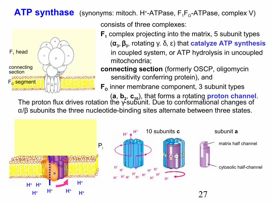
ATP synthase (synonyms: mitoch. H+-ATPase, F1FO-ATPase, complex V)
F1 head
FO segment
connectingsection
consists of three complexes:F1 complex projecting into the matrix, 5 subunit types (α3, β3, rotating γ. δ, ε) that catalyze ATP synthesis
in coupled system, or ATP hydrolysis in uncoupled mitochondria;
connecting section (formerly OSCP, oligomycin sensitivity conferring protein), and
FO inner membrane component, 3 subunit types (a, b2, c10), that forms a rotating proton channel.
The proton flux drives rotation the γ-subunit. Due to conformational changes ofα/β subunits the three nucleotide-binding sites alternate between three states.
28
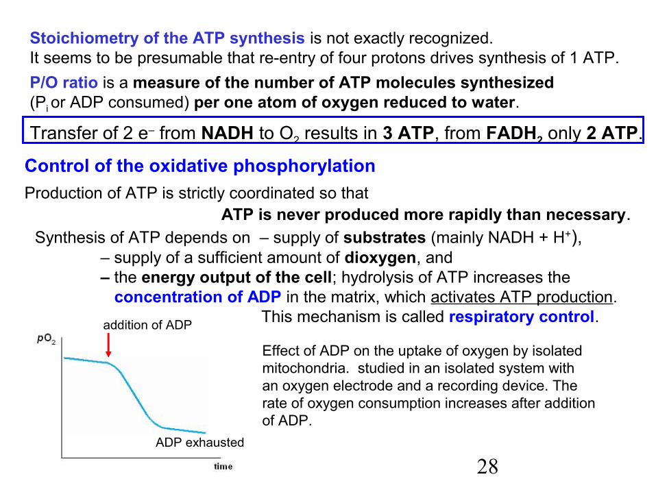
addition of ADP
ADP exhausted
Stoichiometry of the ATP synthesis is not exactly recognized.It seems to be presumable that re-entry of four protons drives synthesis of 1 ATP.P/O ratio is a measure of the number of ATP molecules synthesized (Pi or ADP consumed) per one atom of oxygen reduced to water.
Transfer of 2 e– from NADH to O2 results in 3 ATP, from FADH2 only 2 ATP. Control of the oxidative phosphorylation
Production of ATP is strictly coordinated so thatATP is never produced more rapidly than necessary.
Synthesis of ATP depends on – supply of substrates (mainly NADH + H+),– supply of a sufficient amount of dioxygen, and– the energy output of the cell; hydrolysis of ATP increases the concentration of ADP in the matrix, which activates ATP production. This mechanism is called respiratory control.
Effect of ADP on the uptake of oxygen by isolated mitochondria. studied in an isolated system withan oxygen electrode and a recording device. Therate of oxygen consumption increases after additionof ADP.
29
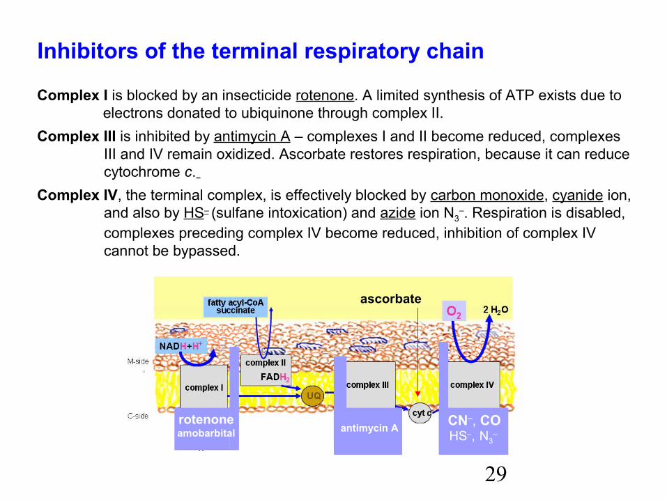
Inhibitors of the terminal respiratory chain
Complex I is blocked by an insecticide rotenone. A limited synthesis of ATP exists due to electrons donated to ubiquinone through complex II.Complex III is inhibited by antimycin A – complexes I and II become reduced, complexes
III and IV remain oxidized. Ascorbate restores respiration, because it can reducecytochrome c.
Complex IV, the terminal complex, is effectively blocked by carbon monoxide, cyanide ion,and also by HS– (sulfane intoxication) and azide ion N3
–. Respiration is disabled,complexes preceding complex IV become reduced, inhibition of complex IV cannot be bypassed.
CN–, COHS–, N3
– rotenoneamobarbital antimycin A
ascorbate
30
occurs after ingestion of alkali cyanides or inhalation of hydrogen cyanide.Bitter almonds or apricot kernels contain amygdalin, which can release HCN.Cyanide ion, besides inhibition of cytochrome c oxidase, binds with high affinity onto methaemoglobin (haemin, Fe3+).The lethal dose LD50 of alkali cyanide is about 250 mg. Symptoms - dizziness, gasping for breath, cramps, and unconsciousness follow rapidly.Antidotes may be effective, when applied without any delay:Hydroxycobalamin (a semisynthetic compound) exhibits high affinity to CN– ions, binds them in the form of harmless cyanocobalamin (B12).Sodium nitrite NaNO2 or amyl nitrite oxidize haemoglobin (FeII) to methaemoglobin (FeIII),
which is not able to transport oxygen, but binds CN– and may so prevent inhibition of cytochrome c oxidase.Sodium thiosulfate Na2S2O3, administered intravenously, can convert cyanide to the relatively harmless thiocyanate ion: CN– + S2O3
2– → SCN– + SO32– .
Cyanide poisoning
Carbon monoxide poisoningCO binds primarily to haemoglobin (FeII) and inhibits oxygen transport, but it alsoblocks the respiratory chain by inhibiting cytochrome oxidase (complex IV).Oxygenotherapy can improve blood oxygen transport, administered methylene blue serves as acceptor of electrons from complex III so that limited ATP synthesis can continue.
31
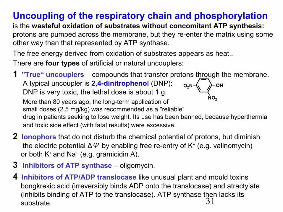
Uncoupling of the respiratory chain and phosphorylation is the wasteful oxidation of substrates without concomitant ATP synthesis:protons are pumped across the membrane, but they re-enter the matrix using some other way than that represented by ATP synthase.The free energy derived from oxidation of substrates appears as heat..
DNP is very toxic, the lethal dose is about 1 g.More than 80 years ago, the long-term application ofsmall doses (2.5 mg/kg) was recommended as a "reliable“ drug in patients seeking to lose weight. Its use has been banned, because hyperthermia and toxic side effect (with fatal results) were excessive.
2 Ionophors that do not disturb the chemical potential of protons, but diminish the electric potential ΔΨ by enabling free re-entry of K+ (e.g. valinomycin) or both K+ and Na+ (e.g. gramicidin A).3 Inhibitors of ATP synthase – oligomycin. 4 Inhibitors of ATP/ADP translocase like unusual plant and mould toxins bongkrekic acid (irreversibly binds ADP onto the translocase) and atractylate (inhibits binding of ATP to the translocase). ATP synthase then lacks its substrate.
There are four types of artificial or natural uncouplers:1 "True“ uncouplers – compounds that transfer protons through the membrane. A typical uncoupler is 2,4-dinitrophenol (DNP):
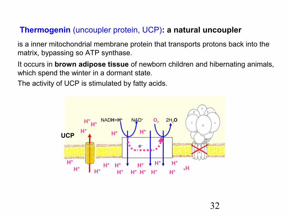
32
is a inner mitochondrial membrane protein that transports protons back into the matrix, bypassing so ATP synthase.It occurs in brown adipose tissue of newborn children and hibernating animals, which spend the winter in a dormant state.The activity of UCP is stimulated by fatty acids.
Thermogenin (uncoupler protein, UCP): a natural uncoupler
H+ H+H+
H+H+
H+
H+H+
H+ H+
NADH+H+ 2H2O
H+
H+
H+
H+
H+
H+
H+
H+
H+
O2NAD+
e–
UCP