Embed Size (px)
Citation preview
LUND UNIVERSITY
PO Box 117221 00 Lund+46 46-222 00 00
Oxidative stress in preeclampsia and the role of free fetal hemoglobin.
Hansson, Stefan; Nääv, Åsa; Erlandsson, Lena
Published in:Frontiers in Physiology
DOI:10.3389/fphys.2014.00516
2015
Link to publication
Citation for published version (APA):Hansson, S., Nääv, Å., & Erlandsson, L. (2015). Oxidative stress in preeclampsia and the role of free fetalhemoglobin. Frontiers in Physiology, 5, [516]. https://doi.org/10.3389/fphys.2014.00516
Total number of authors:3
General rightsUnless other specific re-use rights are stated the following general rights apply:Copyright and moral rights for the publications made accessible in the public portal are retained by the authorsand/or other copyright owners and it is a condition of accessing publications that users recognise and abide by thelegal requirements associated with these rights. • Users may download and print one copy of any publication from the public portal for the purpose of private studyor research. • You may not further distribute the material or use it for any profit-making activity or commercial gain • You may freely distribute the URL identifying the publication in the public portal
Read more about Creative commons licenses: https://creativecommons.org/licenses/Take down policyIf you believe that this document breaches copyright please contact us providing details, and we will removeaccess to the work immediately and investigate your claim.
REVIEW ARTICLEpublished: 13 January 2015
doi: 10.3389/fphys.2014.00516
Oxidative stress in preeclampsia and the role of free fetalhemoglobinStefan R. Hansson*, Åsa Nääv and Lena Erlandsson
Department of Obstetrics and Gynecology, Institute for Clinical Sciences, Lund University, Lund, Sweden
Edited by:
Sruti Shiva, University of Pittsburgh,USA
Reviewed by:
Cameron Rink, The Ohio StateUniversity Wexner Medical Center,USADeepesh Pandey, Georgia HealthSciences University, USA
*Correspondence:
Stefan R. Hansson, Department ofObstetrics and Gynecology, Institutefor Clinical Sciences, LundUniversity, BMC C14, Klinikgatan 28,SE-221 85 Lund, Swedene-mail: [email protected]
Preeclampsia is a leading cause of pregnancy complications and affects 3–7% of pregnantwomen. This review summarizes the current knowledge of a new potential etiology ofthe disease, with a special focus on hemoglobin-induced oxidative stress. Furthermore,we also suggest hemoglobin as a potential target for therapy. Gene and protein profilingstudies have shown increased expression and accumulation of free fetal hemoglobinin the preeclamptic placenta. Predominantly due to oxidative damage to the placentalbarrier, fetal hemoglobin leaks over to the maternal circulation. Free hemoglobin and itsmetabolites are toxic in several ways; (a) ferrous hemoglobin (Fe2+) binds strongly tothe vasodilator nitric oxide (NO) and reduces the availability of free NO, which resultsin vasoconstriction, (b) hemoglobin (Fe2+) with bound oxygen spontaneously generatesfree oxygen radicals, and (c) the heme groups create an inflammatory response byinducing activation of neutrophils and cytokine production. The endogenous proteinα1-microglobulin, with radical and heme binding properties, has shown both ex vivo andin vivo to have the ability to counteract free hemoglobin-induced placental and kidneydamage. Oxidative stress in general, and more specifically fetal hemoglobin-inducedoxidative stress, could play a key role in the pathology of preeclampsia seen both in theplacenta and ultimately in the maternal endothelium.
Keywords: placenta, oxidative stress, fetal hemoglobin, hemolysis, alpha-1-microglobulin
INTRODUCTIONPreeclampsia is a clinical syndrome that manifests during thesecond half of pregnancy, afflicting 3–7% of pregnant women.Preeclampsia is one of the leading causes of maternal mortal-ity and morbidity, especially in developing countries (Mackayet al., 2001; Berg et al., 2009). The disease is clinically mani-fested and defined as de novo hypertension with proteinuria after20 gestational weeks (Redman, 2011). Preeclampsia can developinto a life-threatening condition with general hemolysis, elevatedliver enzymes, low platelet counts, and elevated levels of freeadult hemoglobin (Hb), classified as the HELLP syndrome (i.e.,Hemolysis, Elevated Liver enzymes, Low Platelets) (Schroederet al., 2002). Preeclampsia can further develop into eclampsia,a severe complication that is defined by the presence of seizures(Mackay et al., 2001). Currently, only symptomatic blood pres-sure treatment is available for preeclampsia and the only knowncure to date is delivery. Hence, preeclampsia causes ∼15% ofpre-term deliveries. Also, in 25% of cases, preeclampsia leadsto intrauterine growth restriction (IUGR) of the fetus. Both
Abbreviations: A1M, alpha-1-microglobulin; IUGR, intrauterine growth restric-tion; HELLP, hemolysis, elevated liver enzymes, and low platelets syndrome; SOD,superoxide dismutase; HO, hemoxygenase; O−
2 , superoxide; H2O2, hydrogen per-oxide; ROS, reactive oxygen species; ER, endoplasmatic reticulum; NO, nitric oxide;sFlt1, soluble fms-like tyrosine kinase 1; VEGF, vascular endothelial growth fac-tor; PlGF, placental growth factor; TGFβ, transforming growth factor-beta; sEng,soluble endoglin; RAS, renin-angiotensin system; DIC, disseminated intravascularcoagulation.
pre-term delivery and IUGR result in infant morbidity and sub-stantial health care expenditure (Shmueli et al., 2012). To datethere is neither a diagnostic test nor screening tool available forearly identification of women at risk of preeclampsia.
Several varying hypotheses and theories regarding the etiol-ogy have been put forward over the years. One hypothesis, thatwas suggested about 25 years ago, is that the extensive vascularendothelial dysfunction in preeclampsia is the result of circulatingfactors released from the placenta (Rodgers et al., 1988; Robertset al., 1989). The fact that removal of the placenta is necessary forsymptoms to regress has resulted in the theory that the placentais central in the etiology of preeclampsia (Roberts and Hubel,2009; Roberts and Escudero, 2012). According to the current the-ory, preeclampsia evolves in two stages. In brief, the first stageis characterized by a defective placentation, involving incompleteconversion of the spiral arteries by superficial ingrowth of tro-phoblasts in the decidua (Brosens et al., 2002). As a consequence,uneven blood perfusion, hypoxia, and oxidative stress follow. Thesecond stage of the disease is characterized by the clinical manifes-tations hypertension, proteinuria, and edema, caused by maternalendothelial damage and systemic inflammation. The endothelialdamage is suggested to be caused by placental-derived materialor factors (Roberts et al., 1989; Tjoa et al., 2006; Redman andSargent, 2009).
Despite intense research efforts to unveil the etiology ofpreeclampsia, it remains enigmatic. However, a growing body ofevidence supports the understanding that the disease begins in
www.frontiersin.org January 2015 | Volume 5 | Article 516 | 1
Hansson et al. Fetal hemoglobin in preeclampsia
the utero-placental unit, is amplified by oxidative stress, and endsin the maternal endothelium.
OXIDATIVE STRESSOxidative stress reflects an imbalance between the formationof oxidative substances and the innate antioxidants that makeup the endogenous defense system. Oxidative substances areoften free oxygen radicals and peroxides that normally form insmall amounts (Buonocore et al., 2010). They can be formedin e.g., the mitochondrial respiratory chain, and when the tis-sue is exposed to ischemia/reperfusion injury they are producedin larger amounts. Due to their highly reactive properties, theycan cause structural and functional damage to cellular DNA, pro-teins and cell membranes (Tjoa et al., 2006; Valko et al., 2007).In addition to oxidative stress, nitrative stress has also been doc-umented in preeclampsia, which is the covalent modification andnitration of proteins and DNA by peroxynitrite. This occurs inthe preeclamptic placenta by the release of reactive oxygen species(ROS) such as superoxide, which interacts with nitric oxide (NO)to form peroxynitrite (Myatt, 2010).
Antioxidants are molecules that inhibit the oxidation causedby oxidative substances, and they are made up of two sub-groups;enzymatic and non-enzymatic antioxidants. Examples of enzy-matic antioxidants are superoxide dismutase (SOD), hemoxyge-nase (HO), and catalase. SOD’s enzymatic activity catalyzes thedismutation of superoxide (O−
2 ) into oxygen and hydrogen per-oxide (H2O2) (McCord and Fridovich, 1988). Thus, SOD is animportant antioxidant defense in nearly all cells exposed to oxy-gen. Three isoforms of HO are known, where HO-1 is induciblein response to oxidative stress and catalyzes the degradationof heme. This produces biliverdin, iron, and carbon monoxide(Kikuchi et al., 2005; Ryter et al., 2006). Catalase is a commonenzyme found in nearly all living organisms exposed to oxy-gen. It catalyzes the decomposition of H2O2 to water and oxygen(Chelikani et al., 2004).
Examples of non-enzymatic antioxidants are glutathione,thioredoxin, NADH, and NADPH. Glutathione is a key endoge-nous antioxidant that serves several functions, such as neutraliza-tion of free radicals and ROS (Pompella et al., 2003). In addition,glutathione also plays a role in keeping vitamins C and E in theirreduced forms (Hughes, 1964; Scholz et al., 1989). Thioredoxinis an oxidoreductase enzyme containing a dithiol-disulfide activesite that facilitates the reduction of other proteins by cysteinethiol-disulfide exchange (Holmgren, 1989; Nordberg and Arner,2001). NADH is a coenzyme found in all living cells, and ithas a role in metabolism where it is involved in redox reactions(Belenky et al., 2007). NADPH is, among other things, involvedin the protection against ROS by allowing for the regenerationof reduced glutathione (Rush et al., 1985). The NADPH systemis also responsible for generating free radicals in neutrophils,which is used in the destruction of pathogens (Ogawa et al.,2008).
α1-microglobulin (A1M) is both an enzymatic and a non-enzymatic antioxidant, present both intra- and extracellularly(Akerstrom and Gram, 2014). It has it’s defensive function byworking as an antioxidant as well as a binding protein for freeradicals and heme (Allhorn et al., 2002; Olsson et al., 2008).
A1M is mainly produced in the liver, but can be found in allorgans. Once produced, it is deployed to the extracellular flu-ids and compartments via the blood stream. The synthesis ofA1M is up-regulated in response to elevated levels of free rad-icals, heme, and in particular by free Hb (Olsson et al., 2007).A1M levels are seen to increase in the preeclamptic placenta in aneffort by the placenta to up-regulate its defense against increasedoxidative stress (Olsson et al., 2010). The protein prevents oxi-dation and oxidative damage to cells and matrix molecules, andremoves heme from cell membranes and cytosol (Olsson et al.,2008).
OXIDATIVE STRESS AND PREECLAMPSIAIn preeclampsia, evidence of oxidative stress can be seen bothin the maternal circulation and in the placenta. Placentas fromwomen with preeclampsia have reduced antioxidant capacitycompared to normal placentas (Wang and Walsh, 1996; Walsh,1998). Furthermore, levels of antioxidants in blood from womenwith preeclampsia have been shown to be reduced, as well as lev-els of oxidative modifications of proteins and lipoprotein particles(Hubel, 1999; Raijmakers et al., 2004).
Placental trophoblasts and endothelial cells constitute the pla-cental barrier, which effectively separates the fetal and maternalcirculation. Oxidative stress and tissue damage is suggested tocause a breach in the barrier and create a leakage of fetal andplacenta-derived factors and/or material into the maternal cir-culation. In stage two of the disease the shedding of placentaldebris leads to maternal endothelial damage, elevated oxidativestress, and systemic inflammation (Smarason et al., 1993; Knightet al., 1998; Hahn and Holzgreve, 2002; Tjoa et al., 2006). Anadditional cause to inflammation and vascular damage is theshedding of microparticles from the placenta, induced by placen-tal abnormalities and utero-placental ischemia. The content of themicroparticles, such as free Hb and miRNA can further aggra-vate the systemic oxidative stress (Redman and Sargent, 2008;Tannetta et al., 2013; Cronqvist et al., 2014; Rudov et al., 2014).
Women living at high altitude alter the normal oxygen balanceand are shown to have increased risk of developing preeclamp-sia (Palmer et al., 1999). The effect of oxidative stress on femalereproduction has been explored in relation to infertility andassisted reproductive techniques (Agarwal et al., 2012). Pregnancycomplications such as recurrent pregnancy loss and spontaneousabortion can develop in response to oxidative stress (Jauniauxet al., 2000; Poston and Raijmakers, 2004). Maternal obesity hasalso been associated with an increased risk of preeclampsia (Wolfet al., 2001), which could be partly mediated by the increasedlevel of oxidative stress present in obesity (Zavalza-Gomez, 2011).Smoking causes oxidative stress and is an established risk fac-tor for cardio-vascular disease. In pregnant women, smokingcauses increased risk of IUGR. However, smoking has also beenassociated with a protective effect against preeclampsia (Conde-Agudelo et al., 1999). A suggested explanation could be thatthe observed lower level of sFlt1 in smokers compared to non-smokers is due to the suppression of sFlt1 by carbon monoxide(Karumanchi and Levine, 2010). This is in line with the elevatedlevels of sFlt1 observed in women with preeclampsia (Levine et al.,2006).
Frontiers in Physiology | Oxidant Physiology January 2015 | Volume 5 | Article 516 | 2
Hansson et al. Fetal hemoglobin in preeclampsia
OXIDATIVE STRESS CAUSES CELLULAR AND SYSTEMICEFFECTSCELLULAR LEVELThe degree to which the implantation and the conversion of thespiral arteries are deficient has been postulated to influence thedegree of oxidative stress and endoplasmic reticulum (ER) stress.Oxidative stress disturbs the normal redox state of the cell, whichcauses toxic effects on all cellular components, including proteins,lipids, and DNA, and more severe oxidative stress can result incell death (Valko et al., 2007). During ER stress, unfolded andmisfolded proteins accumulate in the ER and activate ER stress-response pathways, also known as the unfolded protein response(Yung et al., 2014). Preeclamptic placentas have been shown toaccumulate clusters of misfolded proteins and it is speculated thatthese aggregates may contribute to the pathophysiology of the dis-ease. The misfolded proteins can also be detected in urine, as apotential biomarker by the Congo red dot test (Buhimschi et al.,2014). ER stress has been shown to be associated with the smallplacental phenotype in both preeclampsia and IUGR (Burtonet al., 2009).
SYSTEMIC LEVELInflammation in response to oxidative stress is mediated bythe recruitment of leukocytes and the production of pro-inflammatory cytokines, adhesion molecules (ICAM-1 andVCAM-1), and chemokines. This cascade of events is precededby the presence of heme, which causes production of ROS(Lavrovsky et al., 1994; Markus et al., 2007). Heme has also beenshown to induce inflammation via the Toll-like receptors (Belcheret al., 2014).
Endothelial function is affected by the oxidative burden.Superoxide inactivates NO, leading to an increase in peroxyni-trite as well as a reduced bioactivity of NO. Furthermore, freeHb can also bind NO (Kim-Shapiro et al., 2006). The deple-tion of NO results in a series of downstream events, suchas impaired endothelium-dependent vasodilation or vasocon-striction, lipoprotein accumulation and oxidative modification,intravascular inflammation and release of pro-inflammatory andvasoactive mediators (Motterlini et al., 1995; Madamanchi et al.,2005). The contents of placental microparticles released into thematernal circulation during preeclampsia also contributes to theelevated level of systemic oxidative stress (Redman and Sargent,2008; Tannetta et al., 2013; Cronqvist et al., 2014; Rudov et al.,2014).
Primipara women have an increased risk of preeclampsia whilepre-conception exposure to paternal antigen gives a reduced risk,implicating an immunological component in the etiology of thesyndrome. During the first stage of preeclampsia, maternal adap-tation and tolerance to fetal antigen has been suggested to becrucial (Redman and Sargent, 2010).
ANTIOXIDANT PROTECTIVE SYSTEMSThe human body relies on several antioxidant protective systems.Uric acid is a powerful antioxidant in the human plasma andis a scavenger of free oxygen and radicals. It reduces the oxo-heme oxidant formed by peroxide reaction with Hb and protectserythrocytes from peroxidative damage leading to lysis (Ames
et al., 1981). The uric acid level both predicts and correlates withthe development of conditions associated with oxidative stress(Sautin and Johnson, 2008) and is suggested to be a marker foroxidative stress (Becker, 1993). Therefore, the concentration ofserum uric acid in pregnant women with preeclampsia has beensuggested to be associated with disease severity (Pereira et al.,2014).
Vitamins C and E are described as antioxidants against oxida-tive stress, and their effect on the reduced antioxidant capacity inplacentas from women with preeclampsia has been tested in largerandomized trials (Poston et al., 2006; Rumbold et al., 2006; Villaret al., 2009; Conde-Agudelo et al., 2011). However, supplementa-tion with vitamins C and E during pregnancy could not be shownto reduce the risk of preeclampsia (Spinnato, 2006).
The compound magnesium sulfate (MgSO4) is used as a drugto treat and prevent seizures in severe preeclampsia and eclamp-sia (Sibai, 2005; Kassie et al., 2014). It has been shown to havea mild vasodilator effect in the maternal circulation (Sontia andTouyz, 2007; Souza et al., 2010) as well as a protective effect onendothelial cells (Kharitonova et al., 2015). It has also been shownto restore cerebral oxygenation impairment seen in patients withsevere preeclampsia (Guerci et al., 2014). The exact mechanismof action for MgSO4 is not yet understood, but a role as anantioxidant has recently been suggested.
From a clinical perspective it would be of great interest to gaina deeper understanding of the impact of environmental pollu-tants as well as the influence of nutrients and micronutrients onthe oxidative balance in pregnancy in general and preeclampsiain particular (Mistry and Williams, 2011). Exposure to air pollu-tion, especially to ultra-fine particles has been shown to induceoxidative stress (Terzano et al., 2010). Furthermore, a positiveassociation between preeclampsia and exposure to air pollutants,such as nitrogen oxides, nitrogen monoxide, nitrogen dioxide,carbon monoxide, ozone, particulate matter of <2.5 μm andparticulate matter of <10 μm, has been shown in recent stud-ies (Wu et al., 2009, 2011; Lee et al., 2013; Malmqvist et al.,2013).
Deficiency of micronutrients such as selenium, copper(Cu), zinc (Zn), and manganese (Mn) during pregnancy canaffect antioxidant capacities in the mother and contribute topoor pregnancy outcomes e.g., preeclampsia (Rumbold et al.,2008). The antioxidant defenses found in placenta includes theselenium-dependent glutathione peroxidases, thioredoxin reduc-tases, selenoprotein-P, and Cu/Zn and Mn SOD’s (Wang andWalsh, 1996; Vanderlelie et al., 2005; Rayman, 2009). Studieshave shown a correlation between low serum selenium con-centrations and reduced antioxidant function in preeclampsia,suggesting that selenium status is important. This is further sup-ported by placebo-controlled randomized controls trials wherepregnant women were given selenium supplementation, result-ing in lower incidence of pregnancy-induced hypertension and/orpreeclampsia (Han and Zhou, 1994; Rumiris et al., 2006). Cuis an essential cofactor for several important enzymes includ-ing catalase and SOD (Gambling et al., 2008). Cu is a redox-active transition metal with pro-oxidant properties and cancatalyze the formation of free radicals, thereby inducing oxida-tive stress in preeclampsia (Serdar et al., 2006). However, when
www.frontiersin.org January 2015 | Volume 5 | Article 516 | 3
Hansson et al. Fetal hemoglobin in preeclampsia
Cu is associated in the Cu/Zn SOD it functions as an antiox-idant expressed in both maternal and fetal tissue (Ali Akbaret al., 1998). Studies have shown increased levels of Cu inplacenta, maternal serum, and amniotic fluid in preeclamp-tic women compared to controls (Brophy et al., 1985; Dawsonet al., 1999; Serdar et al., 2006). However, data on Cu statusthroughout normal pregnancies as well as early preeclampticpregnancies is inadequate, and it remains unclear whether Cudeficiency is a public health problem (Arredondo and Nunez,2005).
Zn has antioxidant functions through the Cu/Zn SOD(Izquierdo Alvarez et al., 2007). Zn supplementation studiesshow reduced incidence of pregnancy induced hypertension(Goldenberg et al., 1995), and Zn deficiency has been associ-ated with preeclampsia with decreased levels in placental tissueand maternal serum (Brophy et al., 1985; Dawson et al., 1999;Kumru et al., 2003). Mn is also a cofactor for important enzymes,such as the antioxidant Mn SOD which is involved in protectionagainst oxidative stress in the placenta (Ademuyiwa et al., 2007).Studies have indicated that whole blood Mn concentrations arelower in women with fetal growth restriction (Vigeh et al., 2008).Furthermore, umbilical cord whole blood in preeclampsia hasbeen shown to have reduced levels of Mn (Jones et al., 2010).
Dietary nitrate, in e.g., beetroot juice, is converted in vivo tobioactive nitrogen oxides such as NO and may be used as nitratesupplementation (Lundberg et al., 2008). It has been shown toreduce blood pressure, increase blood flow, and modulate oxida-tive stress (Webb et al., 2008; Carlstrom et al., 2011; Lundberget al., 2011). Beetroot juice has recently been shown to improveplacental vascular function during pregnancy in mice (Cottrellet al., 2014).
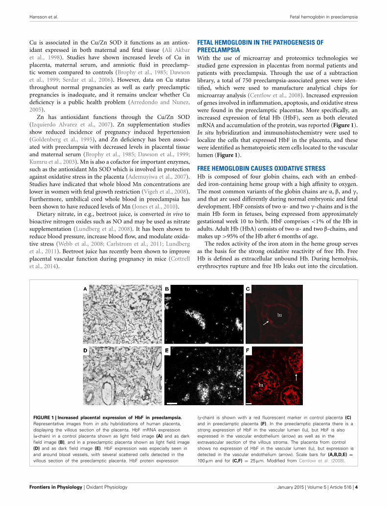
FETAL HEMOGLOBIN IN THE PATHOGENESIS OFPREECLAMPSIAWith the use of microarray and proteomics technologies westudied gene expression in placentas from normal patients andpatients with preeclampsia. Through the use of a subtractionlibrary, a total of 750 preeclampsia-associated genes were iden-tified, which were used to manufacture analytical chips formicroarray analysis (Centlow et al., 2008). Increased expressionof genes involved in inflammation, apoptosis, and oxidative stresswere found in the preeclamptic placentas. More specifically, anincreased expression of fetal Hb (HbF), seen as both elevatedmRNA and accumulation of the protein, was reported (Figure 1).In situ hybridization and immunohistochemistry were used tolocalize the cells that expressed HbF in the placenta, and thesewere identified as hematopoietic stem cells located to the vascularlumen (Figure 1).
FREE HEMOGLOBIN CAUSES OXIDATIVE STRESSHb is composed of four globin chains, each with an embed-ded iron-containing heme group with a high affinity to oxygen.The most common variants of the globin chains are α, β, and γ,and that are used differently during normal embryonic and fetaldevelopment. HbF consists of two α- and two γ-chains and is themain Hb form in fetuses, being expressed from approximatelygestational week 10 to birth. HbF comprises <1% of the Hb inadults. Adult Hb (HbA) consists of two α- and two β-chains, andmakes up >95% of the Hb after 6 months of age.
The redox activity of the iron atom in the heme group servesas the basis for the strong oxidative reactivity of free Hb. FreeHb is defined as extracellular unbound Hb. During hemolysis,erythrocytes rupture and free Hb leaks out into the circulation.
FIGURE 1 | Increased placental expression of HbF in preeclampsia.
Representative images from in situ hybridizations of human placenta,displaying the villous section of the placenta. HbF mRNA expression(α-chain) in a control placenta shown as light field image (A) and as darkfield image (B), and in a preeclamptic placenta shown as light field image(D) and as dark field image (E). HbF expression was especially seen inand around blood vessels, with several scattered cells detected in thevillous section of the preeclamptic placenta. HbF protein expression
(γ-chain) is shown with a red fluorescent marker in control placenta (C)
and in preeclamptic placenta (F). In the preeclamptic placenta there is astrong expression of HbF in the vascular lumen (lu), but HbF is alsoexpressed in the vascular endothelium (arrow) as well as in theextravascular section of the villous stroma. The placenta from controlshows no expression of HbF in the vascular lumen (lu), but expression isdetected in the vascular endothelium (arrow). Scale bars for (A,B,D,E) =100 μm and for (C,F) = 25 μm. Modified from Centlow et al. (2008).
Frontiers in Physiology | Oxidant Physiology January 2015 | Volume 5 | Article 516 | 4

Hansson et al. Fetal hemoglobin in preeclampsia
Eryptosis is a suicidal erythrocyte death, stimulated by oxidativestress, energy depletion, calcium imbalance, and a wide range ofxenobiotic compounds. It is inhibited by erythropoietin and NO(Lang et al., 2012). Eryptosis affects the capillary beds by foster-ing thrombosis. It is seen in several systemic conditions such asdiabetes, renal insufficiency and malaria, conditions well-knownas risk factors for preeclampsia.
The vasoconstrictive effect of free Hb is a result of the fer-rous Hb (Fe2+) binding strongly to the vasodilator NO, therebyreducing the availability of NO. Currently, NO donors arebeing investigated in clinical trials as therapeutics for IUGR andpreeclampsia (Cindrova-Davies, 2014). Hb (Fe2+) with boundoxygen (OxyHb) spontaneously generates free oxygen radicals.This results in aggregated and oxidized forms of the molecule,degradation products, and free iron and heme groups. As a result,Hb and its degradation products are toxic and can cause oxidativestress, hemolysis, vasoconstriction, kidney, and vascular endothe-lial damage (Buehler and D’Agnillo, 2010). Heme groups have adirect effect on the inflammatory response by inducing activationof neutrophils and induction of cytokine production (Kumar andBandyopadhyay, 2005).
Several defense mechanisms exist to protect against the harm-ful effects of free Hb. Antioxidants such as vitamins C and E pro-tect against oxidative stress. The blood plasma scavenger proteinshaptoglobin and hemopexin bind free Hb and free heme groups,respectively, and the formed complexes are eliminated from theblood by cellular uptake via the two receptor-mediated pathwaysCD163 on macrophages and CD91 in the liver (Kristiansen et al.,2001; Ascenzi et al., 2005). In fact, haptoglobin levels are reducedin preeclampsia, indicating a depletion of the protective systemsagainst free Hb (Olsson et al., 2010).
FREE HEMOGLOBIN CAUSES PLACENTAL DAMAGERecently our group has shown that free Hb is a potential key fac-tor in the pathogenesis of preeclampsia. By aggravating oxidativestress, causing damage to the placental barrier and leaking into thematernal bloodstream, it causes endothelial damage and vasocon-striction (Centlow et al., 2008; May et al., 2011). To study the toxiceffect of free Hb on placental tissue, the placenta perfusion modelwas used. This well-documented ex vivo system mimics the preg-nant in vivo situation in humans in a way not possible by usingplacenta cell culture or animal models (Schneider and Huch,
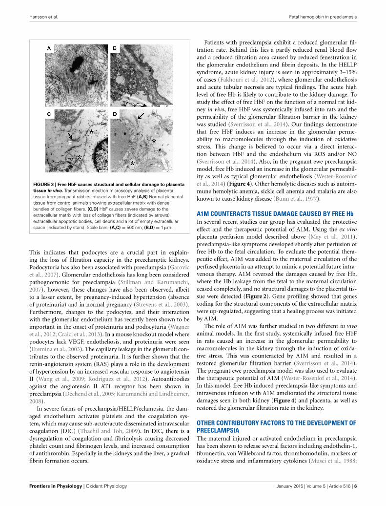
1985; Di Santo et al., 2007). In this system, free Hb was addedto the fetal circulation. A preeclampsia-like situation developedwithin 10 min, displaying a steep increase in blood pressure. Signsof placental barrier breach appeared after ∼1 h of perfusion, withleakage of nutrients from the fetal circulation into the maternalcirculation. Transmission electron microscopy (TEM) confirmedthe tissue damage with considerable damage to the extracellularmatrix, with an almost complete elimination of the collagen fib-rils that maintain the tissue structure (Figure 2). Further, therewere widespread cellular changes with damage to membranes,nuclei and mitochondria, and formation of apoptotic vesiclesand vacuoles. Gene profiling of placentas perfused with free Hbshowed a similar genetic profile as women with preeclampsia.These ex vivo findings suggest that free Hb plays an importantrole in the disease etiology (Centlow et al., 2009; May et al.,2011). To further study the effects of free HbF, we established apregnant rabbit preeclampsia model where pregnant rabbits wereinfused with species-specific free HbF, starting at mid-gestationuntil term. TEM analysis of the placenta revealed structural dam-ages with a dramatic reduction of the collagen fibers in theextracellular matrix, as well as cellular damages such as mito-chondrial swelling, aberrant intracellular nuclear membranes,damaged electron dense barrier, and high levels of apoptoticbodies (Figure 3). In the pregnant ewe preeclampsia model, star-vation induces preeclampsia-like symptoms by causing hemolysiswith subsequent release of free Hb (Wester-Rosenlof et al., 2014).Also in this model TEM analysis could confirm tissue damage inplacentas from starved ewes, with an almost complete eliminationof the collagen fibers as well as cellular damages (Wester-Rosenlofet al., 2014).
FREE HEMOGLOBIN CAUSES KIDNEY DAMAGEThe proteinuria in preeclampsia is associated with glomeru-lar injuries in the kidney, known as glomerular endotheliosis.Endotheliosis includes swelling of endothelial cells and loss ofendothelial fenestration, as well as occlusion of capillary lumensin some cases (Lafayette et al., 1998; Stillman and Karumanchi,2007). Podocytes form the slit diaphragm, which is crucial formaintaining the size-selective nature of the glomerular filtra-tion barrier in the kidney, and alterations in the podocyteshave been associated with proteinuria in preeclampsia (Garovicet al., 2007; Henao et al., 2008; Henao and Saleem, 2013).
FIGURE 2 | Free Hb causes placental damage ex vivo which can be
ameliorated by A1M. Transmission electron microscopy analysis ofex vivo perfused human placenta. (A) Non-perfused human placenta.(B) Free Hb was added to the fetal circulation and caused severedamage to the extracellular matrix with an almost complete
elimination of the collagen fibers. (C) A1M was added to thematernal circulation at the same time as Hb was added to the fetalcirculation, and prevented the damaging effects of Hb on theextracellular matrix, displaying normal collagen fibers. Scale bars:200 μm. Adapted from May et al. (2011).
www.frontiersin.org January 2015 | Volume 5 | Article 516 | 5
Hansson et al. Fetal hemoglobin in preeclampsia
FIGURE 3 | Free HbF causes structural and cellular damage to placenta
tissue in vivo. Transmission electron microscopy analysis of placentatissue from pregnant rabbits infused with free HbF. (A,B) Normal placentaltissue from control animals showing extracellular matrix with densebundles of collagen fibers. (C,D) HbF causes severe damage to theextracellular matrix with loss of collagen fibers (indicated by arrows),extracellular apoptotic bodies, cell debris and a lot of empty extracellularspace (indicated by stars). Scale bars: (A,C) = 500 nm; (B,D) = 1 μm.
This indicates that podocytes are a crucial part in explain-ing the loss of filtration capacity in the preeclamptic kidneys.Podocyturia has also been associated with preeclampsia (Garovicet al., 2007). Glomerular endotheliosis has long been consideredpathognomonic for preeclampsia (Stillman and Karumanchi,2007), however, these changes have also been observed, albeitto a lesser extent, by pregnancy-induced hypertension (absenceof proteinuria) and in normal pregnancy (Strevens et al., 2003).Furthermore, changes to the podocytes, and their interactionwith the glomerular endothelium has recently been shown to beimportant in the onset of proteinuria and podocyturia (Wagneret al., 2012; Craici et al., 2013). In a mouse knockout model wherepodocytes lack VEGF, endotheliosis, and proteinuria were seen(Eremina et al., 2003). The capillary leakage in the glomeruli con-tributes to the observed proteinuria. It is further shown that therenin-angiotensin system (RAS) plays a role in the developmentof hypertension by an increased vascular response to angiotensinII (Wang et al., 2009; Rodriguez et al., 2012). Autoantibodiesagainst the angiotensin II AT1 receptor has been shown inpreeclampsia (Dechend et al., 2005; Karumanchi and Lindheimer,2008).
In severe forms of preeclampsia/HELLP/eclampsia, the dam-aged endothelium activates platelets and the coagulation sys-tem, which may cause sub-acute/acute disseminated intravascularcoagulation (DIC) (Thachil and Toh, 2009). In DIC, there is adysregulation of coagulation and fibrinolysis causing decreasedplatelet count and fibrinogen levels, and increased consumptionof antithrombin. Especially in the kidneys and the liver, a gradualfibrin formation occurs.
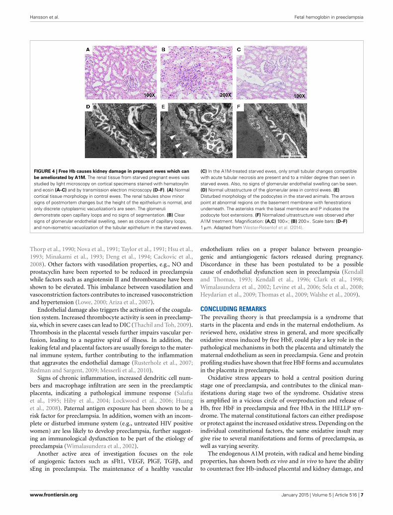
Patients with preeclampsia exhibit a reduced glomerular fil-tration rate. Behind this lies a partly reduced renal blood flowand a reduced filtration area caused by reduced fenestration inthe glomerular endothelium and fibrin deposits. In the HELLPsyndrome, acute kidney injury is seen in approximately 3–15%of cases (Fakhouri et al., 2012), where glomerular endotheliosisand acute tubular necrosis are typical findings. The acute highlevel of free Hb is likely to contribute to the kidney damage. Tostudy the effect of free HbF on the function of a normal rat kid-ney in vivo, free HbF was systemically infused into rats and thepermeability of the glomerular filtration barrier in the kidneywas studied (Sverrisson et al., 2014). Our findings demonstratethat free HbF induces an increase in the glomerular perme-ability to macromolecules through the induction of oxidativestress. This change is believed to occur via a direct interac-tion between HbF and the endothelium via ROS and/or NO(Sverrisson et al., 2014). Also, in the pregnant ewe preeclampsiamodel, free Hb induced an increase in the glomerular permeabil-ity as well as typical glomerular endotheliosis (Wester-Rosenlofet al., 2014) (Figure 4). Other hemolytic diseases such as autoim-mune hemolytic anemia, sickle cell anemia and malaria are alsoknown to cause kidney disease (Bunn et al., 1977).
A1M COUNTERACTS TISSUE DAMAGE CAUSED BY FREE HbIn several recent studies our group has evaluated the protectiveeffect and the therapeutic potential of A1M. Using the ex vivoplacenta perfusion model described above (May et al., 2011),preeclampsia-like symptoms developed shortly after perfusion offree Hb to the fetal circulation. To evaluate the potential thera-peutic effect, A1M was added to the maternal circulation of theperfused placenta in an attempt to mimic a potential future intra-venous therapy. A1M reversed the damages caused by free Hb,where the Hb leakage from the fetal to the maternal circulationceased completely, and no structural damages to the placental tis-sue were detected (Figure 2). Gene profiling showed that genescoding for the structural components of the extracellular matrixwere up-regulated, suggesting that a healing process was initiatedby A1M.
The role of A1M was further studied in two different in vivoanimal models. In the first study, systemically infused free HbFin rats caused an increase in the glomerular permeability tomacromolecules in the kidney through the induction of oxida-tive stress. This was counteracted by A1M and resulted in arestored glomerular filtration barrier (Sverrisson et al., 2014).The pregnant ewe preeclampsia model was also used to evaluatethe therapeutic potential of A1M (Wester-Rosenlof et al., 2014).In this model, free Hb induced preeclampsia-like symptoms andintravenous infusion with A1M ameliorated the structural tissuedamages seen in both kidney (Figure 4) and placenta, as well asrestored the glomerular filtration rate in the kidney.
OTHER CONTRIBUTORY FACTORS TO THE DEVELOPMENT OFPREECLAMPSIAThe maternal injured or activated endothelium in preeclampsiahas been shown to release several factors including endothelin-1,fibronectin, von Willebrand factor, thrombomodulin, markers ofoxidative stress and inflammatory cytokines (Musci et al., 1988;
Frontiers in Physiology | Oxidant Physiology January 2015 | Volume 5 | Article 516 | 6
Hansson et al. Fetal hemoglobin in preeclampsia
FIGURE 4 | Free Hb causes kidney damage in pregnant ewes which can
be ameliorated by A1M. The renal tissue from starved pregnant ewes wasstudied by light microscopy on cortical specimens stained with hematoxylinand eosin (A–C) and by transmission electron microscopy (D–F). (A) Normalcortical tissue morphology in control ewes. The renal tubules show minorsigns of postmortem changes but the height of the epithelium is normal, andonly discrete cytoplasmic vacuolization’s are seen. The glomerulidemonstrate open capillary loops and no signs of segmentation. (B) Clearsigns of glomerular endothelial swelling, seen as closure of capillary loops,and non-isometric vacuolization of the tubular epithelium in the starved ewes.
(C) In the A1M-treated starved ewes, only small tubular changes compatiblewith acute tubular necrosis are present and to a milder degree than seen instarved ewes. Also, no signs of glomerular endothelial swelling can be seen.(D) Normal ultrastructure of the glomerular area in control ewes. (E)
Disturbed morphology of the podocytes in the starved animals. The arrowspoint at abnormal regions on the basement membrane with fenestrationsunderneath. The asterisks mark the basal membrane and P indicates thepodocyte foot extensions. (F) Normalized ultrastructure was observed afterA1M treatment. Magnification: (A,C) 100×; (B) 200×. Scale bars: (D–F)
1 μm. Adapted from Wester-Rosenlof et al. (2014).
Thorp et al., 1990; Nova et al., 1991; Taylor et al., 1991; Hsu et al.,1993; Minakami et al., 1993; Deng et al., 1994; Cackovic et al.,2008). Other factors with vasodilation properties, e.g., NO andprostacyclin have been reported to be reduced in preeclampsiawhile factors such as angiotensin II and thromboxane have beenshown to be elevated. This imbalance between vasodilation andvasoconstriction factors contributes to increased vasoconstrictionand hypertension (Lowe, 2000; Ariza et al., 2007).
Endothelial damage also triggers the activation of the coagula-tion system. Increased thrombocyte activity is seen in preeclamp-sia, which in severe cases can lead to DIC (Thachil and Toh, 2009).Thrombosis in the placental vessels further impairs vascular per-fusion, leading to a negative spiral of illness. In addition, theleaking fetal and placental factors are usually foreign to the mater-nal immune system, further contributing to the inflammationthat aggravates the endothelial damage (Rusterholz et al., 2007;Redman and Sargent, 2009; Messerli et al., 2010).
Signs of chronic inflammation, increased dendritic cell num-bers and macrophage infiltration are seen in the preeclampticplacenta, indicating a pathological immune response (Salafiaet al., 1995; Hiby et al., 2004; Lockwood et al., 2006; Huanget al., 2008). Paternal antigen exposure has been shown to be arisk factor for preeclampsia. In addition, women with an incom-plete or disturbed immune system (e.g., untreated HIV positivewomen) are less likely to develop preeclampsia, further suggest-ing an immunological dysfunction to be part of the etiology ofpreeclampsia (Wimalasundera et al., 2002).
Another active area of investigation focuses on the roleof angiogenic factors such as sFlt1, VEGF, PlGF, TGFβ, andsEng in preeclampsia. The maintenance of a healthy vascular
endothelium relies on a proper balance between proangio-genic and antiangiogenic factors released during pregnancy.Discordance in these has been postulated to be a possiblecause of endothelial dysfunction seen in preeclampsia (Kendalland Thomas, 1993; Kendall et al., 1996; Clark et al., 1998;Wimalasundera et al., 2002; Levine et al., 2006; Sela et al., 2008;Heydarian et al., 2009; Thomas et al., 2009; Walshe et al., 2009).
CONCLUDING REMARKSThe prevailing theory is that preeclampsia is a syndrome thatstarts in the placenta and ends in the maternal endothelium. Asreviewed here, oxidative stress in general, and more specificallyoxidative stress induced by free HbF, could play a key role in thepathological mechanisms in both the placenta and ultimately thematernal endothelium as seen in preeclampsia. Gene and proteinprofiling studies have shown that free HbF forms and accumulatesin the placenta in preeclampsia.
Oxidative stress appears to hold a central position duringstage one of preeclampsia, and contributes to the clinical man-ifestations during stage two of the syndrome. Oxidative stressis amplified in a vicious circle of overproduction and release ofHb, free HbF in preeclampsia and free HbA in the HELLP syn-drome. The maternal constitutional factors can either predisposeor protect against the increased oxidative stress. Depending on theindividual constitutional factors, the same oxidative insult maygive rise to several manifestations and forms of preeclampsia, aswell as varying severity.
The endogenous A1M protein, with radical and heme bindingproperties, has shown both ex vivo and in vivo to have the abilityto counteract free Hb-induced placental and kidney damage, and
www.frontiersin.org January 2015 | Volume 5 | Article 516 | 7

Hansson et al. Fetal hemoglobin in preeclampsia
to restore both placental barrier and glomerular filtration barrierfunctions.
REFERENCESAdemuyiwa, O., Odusoga, O. L., Adebawo, O. O., and Ugbaja, R. (2007).
Endogenous antioxidant defences in plasma and erythrocytes of pregnantwomen during different trimesters of pregnancy. Acta Obstet. Gynecol. Scand.86, 1175–1182. doi: 10.1080/00016340701515357
Agarwal, A., Aponte-Mellado, A., Premkumar, B. J., Shaman, A., and Gupta, S.(2012). The effects of oxidative stress on female reproduction: a review. Reprod.Biol. Endocrinol. 10:49. doi: 10.1186/1477-7827-10-49
Akerstrom, B., and Gram, M. (2014). A1M, an extravascular tissue clean-ing and housekeeping protein. Free Radic. Biol. Med. 74C, 274–282. doi:10.1016/j.freeradbiomed.2014.06.025
Ali Akbar, S., Nicolaides, K. H., and Brown, P. R. (1998). Measurement of Cu/ZnSOD in placenta, cultured cells, various fetal tissues, decidua and semen byELISA. J. Obstet. Gynaecol. 18, 331–335. doi: 10.1080/01443619867056
Allhorn, M., Berggard, T., Nordberg, J., Olsson, M. L., and Akerstrom, B. (2002).Processing of the lipocalin alpha(1)-microglobulin by hemoglobin inducesheme-binding and heme-degradation properties. Blood 99, 1894–1901. doi:10.1182/blood.V99.6.1894
Ames, B. N., Cathcart, R., Schwiers, E., and Hochstein, P. (1981). Uric acid pro-vides an antioxidant defense in humans against oxidant- and radical-causedaging and cancer: a hypothesis. Proc. Natl. Acad. Sci. U.S.A. 78, 6858–6862. doi:10.1073/pnas.78.11.6858
Ariza, A. C., Bobadilla, N. A., and Halhali, A. (2007). [Endothelin 1 and angiotensinII in preeeclampsia]. Rev. Invest. Clin. 59, 48–56.
Arredondo, M., and Nunez, M. T. (2005). Iron and copper metabolism. Mol. AspectsMed. 26, 313–327. doi: 10.1016/j.mam.2005.07.010
Ascenzi, P., Bocedi, A., Visca, P., Altruda, F., Tolosano, E., Beringhelli, T., et al.(2005). Hemoglobin and heme scavenging. IUBMB Life 57, 749–759. doi:10.1080/15216540500380871
Becker, B. F. (1993). Towards the physiological function of uric acid. Free Radic.Biol. Med. 14, 615–631. doi: 10.1016/0891-5849(93)90143-I
Belcher, J. D., Chen, C., Nguyen, J., Milbauer, L., Abdulla, F., Alayash, A. I.,et al. (2014). Heme triggers TLR4 signaling leading to endothelial cell activa-tion and vaso-occlusion in murine sickle cell disease. Blood 123, 377–390. doi:10.1182/blood-2013-04-495887
Belenky, P., Bogan, K. L., and Brenner, C. (2007). NAD+ metabolism in health anddisease. Trends Biochem. Sci. 32, 12–19. doi: 10.1016/j.tibs.2006.11.006
Berg, C. J., Mackay, A. P., Qin, C., and Callaghan, W. M. (2009). Overviewof maternal morbidity during hospitalization for labor and delivery in theUnited States: 1993-1997 and 2001-2005. Obstet. Gynecol. 113, 1075–1081. doi:10.1097/AOG.0b013e3181a09fc0
Brophy, M. H., Harris, N. F., and Crawford, I. L. (1985). Elevated copper and low-ered zinc in the placentae of pre-eclamptics. Clin. Chim. Acta 145, 107–111. doi:10.1016/0009-8981(85)90024-5
Brosens, J. J., Pijnenborg, R., and Brosens, I. A. (2002). The myometrial junctionalzone spiral arteries in normal and abnormal pregnancies: a review of the litera-ture. Am. J. Obstet. Gynecol. 187, 1416–1423. doi: 10.1067/mob.2002.127305
Buehler, P. W., and D’Agnillo, F. (2010). Toxicological consequences of extracel-lular hemoglobin: biochemical and physiological perspectives. Antioxid. RedoxSignal. 12, 275–291. doi: 10.1089/ars.2009.2799
Buhimschi, I. A., Nayeri, U. A., Zhao, G., Shook, L. L., Pensalfini, A., Funai, E.F., et al. (2014). Protein misfolding, congophilia, oligomerization, and defec-tive amyloid processing in preeclampsia. Sci. Transl. Med. 6, 245ra92. doi:10.1126/scitranslmed.3008808
Bunn, H. F., Forget, B. G., and Ranney, H. M. (1977). Hemoglobinopathies. MajorProbl. Intern. Med. 12, 1–291.
Buonocore, G., Perrone, S., and Tataranno, M. L. (2010). Oxygen toxicity: chem-istry and biology of reactive oxygen species. Semin. Fetal Neonatal Med. 15,186–190. doi: 10.1016/j.siny.2010.04.003
Burton, G. J., Yung, H. W., Cindrova-Davies, T., and Charnock-Jones, D. S.(2009). Placental endoplasmic reticulum stress and oxidative stress in thepathophysiology of unexplained intrauterine growth restriction and early onsetpreeclampsia. Placenta 30, S43–S48. doi: 10.1016/j.placenta.2008.11.003
Cackovic, M., Buhimschi, C. S., Zhao, G., Funai, E. F., Norwitz, E. R.,Kuczynski, E., et al. (2008). Fractional excretion of tumor necrosis factor-alpha
in women with severe preeclampsia. Obstet. Gynecol. 112, 93–100. doi:10.1097/AOG.0b013e31817c4304
Carlstrom, M., Persson, A. E., Larsson, E., Hezel, M., Scheffer, P. G., Teerlink,T., et al. (2011). Dietary nitrate attenuates oxidative stress, prevents cardiacand renal injuries, and reduces blood pressure in salt-induced hypertension.Cardiovasc. Res. 89, 574–585. doi: 10.1093/cvr/cvq366
Centlow, M., Carninci, P., Nemeth, K., Mezey, E., Brownstein, M., and Hansson, S.R. (2008). Placental expression profiling in preeclampsia: local overproductionof hemoglobin may drive pathological changes. Fertil. Steril. 90, 1834–1843. doi:10.1016/j.fertnstert.2007.09.030
Centlow, M., Junus, K., Nystrom, H., May, K., Larsson, I., Olsson, M. G., et al.(2009). Perfusion of the human placenta with red blood cells and xanthine oxi-dase mimics preeclampsia in-vitro. Z. Geburtshilfe Neonatol. 213, 89–95. doi:10.1055/s-0029-1224196
Chelikani, P., Fita, I., and Loewen, P. C. (2004). Diversity of structures andproperties among catalases. Cell. Mol. Life Sci. 61, 192–208. doi: 10.1007/s00018-003-3206-5
Cindrova-Davies, T. (2014). The therapeutic potential of antioxidants, ER chap-erones, NO and H2S donors, and statins for treatment of preeclampsia. Front.Pharmacol. 5:119. doi: 10.3389/fphar.2014.00119
Clark, D. E., Smith, S. K., He, Y., Day, K. A., Licence, D. R., Corps, A. N., et al.(1998). A vascular endothelial growth factor antagonist is produced by thehuman placenta and released into the maternal circulation. Biol. Reprod. 59,1540–1548. doi: 10.1095/biolreprod59.6.1540
Conde-Agudelo, A., Althabe, F., Belizan, J. M., and Kafury-Goeta, A. C. (1999).Cigarette smoking during pregnancy and risk of preeclampsia: a system-atic review. Am. J. Obstet. Gynecol. 181, 1026–1035. doi: 10.1016/S0002-9378(99)70341-8
Conde-Agudelo, A., Romero, R., Kusanovic, J. P., and Hassan, S. S. (2011).Supplementation with vitamins C and E during pregnancy for the preventionof preeclampsia and other adverse maternal and perinatal outcomes: a system-atic review and metaanalysis. Am. J. Obstet. Gynecol. 204, 503.e1–503.12. doi:10.1016/j.ajog.2011.02.020
Cottrell, E., Garrod, A., Wareing, M., Dilworth, M., Finn-Sell, S., Greenwood,S., et al. (2014). Supplementation with inorganic nitrate during pregnancyimproves maternal uterine artery function and placental efficiency in mice.Placenta 35, A21. doi: 10.1016/j.placenta.2014.06.071
Craici, I. M., Wagner, S. J., Bailey, K. R., Fitz-Gibbon, P. D., Wood-Wentz, C. M.,Turner, S. T., et al. (2013). Podocyturia predates proteinuria and clinical featuresof preeclampsia: longitudinal prospective study. Hypertension 61, 1289–1296.doi: 10.1161/HYPERTENSIONAHA.113.01115
Cronqvist, T., Salje, K., Familari, M., Guller, S., Schneider, H., Gardiner, C., et al.(2014). Syncytiotrophoblast vesicles show altered micro-RNA and haemoglobincontent after ex-vivo perfusion of placentas with haemoglobin to mimicpreeclampsia. PLoS ONE 9:e90020. doi: 10.1371/journal.pone.0090020
Dawson, E. B., Evans, D. R., and Nosovitch, J. (1999). Third-trimester amni-otic fluid metal levels associated with preeclampsia. Arch. Environ. Health 54,412–415. doi: 10.1080/00039899909603372
Dechend, R., Gratze, P., Wallukat, G., Shagdarsuren, E., Plehm, R., Brasen,J. H., et al. (2005). Agonistic autoantibodies to the AT1 receptor ina transgenic rat model of preeclampsia. Hypertension 45, 742–746. doi:10.1161/01.HYP.0000154785.50570.63
Deng, L., Bremme, K., Hansson, L. O., and Blomback, M. (1994). Plasma levelsof von Willebrand factor and fibronectin as markers of persisting endothelialdamage in preeclampsia. Obstet. Gynecol. 84, 941–945.
Di Santo, S., Sager, R., Andres, A. C., Guller, S., and Schneider, H. (2007). Dualin vitro perfusion of an isolated cotyledon as a model to study the implication ofchanges in the third trimester placenta on preeclampsia. Placenta 28, S23–S32.doi: 10.1016/j.placenta.2007.01.009
Eremina, V., Sood, M., Haigh, J., Nagy, A., Lajoie, G., Ferrara, N., et al.(2003). Glomerular-specific alterations of VEGF-A expression lead to dis-tinct congenital and acquired renal diseases. J. Clin. Invest. 111, 707–716. doi:10.1172/JCI17423
Fakhouri, F., Vercel, C., and Fremeaux-Bacchi, V. (2012). Obstetric nephrology:AKI and thrombotic microangiopathies in pregnancy. Clin. J. Am. Soc. Nephrol.7, 2100–2106. doi: 10.2215/CJN.13121211
Gambling, L., Andersen, H. S., and McArdle, H. J. (2008). Iron and copper, andtheir interactions during development. Biochem. Soc. Trans. 36, 1258–1261. doi:10.1042/BST0361258
Frontiers in Physiology | Oxidant Physiology January 2015 | Volume 5 | Article 516 | 8
Hansson et al. Fetal hemoglobin in preeclampsia
Garovic, V. D., Wagner, S. J., Turner, S. T., Rosenthal, D. W., Watson, W. J., Brost, B.C., et al. (2007). Urinary podocyte excretion as a marker for preeclampsia. Am.J. Obstet. Gynecol. 196, 320.e1–320.e7. doi: 10.1016/j.ajog.2007.02.007
Goldenberg, R. L., Tamura, T., Neggers, Y., Copper, R. L., Johnston, K. E., Dubard,M. B., et al. (1995). The effect of zinc supplementation on pregnancy outcome.JAMA 274, 463–468. doi: 10.1001/jama.1995.03530060037030
Guerci, P., Vial, F., Feugeas, J., Pop, M., Baka, N. E., Bouaziz, H., et al.(2014). Cerebral oximetry assessed by near-infrared spectrometry duringpreeclampsia: an observational study: a preliminary study of the impactof magnesium sulfate administration. Crit. Care Med. 42, 2379–2386. doi:10.1097/CCM.0000000000000519
Hahn, S., and Holzgreve, W. (2002). Fetal cells and cell-free fetal DNA in maternalblood: new insights into pre-eclampsia. Hum. Reprod. Update 8, 501–508. doi:10.1093/humupd/8.6.501
Han, L., and Zhou, S. M. (1994). Selenium supplement in the prevention ofpregnancy induced hypertension. Chin. Med. J. 107, 870–871.
Henao, D. E., Arias, L. F., Mathieson, P. W., Ni, L., Welsh, G. I., Bueno, J. C.,et al. (2008). Preeclamptic sera directly induce slit-diaphragm protein redistri-bution and alter podocyte barrier-forming capacity. Nephron Exp. Nephrol. 110,e73–e81. doi: 10.1159/000166993
Henao, D. E., and Saleem, M. A. (2013). Proteinuria in preeclampsia froma podocyte injury perspective. Curr. Hypertens. Rep. 15, 600–605. doi:10.1007/s11906-013-0400-1
Heydarian, M., McCaffrey, T., Florea, L., Yang, Z., Ross, M. M., Zhou, W., et al.(2009). Novel splice variants of sFlt1 are upregulated in preeclampsia. Placenta30, 250–255. doi: 10.1016/j.placenta.2008.12.010
Hiby, S. E., Walker, J. J., O’Shaughnessy, K., M., Redman, C. W., Carrington, M.,Trowsdale, J., et al. (2004). Combinations of maternal KIR and fetal HLA-Cgenes influence the risk of preeclampsia and reproductive success. J. Exp. Med.200, 957–965. doi: 10.1084/jem.20041214
Holmgren, A. (1989). Thioredoxin and glutaredoxin systems. J. Biol. Chem. 264,13963–13966.
Hsu, C. D., Iriye, B., Johnson, T. R., Witter, F. R., Hong, S. F., and Chan, D. W.(1993). Elevated circulating thrombomodulin in severe preeclampsia. Am. J.Obstet. Gynecol. 169, 148–149. doi: 10.1016/0002-9378(93)90151-8
Huang, S. J., Chen, C. P., Schatz, F., Rahman, M., Abrahams, V. M., and Lockwood,C. J. (2008). Pre-eclampsia is associated with dendritic cell recruitment into theuterine decidua. J. Pathol. 214, 328–336. doi: 10.1002/path.2257
Hubel, C. A. (1999). Oxidative stress in the pathogenesis of preeclampsia. Proc. Soc.Exp. Biol. Med. 222, 222–235. doi: 10.1046/j.1525-1373.1999.d01-139.x
Hughes, R. E. (1964). Reduction of dehydroasorbic acid by animal tissues. Nature203, 1068–1069. doi: 10.1038/2031068a0
Izquierdo Alvarez, S., Castanon, S. G., Ruata, M. L., Aragues, E. F., Terraz, P. B.,Irazabal, Y. G., et al. (2007). Updating of normal levels of copper, zinc and sele-nium in serum of pregnant women. J. Trace Elem. Med. Biol. 21, 49–52. doi:10.1016/j.jtemb.2007.09.023
Jauniaux, E., Watson, A. L., Hempstock, J., Bao, Y. P., Skepper, J. N., and Burton, G.J. (2000). Onset of maternal arterial blood flow and placental oxidative stress. Apossible factor in human early pregnancy failure. Am. J. Pathol. 157, 2111–2122.doi: 10.1016/S0002-9440(10)64849-3
Jones, E. A., Wright, J. M., Rice, G., Buckley, B. T., Magsumbol, M. S., Barr, D. B.,et al. (2010). Metal exposures in an inner-city neonatal population. Environ. Int.36, 649–654. doi: 10.1016/j.envint.2010.04.007
Karumanchi, S. A., and Levine, R. J. (2010). How does smokingreduce the risk of preeclampsia? Hypertension 55, 1100–1101. doi:10.1161/HYPERTENSIONAHA.109.148973
Karumanchi, S. A., and Lindheimer, M. D. (2008). Preeclampsia pathogenesis:“triple a rating”-autoantibodies and antiangiogenic factors. Hypertension 51,991–992. doi: 10.1161/HYPERTENSIONAHA.107.100735
Kassie, G. M., Negussie, D., and Ahmed, J. H. (2014). Maternal outcomes of mag-nesium sulphate and diazepam use in women with severe pre-eclampsia andeclampsia in Ethiopia. Pharm. Pract. (Granada) 12:400. doi: 10.4321/S1886-36552014000200006
Kendall, R. L., and Thomas, K. A. (1993). Inhibition of vascular endothe-lial cell growth factor activity by an endogenously encoded soluble recep-tor. Proc. Natl. Acad. Sci. U.S.A. 90, 10705–10709. doi: 10.1073/pnas.90.22.10705
Kendall, R. L., Wang, G., and Thomas, K. A. (1996). Identification of a naturalsoluble form of the vascular endothelial growth factor receptor, FLT-1, and its
heterodimerization with KDR. Biochem. Biophys. Res. Commun. 226, 324–328.doi: 10.1006/bbrc.1996.1355
Kharitonova, M., Iezhitsa, I., Zheltova, A., Ozerov, A., Spasov, A., and Skalny, A.(2015). Comparative angioprotective effects of magnesium compounds. J. TraceElem. Med. Biol. 29, 227–234. doi: 10.1016/j.jtemb.2014.06.026
Kikuchi, G., Yoshida, T., and Noguchi, M. (2005). Heme oxygenase andheme degradation. Biochem. Biophys. Res. Commun. 338, 558–567. doi:10.1016/j.bbrc.2005.08.020
Kim-Shapiro, D. B., Schechter, A. N., and Gladwin, M. T. (2006). Unravelingthe reactions of nitric oxide, nitrite, and hemoglobin in physiologyand therapeutics. Arterioscler. Thromb. Vasc. Biol. 26, 697–705. doi:10.1161/01.ATV.0000204350.44226.9a
Knight, M., Redman, C. W., Linton, E. A., and Sargent, I. L. (1998). Shedding ofsyncytiotrophoblast microvilli into the maternal circulation in pre-eclampticpregnancies. Br. J. Obstet. Gynaecol. 105, 632–640. doi: 10.1111/j.1471-0528.1998.tb10178.x
Kristiansen, M., Graversen, J. H., Jacobsen, C., Sonne, O., Hoffman, H. J., Law, S.K., et al. (2001). Identification of the haemoglobin scavenger receptor. Nature409, 198–201. doi: 10.1038/35051594
Kumar, S., and Bandyopadhyay, U. (2005). Free heme toxicity and its detoxifica-tion systems in human. Toxicol. Lett. 157, 175–188. doi: 10.1016/j.toxlet.2005.03.004
Kumru, S., Aydin, S., Simsek, M., Sahin, K., Yaman, M., and Ay, G. (2003).Comparison of serum copper, zinc, calcium, and magnesium levels inpreeclamptic and healthy pregnant women. Biol. Trace Elem. Res. 94, 105–112.doi: 10.1385/BTER:94:2:105
Lafayette, R. A., Druzin, M., Sibley, R., Derby, G., Malik, T., Huie, P., et al. (1998).Nature of glomerular dysfunction in pre-eclampsia. Kidney Int. 54, 1240–1249.doi: 10.1046/j.1523-1755.1998.00097.x
Lang, F., Lang, E., and Foller, M. (2012). Physiology and pathophysiology oferyptosis. Transfus. Med. Hemother. 39, 308–314. doi: 10.1159/000342534
Lavrovsky, Y., Schwartzman, M. L., Levere, R. D., Kappas, A., and Abraham, N. G.(1994). Identification of binding sites for transcription factors NF-kappa B andAP-2 in the promoter region of the human heme oxygenase 1 gene. Proc. Natl.Acad. Sci. U.S.A. 91, 5987–5991. doi: 10.1073/pnas.91.13.5987
Lee, P. C., Roberts, J. M., Catov, J. M., Talbott, E. O., and Ritz, B. (2013). Firsttrimester exposure to ambient air pollution, pregnancy complications andadverse birth outcomes in Allegheny County, PA. Matern. Child Health J. 17,545–555. doi: 10.1007/s10995-012-1028-5
Levine, R. J., Lam, C., Qian, C., Yu, K. F., Maynard, S. E., Sachs, B. P., et al. (2006).Soluble endoglin and other circulating antiangiogenic factors in preeclampsia.N. Engl. J. Med. 355, 992–1005. doi: 10.1056/NEJMoa055352
Lockwood, C. J., Matta, P., Krikun, G., Koopman, L. A., Masch, R., Toti, P.,et al. (2006). Regulation of monocyte chemoattractant protein-1 expressionby tumor necrosis factor-alpha and interleukin-1beta in first trimester humandecidual cells: implications for preeclampsia. Am. J. Pathol. 168, 445–452. doi:10.2353/ajpath.2006.050082
Lowe, D. T. (2000). Nitric oxide dysfunction in the pathophysiology of preeclamp-sia. Nitric Oxide 4, 441–458. doi: 10.1006/niox.2000.0296
Lundberg, J. O., Carlstrom, M., Larsen, F. J., and Weitzberg, E. (2011). Roles ofdietary inorganic nitrate in cardiovascular health and disease. Cardiovasc. Res.89, 525–532. doi: 10.1093/cvr/cvq325
Lundberg, J. O., Weitzberg, E., and Gladwin, M. T. (2008). The nitrate-nitrite-nitric oxide pathway in physiology and therapeutics. Nat. Rev. Drug Discov. 7,156–167. doi: 10.1038/nrd2466
Mackay, A. P., Berg, C. J., and Atrash, H. K. (2001). Pregnancy-related mor-tality from preeclampsia and eclampsia. Obstet. Gynecol. 97, 533–538. doi:10.1016/S0029-7844(00)01223-0
Madamanchi, N. R., Vendrov, A., and Runge, M. S. (2005). Oxidative stressand vascular disease. Arterioscler. Thromb. Vasc. Biol. 25, 29–38. doi:10.1161/01.ATV.0000150649.39934.13
Malmqvist, E., Jakobsson, K., Tinnerberg, H., Rignell-Hydbom, A., and Rylander,L. (2013). Gestational diabetes and preeclampsia in association with air pollu-tion at levels below current air quality guidelines. Environ. Health Perspect. 121,488–493. doi: 10.1289/ehp.1205736
Markus, T., Hansson, S., Amer-Wahlin, I., Hellstrom-Westas, L., Saugstad, O.D. and Ley, D. (2007). Cerebral inflammatory response after fetal asphyxiaand hyperoxic resuscitation in newborn sheep. Pediatr. Res. 62, 71–77. doi:10.1203/PDR.0b013e31811ead6e
www.frontiersin.org January 2015 | Volume 5 | Article 516 | 9
Hansson et al. Fetal hemoglobin in preeclampsia
May, K., Rosenlof, L., Olsson, M. G., Centlow, M., Morgelin, M., Larsson,I., et al. (2011). Perfusion of human placenta with hemoglobin introducespreeclampsia-like injuries that are prevented by alpha1-microglobulin. Placenta32, 323–332. doi: 10.1016/j.placenta.2011.01.017
McCord, J. M., and Fridovich, I. (1988). Superoxide dismutase: the first twentyyears (1968-1988). Free Radic. Biol. Med. 5, 363–369. doi: 10.1016/0891-5849(88)90109-8
Messerli, M., May, K., Hansson, S. R., Schneider, H., Holzgreve, W., Hahn, S.,et al. (2010). Feto-maternal interactions in pregnancies: placental micropar-ticles activate peripheral blood monocytes. Placenta 31, 106–112. doi:10.1016/j.placenta.2009.11.011
Minakami, H., Takahashi, T., Izumi, A., and Tamada, T. (1993). Increased levels ofplasma thrombomodulin in preeclampsia. Gynecol. Obstet. Invest. 36, 208–210.doi: 10.1159/000292631
Mistry, H. D., and Williams, P. J. (2011). The importance of antioxidantmicronutrients in pregnancy. Oxid. Med. Cell. Longev. 2011:841749. doi:10.1155/2011/841749
Motterlini, R., Foresti, R., Vandegriff, K., Intaglietta, M., and Winslow, R. M.(1995). Oxidative-stress response in vascular endothelial cells exposed to acel-lular hemoglobin solutions. Am. J. Physiol. 269, H648–H655.
Musci, T. J., Roberts, J. M., Rodgers, G. M., and Taylor, R. N. (1988). Mitogenicactivity is increased in the sera of preeclamptic women before delivery. Am. J.Obstet. Gynecol. 159, 1446–1451. doi: 10.1016/0002-9378(88)90572-8
Myatt, L. (2010). Review: reactive oxygen and nitrogen species and functional adap-tation of the placenta. Placenta 31, S66–S69. doi: 10.1016/j.placenta.2009.12.021
Nordberg, J., and Arner, E. S. (2001). Reactive oxygen species, antioxidants, andthe mammalian thioredoxin system. Free Radic. Biol. Med. 31, 1287–1312. doi:10.1016/S0891-5849(01)00724-9
Nova, A., Sibai, B. M., Barton, J. R., Mercer, B. M., and Mitchell, M. D. (1991).Maternal plasma level of endothelin is increased in preeclampsia. Am. J. Obstet.Gynecol. 165, 724–727. doi: 10.1016/0002-9378(91)90317-K
Ogawa, K., Suzuki, K., Okutsu, M., Yamazaki, K., and Shinkai, S. (2008). The associ-ation of elevated reactive oxygen species levels from neutrophils with low-gradeinflammation in the elderly. Immun. Ageing 5:13. doi: 10.1186/1742-4933-5-13
Olsson, M. G., Allhorn, M., Olofsson, T., and Akerstrom, B. (2007). Up-regulation of alpha1-microglobulin by hemoglobin and reactive oxygen speciesin hepatoma and blood cell lines. Free Radic. Biol. Med. 42, 842–851. doi:10.1016/j.freeradbiomed.2006.12.017
Olsson, M. G., Centlow, M., Rutardottir, S., Stenfors, I., Larsson, J., Hosseini-Maaf,B., et al. (2010). Increased levels of cell-free hemoglobin, oxidation markers,and the antioxidative heme scavenger alpha(1)-microglobulin in preeclamp-sia. Free Radic. Biol. Med. 48, 284–291. doi: 10.1016/j.freeradbiomed.2009.10.052
Olsson, M. G., Olofsson, T., Tapper, H., and Akerstrom, B. (2008). The lipocalinalpha1-microglobulin protects erythroid K562 cells against oxidative damageinduced by heme and reactive oxygen species. Free Radic. Res. 42, 725–736. doi:10.1080/10715760802337265
Palmer, S. K., Moore, L. G., Young, D., Cregger, B., Berman, J. C., and Zamudio, S.(1999). Altered blood pressure course during normal pregnancy and increasedpreeclampsia at high altitude (3100 meters) in Colorado. Am. J. Obstet. Gynecol.180, 1161–1168. doi: 10.1016/S0002-9378(99)70611-3
Pereira, K. N., Knoppka, C. K., and Da Silva, J. E. (2014). Association between uricacid and severity of pre-eclampsia. Clin. Lab. 60, 309–314.
Pompella, A., Visvikis, A., Paolicchi, A., De Tata, V., and Casini, A. F. (2003). Thechanging faces of glutathione, a cellular protagonist. Biochem. Pharmacol. 66,1499–1503. doi: 10.1016/S0006-2952(03)00504-5
Poston, L., Briley, A. L., Seed, P. T., Kelly, F. J., Shennan, A. H., and Vitamins InPre-Eclampsia Trial, C. (2006). Vitamin C and vitamin E in pregnant women atrisk for pre-eclampsia (VIP trial): randomised placebo-controlled trial. Lancet367, 1145–1154. doi: 10.1016/S0140-6736(06)68433-X
Poston, L., and Raijmakers, M. T. (2004). Trophoblast oxidative stress,antioxidants and pregnancy outcome–a review. Placenta 25, S72–S78. doi:10.1016/j.placenta.2004.01.003
Raijmakers, M. T., Dechend, R., and Poston, L. (2004). Oxidative stress andpreeclampsia: rationale for antioxidant clinical trials. Hypertension 44, 374–380.doi: 10.1161/01.HYP.0000141085.98320.01
Rayman, M. P. (2009). Selenoproteins and human health: insights fromepidemiological data. Biochim. Biophys. Acta 1790, 1533–1540. doi:10.1016/j.bbagen.2009.03.014
Redman, C. W. (2011). Hypertension in pregnancy: the NICE guidelines. Heart 97,1967–1969. doi: 10.1136/heartjnl-2011-300949
Redman, C. W., and Sargent, I. L. (2008). Circulating microparticlesin normal pregnancy and pre-eclampsia. Placenta 29, S73–S77. doi:10.1016/j.placenta.2007.11.016
Redman, C. W., and Sargent, I. L. (2009). Placental stress and pre-eclampsia: a revised view. Placenta 30(Suppl. A), S38–S42. doi:10.1016/j.placenta.2008.11.021
Redman, C. W., and Sargent, I. L. (2010). Immunology of pre-eclampsia. Am. J.Reprod. Immunol. 63, 534–543. doi: 10.1111/j.1600-0897.2010.00831.x
Roberts, J. M., and Escudero, C. (2012). The placenta in preeclampsia. PregnancyHypertens. 2, 72–83. doi: 10.1016/j.preghy.2012.01.001
Roberts, J. M., and Hubel, C. A. (2009). The two stage model of preeclamp-sia: variations on the theme. Placenta, 30 Suppl. A, S32–S37. doi:10.1016/j.placenta.2008.11.009
Roberts, J. M., Taylor, R. N., Musci, T. J., Rodgers, G. M., Hubel, C. A., andMcLaughlin, M. K. (1989). Preeclampsia: an endothelial cell disorder. Am. J.Obstet. Gynecol. 161, 1200–1204. doi: 10.1016/0002-9378(89)90665-0
Rodgers, G. M., Taylor, R. N., and Roberts, J. M. (1988). Preeclampsia is associatedwith a serum factor cytotoxic to human endothelial cells. Am. J. Obstet. Gynecol.159, 908–914. doi: 10.1016/S0002-9378(88)80169-8
Rodriguez, M., Moreno, J., and Hasbun, J. (2012). RAS in pregnancyand preeclampsia and eclampsia. Int. J. Hypertens. 2012:739274. doi:10.1155/2012/739274
Rudov, A., Balduini, W., Carloni, S., Perrone, S., Buonocore, G., and Albertini, M.C. (2014). Involvement of miRNAs in placental alterations mediated by oxida-tive stress. Oxid. Med. Cell. Longev. 2014:103068. doi: 10.1155/2014/103068
Rumbold, A., Duley, L., Crowther, C. A., and Haslam, R. R. (2008). Antioxidantsfor preventing pre-eclampsia. Cochrane Database Syst. Rev. CD004227. doi:10.1002/14651858.CD004227.pub3
Rumbold, A. R., Crowther, C. A., Haslam, R. R., Dekker, G. A., Robinson, J.S., and Group, A. S. (2006). Vitamins C and E and the risks of preeclamp-sia and perinatal complications. N. Engl. J. Med., 354, 1796–1806. doi:10.1056/NEJMoa054186
Rumiris, D., Purwosunu, Y., Wibowo, N., Farina, A., and Sekizawa, A. (2006).Lower rate of preeclampsia after antioxidant supplementation in pregnantwomen with low antioxidant status. Hypertens. Pregnancy 25, 241–253. doi:10.1080/10641950600913016
Rush, G. F., Gorski, J. R., Ripple, M. G., Sowinski, J., Bugelski, P., and Hewitt, W.R. (1985). Organic hydroperoxide-induced lipid peroxidation and cell death inisolated hepatocytes. Toxicol. Appl. Pharmacol. 78, 473–483. doi: 10.1016/0041-008X(85)90255-8
Rusterholz, C., Hahn, S., and Holzgreve, W. (2007). Role of placentally pro-duced inflammatory and regulatory cytokines in pregnancy and the etiologyof preeclampsia. Semin. Immunopathol. 29, 151–162. doi: 10.1007/s00281-007-0071-6
Ryter, S. W., Alam, J., and Choi, A. M. (2006). Heme oxygenase-1/carbon monox-ide: from basic science to therapeutic applications. Physiol. Rev. 86, 583–650.doi: 10.1152/physrev.00011.2005
Salafia, C. M., Pezzullo, J. C., Lopez-Zeno, J. A., Simmens, S., Minior, V. K., andVintzileos, A. M. (1995). Placental pathologic features of preterm preeclampsia.Am. J. Obstet. Gynecol. 173, 1097–1105. doi: 10.1016/0002-9378(95)91333-5
Sautin, Y. Y., and Johnson, R. J. (2008). Uric acid: the oxidant-antioxidantparadox. Nucleosides Nucleotides Nucleic Acids 27, 608–619. doi:10.1080/15257770802138558
Schneider, H., and Huch, A. (1985). Dual in vitro perfusion of an isolated lobeof human placenta: method and instrumentation. Contrib. Gynecol. Obstet. 13,40–47.
Scholz, R. W., Graham, K. S., Gumpricht, E., and Reddy, C. C. (1989). Mechanismof interaction of vitamin E and glutathione in the protection against membranelipid peroxidation. Ann. N.Y. Acad. Sci. 570, 514–517. doi: 10.1111/j.1749-6632.1989.tb14973.x
Schroeder, B. M., American College Of, O., and Gynecologists (2002). ACOGpractice bulletin on diagnosing and managing preeclampsia and eclampsia.American College of Obstetricians and Gynecologists. Am. Fam. Physician 66,330–331.
Sela, S., Itin, A., Natanson-Yaron, S., Greenfield, C., Goldman-Wohl, D., Yagel, S.,et al. (2008). A novel human-specific soluble vascular endothelial growth factorreceptor 1: cell-type-specific splicing and implications to vascular endothelial
Frontiers in Physiology | Oxidant Physiology January 2015 | Volume 5 | Article 516 | 10
Hansson et al. Fetal hemoglobin in preeclampsia
growth factor homeostasis and preeclampsia. Circ. Res. 102, 1566–1574. doi:10.1161/CIRCRESAHA.108.171504
Serdar, Z., Gur, E., and Develioglu, O. (2006). Serum iron and copper statusand oxidative stress in severe and mild preeclampsia. Cell Biochem. Funct. 24,209–215. doi: 10.1002/cbf.1235
Shmueli, A., Meiri, H., and Gonen, R. (2012). Economic assessment of screeningfor pre-eclampsia. Prenat. Diagn. 32, 29–38. doi: 10.1002/pd.2871
Sibai, B. M. (2005). Magnesium sulfate prophylaxis in preeclampsia: evi-dence from randomized trials. Clin. Obstet. Gynecol. 48, 478–488. doi:10.1097/01.grf.0000160314.59736.d2
Smarason, A. K., Sargent, I. L., Starkey, P. M., and Redman, C. W. (1993). Theeffect of placental syncytiotrophoblast microvillous membranes from normaland pre-eclamptic women on the growth of endothelial cells in vitro. Br. J.Obstet. Gynaecol. 100, 943–949. doi: 10.1111/j.1471-0528.1993.tb15114.x
Sontia, B., and Touyz, R. M. (2007). Role of magnesium in hypertension. Arch.Biochem. Biophys. 458, 33–39. doi: 10.1016/j.abb.2006.05.005
Souza, A. S., Amorim, M. M., Coutinho, I. C., Lima, M. M., Noronha Neto, C., andFigueroa, J. N. (2010). Effect of the loading dose of magnesium sulfate (MgSO4)on the parameters of Doppler flow velocity in the uterine, umbilical and middlecerebral arteries in severe preeclampsia. Hypertens. Pregnancy 29, 123–134. doi:10.3109/10641950902875772
Spinnato, J. A. II (2006). New therapies in the prevention of preeclampsia. Curr.Opin. Obstet. Gynecol. 18, 601–604. doi: 10.1097/01.gco.0000247393.86968.e6
Stillman, I. E., and Karumanchi, S. A. (2007). The glomerular injury of preeclamp-sia. J. Am. Soc. Nephrol. 18, 2281–2284. doi: 10.1681/ASN.2007020255
Strevens, H., Wide-Swensson, D., Hansen, A., Horn, T., Ingemarsson, I., Larsen, S.,et al. (2003). Glomerular endotheliosis in normal pregnancy and pre-eclampsia.BJOG 110, 831–836. doi: 10.1111/j.1471-0528.2003.02162.x
Sverrisson, K., Axelsson, J., Rippe, A., Gram, M., Akerstrom, B., Hansson, S. R.,et al. (2014). Extracellular fetal hemoglobin induces increases in glomerularpermeability: inhibition with alpha1-microglobulin and tempol. Am. J. Physiol.Renal Physiol. 306, F442–F448. doi: 10.1152/ajprenal.00502.2013
Tannetta, D. S., Dragovic, R. A., Gardiner, C., Redman, C. W., and Sargent, I. L.(2013). Characterisation of syncytiotrophoblast vesicles in normal pregnancyand pre-eclampsia: expression of Flt-1 and endoglin. PLoS ONE 8:e56754. doi:10.1371/journal.pone.0056754
Taylor, R. N., Crombleholme, W. R., Friedman, S. A., Jones, L. A., Casal, D. C.,and Roberts, J. M. (1991). High plasma cellular fibronectin levels correlate withbiochemical and clinical features of preeclampsia but cannot be attributed tohypertension alone. Am. J. Obstet. Gynecol. 165, 895–901. doi: 10.1016/0002-9378(91)90435-T
Terzano, C., Di Stefano, F., Conti, V., Graziani, E., and Petroianni, A. (2010). Airpollution ultrafine particles: toxicity beyond the lung. Eur. Rev. Med. Pharmacol.Sci. 14, 809–821.
Thachil, J., and Toh, C. H. (2009). Disseminated intravascular coagulation inobstetric disorders and its acute haematological management. Blood Rev. 23,167–176. doi: 10.1016/j.blre.2009.04.002
Thomas, C. P., Andrews, J. I., Raikwar, N. S., Kelley, E. A., Herse, F., Dechend, R.,et al. (2009). A recently evolved novel trophoblast-enriched secreted form offms-like tyrosine kinase-1 variant is up-regulated in hypoxia and preeclampsia.J. Clin. Endocrinol. Metab. 94, 2524–2530. doi: 10.1210/jc.2009-0017
Thorp, J. M. Jr., White, G. C. II., Moake, J. L., and Bowes, W. A. Jr. (1990).von Willebrand factor multimeric levels and patterns in patients with severepreeclampsia. Obstet. Gynecol. 75, 163–167.
Tjoa, M. L., Cindrova-Davies, T., Spasic-Boskovic, O., Bianchi, D. W., and Burton,G. J. (2006). Trophoblastic oxidative stress and the release of cell-free feto-placental DNA. Am. J. Pathol. 169, 400–404. doi: 10.2353/ajpath,.2006.060161
Valko, M., Leibfritz, D., Moncol, J., Cronin, M. T., Mazur, M., and Telser, J. (2007).Free radicals and antioxidants in normal physiological functions and humandisease. Int. J. Biochem. Cell Biol. 39, 44–84. doi: 10.1016/j.biocel.2006.07.001
Vanderlelie, J., Venardos, K., Clifton, V. L., Gude, N. M., Clarke, F. M., andPerkins, A. V. (2005). Increased biological oxidation and reduced anti-oxidant enzyme activity in pre-eclamptic placentae. Placenta 26, 53–58. doi:10.1016/j.placenta.2004.04.002
Vigeh, M., Yokoyama, K., Ramezanzadeh, F., Dahaghin, M., Fakhriazad,E., Seyedaghamiri, Z., et al. (2008). Blood manganese concentrationsand intrauterine growth restriction. Reprod. Toxicol. 25, 219–223. doi:10.1016/j.reprotox.2007.11.011
Villar, J., Purwar, M., Merialdi, M., Zavaleta, N., Thi Nhu Ngoc, N., Anthony,J., et al. (2009). World Health Organisation multicentre randomised trial of
supplementation with vitamins C and E among pregnant women at high riskfor pre-eclampsia in populations of low nutritional status from developingcountries. BJOG 116, 780–788. doi: 10.1111/j.1471-0528.2009.02158.x
Wagner, S. J., Craici, I. M., Grande, J. P., and Garovic, V. D. (2012). From pla-centa to podocyte: vascular and podocyte pathophysiology in preeclampsia.Clin. Nephrol. 78, 241–249. doi: 10.5414/CN107321
Walsh, S. W. (1998). Maternal-placental interactions of oxidative stress and antiox-idants in preeclampsia. Semin. Reprod. Endocrinol. 16, 93–104. doi: 10.1055/s-2007-1016256
Walshe, T. E., Saint-Geniez, M., Maharaj, A. S., Sekiyama, E., Maldonado, A. E.,and D’Amore, P. A. (2009). TGF-beta is required for vascular barrier function,endothelial survival and homeostasis of the adult microvasculature. PLoS ONE4:e5149. doi: 10.1371/journal.pone.0005149
Wang, A., Rana, S., and Karumanchi, S. A. (2009). Preeclampsia: the role ofangiogenic factors in its pathogenesis. Physiology (Bethesda) 24, 147–158. doi:10.1152/physiol.00043.2008
Wang, Y., and Walsh, S. W. (1996). Antioxidant activities and mRNA expressionof superoxide dismutase, catalase, and glutathione peroxidase in normal andpreeclamptic placentas. J. Soc. Gynecol. Investig. 3, 179–184. doi: 10.1016/1071-5576(96)00015-9
Webb, A. J., Patel, N., Loukogeorgakis, S., Okorie, M., Aboud, Z., Misra, S., et al.(2008). Acute blood pressure lowering, vasoprotective, and antiplatelet prop-erties of dietary nitrate via bioconversion to nitrite. Hypertension 51, 784–790.doi: 10.1161/HYPERTENSIONAHA.107.103523
Wester-Rosenlof, L., Casslen, V., Axelsson, J., Edstrom-Hagerwall, A., Gram, M.,Holmqvist, M., et al. (2014). A1M/alpha1-microglobulin protects from heme-induced placental and renal damage in a pregnant sheep model of preeclampsia.PLoS ONE 9:e86353. doi: 10.1371/journal.pone.0086353
Wimalasundera, R. C., Larbalestier, N., Smith, J. H., De Ruiter, A., McG Thom,S. A., Hughes, A. D., et al. (2002). Pre-eclampsia, antiretroviral therapy,and immune reconstitution. Lancet 360, 1152–1154. doi: 10.1016/S0140-6736(02)11195-0
Wolf, M., Kettyle, E., Sandler, L., Ecker, J. L., Roberts, J., and Thadhani, R. (2001).Obesity and preeclampsia: the potential role of inflammation. Obstet. Gynecol.98, 757–762. doi: 10.1016/S0029-7844(01)01551-4
Wu, J., Ren, C., Delfino, R. J., Chung, J., Wilhelm, M., and Ritz, B. (2009).Association between local traffic-generated air pollution and preeclampsia andpreterm delivery in the south coast air basin of California. Environ. HealthPerspect. 117, 1773–1779. doi: 10.1289/ehp.0800334
Wu, J., Wilhelm, M., Chung, J., and Ritz, B. (2011). Comparing exposureassessment methods for traffic-related air pollution in an adverse preg-nancy outcome study. Environ. Res. 111, 685–692. doi: 10.1016/j.envres.2011.03.008
Yung, H. W., Atkinson, D., Campion-Smith, T., Olovsson, M., Charnock-Jones, D.S., and Burton, G. J. (2014). Differential activation of placental unfolded proteinresponse pathways implies heterogeneity in causation of early- and late-onsetpre-eclampsia. J. Pathol. 234, 262–276. doi: 10.1002/path.4394
Zavalza-Gomez, A. B. (2011). Obesity and oxidative stress: a direct link topreeclampsia? Arch. Gynecol. Obstet. 283, 415–422. doi: 10.1007/s00404-010-1753-1
Conflict of Interest Statement: Stefan R. Hansson holds patents for the diagnosisand treatment of preeclampsia. Stefan R. Hansson is co-founder of the companiesPreelumina Diagnostics AB and A1M Pharma AB. The other authors declarethat the research was conducted in the absence of any commercial or financialrelationships that could be construed as a potential conflict of interest.
Received: 09 October 2014; accepted: 16 December 2014; published online: 13 January2015.Citation: Hansson SR, Nääv Å and Erlandsson L (2015) Oxidative stress in preeclamp-sia and the role of free fetal hemoglobin. Front. Physiol. 5:516. doi: 10.3389/fphys.2014.00516This article was submitted to Oxidant Physiology, a section of the journal Frontiers inPhysiology.Copyright © 2015 Hansson, Nääv and Erlandsson. This is an open-access articledistributed under the terms of the Creative Commons Attribution License (CC BY).The use, distribution or reproduction in other forums is permitted, provided theoriginal author(s) or licensor are credited and that the original publication in thisjournal is cited, in accordance with accepted academic practice. No use, distribution orreproduction is permitted which does not comply with these terms.
www.frontiersin.org January 2015 | Volume 5 | Article 516 | 11