Embed Size (px)
Citation preview
Accepted Article Preview: Published ahead of advance online publication
Oxytocin Modulation of Amygdala Functional Connectivity to
Fearful Faces in Generalized Social Anxiety Disorder
Stephanie M Gorka, Daniel A Fitzgerald, Izelle Labuschagne,Avinash Hosanagar, Amanda G Wood, Pradeep J Nathan, KLuan Phan
Cite this article as: Stephanie M Gorka, Daniel A Fitzgerald, Izelle Labuschagne,
Avinash Hosanagar, Amanda G Wood, Pradeep J Nathan, K Luan Phan,
Oxytocin Modulation of Amygdala Functional Connectivity to Fearful Faces in
Generalized Social Anxiety Disorder, Neuropsychopharmacology accepted article
preview 7 July 2014; doi: 10.1038/npp.2014.168.
This is a PDF file of an unedited peer-reviewed manuscript that has been accepted
for publication. NPG are providing this early version of the manuscript as a service
to our customers. The manuscript will undergo copyediting, typesetting and a proof
review before it is published in its final form. Please note that during the production
process errors may be discovered which could affect the content, and all legal
disclaimers apply.
Received 16 April 2014; revised 30 June 2014; accepted 1 July 2014; Acceptedarticle preview online 7 July 2014
© 2014 Macmillan Publishers Limited. All rights reserved.
Running head: OXYTOCIN FUNCTIONAL CONNECTIVITY 1
Oxytocin Modulation of Amygdala Functional Connectivity to Fearful Faces in Generalized Social
Anxiety Disorder
Stephanie M. Gorka, M.A.a,b
, Daniel A. Fitzgerald, Ph.D.b, Izelle Labuschagne, Ph.D.
c, Avinash
Hosanagar, M.D.d,e
, Amanda G. Wood, Ph.D.f,g,h
, Pradeep J. Nathan, Ph.D.h,i
, & K. Luan Phan, M.D.a,b,j
a University of Illinois at Chicago, Department of Psychiatry, Chicago, IL 60608
b University of Illinois at Chicago, Department of Psychology, Chicago, IL 60607
c Australian Catholic University, School of Psychology, Melbourne, Australia
d University of Michigan Department of Psychiatry, Ann Arbor, MI 48109
e VA Ann Arbor Healthcare System Mental Health Service Line, Ann Arbor, MI 48104
f University of Birmingham School of Psychology, Birmingham, United Kingdom
g Murdoch Childrens Research Institute, Child Neuropsychology, Melbourne, Australia
h Monash University, Southern Clinical School, Melbourne, Australia
i University of Cambridge, Department of Psychiatry, Cambridge, United Kingdom
j Jesse Brown VA Medical Center, Mental Health Service Line, Chicago, IL 60612
Keywords: social anxiety, oxytocin, functional connectivity, amygdala, threat, fMRI
Corresponding Author:
K. Luan Phan, M.D. (Last Name: Phan; First Name: K. Luan)
Department of Psychiatry
University of Illinois at Chicago
1747 West Roosevelt Road, IJR/WROB Suite 244
Chicago, IL 60608
Phone: 312-355-5954
E-email: [email protected]
© 2014 Macmillan Publishers Limited. All rights reserved.
Running head: OXYTOCIN FUNCTIONAL CONNECTIVITY 2
Abstract
The neuropeptide oxytocin (OXT) is thought to attenuate anxiety by dampening amygdala
reactivity to threat in individuals with generalized social anxiety disorder (GSAD). Because the brain is
organized into networks of interconnected areas, it is likely that OXT impacts functional coupling
between the amygdala and other socio-emotional areas of the brain. Therefore, the aim of the current
study was to examine the effects of OXT on amygdala functional connectivity during the processing of
fearful faces in GSAD subjects and healthy controls. In a randomized, double-blind, placebo-controlled,
within-subjects design, 18 healthy controls and 17 GSAD subjects performed a functional magnetic
resonance imaging (fMRI) task designed to probe amygdala response to fearful faces following acute
intranasal administration of placebo or OXT. Functional connectivity between the amygdala and the
rest of the brain was compared between OXT and placebo sessions using generalized
psychophysiological interaction (gPPI) analyses. Results indicated that within individuals with GSAD,
but not healthy controls, OXT enhanced functional connectivity between the amygdala and the bilateral
insula and middle cingulate/dorsal anterior cingulate gyrus during the processing of fearful faces.
These findings suggest that OXT may have broad pro-social implications such as enhancing the
integration and modulation of social responses.
© 2014 Macmillan Publishers Limited. All rights reserved.
Running head: OXYTOCIN FUNCTIONAL CONNECTIVITY 3
Introduction
Generalized social anxiety disorder (GSAD), also known as generalized social phobia, is highly
prevalent and characterized by extreme fear and avoidance of social situations (APA, 1994). Research
suggests that hyperactive threat response may be involved in the pathophysiology of GSAD (Schultz
and Heimberg, 2008; Shin and Liberzon, 2010). For example, laboratory studies have shown that
GSAD patients exhibit attentional biases for threatening social information (Foa et al, 2000; Mogg et al,
2004), and exaggerated affective responses to aversive social cues (Hermann et al, 2002; Roelofs et al,
2009). Heightened amygdala reactivity has been strongly linked to these social threat-related
impairments and many have posited that the amygdala is an important therapeutic target for the
management of GSAD (Labuschagne et al, 2010; Phan et al, 2006). Indeed, amygdala hyperactivity to
threat has been shown to be modulated by pharmacological treatments including selective serotonin
reuptake inhibitors ([SSRIs]; Harmer et al, 2006) and benzodiazepines (Paulus et al, 2005). Importantly,
however, current pharmacological treatments for GSAD are not universally effective and there is a
critical need to identify novel, effective pharmacological interventions (Taylor et al, 2012).
Oxytocin (OXT), a neuropeptide produced in the hypothalamus, has been increasingly
recognized as a potential therapeutic, anxiolytic agent. For years, OXT agonists have been shown to be
effective in animal models of anxiety (Windle et al, 1997). More recently, studies in humans have
demonstrated that OXT administration increases pro-social behaviors like trust, empathy, and
willingness to take social risks (Kosfeld et al, 2005), while dampening psychophysiological responses
to stressors (Ditzen et al, 2009). Although the mechanisms of these effects are unclear, it has been
shown that OXT can inhibit neurons in the centromedial amygdala through its excitatory effects on γ-
aminobutyric acid (GABA) inhibitory (GABAergic) projections that originate from the lateral and
capsular part of the central nucleus of the amygdala, which disrupts integration of information and
inhibits amygdala output to areas of the brain necessary for fear responding (Huber et al, 2005).
© 2014 Macmillan Publishers Limited. All rights reserved.
Running head: OXYTOCIN FUNCTIONAL CONNECTIVITY 4
Consistent with this evidence, functional magnetic imaging (fMRI) studies with healthy volunteers
have shown that intranasal OXT administration dampens amygdala reactivity to threatening facial
expressions (Domes et al, 2007; Kirsch et al, 2005), and naturalistic social interactions (Baumgartner et
al, 2008).
We have extended these studies to examine the effects of OXT on amygdala reactivity to social
threat in GSAD (Labuschagne et al, 2010) and found that OXT attenuated (or normalized) the
hyperactive amygdala reactivity to fearful faces in GSAD patients. These results suggest that OXT has
a specific effect on fear-related amygdala reactivity and may have an impact on GSAD patients by
modulating neural responses to social signals of threat.
The amygdala is a good candidate target for OXT‟s effects given that, like other anxiolytic
pharmacotherapies, OXT promotes GABAergic inhibition in the amygdala (Huber et al, 2005).
However, brain regions are organized into networks of interconnected areas (van den Heuvel and
Hulshoff Pol, 2010), and neuropeptides, such as OXT, lack spatial specificity and have receptors
located throughout the brain. Researchers have therefore speculated that OXT affects not only specific
regions but also the functional coupling between these regions (Bethlehem et al, 2013). Consequently,
studies utilizing functional connectivity methods are greatly needed to elucidate the circuit-level effects
of OXT.
To date, only a few studies have examined the effects of OXT on functional connectivity in
humans (Dodhia et al, 2014; Domes et al, 2007; Riem et al, 2012; Sripada et al, 2012). Broadly, results
suggest that OXT enhances connectivity between social-emotional areas of the brain, and reduces
connectivity between regions implicated in fear responding. For example, during an interpersonal
cooperation task, Rilling et al. (2012) found that OXT increased functional connectivity between the
amygdala and the anterior insula. Meanwhile, Kirsch and colleagues (2005) found that OXT reduced
functional coupling between the amygdala and the brainstem. Of note, prior studies have exclusively
© 2014 Macmillan Publishers Limited. All rights reserved.
Running head: OXYTOCIN FUNCTIONAL CONNECTIVITY 5
included healthy control subjects and no study to our knowledge has examined OXT-induced, task-
dependent changes in functional connectivity in a patient group characterized by increased fear and
anxiety such as GSAD. We have previously examined resting-state connectivity in patients with GSAD
and demonstrated that OXT reversed or normalized the reduced amygdala-rostral anterior cingulate
cortex (rACC) functional connectivity observed under placebo (Dodhia et al, 2014). Given the
differences in OXT-induced changes in connectivity between healthy controls and GSAD patients
(Kirsch et al, 2005; Dodhia et al, 2014), it is especially critical to explore whether OXT modulates
functional connectivity during actual social threat in this clinical population.
The current study is a novel analysis of existing fMRI data from our previous study on the
neural effects of OXT in a sample of healthy control (HC) and GSAD subjects (Labuschagne et al,
2010). The study utilized a two-session (placebo vs. OXT), double-blind, within-subjects design. Our
prior analyses, from the same cohort, indicated that GSAD patients exhibited hyperactive amygdala
reactivity to fearful faces relative to HCs, which was normalized on OXT. These effects were not
observed in response to angry or happy faces. Thus, in the present secondary analysis we focused on
examining OXT‟s effects on amygdala connectivity to fearful faces. We employed a generalized form
of context-dependent psychophysiological interaction analyses (gPPI; McLaren et al, 2012), using the
amygdala as the seed region of interest, to determine whether the effects of OXT on amygdala
functional connectivity differed between groups (i.e., HCs vs. GSAD). Given the literature reviewed
above, we predicted that OXT‟s effects on amygdala functional connectivity to fearful faces would
differ between HC and GSAD groups and that these differential effects would localize to areas relevant
to socio-emotional functions.
Materials and Methods
Participants
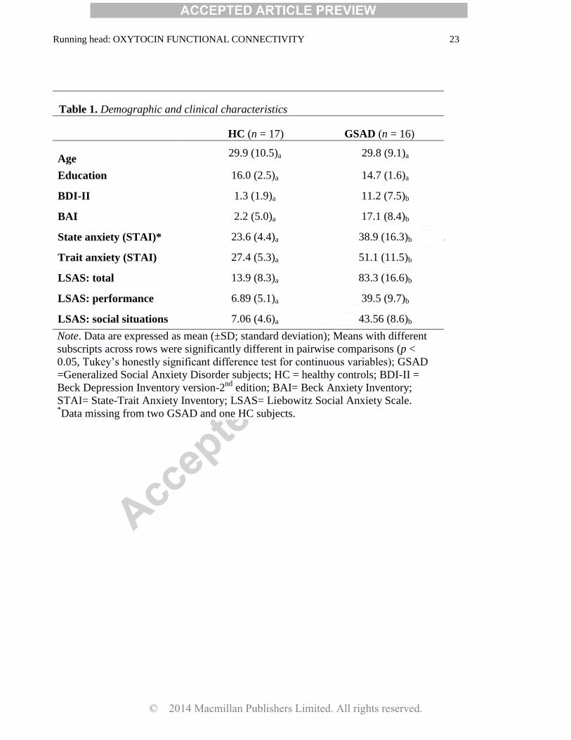
A total of 36 right-handed, male volunteers were enrolled in the study. Eighteen were
© 2014 Macmillan Publishers Limited. All rights reserved.
Running head: OXYTOCIN FUNCTIONAL CONNECTIVITY 6
considered healthy controls and 18 met criteria for current generalized social anxiety disorder (GSAD).
One control and two GSAD patients were excluded from the current study due to poor scan data quality.
Group differences in demographics and clinical characteristics are presented in Table 1. GSAD
diagnoses were confirmed using the Clinical International Diagnostic Interview – Version 2.1 (CIDI;
World Health Organization [WHO], 1997), and probes taken from the Liebowitz Social Anxiety Scale
(LSAS; Liebowitz, 1987). To ensure that GSAD patients were classified as the 'generalized' subtype,
they were required to have a total LSAS score greater than 70, and a 'social situations' subscale score
greater than 30. GSAD participants were excluded if they had a co-occurring anxiety disorder, a
depressive episode within the past six months, alcohol/substance use disorder within the past year, or
any lifetime history of post-traumatic stress disorder, bipolar disorder, psychotic disorder, mental
retardation, or developmental disorders. Healthy controls had no lifetime history of any psychiatric
disorder. All participants were non-smokers, were not taking prescription medications, and had no
known allergies. During study screening, participants completed a brief medical examination. All
participants provided written informed consent after explanation of the protocol, as approved by the
Monash University Human Research Ethics Committee.
Procedure
The study was a within-subjects, randomized, double-blind, placebo-controlled, OXT
administration study. Each participant completed two fMRI scan sessions (order randomized) where
they received either intranasal spray of OXT (24 IU/ 40.32 µg) or placebo (PBO, all ingredients except
for the peptide). Sessions were separated by a minimum of 7 days to allow for proper wash-out.
Participants were instructed to fast for 1-hour prior to each session, and not to ingest any caffeine or
alcohol the day of the scan. The intranasal puffs were self-administered in an alternating order with a
45-sec break between puffs (total of 3 sprays per nostril). The fMRI scan began 45-mins post-
inhalation of OXT or PBO, and lasted for 30-mins. This time frame is consistent with prior OXT
© 2014 Macmillan Publishers Limited. All rights reserved.
Running head: OXYTOCIN FUNCTIONAL CONNECTIVITY 7
administration studies and coincides with the expected timing of OXT's physiological effects (e.g.,
Domes et al, 2007; Kirsch et al, 2005).
Emotional Face Matching Task (EFMT)
The EFMT task has previously been described in detail elsewhere (Labuschagne et al, 2010),
and has been used in previous pharmaco-fMRI studies to probe drug effects on amygdala activation
(Kirsch et al, 2005; Paulus et al, 2005; Phan et al, 2008; Labuschagne et al, 2010) and amygdala
functional connectivity (Gorka et al, 2013). Briefly, using a block-design, participants were shown
three faces and asked to select which of the two faces at the bottom of the screen matched the facial
expression of the target face at the top of the screen. The target face and the congruent probe face
always displayed one of three expressions (angry, fearful, or happy) and the other (i.e., incongruent)
probe face always displayed a neutral expression. Face matching trials were interspersed with a
'control task' where participants were asked to match simple geometric shapes (i.e., circles, rectangles,
or triangles) similar to the instructions above. The paradigm consisted of 18 experimental blocks, each
containing four sequential matching trials of 3-s presentation (i.e., 12-s blocks). There were 9 blocks of
matching emotional faces (3 blocks of each target expression of fearful, angry, and happy), interleaved
with 9 blocks of matching shapes, counterbalanced across two runs. The total task time was 6-minutes.
During the task, participants made responses by pressing the left or right response buttons with their
dominant hand. These responses provided a measure of participants' response accuracy and reaction
time. As noted, the planned fMRI analyses focused on matching fearful faces and shapes. Behavioral
performances of the participants on the EFMT task were previously reported in Labuschagne et al.
(2010). There were no main effects of group or group x drug interactions on accuracy or response times
when matching fearful faces or shapes (all ps > 0.05).
Brain Imaging
Brain images were acquired with a 3T Siemens Tim Trio scanner using a 12-channel head coil.
© 2014 Macmillan Publishers Limited. All rights reserved.
Running head: OXYTOCIN FUNCTIONAL CONNECTIVITY 8
Functional gradient-echo echo-planar imaging (EPI) data depicting BOLD contrast were acquired
during the experimental task (TE=40 ms, TR=3000 ms, flip-angle=90°, FoV=210 mm, 64×64
matrix, 44 contiguous 3 mm slices parallel to the hippocampus, and interleaved). Whole brain T1-
weighted anatomical reference images were also acquired from all participants for precise anatomical
localization and normalization (TE=2.15 ms, TR=1900 ms, flip-angle=9°, FoV=256 mm, 176
sagittal slices, 1 mm slice thickness, perpendicular to the AC-PC line).
Functional MRI Data Analyses
All data previously processed using Statistical Parametric Mapping software SPM2 (Wellcome
Trust Centre for Neuroimaging, London, UK) (Labuschagne et al, 2010) were re-processed and re-
analyzed using updated SPM8 software. Images were spatially realigned to correct for head motion,
slice-time corrected, warped to standardized Montreal Neurological Institute (MNI) space using the
participant's mean functional image, resampled to 2 mm3 voxels, and smoothed with an 8 mm
3 kernel
to minimize noise and residual differences in gyral anatomy. The general linear model was applied to
the time series, convolved with the canonical hemodynamic response function (HRF) and with a 128
second high-pass filter. Condition effects were modeled with box-car regressors representing the
occurrence of each block type. Effects were estimated at each voxel, and for each subject. Individual
contrast maps for fearful faces (> shapes) were created for each person, and each session.
We first confirmed using a whole-brain, 2 (session: PBO, OXT) x 2 (group: HC, GSAD
analysis of variance (ANOVA) that consistent with Labuschagne et al. (2010), there was a significant
group x drug interaction on left amygdala reactivity to fearful faces [peak MNI coordinates: (-24, -2, -
22), Z=2.77, k =81, p <0.05, small volume corrected), such that OXT attenuated (or normalized) the
hyperactive amygdala reactivity to fearful faces in GSAD patients. There was no group x drug
interaction for right amygdala reactivity to fearful faces.
To examine functional coupling, we used a generalized form of context-dependent
© 2014 Macmillan Publishers Limited. All rights reserved.
Running head: OXYTOCIN FUNCTIONAL CONNECTIVITY 9
psychophysiological interaction analyses (gPPI; http://brainmap.wisc.edu/PPI, McLaren et al, 2012).
The left and right amygdala were used as the seeds of interest and created using anatomically derived
masks (MARINA: http://www.bion.de/Marina.htm; Walter et al., 2003), defined by atlas-based
boundaries (Tzourio-Mazoyer et al., 2002). The volumes of the masks were 1760mm3 on the left and
1984mm3 on the right, and the same masks were applied to all subjects‟ data. The de-convolved time
series from the amygdala was extracted for each subject to create the physiological variable. The
condition onset times for fearful faces, angry faces, happy faces, and shapes were separately convolved
with the canonical HRF for each condition, creating the psychological regressors. The interaction terms
(PPIs) were computed by multiplying the time series from the psychological regressors
(angry/fearful/happy) with the physiological variable. To examine the effect of the interaction terms,
activity within the amygdala was regressed on a voxel-wise basis against the interaction, with the
physiological and psychological variables serving as regressors of interest.
The individual fearful faces (> shapes) contrast images were entered into separate second-level
2 (group) x 2 (drug) ANOVAs for the left and right amygdala to determine whether there were any
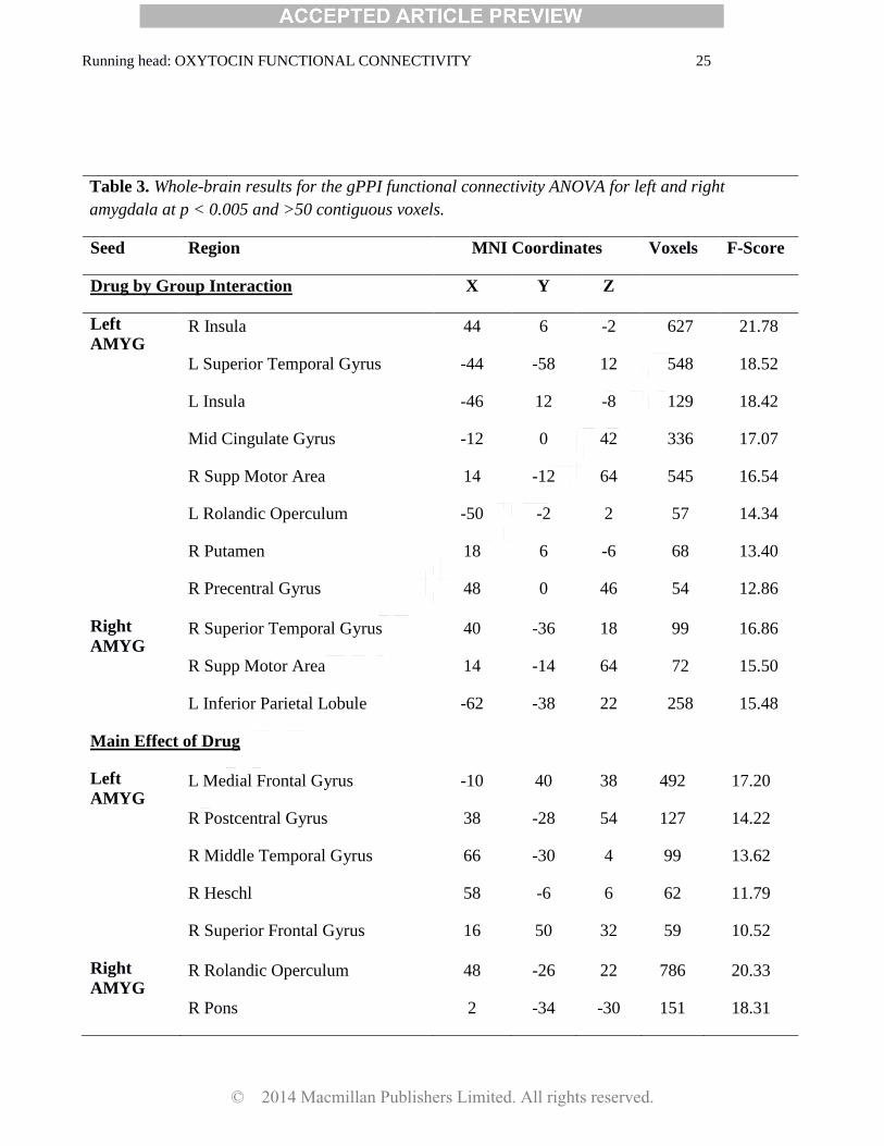
group x drug interactions on functional connectivity. In order to conduct an exploratory whole-brain
analysis, we initially set the significance at p < 0.005 (uncorrected) with a cluster extent threshold of
greater than 50 contiguous voxels to balance between Type I and Type II errors to generate results for
subsequent hypothesis testing (Lieberman and Cunningham, 2009). Next, for a priori regions of
interest, we restricted our analyses to two main areas (anterior to middle cingulate and insula) both
previously shown to exhibit aberrant functional connectivity to the amygdala in GSAD (Etkin and
Wager, 2007; Prater et al, 2013) and affected by OXT (Dodhia et al, 2014; Rilling et al, 2012; Sripada
et al, 2013). Specifically, we applied an anatomically-derived (AAL atlas) partial brain mask of the
anterior and middle cingulate and bilateral insula to our data (search volume = 84cm3, encompassing
10,485 voxels). Cluster-based significance thresholding was used to adjust for multiple comparisons
© 2014 Macmillan Publishers Limited. All rights reserved.
Running head: OXYTOCIN FUNCTIONAL CONNECTIVITY 10
within the search volume. Based on simulations (10,000 iterations) performed with 3DClustSim, an
adaptation of AlphaSim (http://afni.nimh.nih.gov/pub/dist/doc/program_help/3dClustSim.html), a
family wise error correction at α < 0.05 is achieved with a voxel threshold of p <0.005 and a cluster
size of at least 60 contiguous voxels for the left amygdala model and 62 contiguous voxels for the right
amygdala model. Of note, the same search mask was applied to both models, but separate thresholds
were calculated given differences in estimated smoothness. We extracted parameter estimates/β
weights (connectivity values, arbitrary units) from 5mm-radius spheres surrounding peak activations
for each subject to conduct post hoc paired and independent samples t-tests and determine
directionality.
Results
Amygdala Functional Connectivity
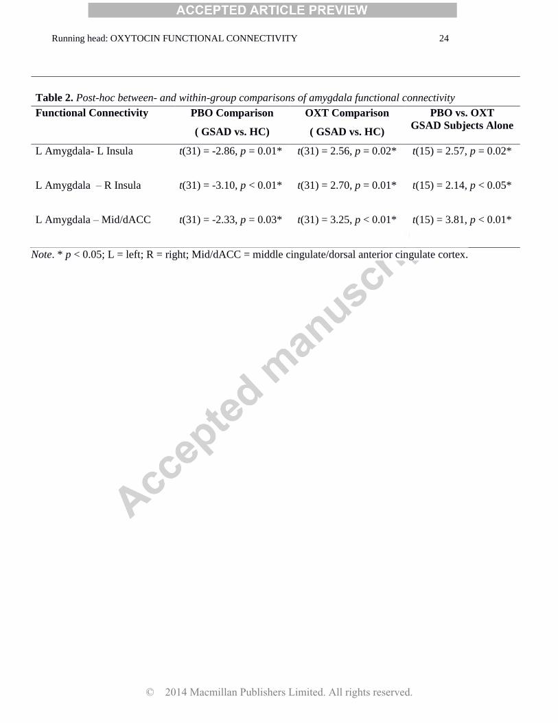
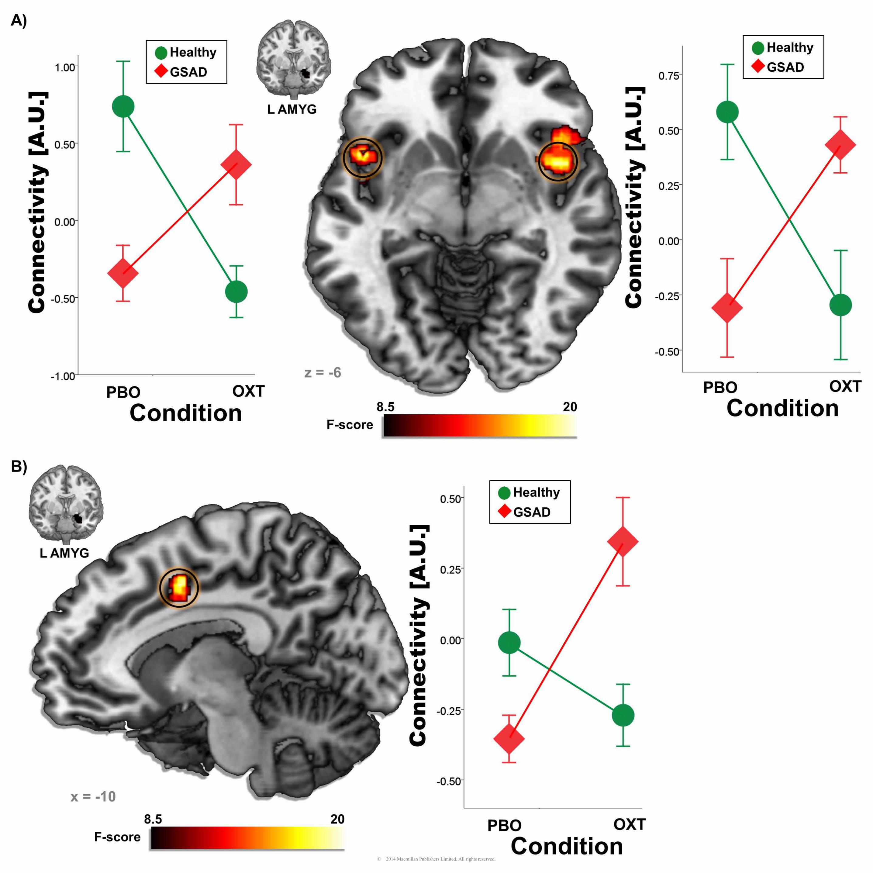
gPPI functional connectivity analyses indicated a significant group x drug interaction for left
amygdala to bilateral insula (right MNI[44, 6, -2], Z = 4.16, k = 627, p < 0.05, corrected; left MNI [-46,
12, -8], Z = 3.84, k = 129, p < 0.05, corrected) and middle cingulate/dorsal anterior cingulate gyrus
(mid/dACC) connectivity (MNI [-12, 0, 42]; Z = 3.70, k = 336, p <0.05, corrected). Post hoc
comparisons of extracted gPPI parameter estimates of connectivity from insula and mid/dACC are
presented in Table 2. In all three cases, GSAD subjects had significantly less functional connectivity
than HC subjects on PBO. However, on OXT, this group difference in connectivity was no longer
evident. In fact, on OXT, GSAD (> HC) subjects displayed enhanced functional connectivity (i.e., the
pattern of reduced connectivity on PBO was reversed on OXT). For completeness, and to obviate bias,
we report all main effects of group, main effects of drug, and group x drug interactions from the left
and right amygdala gPPI ANOVAs across the entire brain (Table 3).
Additional Analyses
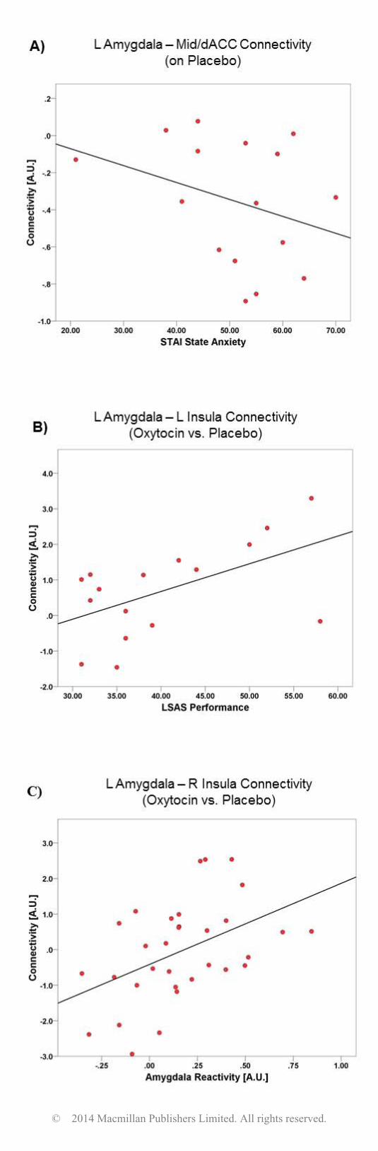
Post-hoc we conducted analyses to determine whether amygdala reactivity to fearful faces on
© 2014 Macmillan Publishers Limited. All rights reserved.
Running head: OXYTOCIN FUNCTIONAL CONNECTIVITY 11
PBO (a potential neuronal endophenotype for arousal/anxiety; Shin and Liberzon, 2010) was correlated
with OXT-induced changes in amygdala functional connectivity. We found that greater left amygdala
reactivity was associated with greater magnitude of OXT-induced changes in left amygdala-right insula
connectivity (r = 0.46, p = 0.01). We also conducted correlational analyses between self-reported
anxiety measures and our amygdala connectivity findings, within GSAD subjects, using measures of
social anxiety severity (i.e., LSAS scores) and state and trait anxiety levels (i.e., State-Trait Anxiety
Inventory scores; Spielberger et al., 1983). Results indicated that within GSAD individuals, greater
state anxiety on PBO scan day was correlated with less left amygdala-mid/dACC functional
connectivity on PBO (r = -0.56, p = 0.02). Moreover, within GSAD individuals, greater LSAS
performance-based social anxiety scores were correlated with greater magnitude of OXT-induced
changes in left amygdala-left insula functional connectivity (r = 0.55, p = 0.03). A similar pattern of
results was found with LSAS total scores, albeit at a trend level (r = 0.38, p = 0.12). These findings are
illustrated in Figure 2. Of note, as has been published elsewhere, there were no group x session, group
x time (pre- post- ingestion), or group x session x time interactions on mood (e.g., anxiety) or self-
reported drug effects (e.g., calmness, alertness, feel high; Labuschagne et al, 2010; Labuschagneet al,
2012).
Discussion
The aim of the current study was to examine the effects of OXT on amygdala functional
connectivity during the processing of fearful faces in GSAD subjects and HCs. Results indicated that
on PBO, GSAD subjects exhibited reduced functional connectivity, relative to HC subjects, between
amygdala-bilateral insula and amygdala-mid/dACC. This pattern of reduced functional connectivity
was no longer evident on OXT and individuals with GSAD exhibited greater functional connectivity
compared with HC subjects. This suggests that OXT attenuates amygdala reactivity while
simultaneously enhancing functional connectivity between the amygdala and the insula and mid/dACC
© 2014 Macmillan Publishers Limited. All rights reserved.
Running head: OXYTOCIN FUNCTIONAL CONNECTIVITY 12
– regions heavily implicated in processing socio-emotional stimuli.
The current pattern of results is noteworthy given that prior studies have shown reduced or
disrupted amygdala-insula and amygdala-dACC connectivity to threatening faces in individuals with
GSAD (Prater et al, 2013). Moreover, higher levels of social inhibition, defined as the tendency to
withdrawal from people or avoid social situations, is associated with reduced connectivity between the
left amygdala and the insula, dACC, and hippocampus (Blackford et al, 2014). These findings suggest
that OXT may impact anxiety by „normalizing‟ (i.e., enhancing reduced connectivity) coupling
between the amygdala, insula, and ACC. In support of these ideas, we found that greater amygdala
reactivity to fearful faces (an indicator of anxious responding) was associated with less amygdala-
insula functional connectivity at baseline. Additionally, within GSAD subjects, greater state anxiety
was related to less baseline amygdala-mid/dACC connectivity and greater performance-based social
anxiety was associated with the magnitude of OXT‟s effects on amygdala-insula connectivity. These
findings together suggest that reduced amygdala to insula and mid/dACC functional connectivity in
response to fearful stimuli may be an indicator of maladaptive or anxious responding, whereas
enhanced connectivity may reflect „normal‟ or typical emotional processing.
There are several potential mechanisms that may explain the current pattern of results. First,
resting-state studies have suggested that the insula, dACC, and amygdala are part of a „salience
network‟ that identifies the most emotionally salient information amongst internal and external stimuli
(e.g., interoceptive awareness) and uses it to guide behavior (Seeley et al, 2007). Cross-talk between
these regions is necessary to execute these functions and reduced connectivity may reflect
dysfunctional salience processing of threat stimuli. Related to the broader social phobia literature, it is
possible that dysfunctional salience processing underlies maladaptive attentional biases for social-
evaluative stimuli that are characteristic of GSAD (Asmundson and Stein, 1994). Notably, these three
regions in particular – amygdala, insula, and ACC – are increasingly implicated in the pathophysiology
© 2014 Macmillan Publishers Limited. All rights reserved.
Running head: OXYTOCIN FUNCTIONAL CONNECTIVITY 13
of GSAD (Etkin and Wager, 2007; Marazziti et al, 2014; Miskovic et al, 2012).
It is also possible that the current findings are related to the broader functions of insula and
mid/dACC functional connectivity with the amygdala. The insula has direct projections to the
amygdala, and functional coupling between these areas is necessary for the regulation of autonomic
responses and the awareness of interoceptive states (Craig, 2006). As such, reduced connectivity may
reflect diminished insula modulatory influences on amygdala reactivity, which could impair
interoception and potentiate autonomic anxiety responses during social threat. Enhanced connectivity
could therefore have the opposite effect and increase autonomic regulation. Of note, Rilling et al.
(2012) also found that OXT enhanced amygdala-insula functional connectivity and posited that this
reflected an increased ability to assess internal states (e.g., somatic markers) to guide decision-making.
As for the mid/dACC, it is activated in response to numerous social contexts, and through functional
interactions with other limbic areas, recruits regions necessary for affect regulation, directs attentional
control, and ameliorates cognitive conflicts (Kerns et al, 2004). Moreover, acute OXT has been shown
to reduce amygdala reactivity while enhancing connectivity to ACC and insula to infant laughter in
nulliparous women and the authors suggested that “increased functional connectivity between the
amygdala and regions involved in emotion regulation may reduce negative emotional arousal while
enhancing the incentive salience of the infant laughter” (Reim et al, 2012). Taken together, reduced
amygdala-insula and mid/dACC functional connectivity at baseline may reflect global deficits in
detecting social information and processing and modulating affective and attentional responses. It is
necessary to note that although we observed these amygdala effects in the context of threat processing,
interpretations of the functional relevance of OXT‟s actions in alleviating the symptoms or behaviors of
social anxiety are speculative and future studies are needed to delineate these putative effects.
It is also important to highlight that within individuals with GSAD, OXT may not „enhance‟
amygdala to insula and mid/dACC connectivity per se. Rather, the findings could be interpreted as
© 2014 Macmillan Publishers Limited. All rights reserved.
Running head: OXYTOCIN FUNCTIONAL CONNECTIVITY 14
OXT changing the direction of connectivity effects. On PBO, individuals with GSAD may exhibit
strong inverse connections between the amygdala and insula and mid/dACC, whereas on OXT they
exhibit strong positive connections. This would imply that hyperactive amygdala responding is coupled
with reduced insula and mid/dACC activation on PBO and the opposite pattern on OXT - reduced
amygdala reactivity is coupled with enhanced insula and mid/dACC activation. Importantly, if this
were indeed the case, many of the interpretations discussed above would similarly apply. Inverse
connectivity may reflect aberrant interactions between these socio-emotional regions, leading to
anxious responding and/or deficits in social processing. Conversely, positive connectivity may be an
indicator of adaptive functional interactions which facilitate social recognition and affect modulation.
It is interesting that the aforementioned effects of OXT are specific to individuals with GSAD
and differ from that observed in healthy volunteers. In fact, OXT reduces amygdala connectivity to
insula and mid/dACC in HC subjects. This finding of divergent effects is consistent with the two other
OXT administration studies from our group (Dodhia et al, 2014; Labuschagne et al, 2010) and
underscores that acute administration of OXT can be influenced by individual differences in anxiety
psychopathology. It is likely that this phenomenon has contributed to some of the mixed findings in the
OXT literature (see Meyer-Lindenberg et al, 2011; but also Grillon et al, 2013). Together, these
findings suggest that the relation between OXT and anxious responding is nuanced and that OXT‟s
effects may be most consistent in the context of dysfunctional physiological states. However, future
work is greatly needed to examine this question and to elucidate the moderators and mediators of
OXT‟s acute effects on typical and atypical brain function.
While these results extend the literature on the neural effects of OXT, there are a few limitations
worth noting. First, although a within-subjects study, the sample size was small and this likely reduced
statistical power. Second, our sample was restricted to males, given the role of sex hormones on OXT
neural effect‟s (Domes et al, 2010), and may not generalize to females. Third, gPPI analyses are
© 2014 Macmillan Publishers Limited. All rights reserved.
Running head: OXYTOCIN FUNCTIONAL CONNECTIVITY 15
correlational and therefore directionality between amygdala and the insula and mid/dACC cannot be
inferred. Likewise, functional relevance of OXT‟s effects on amygdala reactivity and functional
connectivity remain unclear and more work is needed to link these effects to functional outcomes.
Future studies are also needed to determine whether the current findings are specific to fear or similarly
apply to other social stimuli (e.g., anger, disgust), and how these findings relate to OXT induced
changes in GABAergic transmission within the amygdala.
In sum, the results suggest that within individuals with GSAD, OXT simultaneously dampens
amygdala reactivity and enhances amygdala functional connectivity with the insula and mid/dACC
during the processing of fearful faces. These neural effects may have broad pro-social implications
such as enhancing the integration and modulation of social responses. These data add to the growing
evidence of OXT‟s acute effects on reactivity and connectivity within a socio-emotional, salience brain
network in GSAD and prompt further studies to further investigate specific mechanisms of OXT‟s
actions in anxious states and social behaviors.
© 2014 Macmillan Publishers Limited. All rights reserved.
Running head: OXYTOCIN FUNCTIONAL CONNECTIVITY 16
Funding and Disclosure
This research was funded by an Independent Investigator Award to PJN from the Brain and Behavior
Research Foundation (formally National Alliance for Research in Schizophrenia and Depression
[NARSAD]). PJN was an employee of GSK and UCB Pharma during the conduct of the study but
received no funding from either company for the study. AGW was supported by an Australian Rotary
Health fellowship (Geoffrey Betts Award) but ARH had no role in the current study. All authors
declare no conflict of interests.
© 2014 Macmillan Publishers Limited. All rights reserved.
Running head: OXYTOCIN FUNCTIONAL CONNECTIVITY 17
References
American Psychiatric Association (1994). Diagnostic and statistical manual of mental disorders
(4th ed.). Washington, DC: Author.
Asmundson GJ, Stein MB (1994). Selective processing of social threat in patients with
generalized social phobia: Evaluation using a dot-probe paradigm. J Anxiety
Disord 8: 107-117.
Baumgartner T, Heinrichs M, Vonlanthen A, Fischbacher U, Fehr E (2008). Oxytocin shapes the neural
circuitry of trust and trust adaptation in humans. Neuron 58: 639–650.
Bethlehem RA, van Honk J, Auyeung B, Baron-Cohen S (2013). Oxytocin, brain physiology,
and functional connectivity: a review of intranasal oxytocin fMRI
studies. Psychoneuroendocrinology 38: 962-974.
Blackford JU, Clauss JA, Avery SN, Cowan RL, Benningfield MM, VanDerKlok RM
(2014). Amygdala-cingulate intrinsic connectivity is associated with degree of social
inhibition. Biol Psychol E-pub ahead of print.
Craig (2009). How do you feel – now? The anterior insula and human awareness. Nat Rev
Neurosci 10: 59-70.
Ditzen B, Schaer M, Gabriel B, Bodenmann G, Ehlert U, Heinrichs M (2009). Intranasal
oxytocin increases positive communication and reduces cortisol levels during couple conflict.
Biol Psychiatry 65: 728-731.
Dodhia S, Hosanagar A, Fitzgerald DA, Labuschagne I, Wood AG, Nathan PJ, Phan K.L.
(2014). Moduation of resting-state amygdala-frontal functional connectivity by oxytocin in
generalized social anxiety disorder. Neuropsychopharmacology E-pub ahead of print.
Domes G, Heinrichs M, Gläscher J, Büchel C, Braus DF, Herpertz SC (2007). Oxytocin attenuates
© 2014 Macmillan Publishers Limited. All rights reserved.
Running head: OXYTOCIN FUNCTIONAL CONNECTIVITY 18
amygdala responses to emotional faces regardless of valence. Biol Psychiatry 62: 1187–1190.
Domes G, Lischke A, Berger C, Grossmann A, Hauenstein K, Heinrichs M et al (2010). Effects of
intranasal oxytocin on emotional face processing in women. Psychoneuroendocrinology 35: 83–
93.
Etkin A, Wager T (2007). Functional neuroimaging of anxiety: a meta-analysis of emotional processing
in PTSD, social anxiety disorder, and specific phobia. Am J Psychiatry 164(10): 1476-1488.
Foa EB, Gilboa-Schechtman E, Amir N, Freshman M (2000). Memory bias in generalized social
phobia: remembering negative emotional expressions. J Anxiety Disord 14: 501–519.
Gorka SM, Fitzgerald DA, King AC, Phan KL (2013). Alcohol attenuates amygdala–frontal
connectivity during processing social signals in heavy social
drinkers. Psychopharmacology 229(1): 141-154.
Grillon C, Krimsky M, Charney DR, Vytal K, Ernst M, Cornwell B (2013). Oxytocin
increases anxiety to unpredictable threat. Mol Psychiatry 18: 958-960.
Harmer CJ, Mackay CE, Reid CB, Cowen PJ, Goodwin GM (2006). Antidepressant drug treatment
modifies the neural processing of nonconscious threat cues. Biol Psychiatry 59(9): 816–820.
Hermann C, Ziegler S, Birbaumer N, Flor H (2002). Psychophysiological and subjective
indicators of aversive Pavlovian conditioning in generalized social phobia. Biol
Psychiatry 52(4): 328-337.
Huber D, Veinante P, Stoop R (2005). Vasopressin and oxytocin excite distinct neuronal
populations in the central amygdala. Science 308: 245–248.
Kerns JG, Cohen JD, MacDonald AW, Cho RY, Stenger VA. Carter CS (2004). Anterior cingulate
conflict monitoring and adjustments in control. Science 303(5660): 1023-1026.
Kirsch P, Esslinger C, Chen Q, Mier D, Lis S, Siddhanti S et al (2005). Oxytocin modulates neural
circuitry for social cognition and fear in humans. J Neurosci 25: 11489–11493.
© 2014 Macmillan Publishers Limited. All rights reserved.
Running head: OXYTOCIN FUNCTIONAL CONNECTIVITY 19
Kosfeld M, Heinrichs M, Zak PJ, Fischbacher U, Fehr E (2005). Oxytocin increases trust in
humans. Nature 435: 673–676.
Labuschagne I, Phan KL, Wood A, Angstadt M, Chua P, Heinrichs M, et al
(2010). Oxytocin attenuates amygdala reactivity to fear in generalized social anxiety
disorder. Neuropsychopharmacology 35(12): 2403-2413.
Labuschagne I, Phan KL, Wood A, Angstadt M, Chua P, Heinrichs M, Stout JC, Nathan PJ
(2012). Medial frontal hyperactivity to sad faces in generalized social anxiety disorder and
modulation by oxytocin. Int J Neuropsychopharmacol 15(7): 883-896.
Lieberman MD, Cunningham WA (2009). Type I and Type II error concerns in fMRI research:
Rebalancing the scale. Soc Cogn Affect Neurosci 4: 423–428
Liebowitz MR (1987). Social Phobia. Mod Probl Pharmacopsychiatry 22: 141–173.
Marazziti D, Abelli M, Baroni S, Carpita B, Ramacciotti CE, Dell'Osso L. (2014). Neurobiological
correlates of social anxiety disorder: an update. CNS Spectrums E-pub ahead of print.
McLaren DG, Ries ML, Xu G, Johnson SC (2012). A generalized form of context-
dependent psychophysiological interactions (gPPI): a comparison to standard
approaches. Neuroimage 61(4): 1277-1286.
Meyer-Lindenberg A, Domes G, Kirsch P, Heinrichs M (2011). Oxytocin and vasopressin in the human
brain: Social neuropeptides for translational medicine. Nat Rev Neurosci 12(9): 524-538.
Miskovic V, Schmidt LA (2012). Social fearfulness in the human brain. Neurosci Biobehav R 36(1):
459-478.
Mogg K, Philippot P, Bradley BP (2004). Selective attention to angry faces in clinical social
phobia. J Abnorm Psychol 113(1): 160.
Parkes SL, Westbrook RF (2010). The basolateral amygdala is critical for the acquisition and
extinction of associations between a neutral stimulus and a learned danger signal but not
© 2014 Macmillan Publishers Limited. All rights reserved.
Running head: OXYTOCIN FUNCTIONAL CONNECTIVITY 20
between two neutral stimuli. J Neurosci 30: 12608–12618.
Paulus MP, Feinstein JS, Castillo G, Simmons AN, Stein MB (2005). Dose-dependent decrease of
activation in bilateral amygdala and insula by lorazepam during emotion processing. Arch Gen
Psychiatry 62(3): 282-288.
Phan KL, Angstadt M, Golden J, Onyewuenui I, Povpovska A, de Wit H (2008)
Cannabinoid modulation of amygdala reactivity to social signals of threat in humans. J Neurosci
28: 2313–2319.
Phan KL, Fitzgerald DA, Nathan PJ, Tancer ME (2006). Association between amygdala hyperactivity
to harsh faces and severity of social anxiety in generalised social phobia. Biol Psychiatry 59:
424–429
Prater KE, Hosanagar A, Klumpp H, Angstadt M, & Phan KL (2013). Aberrant amygdala–frontal
cortex connectivity during perception of fearful faces and at rest in generalized social anxiety
disorder. Depress Anxiety 30(3): 234-241.
Riem MM, van IJzendoorn MH, Tops M, Boksem MA, Rombouts SA, Bakermans-
Kranenburg MJ (2012). No laughing matter: intranasal oxytocin administration changes
functional brain connectivity during exposure to infant
laughter. Neuropsychopharmacology 37(5): 1257-1266.
Rilling JK, DeMarco AC, Hackett PD, Thompson R, Ditzen B, Patel R, et al
(2012). Effects of intranasal oxytocin and vasopressin on cooperative behavior and associated
brain activity in men. Psychoneuroendocrinology 37(4): 447-461.
Roelofs K, van Peer J, Berretty E, de Jong P, Spinhoven P, Elzinga BM (2009). HPA-axis
hyperresponsiveness is associated with increased social avoidance behavior in social phobia.
Biol Psychiatry 65: 336-343.
Schultz LT, Heimberg RG (2008). Attentional focus in social anxiety disorder: potential for
© 2014 Macmillan Publishers Limited. All rights reserved.
Running head: OXYTOCIN FUNCTIONAL CONNECTIVITY 21
interactive processes. Clin Psychol Rev 28: 1206–1221
Seeley WW, Menon V, Schatzberg AF, Keller J, Glover GH, Kenna H, et al (2007). Dissociable
intrinsic connectivity networks for salience processing and executive control. J
Neurosci. 27(9): 2349–2356.
Shin LM, Liberzon I (2010). The neurocircuitry of fear, stress, and anxiety
disorders. Neuropsychopharmacology 35: 169–191.
Spielberger CD, Gorsuch RL, Lushene RE (1983). Manual for the State-Trait Anxiety Inventory. Palo
Alto, CA: Consulting Psychologist Press.
Sripada CS, Phan KL, Labuschagne I, Welsh R, Nathan PJ, Wood AG (2013). Oxytocin enhances
resting-state connectivity between amygdala and medial frontal cortex. Int J
Neuropsychopharmacology 16(02): 255-260.
Taylor S, Abramowitz JS, McKay D (2012). Non-adherence and non-response in the treatment of
anxiety disorders. J Anxiety Disord. 26(5): 583-589.
Tzourio-Mazoyer N, Landeau B, Papathanassiou D, Crivello F, Etard O, Delcroix N, Mazoyer B, Joliot
M (2002). Automated anatomical labeling of activations in SPM using a macroscopic
anatomical parcellation of the MNI MRI single-subject brain. Neuroimage 15(1): 273-289.
Van Den Heuvel MP, Hulshoff Pol HE (2010). Exploring the brain network: a review on
resting-state fMRI functional connectivity. Eur Neuropsychopharmacology 20(8): 519-
534.
Walter, B, Blecker C, Kirsch P, Sammer G, Schienle A, Stark R, Vaitl D (2003). Marina: An easy to
use tool for the creation of masks for interest analyses. Neuroimage 19:S47.
WHO, World Health Organization Composite International Diagnostic Interview (CIDI, Version 2.1,
1997 World Health Organization: Geneva.
Windle RJ, Shanks N, Lightman SL, Ingram CD (1997). Central oxytocin administration reduces
© 2014 Macmillan Publishers Limited. All rights reserved.
Running head: OXYTOCIN FUNCTIONAL CONNECTIVITY 22
stress-induced corticosterone release and anxiety behavior in rats 1. Endocrinology 138(7):
2829-2834.
© 2014 Macmillan Publishers Limited. All rights reserved.
Running head: OXYTOCIN FUNCTIONAL CONNECTIVITY 23
Table 1. Demographic and clinical characteristics
HC (n = 17) GSAD (n = 16)
Age 29.9 (10.5)a 29.8 (9.1)a
Education 16.0 (2.5)a 14.7 (1.6)a
BDI-II 1.3 (1.9)a 11.2 (7.5)b
BAI 2.2 (5.0)a 17.1 (8.4)b
State anxiety (STAI)* 23.6 (4.4)a 38.9 (16.3)b
Trait anxiety (STAI) 27.4 (5.3)a 51.1 (11.5)b
LSAS: total 13.9 (8.3)a 83.3 (16.6)b
LSAS: performance 6.89 (5.1)a 39.5 (9.7)b
LSAS: social situations 7.06 (4.6)a 43.56 (8.6)b
Note. Data are expressed as mean (±SD; standard deviation); Means with different
subscripts across rows were significantly different in pairwise comparisons (p <
0.05, Tukey‟s honestly significant difference test for continuous variables); GSAD
=Generalized Social Anxiety Disorder subjects; HC = healthy controls; BDI-II =
Beck Depression Inventory version-2nd
edition; BAI= Beck Anxiety Inventory;
STAI= State-Trait Anxiety Inventory; LSAS= Liebowitz Social Anxiety Scale. *Data missing from two GSAD and one HC subjects.
© 2014 Macmillan Publishers Limited. All rights reserved.
Running head: OXYTOCIN FUNCTIONAL CONNECTIVITY 24
Table 2. Post-hoc between- and within-group comparisons of amygdala functional connectivity
Functional Connectivity PBO Comparison
( GSAD vs. HC)
OXT Comparison
( GSAD vs. HC)
PBO vs. OXT
GSAD Subjects Alone
L Amygdala- L Insula t(31) = -2.86, p = 0.01* t(31) = 2.56, p = 0.02* t(15) = 2.57, p = 0.02*
L Amygdala – R Insula t(31) = -3.10, p < 0.01* t(31) = 2.70, p = 0.01* t(15) = 2.14, p < 0.05*
L Amygdala – Mid/dACC t(31) = -2.33, p = 0.03* t(31) = 3.25, p < 0.01* t(15) = 3.81, p < 0.01*
Note. * p < 0.05; L = left; R = right; Mid/dACC = middle cingulate/dorsal anterior cingulate cortex.
© 2014 Macmillan Publishers Limited. All rights reserved.
Running head: OXYTOCIN FUNCTIONAL CONNECTIVITY 25
Table 3. Whole-brain results for the gPPI functional connectivity ANOVA for left and right
amygdala at p < 0.005 and >50 contiguous voxels.
Seed Region MNI Coordinates Voxels F-Score
Drug by Group Interaction X Y Z
Left
AMYG R Insula 44 6 -2 627 21.78
L Superior Temporal Gyrus -44 -58 12 548 18.52
L Insula -46 12 -8 129 18.42
Mid Cingulate Gyrus -12 0 42 336 17.07
R Supp Motor Area 14 -12 64 545 16.54
L Rolandic Operculum -50 -2 2 57 14.34
R Putamen 18 6 -6 68 13.40
R Precentral Gyrus 48 0 46 54 12.86
Right
AMYG R Superior Temporal Gyrus 40 -36 18 99 16.86
R Supp Motor Area 14 -14 64 72 15.50
L Inferior Parietal Lobule -62 -38 22 258 15.48
Main Effect of Drug
Left
AMYG L Medial Frontal Gyrus -10 40 38 492 17.20
R Postcentral Gyrus 38 -28 54 127 14.22
R Middle Temporal Gyrus 66 -30 4 99 13.62
R Heschl 58 -6 6 62 11.79
R Superior Frontal Gyrus 16 50 32 59 10.52
Right
AMYG R Rolandic Operculum 48 -26 22 786 20.33
R Pons 2 -34 -30 151 18.31
© 2014 Macmillan Publishers Limited. All rights reserved.
Running head: OXYTOCIN FUNCTIONAL CONNECTIVITY 26
R Middle Frontal Gyrus 28 -12 50 777 17.36
L Parahippocampal Gyrus -26 -52 -8 56 17.22
L Parietal Lobe -30 -48 26 63 16.11
R Superior Occipital Gyrus 26 -62 30 229 15.36
L Postcentral Gyrus -56 -20 26 84 13.68
Mid Cingulate Gyrus -8 -22 40 180 13.27
L Precentral Gyrus -22 -20 66 68 12.06
Main Effect of Group
Left
AMYG R Lingual Gyrus 18 -84 -12 117 16.37
L Middle Occipital Gyrus -32 -72 6 104 15.13
L Fusiform Gyrus -32 -72 -16 140 12.02
Right
AMYG L Declive -8 -66 -22 90 20.88
R Parietal Lobe 24 -44 32 113 19.00
L Frontal Lobe -28 18 18 373 17.89
L Cuneus -24 -78 12 181 17.15
Mid Cingulate Gyrus -16 -2 28 205 16.34
L Posterior Cingulate Gyrus -16 -44 34 66 16.12
R Cuneus 20 -82 10 166 13.49
Note. gPPI= generalized psychophysiological interaction; MNI = Montreal Neurological Institute; L =
left; R = right; AMYG = amygdala; results at p < 0.005, uncorrected, >50 voxel minimum.
© 2014 Macmillan Publishers Limited. All rights reserved.
Running head: OXYTOCIN FUNCTIONAL CONNECTIVITY 27
Figure Captions
Figure 1. Voxel-wise statistical F-maps displayed on canonical brains illustrating significant group by
drug interactions. Line graphs illustrating extracted connectivity parameter estimates during the
oxytocin and placebo conditions for GSAD and HC subjects; A) left amygdala-left insula and left
amygdala- right insula; B) left amygdala-mid/dACC; GSAD = generalized social anxiety disorder; HC
= healthy control; PBO = placebo condition; OXT = oxytocin condition; mid/dACC = middle
cingulate/dorsal anterior cingulate cortex.
Figure 2. Scatter plot of correlation between: A) left amygdala-mid/dACC functional connectivity on
placebo and STAI state anxiety scores within GSAD subjects; B) left amygdala-right insula functional
connectivity during oxytocin (>placebo) and LSAS performance scores within GSAD subjects; C) left
amygdala-left insula functional connectivity during oxytocin (>placebo) and left amygdala reactivity
on placebo within all subjects; L = left; R = right; STAI = State-Trait Anxiety Inventory; LSAS =
Liebowitz Social Anxiety Scale; Mid/dACC= middle cingulate/dorsal anterior cingulate cortex.
© 2014 Macmillan Publishers Limited. All rights reserved.
Running head: OXYTOCIN FUNCTIONAL CONNECTIVITY 28
© 2014 Macmillan Publishers Limited. All rights reserved.
© 2014 Macmillan Publishers Limited. All rights reserved.
© 2014 Macmillan Publishers Limited. All rights reserved.


![Self-Regulation of Amygdala Activation Using Real-Time ...€¦ · amygdala participates in more detailed and elaborate stimulus evaluation [20,26,27]. The involvement of the amygdala](https://img.pdfslide.net/doc/110x75/5fa8a495e8acaa50d8405bd2/self-regulation-of-amygdala-activation-using-real-time-amygdala-participates.jpg)