Embed Size (px)
Citation preview

~ 30 ~
International Journal of Orthopaedics Sciences 2021; 7(2): 30-37
E-ISSN: 2395-1958
P-ISSN: 2706-6630
IJOS 2021; 7(2): 30-37
© 2021 IJOS
www.orthopaper.com
Received: 15-02-2021
Accepted: 19-03-2021
Dr. Harbir Kaur
Junior resident third year,
Department of orthopaedics,
University Shri Guru Ram das
Institute of medical Sciences and
research, Vallah, Amritsar,
Punjab, India
Dr. Raj Kumar Aggarwal
Professor and head of
department, Department of
orthopaedics, University Shri
Guru Ram das Institute of
medical Sciences and research,
Vallah, Amritsar, Punjab, India
Dr. Aarti Dewan
Associate Professor, Department
of orthopaedics, University Shri
Guru Ram das Institute of
medical Sciences and research,
Vallah, Amritsar, Punjab, India
Dr. Rajan Sarad
Associate Professor, Department
of orthopaedics, University Shri
Guru Ram das Institute of
medical Sciences and research,
Vallah, Amritsar, Punjab, India
Dr. Chandan Jasrotia
Associate Professor, Department
of orthopaedics, University Shri
Guru Ram das Institute of
medical Sciences and research,
Vallah, Amritsar, Punjab, India
Corresponding Author:
Dr. Harbir Kaur
Junior resident third year,
Department of orthopaedics,
University Shri Guru Ram das
Institute of medical Sciences and
research, Vallah, Amritsar,
Punjab, India
To study risk factors of degenerative rotator cuff tear
Dr. Harbir Kaur, Dr. Raj Kumar Aggarwal, Dr. Aarti Dewan, Dr. Rajan
Sarad and Dr. Chandan Jasrotia
DOI: https://doi.org/10.22271/ortho.2021.v7.i2a.2610
Abstract Background: Rotator cuff has been a known entity for orthopaedic surgeons for more than two hundred
years [1]. Rotator cuff disease is a common condition that causes shoulder pain and functional disability
with a prevalence rate of 2.8-15%, affecting approximately 40% of population who are more than 60
years of age and substantially impacting on individual’s function [2].
Aims and Objectives: To study risk factors in patients with symptomatic atraumatic rotator cuff tear –
Age, Obesity, Gender, Smoking, Diabetes mellitus, Hypertension, Hand dominance, Use of
corticosteroid injections, Hyperlipidemia, Osteoarthritis of knee.
Materials and Methods:
Inclusion criteria
Pain in the shoulder of duration more than three weeks.
Normal range of motion of the shoulder.
One or more than one positive clinical tests for rotator cuff tear.
Diagnostic imaging like ultrasound and magnetic resonance imaging pointing to diagnosis of rotator cuff
tear.
Exclusion criteria
History of trauma.
History of any surgery.
Patients using assisted devices for walking.
Results: Our study proved that the metabolic factors of diabetes, BMI, age, hand dominance,
osteoarthritis of knee, hypertension, gender were independent significant risk factors associated with the
development of atraumatic rotator cuff tears. Out of all the risk factors evaluated, osteoarthritis of knee,
age, obesity were the most significant risk factors for atraumatic rotator cuff tear.
Conclusions: Out of all risk factors evaluated, osteoarthritis of knee, obesity and age were the most
important risk factors for atraumatic rotator cuff tear
Evaluation of various risk factors results in enhanced understanding of the prevention and management
of various other common conditions and comorbidities associated with degenerative rotator cuff tears [26].
Keywords: Degenerative rotator cuff, BMI, atraumatic, osteoarthritis, hyperlipdemia
Introduction
Rotator cuff has been a known entity for orthopaedic surgeons for more than two hundred
years [1]. Rotator cuff disease is a common condition that causes shoulder pain and functional
disability with a prevalence rate of 2.8-15%, affecting approximately 40% of population who
are more than 60 years of age and substantially impacting on individual’s function [2].
The prevalence of rotator cuff tendon tears both symptomatic and asymptomatic increases with
age ranging widely from 0% to 15% after the 50’s to 30% to 50% in the 80’s.
Rotator cuff tears are either traumatic or degenerative, degenerative tears are far more frequent
and multifactorial in etiology. Various intrinsic and extrinsic factors contribute to the causation
of rotator cuff pathology [3].
Aging can contribute to dysfunction of scapular muscles and result in degenerative rotator cuff
tears [4]. An age related decrease in vascularity is consistent with the increasing prevalence of
cuff pathology with aging. There is relative hypovascularity of the deep surface of the
supraspinatus insertion.
Effect of obesity on tendon degeneration is cellular inflammation through the disturbed
production of adiponectin and increased leptin resistance induce intracellular reactive oxygen

~ 31 ~
International Journal of Orthopaedics Sciences www.orthopaper.com species which induce inflammation and apoptosis of cells.
Keener et al. reported that hand dominance is related to
symptomatic rotator cuff tears especially tears involving
shoulder pain [5].
Djerbi et al. reported that the prevalence of rotator cuff tears
was greater among patients with dyslipidemia [6]. In
hyperlipidemia environments, lipids accumulate within the
extracellular matrix of the tendon and thus affect the
mechanical properties of tendon.
Baumgarten et al. found that 61.9% of patients with
degenerative rotator cuff tear had a history of daily tobacco
smoking [7]. Tobacco smoking has been associated with
musculoskeletal pain dysfunction with nicotine implicated in
delaying tendon to bone healing.
Degenerative rotator cuff tears are more common in females
as compared to males as there are differences in activities of
daily living between men and women that may require
different ranges of shoulder motion [8]. Atraumatic rotator cuff
tear is more common in post-menopausal women.
Denaro V et al. showed that there is a possible relationship
between hyperglycemia and collagen structure alteration [9].
Tendon degeneration in diabetes results from the formation of
advanced glycation end products and from subsequent cross
linking within collagen fibres which can deteriorate
mechanical function [10].
People who use upper extremity joints as the primary weight
bearing joints of the body creates a dynamic relationship
between mobility and stability, which can lead to specific
subset of pathology [11]. Patients who have osteoarthritis knee
might cause additional strain to their shoulders while getting
up from standing or sitting position.
Arterial hypertension is a cause of peripheral hypovascularity
so patients with hypertension have a risk of degenerative
rotator cuff tear [12]. High blood pressure is associated with a
twofold higher risk of having a complete rotator cuff tear, two
fold higher risk of having a large tendon tear involving more
than one tendon. Identification of degenerative rotator cuff
pathology, its etiological risk factors and initiation of
measures designed to maintain acceptable shoulder function is
must to reduce the prevalence of degenerative rotator cuff
pathology [13].
Targeting and reducing costs of rotator cuff repair can reduce
overall health care expenditure by as much as an estimated
$80 to $ 262 million per year [14].
We conducted a study involving 50 patients coming to
orthopaedic OPD of Shri Guru Ram Das Hospital Vallah,
Amritsar was to evaluate the risk factors for degenerative
rotator cuff tear in general population. The study involved a
group of Indian rural population (50 subjects) diagnosed with
atraumatic rotator cuff tear on clinical and radiological basis
for evaluation of following risk factors.
Age
Gender
Obesity
Smoking
Diabetes mellitus
Hypertension
Osteoarthritis of knee
Hand dominance
Hyperlipidemia
Repeated corticosteroid injections
Evaluation of various risk factors results in enhanced
understanding of the prevention and management of various
other common conditions and comorbidities associated with
degenerative rotator cuff tears [15].
Degenerative rotator cuff tear causes high healthcare costs in
industrialized countries, so better understanding and
evaluation of risk factors, associated comorbidities will
reduce the psychological distress, improve people quality of
life and capacity to work [16].
Aim and Objectives
To study risk factors in patients with symptomatic atraumatic
rotator cuff tear -
Age
Obesity
Gender
Smoking
Diabetes mellitus
Hypertension
Hand dominance
Use of corticosteroid injections
Hyperlipidemia
Osteoarthritis of knee
Material and Methods
Source of data
Out of patients presenting to the outpatient department of
Orthopaedics of Sri Guru Ram Das Institute of medical
sciences and research, Amritsar, patients with signs and
symptoms of shoulder, subjected to ultrasound and magnetic
resonance imaging evidence as having rotator cuff tear were
selected for the study.
Inclusion criteria
Pain in the shoulder of duration more than three weeks.
Normal range of motion of the shoulder.
One or more than one positive clinical tests for rotator
cuff tear.
Diagnostic imaging like ultrasound and magnetic
resonance imaging pointing to diagnosis of rotator cuff
tear.
Following clinical examination was done in the shoulder for
clinical diagnosis of rotator cuff tear-
Test for supraspinatus
Jobe test
The test was performed by placing the shoulder in 90 degrees
of abduction and 30 degrees of forward flexion and internally
rotated so that the thumb was pointing toward the floor.
Muscle testing against resistance showed weakness or
insufficiency of the supraspinatus owing to a tear or pain
associated with rotator cuff impingement.
Test for subscapularis
Belly press test
Used for patients who had decreased internal rotation, the
patient pressed the abdomen with the flat of the hand and
attempted to keep the arm in maximal internal rotation. If
active internal rotation was strong, the elbow did not drop
backward. If the strength of subscapular was impaired,
maximal internal rotation could not be obtained. The patient
exerted pressure on the abdomen by extending the shoulder
rather than by internally rotating it.
Lift off test Used for detection of isolated rupture of the subscapularis
tendon. With the patient seated or standing, the arm was

~ 32 ~
International Journal of Orthopaedics Sciences www.orthopaper.com internally rotated and the dorsum of hand was placed against
the lower back. If the patient was unable to lift off the dorsum
of hand off the back, the test was positive.
Test for teres minor
Patte Hornblower sign The Patte sign was used to determine the strength of the teres
minor. With the patient standing, the examiner elevated the
patient’s arm to 90 degrees in the scapular plane and flexed
the elbow to 90 degrees. The patient was then asked to
laterally rotate the shoulder.
Test for infraspinatus
External rotation stress test The test was used to test the integrity of external rotators of
the shoulder, specially the infraspinatus and teres minor. With
the patient arms by his or her side in neutral flexion and
abduction, shoulders were externally rotated 45 to 60 degrees.
The examiner applied force against the dorsum of hands,
while the patient was asked to resist. Pain and weakness
depicted inflammation or tearing of infraspinatus or teres
minor or both supraspinatus.
Diagnostic imaging pointing to the diagnosis of rotator
cuff tears
Ultrasound imaging
USG as an imaging modality was utilized to perform dynamic
evaluation of the rotator cuff and long head of biceps
Dynamic ultrasound was used in the diagnosis of degenerative
rotator cuff pathology. A 7.5-MHz transducer was used and
the deltoid muscle, subacromial-subdeltoid bursa, long head
of the biceps tendon, and entire rotator cuff were examined,
with special emphasis on the integrity of the subscapularis
tendon (SSC), supraspinatus tendon (SSP), infraspinatus
tendon (ISP), and teres minor tendon (TM).
The ultrasound criteria used for the diagnosis of partial-
thickness rotator cuff tears were focal partial-thickness rotator
cuff discontinuity (i.e. a focal hypoechoic zone or mixed
hyper- and hypoechoic zone) involving the articular or bursal
side or located within the tendon, and focal thinning
(flattening) of the rotator cuff or loss of convexity of the outer
border (bursal surface) of the rotator cuff.
Fig 1: Ultrasound imaging of rotator cuff tear showing supraspinatus full thickness tear

~ 33 ~
International Journal of Orthopaedics Sciences www.orthopaper.com Magnetic resonance imaging
The accuracy of MRI as a diagnostic tool for full thickness
rotator cuff tears was excellent. 1.5 tesla MRI machine was
used for imaging. MRI imaging of shoulder was done in
FSPD (fat sac protein density) including combination of T1
and T2 weighted images. Supraspinatus tendon tear was best
appreciated on coronal views, subscapularis on axial views
and sagittal view was used for overall assessment of
involvement of all tendon tears.
Fig 2: Focal full-thickness supraspinatus tendon tears. Double-
headed arrow indicates the greatest dimension of the tears. GT =
greater tuberosity
Fig 3: Partial-thickness supraspinatus tendon tears. Coronal oblique
fat-saturated T1-weighted MR image (570/11) shows an articular-
surface partial-thickness tear (arrow) in the distal supraspinatus
tendon (SST)
Exclusion criteria
History of trauma.
History of any surgery.
Patients using assisted devices for walking.
Design of study
Observational study
Duration and purpose of the study The study was conducted from January 2019 to September
2020 at Sri Guru Ram Das Institute of Medical Sciences and
Research, Vallah, Amritsar and the purpose of study was
descriptive analysis of 50 cases of degenerative
(symptomatic) rotator cuff tear for assessment of following
risk factors-
Age
Gender
Obesity-Measured using body mass index values
Table 1: BMI = Weight (KGS) / Height (M2)
WHO classification BMI (World)
Underweight <18.5
Normal BMI 18.5-24.99
Overweight 25-29.99
Obesity >30
Smoking- A smoker is someone who smokes any tobacco
product either daily or occasionally according to WHO
smoking and tobacco use policy.
Diabetes- Measured using HBAIC VALUES (value more
than 6 suggested diabetes as a risk factor for atraumatic
rotator cuff tear)
Hypertension- Value of hypertension (systolic blood
pressure value more than 120mm/hg)
Hyperlipidemia- Total cholesterol values was used to rule
out hyperlipidemia as a risk factor
Value more than 200 mg/dl suggested hyperlipidemia
Use of corticosteroid injections- History of Number of
Corticosteroid Injections in past
Osteoarthritis of knee
Table 2: Kellgren Lawrence classification for osteoarthritis of knee
(Based on weight bearing AP X-ray)
Grade Description
0 Normal
I Unlikely narrowing of the joint space, doubtful
osteophyte
II Small osteophytes, possible narrowing of the joint
III
Multiple, moderately sized osteophytes, definite joint
space narrowing, some sclerotic areas, possible
deformation of bone ends
IV Multiple large osteophytes, severe joint space narrowing,
marked sclerosis and definite bony end deformity
Hand dominance
.
Discussion
The natural history of atraumatic rotator cuff disease follows
a predictable clinical path. The last half-decade has seen
significant advances in our understanding of the natural
history of rotator cuff disease [17]. Studies have continued to
provide valuable insight into the clinical, radiographic, and
anatomic features of these atraumatic tears. The risk factors
for tear progression in symptomatic rotator cuff tears have not
been clarified yet. It is important for orthopaedic surgeons to
know the natural course of tear progression when non-
operative management is to be chosen. The identification of
risk factors of atraumatic rotator cuff tear characterized by
few and mild symptoms, would allow early preventive
interventions, with the aim of modifying the course of the
disease [18].
In the present study, 50 cases of atraumatic rotator cuff tear
presenting to the outpatient department of Sri Guru Ram Das
Institute of Medical Sciences and Research, Vallah, Amritsar
were diagnosed clinically and radiologically for evaluation of
following risk factors-
~ 34 ~
International Journal of Orthopaedics Sciences www.orthopaper.com Age
Gender
Smoking
Obesity
Hyperlipidemia
Hypertension
Hand dominance
Diabetes
Osteoarthritis of knee
Use of corticosteroid injections
Our study proved that the metabolic factors of diabetes, BMI,
age, hand dominance, osteoarthritis of knee, hypertension,
gender were independent significant risk factors associated
with the development of atraumatic rotator cuff tears. Out of
all the risk factors evaluated, osteoarthritis of knee, age,
obesity were the most significant risk factors for atraumatic
rotator cuff tear.
In our present study, p value for osteoarthritis of knee as a
risk factor was significant. Majority cases of degenerative
rotator cuff tear-56 percent (28) had osteoarthritis of knee and
22 patients had no such history, proving it as a significant risk
factor for degenerative rotator cuff tear. People who used
upper extremity joints as the primary weight- bearing joints of
body created a dynamic relationship between mobility and
stability, which led to atraumatic rotator cuff tear. Patients
who had osteoarthritis knee might cause additional strain to
their shoulders while getting up from standing or sitting
position [11]. This risk factor evaluated was in discordance to
the study conducted by Hyung et al. in 2018 on risk factors of
posterosuperior atraumatic rotator cuff tear in which this
factor had no place in tear causation.
The present study proved age to be a significant risk factor for
atraumatic rotator cuff tear six patients were in age group of
30 to 40 years, 28 patients were in the age group of 41 to 50
years and 28 patients in age group 51 to 60 years. Our study
further establishes that age is an independent risk factor for an
atraumatic rotator cuff tear, supporting the consensus that an
atraumatic rotator cuff tear is a result of the degenerative
process that accompanies aging. Tendons in the elderly have a
significantly compromised microvascular system making the
patient more prone to degeneration, microtears, calcification,
fibrovascular proliferation and general insult. The result
proved here is in concordance to the study conducted by
Sayampanathan al in 2017 and Moosmayer et al. on systemic
risk factors for rotator cuff tear in which the odds of an
individual aged 60 years and above sustaining a RCT was
5.07 times higher than an individual less than 60 years old (p<
0.001).
Secondly, obesity and hypertension were significant risk
factor for atraumatic rotator cuff tear according to p value.
Majority of patients (36) had BMI more than 25 and only 28
percent cases had BMI less than 25 proving it to be the second
most important risk factor after osteoarthritis of knee for
atraumatic rotator cuff tear. 36 patients were hypertensive and
only 14 patients were non-hypertensive. Data collected from
present study was in accordance to study conducted by
Gumina et al. in 2013 and in 2014 on association between,
body fat and rotator cuff tear in which patients with RCT had
a BMI higher than that of subjects with no RCT in both
groups, (P=0.031). It has been BMI and % BF, is a risk factor
for the occurrence and severity of degenerative rotator cuff
tear and hypertension was associated with a 2-fold higher risk
of tear occurrence [14].
This study proved diabetes as a significant risk factor for
atraumatic rotator cuff tear, majority of patients were
diabetics (37) with HBAIC value more than 6 and only 13
patients had HBAIC value less than 6. The finding that
diabetes is strongly associated with atraumatic rotator cuff
tears is consistent with the findings of several previous studies
conducted by Hyung et al. in 2018 noting diabetes as a risk
factor for a atraumatic rotator cuff tear and metabolic
syndrome, which is strongly related to insulin resistance and
hyperglycaemia, is also associated with it.
In this present study, sixty percent of degenerative rotator cuff
tear patients were females and only 40 percent patients were
males This result is in discordance to the study conducted by
Gumina et al. in 2013 who proved that gender was not a
significant risk factor for degenerative rotator cuff tear.
Moreover, hand dominance was proved as a risk factor of
degenerative rotator cuff tear This result was same according
to the data collected from previous study conducted by
Keener et al. and Hyung et al, in 2018 who reported that hand
dominance was related to symptomatic rotator cuff tears,
especially tears involving shoulder pain and present study
proved that degenerative rotator was more common in
dominant shoulder of patients [16].
In this study, hyperlipidemia, use of corticosteroid injections
and smoking were not significant risk factor for atraumatic
rotator cuff tear Majority of patients of atraumatic rotator cuff
tear had hypolipidemia and only 16 patients were
hyperlipidemic. Our study gave results similar to the study
conducted by Djerbi et al. 2015 in a case-control study, who
found that patients with dyslipidemia had a higher prevalence
of rotator cuff tears [19]. 32 patients of atraumatic rotator cuff
tear had no history of corticosteroid injections in past and 18
patients had such history. This result is in discordance to
previous study conducted by Gialanella B et al. to study
the effect of intraarticular injections of corticosteroids
(triamcinolone) in patients with symptomatic rotator cuff tears [20] and Keith et al. who hypothesized that history of cigarette
smoking was more prevalent in patients with rotator cuff tears
compared with patients without rotator cuff tears [21].
Moreover as the present study was conducted in the outpatient
department of the hospital, majority of patients coming were
from Sikh families and smoking was not common in Sikh
population.
Despite several advantages it had few limitations that an
observational study of 50 symptomatic cases of atraumatic
rotator cuff tear presenting to the orthopaedics OPD was
conducted who might not be entirely representative of the
entire population. Sample size of study population was less,
secondly it involved only rural group of population which did
not represent population as a whole. If this study involved a
larger rural group of study population including urban
population, the results would have been different.
Summary and Conclusion
Our study proved that the metabolic factors of diabetes, BMI,
age, hand dominance, osteoarthritis of knee, hypertension,
diabetes were important risk factors associated with the
development of atraumatic rotator cuff tears, Moreover
hyperlipidemia, use of corticosteroid injections, smoking had
no role in increasing the prevalence of degenerative rotator
cuff tear in population. Out of all risk factors evaluated,
osteoarthritis of knee, obesity and age were the most
important risk factors for atraumatic rotator cuff tear.
Moreover as this study was conducted in the outpatient
department of hospital, majority of patients coming were from
Sikh families and smoking is not common in Sikh population,

~ 35 ~
International Journal of Orthopaedics Sciences www.orthopaper.com so smoking was not significant risk factor for atraumatic
rotator cuff tear.
Evaluation of various risk factors results in enhanced
understanding of the prevention and management of various
other common conditions and comorbidities associated with
degenerative rotator cuff tears [15].
Despite several advantages it had few limitations as it was an
observational study so it could not represent the entire
population. Sample size of study population was less,
secondly it involved only rural group of population which did
not represent population as a whole. If this study involved a
larger rural group of study population including urban
population, the results would have been different.
Tables
Table 3: Showing number of cases of degenerative rotator cuff tear according to age
Age (years) No. of cases % age
30-40 6 12.00
41-50 16 32.00
51-60 28 56.00
Total 50 100.00
Mean 50+/-7.4 yrs. R2: 0.200; F: 12.002; p=0.001.
Table 4: Showing number of cases of degenerative rotator cuff tear according to sex
Sex No. of cases % age
Female 30 60.0
Male 20 40.0
Total 50 100.0
Table 5: Showing number of cases of degenerative rotator cuff tear
according to body mass index
BMI (Measured using weight in
kgs/height in metre square) No. of cases % age
<18.5 (Underweight) 0 0
18.5-24.99 (Normal) 14 28.00
25-30 (Obese) 36 72.00
Total 50 100.0
R2: 0.101; F: 5.384; p=0.025
Table 6: Showing number of cases of degenerative rotator
cuff tear according to diabetic history
Diabetes (Measured using HBAIC
value) No. of cases % age
Patients with HBAIC value less than 6 13 26.0
Patients with HBAIC value more than 6 37 74.0
Total 50 100.0
R2: 0.154; F: 8.712; p=0.005
Hyperlipidemia
Table 7: Showing number of cases of degenerative rotator cuff tear
according to hyperlipidemia history
Hyperlipidemia (Total cholesterol value
>200 mg/dL–Hyperlipdemia) No. of cases % age
Patients with total cholesterol value <200 14 28.00
Patients with total cholesterol value >200 36 72.00
Total 50 100.0
R2: 0.061; F: 3.137; p=0.083. Table 8: Showing number of cases of degenerative rotator cuff tear
according to hand dominance
Hand dominance No. of cases % age
Non-dominant 9 18.00
Dominant 41 82.00
Total 50 100.0
Table 9: Showing number of cases of degenerative rotator cuff tear
according to hypertension
Hypertension (Using systolic blood pressure
Values – More than > 120mmHg – Hypertension)
No. of
cases % age
Patients with systolic BP 80-100 mmHg 4 8.00
Patients with systolic BP 100-120 mmHg 10 10.00
Patients with systolic BP 120-140 mmHg 11 22.00
Patients with systolic BP 140-160 mmHg 21 28.00
Patients with systolic BP 160-180 mmHg 4 16.00
Total 50 100.0
R2: 0.753 F: 146.48; p=0.001. Table 10: Showing number of cases of degenerative rotator cuff tear
according to osteoarthritis of knee history
Osteoarthritis of knee
(Kellgren lawrence grading system)
No. of
cases % age
Patients with no history of osteoarthritis knee 22 44.0
Patients with history of osteoarthritis knee 28 56.0
Total 50 100.0
R2: 0.081; F: 4.205; p=0.046. Table 11: Showing number of cases of degenerative rotator cuff tear
according to smoking history
Smoker No. of cases % age
Positive smoking history 9 18.0
No smoking history 41 82.0
Total 50 100.0
Table 12: Showing number of cases of degenerative rotator cuff tear
according to history of corticosteroid injection
History of corticosteroid injection
(No. of injection history in past) No. of cases % age
Patients with no history of injection in past 32 64.0
Patients with one or more than one history of
injection in past 18 36.0
Total 50 100.0
R2: 0.008, F 0.374, P 0.544

~ 36 ~
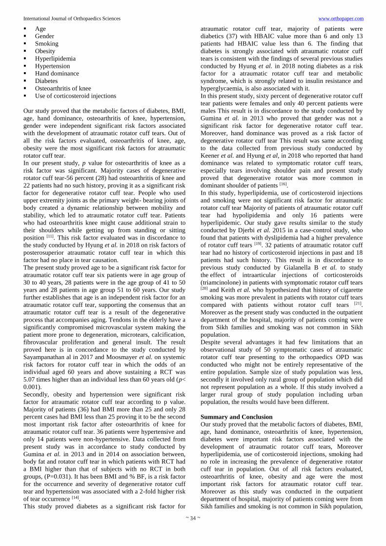
International Journal of Orthopaedics Sciences www.orthopaper.com Case images
Fig 4: Showing 50 year old female with positive lift off and belly press test with subscapularis tendon tear on USG and MRI
References
1. Pandey V, Jaap Willems W. Rotator cuff tear: A detailed
update. Asia Pac J Sports Med Arthrosc Rehabil Technol
2015;2(1):1-14.
2. Yang Y, Qu J. The effects of hyperlipidemia on rotator
cuff diseases: a systematic review. J Orthop Surg Res
2018;13(1):204.
3. Barry JJ, Lansdown DA, Cheung S, Feeley BT, Ma CB.
The relationship between tear severity, fatty infiltration,
and muscle atrophy in the supraspinatus. J Shoulder and
Elbow Surg 2013;22(1):18-25.
4. Edwards P, Ebert J, Joss B, Bhabra G, Ackland T, Wang
A. Exercise rehabilitation in the non-operative
management of rotator cuff tears: a review of the
literature. Int J sports phys therapy 2016;11(2):279.
5. Keener JD, Steger-May K, Stobbs G, Yamaguchi K.
Asymptomatic rotator cuff tears: patient demographics
and baseline shoulder function. J Shoulder and Elbow
Surg 2010;19(8):1191-8.
6. Lai J, Gagnier JJ. The effect of lipid disorders on the risk
of rotator cuff disease: A systematic review and meta-
analysis. JBJS Open Access 2018;3(3).
7. Carbone S, Gumina S, Arceri V, Campagna V, Fagnani
C, Postacchini F. The impact of preoperative smoking
habit on rotator cuff tear: cigarette smoking influences
rotator cuff tear sizes. J Should Elbow Surg
2012;21(1):56-60.
8. Epidemiology of Shoulder Pain. (2018, January 16).
Physiopedia 2020.
9. Minagawa H, Yamamoto N, Abe H et al. Prevalence of
symptomatic and asymptomatic rotator cuff tears in the
general population: From mass-screening in one village. J
Orthop 2013;10(1):8-12.
10. Longo, Umile Giuseppe, Franceschi, Francesco, Ruzzini,
Laura et al. Higher fasting plasma glucose levels within
the normoglycaemic range and rotator cuff tears. Br J
sports med 2008;43:284-7.
11. Li Y, Fessel G, Georgiadis M, Snedeker JG. Advanced
glycation end-products diminish tendon collagen fiber
sliding. Matrix Biol 2013;32(3-4):169-77.
12. Patel RM, Gelber JD, Schickendantz MS. The weight-
bearing shoulder. JAAOS-J Am Acad Orthop Surg
2018;26(1):3-13.
13. Mehta SK, Teefey SA, Middleton W, Steger-May K,
Sefko JA, Keener JD. Prevalence and risk factors for
development of subscapularis and biceps pathology in
shoulders with degenerative rotator cuff disease:
a prospective cohort evaluation. J Shoulder Elbow Surg

~ 37 ~
International Journal of Orthopaedics Sciences www.orthopaper.com 2020;29(3):451-58.
14. Gumina S, Arceri V, Carbone S, Albino P, Passaretti D,
Campagna V et al. The association between arterial
hypertension and rotator cuff tear: the influence on
rotator cuff tear sizes. Journal of shoulder and elbow
surgery 2013;22(2):229-32.
15. Okoroha KR, Mehran N, Duncan J, Washington T,
Spiering T, Bey MJ et al. Characterization of rotator cuff
tears: ultrasound versus magnetic resonance imaging.
Orthopedics 2017;40(1):e124-30.
16. Park HB, Gwark JY, Im JH, Jung J, Na JB, Yoon CH.
Factors Associated with Atraumatic Posterosuperior
Rotator Cuff Tears. J Bone Joint Surg Am
2018;100(16):1397-405.
17. Codding JL, Keener JD. Natural history of degenerative
rotator cuff tears. Current reviews in musculoskeletal
medicine 2018;11(1):77-85.
18. Cuff IO. Rotator Cuff Pathology. ACSM's Sports
Medicine: A Comprehensive Review 2012;10:317.
19. Djerbi I, Chammas M, Mirous MP, Lazerges C, Coulet
B, Shoulder FS. Impact of cardiovascular risk factor on
the prevalence and severity of symptomatic full-thickness
rotator cuff tears. Orthopaedics & Traumatology: Surgery
& Research 2015;101(6):269-73.
20. Gialanella B, Prometti P. Effects of corticosteroids
injection in rotator cuff tears. Pain Medicine
2011;12(10):1559-65.
21. Park HB, Gwark JY, Im JH, Jung J, Na JB, Yoon CH.
Factors associated with atraumatic posterosuperior rotator
cuff tears. The Journal of Bone and Joint Surgery.
American Volume 2018;100(16):1397.