Embed Size (px)
Citation preview

PRACTICE LAB FINAL ASSIGNMENT
RAFAEL LUMAUIG

BODY REGIONS, CAVITIES, AND PLANES
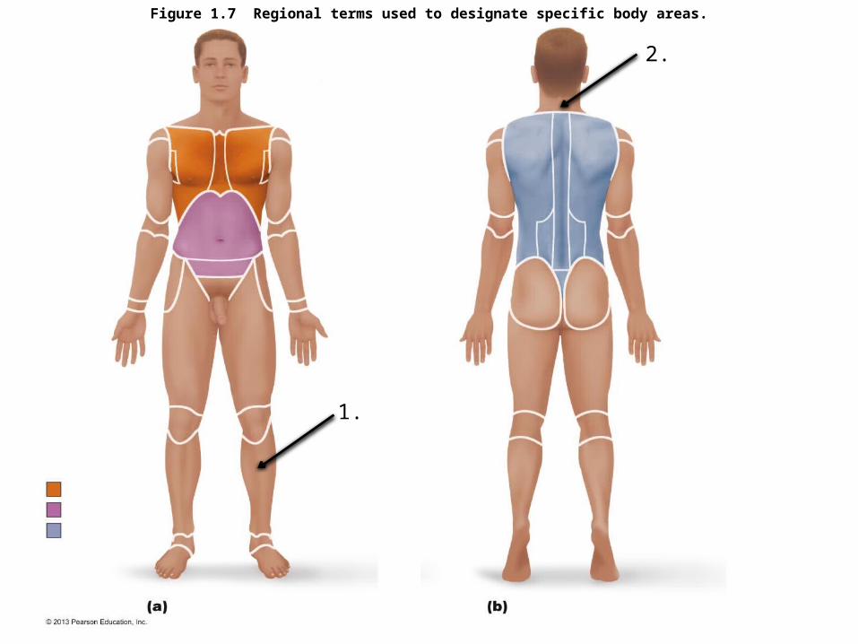
Figure 1.7 Regional terms used to designate specific body areas.
1.
2.
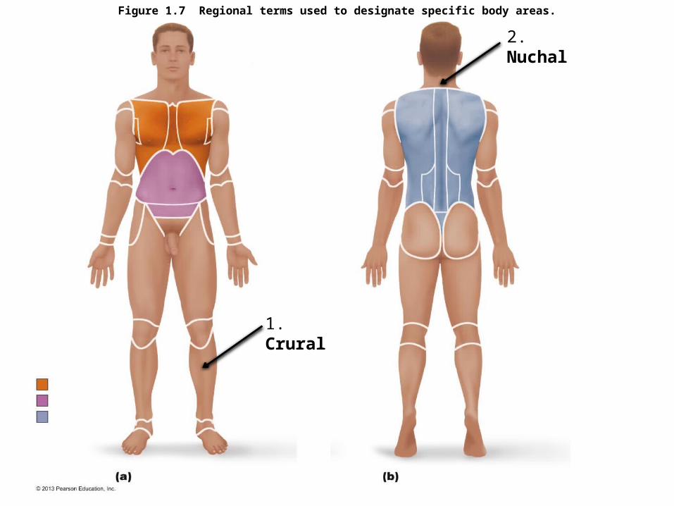
Figure 1.7 Regional terms used to designate specific body areas.
1. Crural
2. Nuchal

Figure 1.9a Dorsal and ventral body cavities and their subdivisions.
3.
4.
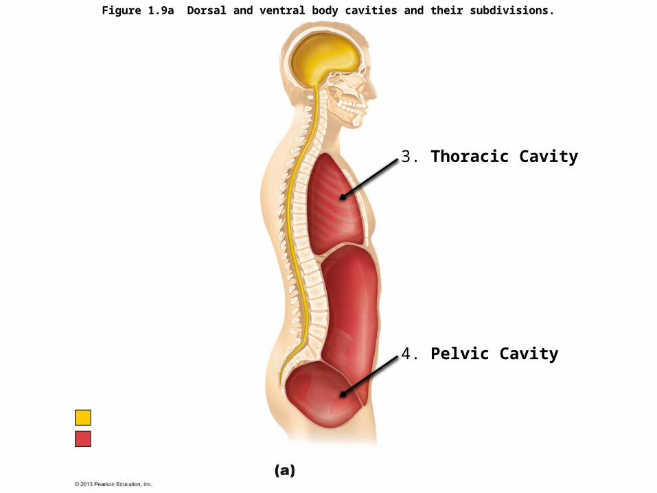
Figure 1.9a Dorsal and ventral body cavities and their subdivisions.
3. Thoracic Cavity
4. Pelvic Cavity
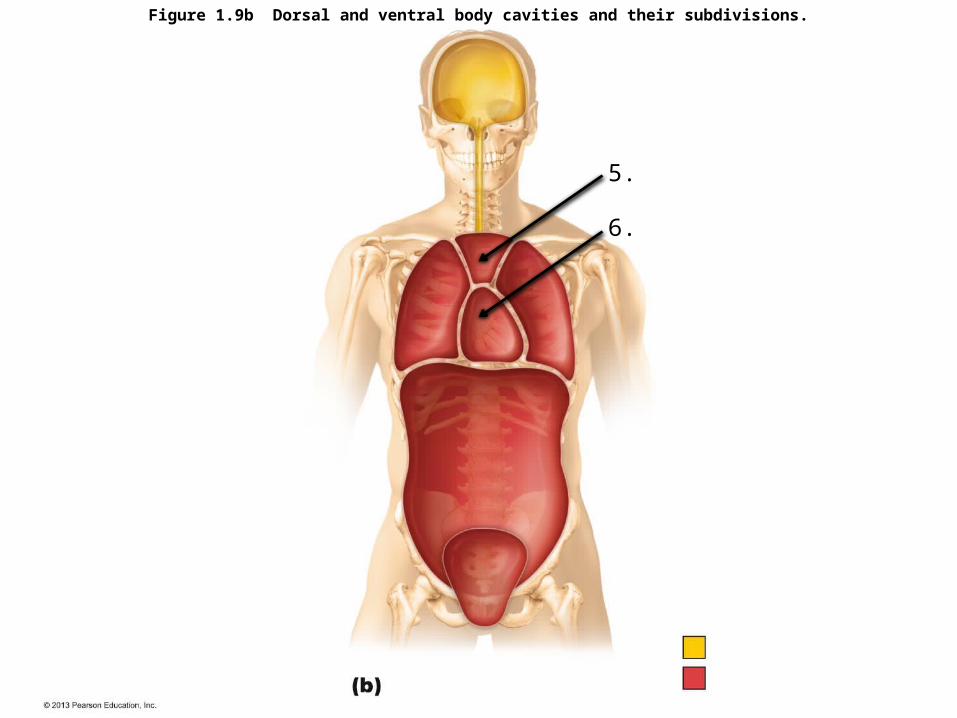
Figure 1.9b Dorsal and ventral body cavities and their subdivisions.
5.
6.

Figure 1.9b Dorsal and ventral body cavities and their subdivisions.
5. Superior Mediastinum
6. Pericardial Cavity
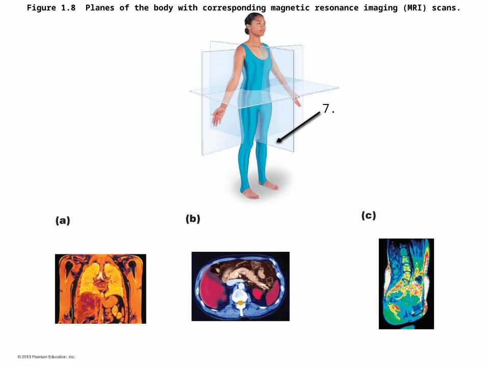
Figure 1.8 Planes of the body with corresponding magnetic resonance imaging (MRI) scans.
7.
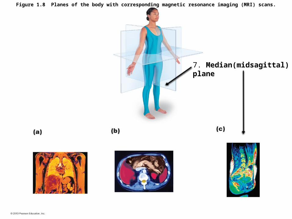
Figure 1.8 Planes of the body with corresponding magnetic resonance imaging (MRI) scans.
7. Median(midsagittal) plane
TISSUES

Figure 4.3d Epithelial tissues.
Goblet Cell
8. ??????????

Figure 4.3d Epithelial tissues.
Goblet Cell
8. Pseudostratified Columnar Epithelium

Figure 4.8h Connective tissues.
9. ??????????
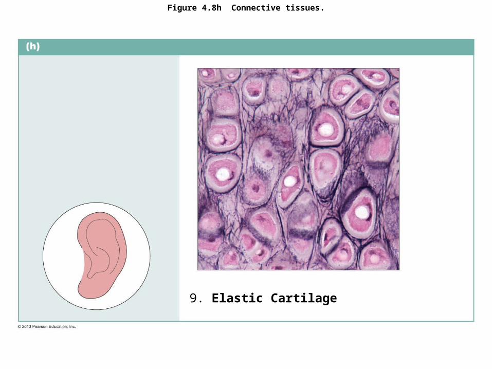
Figure 4.8h Connective tissues.
9. Elastic Cartilage
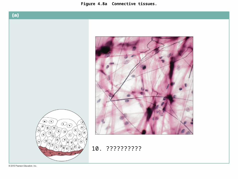
Figure 4.8a Connective tissues.
10. ??????????

Figure 4.8a Connective tissues.
10. Areolar Connective Tissue
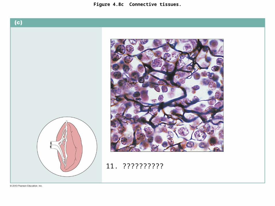
Figure 4.8c Connective tissues.
11. ??????????

Figure 4.8c Connective tissues.
11. Reticular Connective Tissue
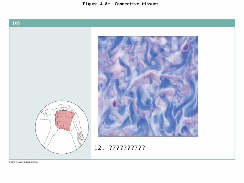
Figure 4.8e Connective tissues.
12. ??????????
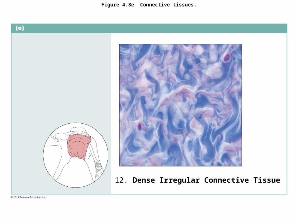
Figure 4.8e Connective tissues.
12. Dense Irregular Connective Tissue

Figure 4.10 Nervous tissue.
13. ??????????
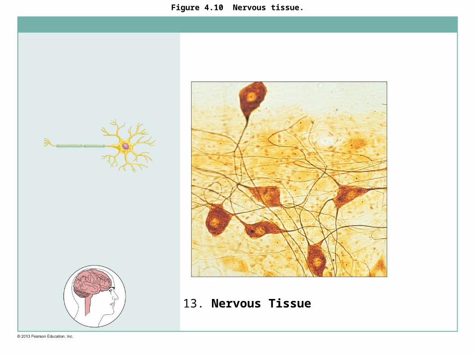
Figure 4.10 Nervous tissue.
13. Nervous Tissue
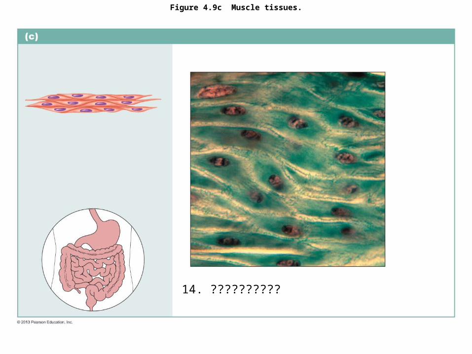
Figure 4.9c Muscle tissues.
14. ??????????

Figure 4.9c Muscle tissues.
14. Smooth Muscle Tissue

BONES
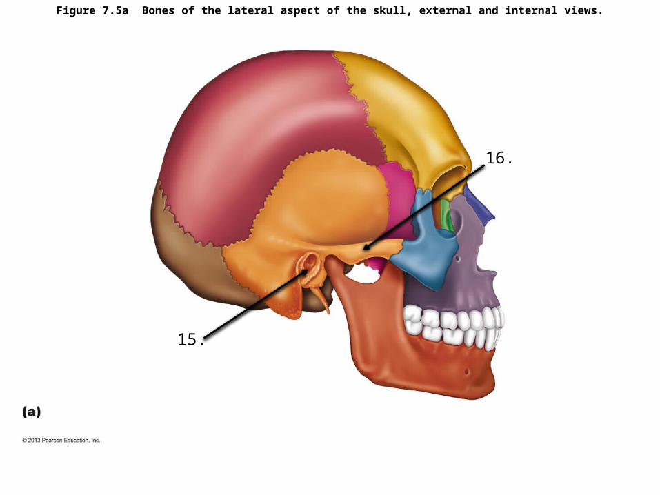
Figure 7.5a Bones of the lateral aspect of the skull, external and internal views.
15.
16.
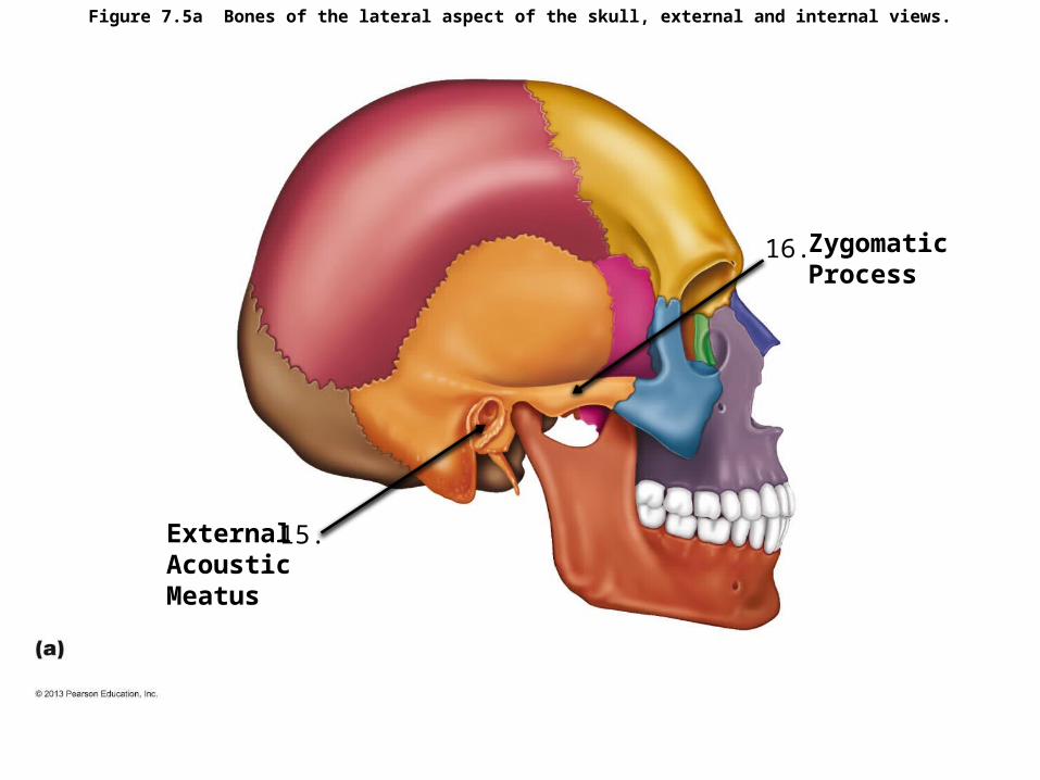
Figure 7.5a Bones of the lateral aspect of the skull, external and internal views.
15.
16.
External Acoustic Meatus
Zygomatic Process

Figure 7.4b Anatomy of the anterior and posterior aspects of the skull.
17.
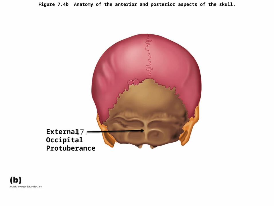
Figure 7.4b Anatomy of the anterior and posterior aspects of the skull.
17. External Occipital Protuberance
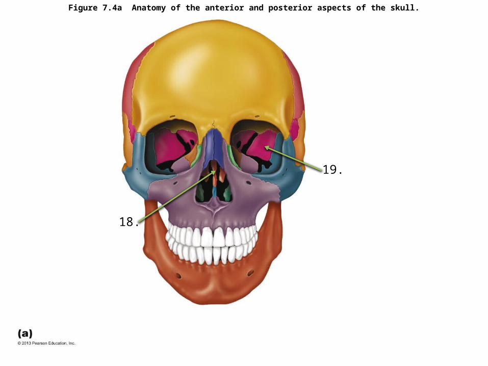
Figure 7.4a Anatomy of the anterior and posterior aspects of the skull.
18.
19.

Figure 7.4a Anatomy of the anterior and posterior aspects of the skull.
18.
19. Sphenoid Bone
Perpendicular Plate of Ethmoid Bone
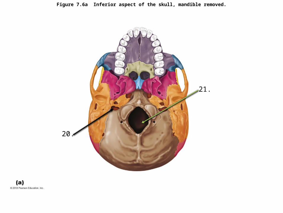
Figure 7.6a Inferior aspect of the skull, mandible removed.
20.
21.

Figure 7.6a Inferior aspect of the skull, mandible removed.
20.
21.
Jugular Canal
Foramen Magnum

Figure 7.15 The hyoid bone, anterior view.
22.

Figure 7.15 The hyoid bone, anterior view.
22. Hyoid Bone

Figure 7.20b The first and second cervical vertebrae.
23.
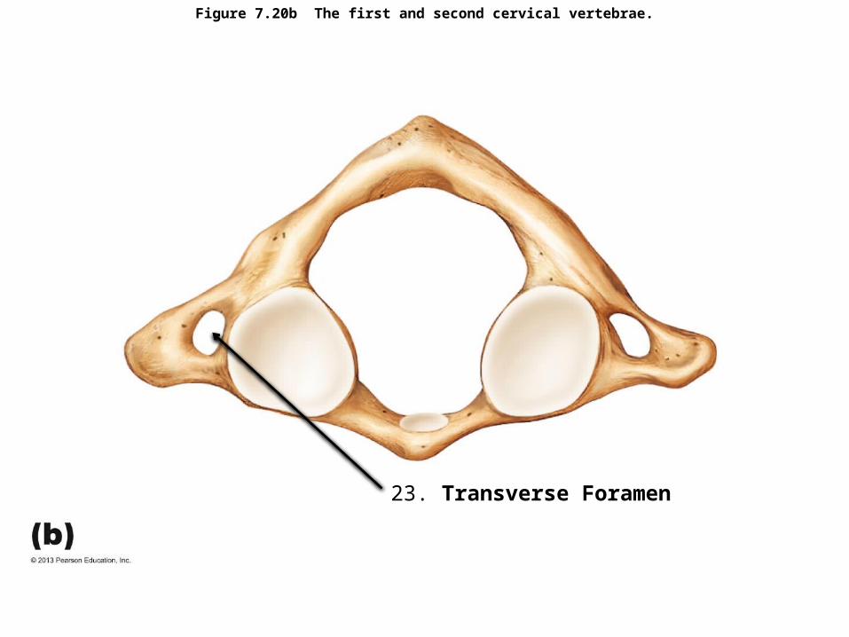
Figure 7.20b The first and second cervical vertebrae.
23. Transverse Foramen

Figure 7.26c The scapula.
24.
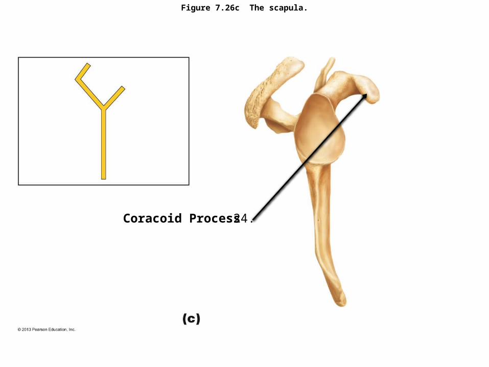
Figure 7.26c The scapula.
24.Coracoid Process
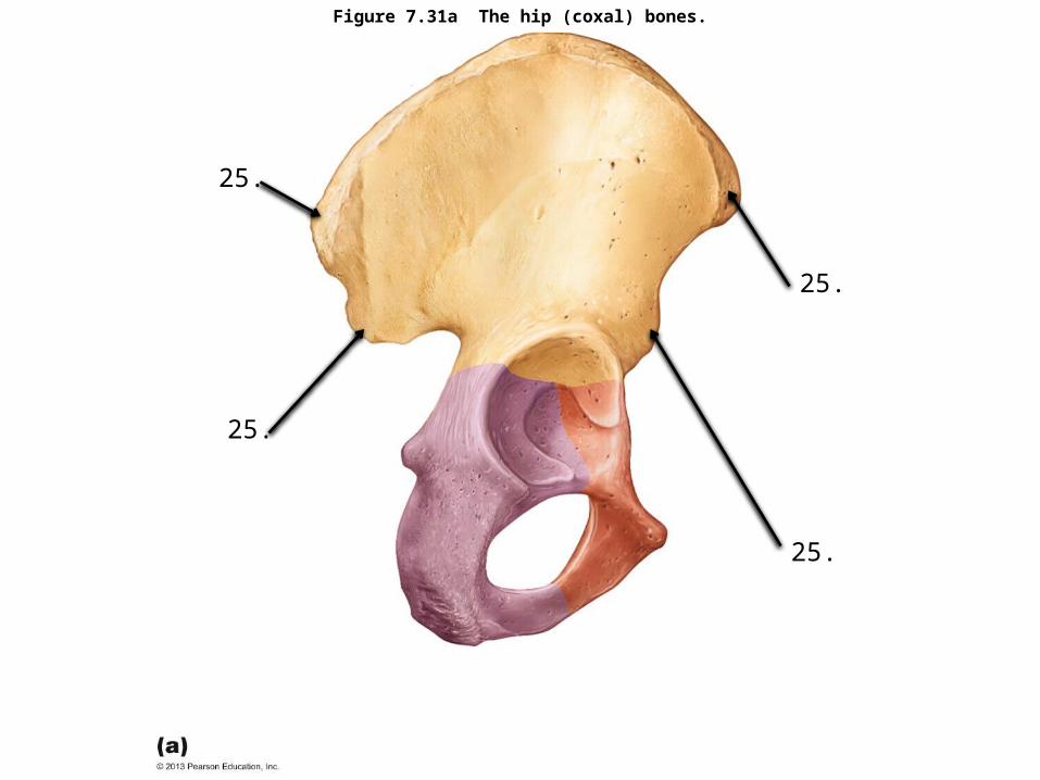
Figure 7.31a The hip (coxal) bones.
25.
25.
25.
25.
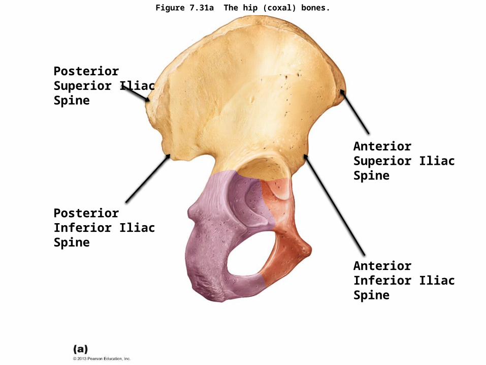
Figure 7.31a The hip (coxal) bones.
Posterior Superior Iliac Spine
Posterior Inferior Iliac Spine
Anterior Superior Iliac Spine
Anterior Inferior Iliac Spine

Figure 7.32b Bones of the right knee and thigh.
26. (ridge)

Figure 7.32b Bones of the right knee and thigh.
26. Linea Aspera

Figure 7.34c Bones of the right foot.
28.
27.
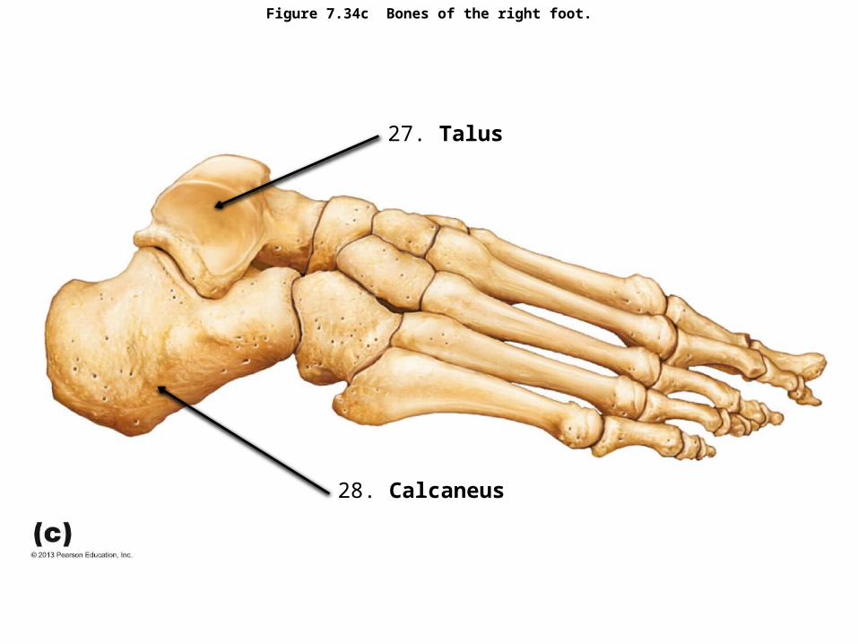
Figure 7.34c Bones of the right foot.
28. Calcaneus
27. Talus
JOINTS AND BODY MOVEMENTS

Figure 8.1a Fibrous joints.
29.

Figure 8.1a Fibrous joints.
29. Synarthrotic Fibrous Suture
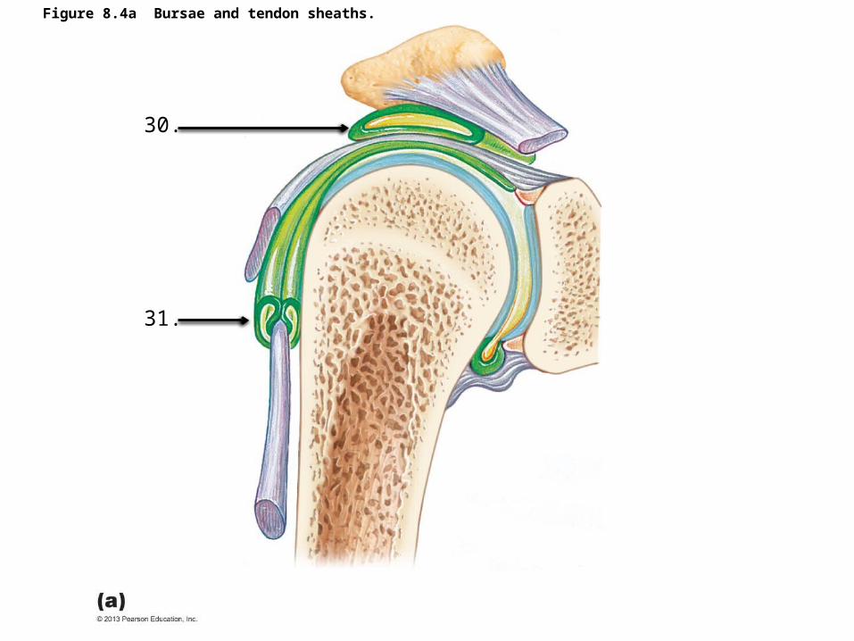
Figure 8.4a Bursae and tendon sheaths.
30.
31.
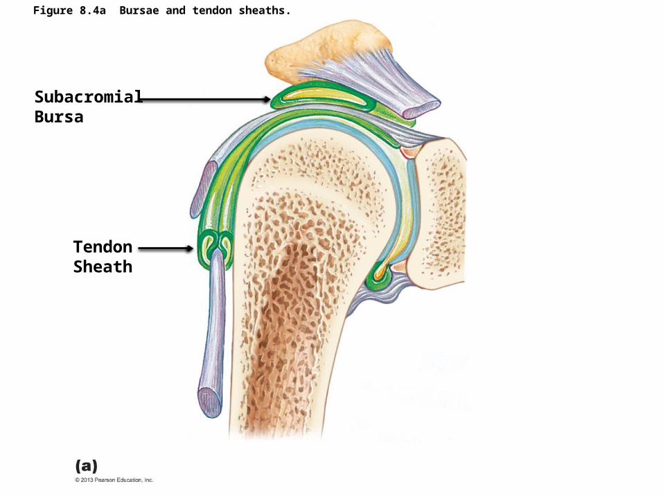
Figure 8.4a Bursae and tendon sheaths.
Tendon Sheath
Subacromial Bursa
Figure 8.5f Movements allowed by synovial joints.
32.
33.

Figure 8.5f Movements allowed by synovial joints.
Lateral Rotation
Medial Rotation

Figure 8.7e The shapes of the joint surfaces define the types of movements that can occur at a synovial joint; they also determine the classification of synovial joints into six structural types.
34. ??????????
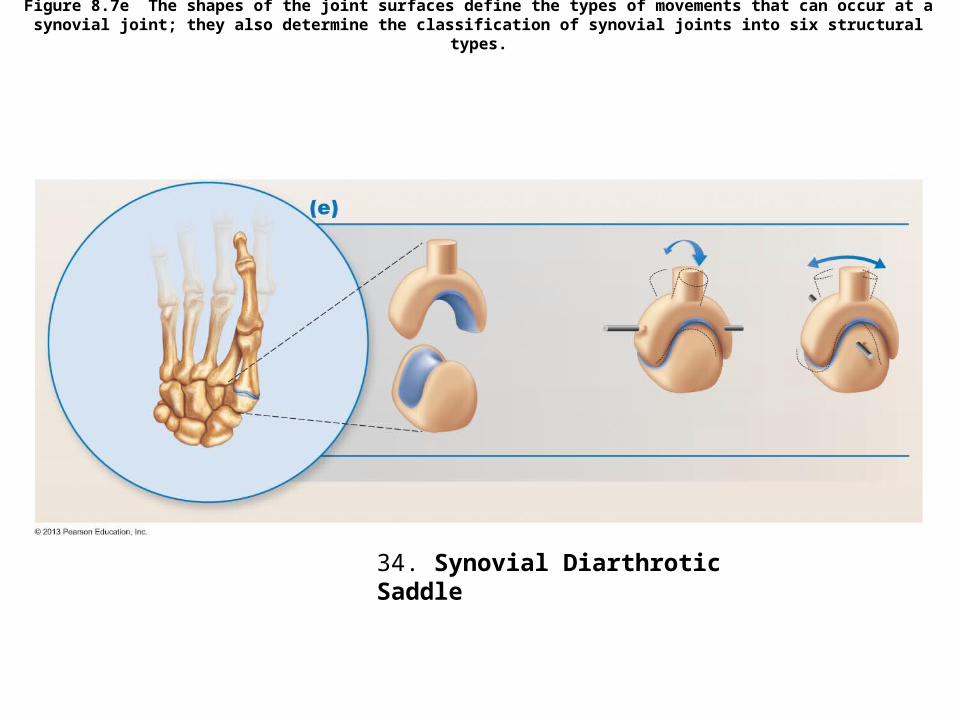
Figure 8.7e The shapes of the joint surfaces define the types of movements that can occur at a synovial joint; they also determine the classification of synovial joints into six structural types.
34. Synovial Diarthrotic Saddle
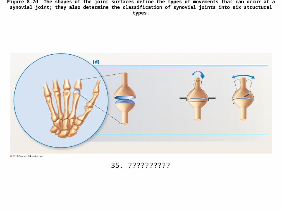
Figure 8.7d The shapes of the joint surfaces define the types of movements that can occur at a synovial joint; they also determine the classification of synovial joints into six structural types.
35. ??????????
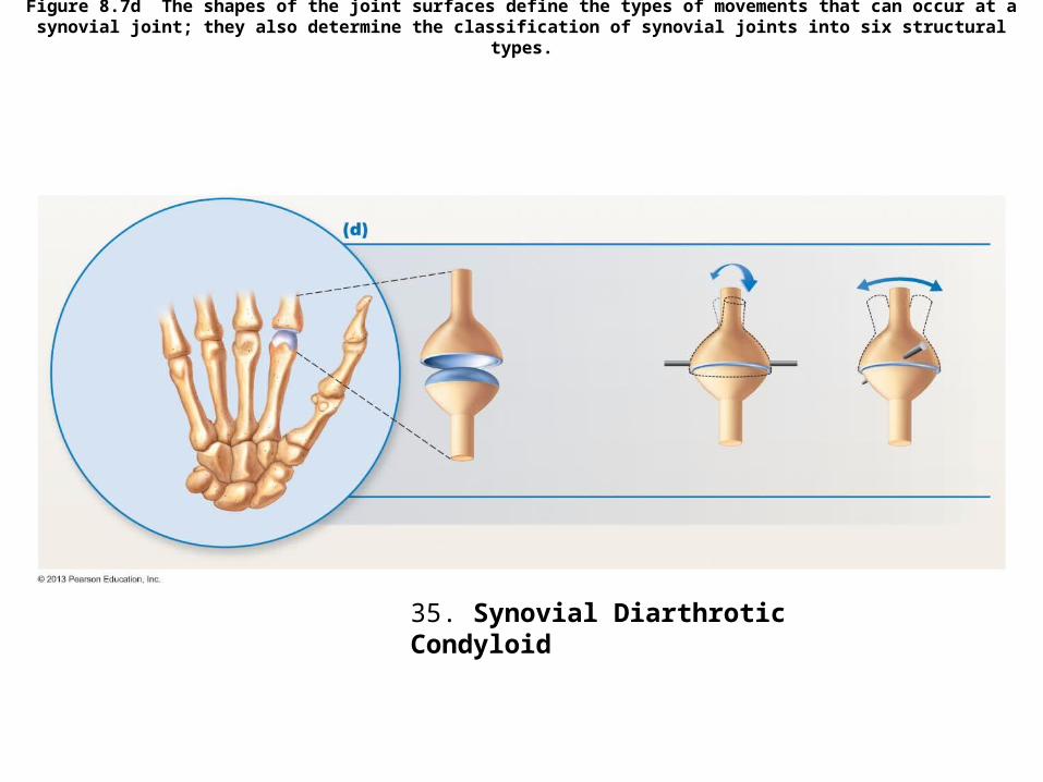
Figure 8.7d The shapes of the joint surfaces define the types of movements that can occur at a synovial joint; they also determine the classification of synovial joints into six structural types.
35. Synovial Diarthrotic Condyloid

MUSCLES

Figure 10.7b Lateral view of muscles of the scalp, face, and neck.
36.
39.
38.37.
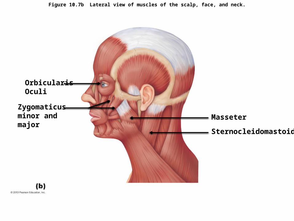
Figure 10.7b Lateral view of muscles of the scalp, face, and neck.
Orbicularis Oculi
Sternocleidomastoid
MasseterZygomaticus minor and major

Figure 10.12a Muscles of the abdominal wall.
40.
41.
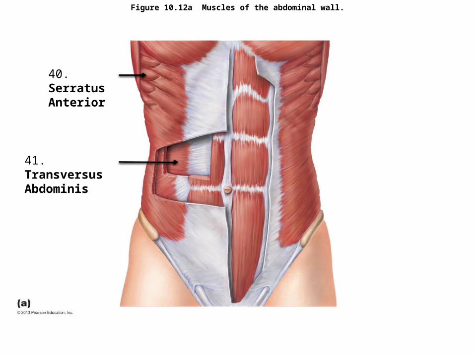
Figure 10.12a Muscles of the abdominal wall.
40. Serratus Anterior
41. Transversus Abdominis

Figure 10.14c Superficial muscles of the thorax and shoulder acting on the scapula and arm.
42.43.
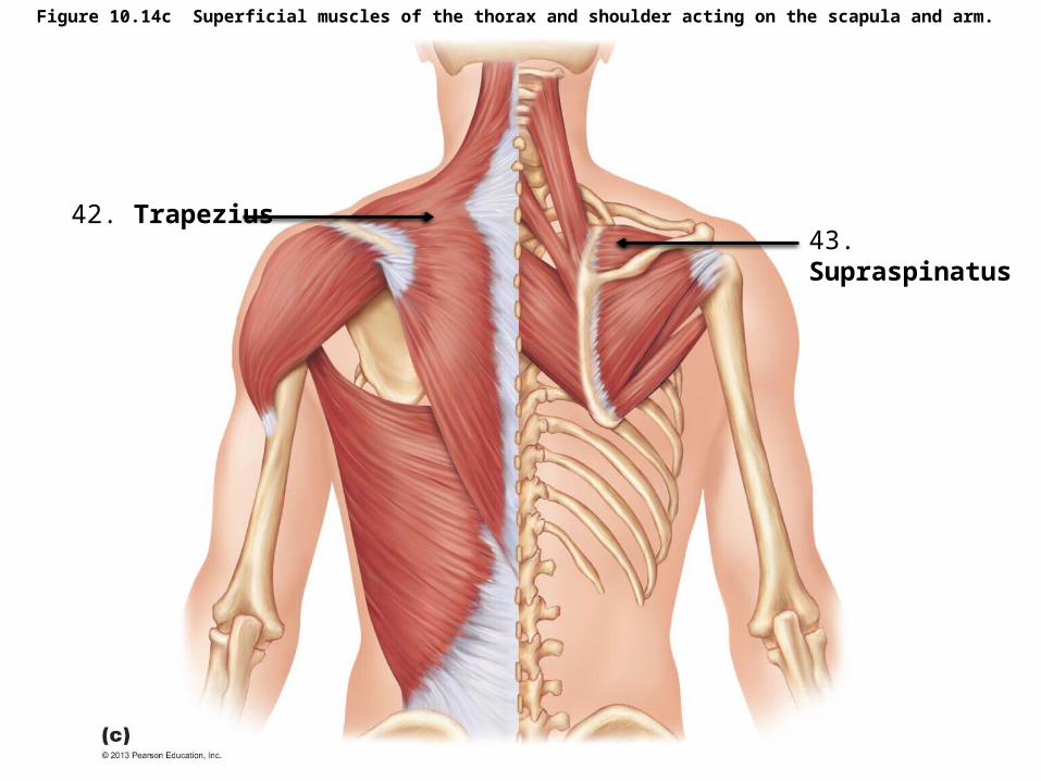
Figure 10.14c Superficial muscles of the thorax and shoulder acting on the scapula and arm.
42. Trapezius43. Supraspinatus
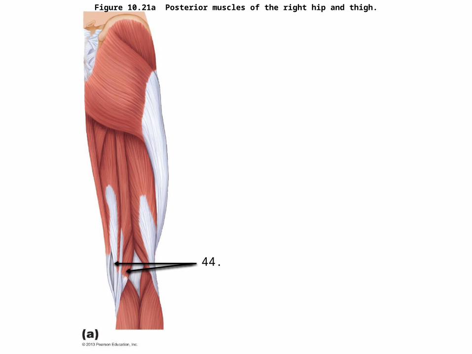
Figure 10.21a Posterior muscles of the right hip and thigh.
44.
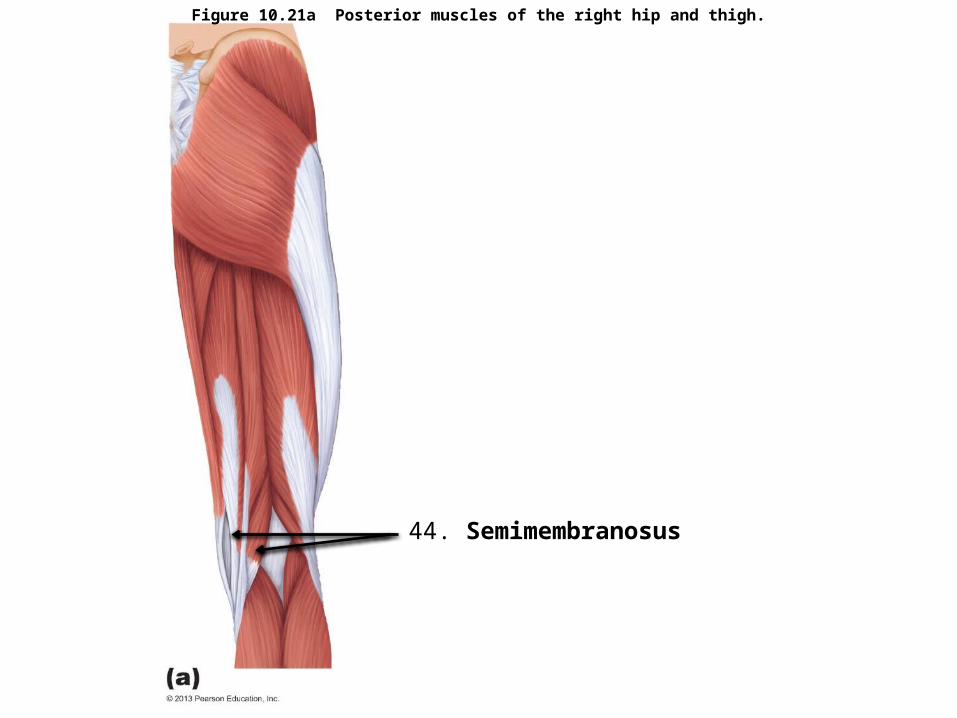
Figure 10.21a Posterior muscles of the right hip and thigh.
44. Semimembranosus
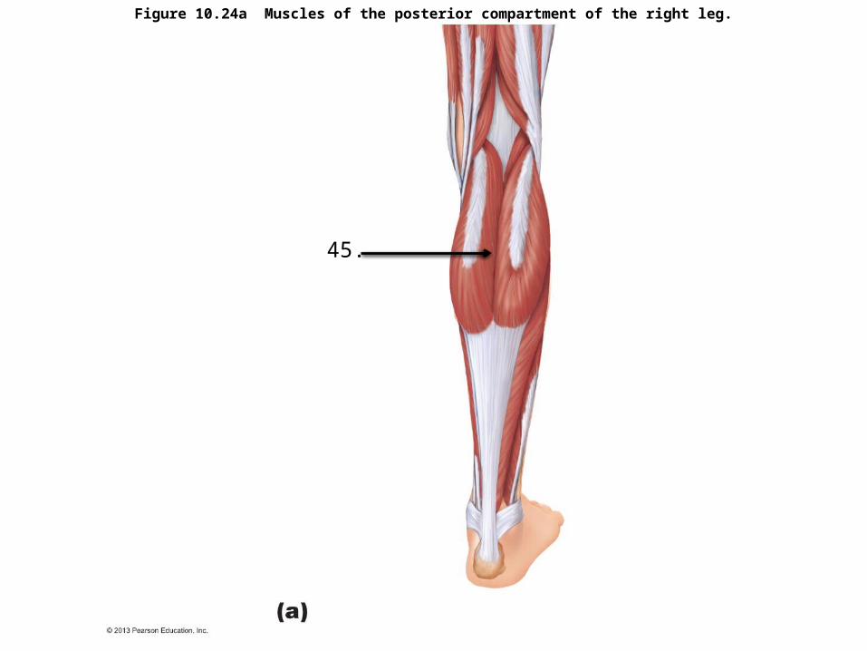
Figure 10.24a Muscles of the posterior compartment of the right leg.
45.

Figure 10.24a Muscles of the posterior compartment of the right leg.
45. Gastrocnemius

BRAIN AND SPINAL CORD

Figure 12.3 Ventricles of the brain.
46.

Figure 12.3 Ventricles of the brain.
Fourth ventricle

Figure 13.6a Location and function of cranial nerves.
47.
48.
49.
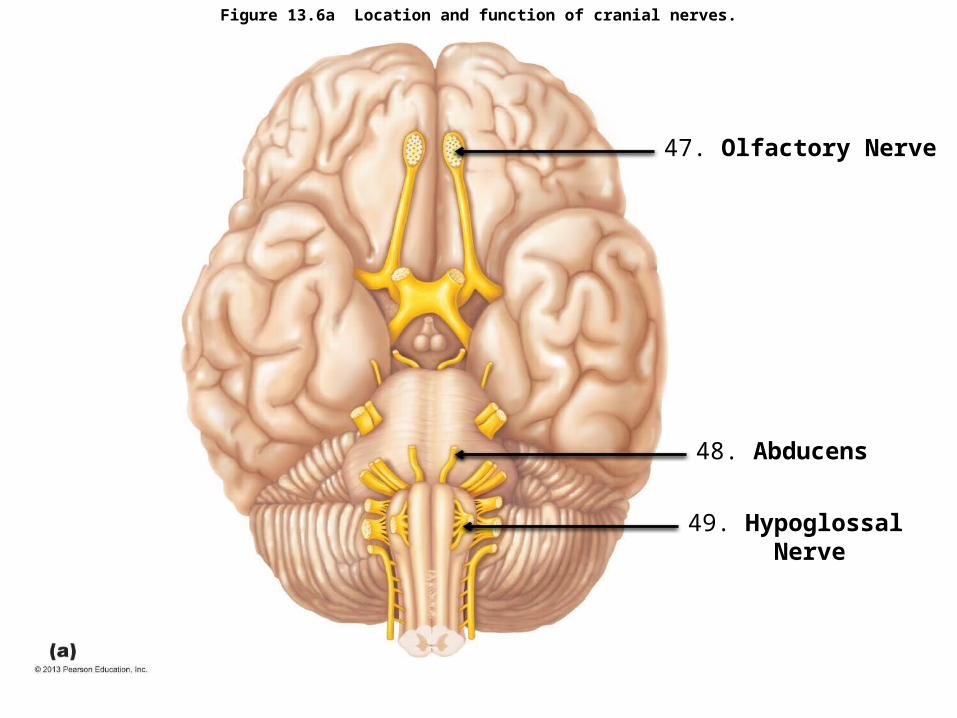
Figure 13.6a Location and function of cranial nerves.
47. Olfactory Nerve
48. Abducens
49. Hypoglossal Nerve
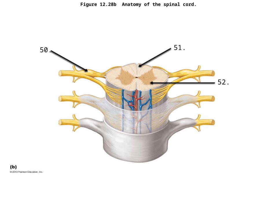
Figure 12.28b Anatomy of the spinal cord.
50. 51.
52.
Figure 12.28b Anatomy of the spinal cord.
50. Dorsal root ganglion
51. Dorsal Median Sulcus
52. Ventral Horn of Gray

EYE

Figure 15.4a Internal structure of the eye (sagittal section).
53.
55.
54.
56.
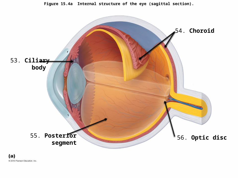
Figure 15.4a Internal structure of the eye (sagittal section).
53. Ciliary body
55. Posterior segment
54. Choroid
56. Optic disc

EAR

Figure 15.24a Structure of the ear.
57.
58.
59. 60.
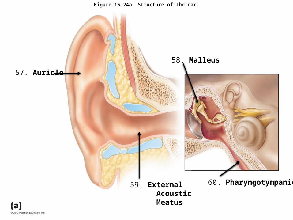
Figure 15.24a Structure of the ear.
57. Auricle
58. Malleus
59. External Acoustic Meatus
60. Pharyngotympanic