Embed Size (px)
Citation preview
Author's personal copy
p75 neurotrophin receptor signalingin nervous system injury anddegeneration: paradox and opportunityCarlos F. Ibanez and Anastasia Simi
Department of Neuroscience, Karolinska Institute, 17177 Stockholm, Sweden
Injury or insult to the adult nervous system often results inreactivation of signaling pathways that are normally onlyactive during development. The p75 neurotrophinreceptor (p75NTR) is one such signaling molecule whoseexpression increases markedly following neural injury inmany of the same cell types that express p75NTR duringdevelopment. A series of studies during the past decadehas demonstrated that p75NTR signaling contributes toneuronal and glial cell damage, axonal degeneration anddysfunction during injury and cellular stress. Why thenervous system reacts to injury by inducing a moleculethat aids the demise of cells and axons is a biologicalparadox that remains to be explained satisfactorily. Onthe other hand, it may offer unique therapeutic opportu-nities for limiting the severity of nervous system injuryand disease.
IntroductionNeurotrophic factors are secreted proteins that play keyroles in most aspects of the life of neurons, including theirgeneration, survival, migration, maturation, axonalgrowth, synaptic connectivity and injury responses [1].Most importantly, neurotrophic factors are required forthe sustained function of the adult nervous system andshow potent neuroprotective and regenerative functions inanimal models of neurodegenerative diseases and neuro-trauma [2]. These properties have made neurotrophicfactors prime candidates for therapeutic interventionagainst diseases of the nervous system, and have attractedtremendous interest from academia, biotech and pharma-ceutical industries.
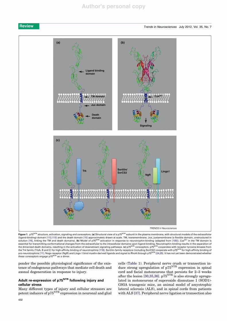
p75NTR is a transmembrane receptor for neurotrophicfactors of the neurotrophin family (Figure 1a,b) whichincludes nerve growth factor (NGF), brain-derived neuro-trophic factor (BDNF), neurotrophin-3 (NT-3) and neuro-trophin-4 (NT-4) [1,3–6]. In addition to p75NTR,neurotrophins signal via TrkA, B and C members of thereceptor tyrosine kinase family [1,7]. The two receptorsystems can function synergistically, antagonistically orindependently of each other in different cell types [8,9](Figure 1c). Neurotrophins are initially synthesized as pre-cursors (pro-neurotrophins) which are then cleaved to pro-duce mature proteins. It has more recently been appreciatedthat pro-neurotrophins can also be released and have
biological actions on cells [10]. The pro-domain interfereswith Trk receptor binding and activation, and thus renderspro-neurotrophins selective p75NTR ligands. On the otherhand, the pro-domain interacts with the sorting receptorsortilin, which then associates and cooperates with p75NTR
to bind pro-neurotrophins with high affinity [11] (Figure 1c).Pro-neurotrophins can induce cell death [12] and decreasesynaptic function [13,14], and thereby in many ways opposethe functions of mature neurotrophins [9].
Similar to other members of the tumor necrosis factorreceptor (TNFR) superfamily, p75NTR contains a so-calleddeath domain in its intracellular region [15] (Figure 1a).The death domain mediates interactions with other cellu-lar components, allowing the receptor to regulate intracel-lular signaling events [3,4]. Some of the pathwaysregulated by p75NTR in response to neurotrophins includeNF-kB [16], c-Jun kinase (JNK) [17,18] and caspases [19].p75NTR can also activate the small GTPase RhoA [20], butthis requires a different set of ligands derived from myelin,such as myelin-associated glycoprotein (MAG) and Nogo[21–23], and two different coreceptors: a lipid-anchoredligand-binding subunit known as the Nogo receptor(NgR), and Lingo-1 [24,25] (Figure 1c). When activatedin cultured neurons, this pathway leads to growth-conecollapse and inhibition of axonal growth [26]. Intriguingly,whereas myelin-derived ligands induce RhoA activitythrough p75NTR, neurotrophin binding to p75NTR has beenshown to downregulate RhoA activity in neurons [20,27].
p75NTR is widely expressed in the developing nervoussystem. Sensory and sympathetic neurons, spinal cord andbrainstem motoneurons, and neurons in the cerebral cortex,cerebellum, hippocampus, basal forebrain and caudateputamen all express p75NTR at some stage of their develop-ment [28–31]. Subpopulations of peripheral and central glialcells, including radial glial and neural stem cells, alsoexpress p75NTR at various stages of development [32]. Inmost cells, p75NTR expression is switched off at adult stages.A few areas, however, retain p75NTR expression at lowerlevels, including basal forebrain cholinergic neurons,sensory neurons and spinal cord motoneurons [28,30,33,34].
After reviewing some of the effects of neural injury onp75NTR expression, this article summarizes some of thefunctions of this receptor in various injury and cell-deathparadigms, as well as axonal pruning and degeneration. Inclosing, we highlight the therapeutic opportunities lyingahead through manipulation of p75NTR signaling, and
Review
Corresponding author: Ibanez, C.F. ([email protected]).Keywords: ALS; Alzheimer’s disease; apoptosis; degeneration; injury; neurotrophin.
0166-2236/$ – see front matter ! 2012 Elsevier Ltd. All rights reserved. doi:10.1016/j.tins.2012.03.007 Trends in Neurosciences, July 2012, Vol. 35, No. 7 431
Author's personal copy
ponder the possible physiological significance of the exis-tence of endogenous pathways that mediate cell death andaxonal degeneration in response to injury.
Adult re-expression of p75NTR following injury andcellular stressMany different types of injury and cellular stressors arepotent inducers of p75NTR expression in neuronal and glial
cells (Table 1). Peripheral nerve crush or transection in-duce strong upregulation of p75NTR expression in spinalcord and facial motoneurons that persists for 2–3 weeksafter the lesion [30,35,36]. p75NTR is also strongly upregu-lated in motoneurons of superoxide dismutase 1 (SOD1)-G93A transgenic mice, an animal model of amyotrophiclateral sclerosis (ALS), and in spinal cords from patientswith ALS [37]. Peripheral nerve ligation or transection also
(a)
Ligand bindingdomain
Cys257
Signaling
TM domain
Jux domain
Deathdomain
Trk Sortilin/SorCS2
NgRLingo1
(b)
(c)
TRENDS in Neurosciences
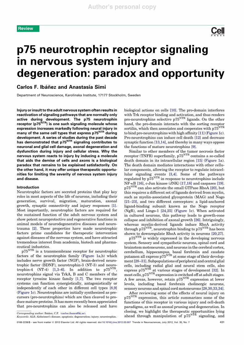
Figure 1. p75NTR structure, activation, signaling and coreceptors. (a) Structural view of a p75NTR subunit in the plasma membrane, with structural models of the extracellular(ligand-binding) domain [112,113] and the death domain [15] approximately drawn at scale. TM, transmembrane. Jux, juxtamembrane (a flexible domain, unstructured insolution [15], linking the TM and death domains). (b) Model of p75NTR activation in response to neurotrophin-binding (adapted from [105]). Cys257 in the TM domain isessential for transmitting conformational changes from the extracellular to the intracellular domains upon ligand-binding. Neurotrophin-binding results in the separation ofthe dimerized death domains, resulting in the activation of downstream signaling pathways. (c) p75NTR coreceptors. p75NTR cooperates with receptor tyrosine kinases fromthe Trk family (TrkA, B and C) for high-affinity binding of neurotrophins [118]. Sortilin-family receptors (including SorCS2) cooperate with p75NTR for high-affinity binding ofpro-neurotrophins [11]. Nogo receptor (NgR) and Lingo-1 bind myelin-derived ligands and signal to RhoA through p75NTR [24,25]. It has not yet been demonstrated whetherthese coreceptors engage p75NTR as a dimer.
Review Trends in Neurosciences July 2012, Vol. 35, No. 7
432
Author's personal copy
lead to increased p75NTR expression in glial cells andsensory neurons [38–40]. Schwann cells in distal sciaticnerve segments upregulate p75NTR expression after axot-omy [41,42] and demyelinating lesions [43]. Thoracic con-tusion of the spinal cord leads to induction of p75NTR
expression in oligodendrocytes [44]. p75NTR is also stronglyupregulated in oligodendrocytes of plaques extracted frombrains of patients with multiple sclerosis [45]. In theretina, optic nerve axotomy [46], ocular hypertension[47] and ischemic injury [48] all result in elevatedp75NTR expression. In the brain, axotomy results in in-creased p75NTR expression in corticospinal neurons [49]and in cerebellar Purkinje cells [50]. p75NTR is stronglyupregulated by neuronal activation following seizures inthe hippocampus [19,51] and excitotoxic insults to basalforebrain cholinergic neurons [52]. Ischemic strokeincreases p75NTR expression in striatal interneurons[53]. Hypo-osmolar stress induces p75NTR in cortical neu-rons by O-GlcNAcylation of the Specificity Protein 1 (Sp1)transcription factor [54,55]. Finally, cortical and basalforebrain cholinergic neurons from patients with Alzhei-mer’s disease (AD) also show elevated levels of p75NTR
[56,57].Intriguingly, neural injury and stress induce p75NTR in
cells that expressed the receptor at some earlier point indevelopment. p75NTR re-expression may thus be part of aprogram of lesion-induced plasticity that to some extentrecapitulates developmental mechanisms. Most of the ear-lier work reporting elevated p75NTR expression after ner-vous system lesions and neuronal stress did not address itsfunctional consequences. Two decades ago it was widelyexpected that, as a neurotrophin receptor, p75NTR wouldplay some kind of a protective role during injury. However,the subsequent finding that p75NTR plays a role in neuro-nal death turned the intellectual tide, leading to the dis-covery of pathogenic activities of this receptor in neuralinjury and disease.
Effects of p75NTR signaling in neuronal and glial celldeath after injuryA role for p75NTR in apoptotic cell death emerged duringthe early 1990s following observations of p75NTR-mediatedcell death, first in transfected cells [58], but later throughendogenous receptors in sensory neurons [59], cells of theisthmo-optic nucleus [60], retinal neurons [61], oligoden-drocytes [62], and sympathetic [63] and hippocampal neu-rons [17]. Two possible physiological scenarios wereenvisaged for the role of p75NTR in cell death, namelydevelopment and injury. Several examples of a role forp75NTR in promoting cell death during development havebeen uncovered [64,65].
With regards to injury, it was first shown by Bartlettand colleagues in 1996 that reduction of p75NTR levelsusing antisense oligonucleotides could prevent the lossof axotomized neurons in dorsal root ganglia, thus impli-cating p75NTR in injury-mediated cell death [66]. Numer-ous subsequent studies have highlighted a role for p75NTR
in neuronal or glial cell death after injury (Table 1). Forinstance, blocking p75NTR signaling with neutralizing anti-bodies prevented the death of corticospinal neurons afteraxotomy [49]. A comparable result has also been obtainedin sortilin knockout mice [67], thus implicating pro-neuro-trophins in this effect. Survival and regeneration of axo-tomized motoneurons were found to be improved in p75NTR
knockout mice compared to control animals [68,69]. Deathof Schwann cells after sciatic nerve axotomy was alsodiminished in p75NTR knockout mice [41]. Intriguingly,in vitro studies have suggested that p75NTR is able toinduce either death or survival in Schwann cells, depend-ing upon expression of receptor-interacting serine/threo-nine-protein kinase 2 (RIP2), an adaptor protein for thereceptor [70]. As pointed out earlier [71], the effects ofp75NTR on Schwann cells after injury may be complex giventhe role of p75NTR in myelination [72]. Indeed, in a model ofremyelination after peripheral nerve injury, transplanted
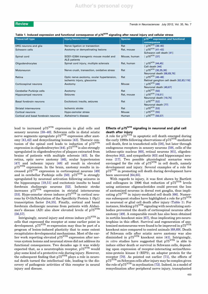
Table 1. Induced expression and functional consequence of p75NTR signaling after neural injury and cellular stress
Tissue/cell type Injury/lesion/model Species p75NTR expression and functionalconsequence
DRG neurons and glia Nerve ligation or transection Rat " p75NTR [38–40]
Schwann cells Axotomy or demyelinating lesions Rat, mouse " p75NTR [41–43]Schwann cell death [41]
Spinal cord SOD1-G39A transgenic mouse model andALS patients
Mouse, human " p75NTR [37]
Oligodendrocytes Spinal cord injury, multiple sclerosis Rat, human " p75NTR [44,45]Cell death [44]
Motoneuron Nerve crush, transection, oxidative stress Rat " p75NTR [30,35,36]Neuronal death [68,69,76]
Retina Optic nerve axotomy, ocular hypertension,ischemic injury, glaucoma
Rat " p75NTR [46–48]Retinal ganglion cell death [82,83,116]
Corticospinal neurons Axotomy Mouse " p75NTR [49]Neuronal death [49,67]
Cerebellar Purkinje cells Axotomy Rat " p75NTR [50]
Hippocampal neurons Seizures Rat, mouse " p75NTR [19,51]Neuronal death [19,75]
Basal forebrain neurons Excitotoxic insults, seizures Rat " p75NTR [52]Neuronal death [77]
Striatal interneurons Ischemic stroke Rat " p75NTR [53]
Cortical neurons Hypo-osmolar stress Mouse " p75NTR [55]
Cortical and basal forebrain neurons Alzheimer’s disease Human " p75NTR [56,57]
Review Trends in Neurosciences July 2012, Vol. 35, No. 7
433
Author's personal copy
wild-type Schwann cells were better at remyelinatinglesioned nerves than were cells lacking p75NTR [73], andspontaneous remyelination after sciatic nerve crush wasimpaired in the absence of p75NTR [74].
In the hippocampus, pilocarpine-induced seizures inducep75NTR expression, caspase-3 activity and neuronal death.Pilocarpine-induced neuronal death was attenuated in micelacking p75NTR or neurotrophin receptor-interacting factor(NRIF), a p75NTR effector protein [19,75]. These studiesestablished for the first time the role of caspase-6 as anupstream activator of caspase-3 in the apoptotic pathwayactivated by p75NTR in hippocampal neurons [19]. Apoptoticdeath of oligodendrocytes after spinal cord injury was alsodiminished in p75NTR knockout mice [44]. In this case, thelesion resulted in the production of the pro-NGF precursor atthe site of injury, which was capable of inducing potentoligodendrocyte death in a p75NTR-dependent manner[44]. In another study, pro-NGF produced by reactive astro-cytes in response to oxidative stress promoted motoneuroncell death in a p75NTR-dependent manner [76]. Pro-neuro-trophins were also found to be produced by basal forebrainastrocytes after kainic acid-induced seizures [77]. The sei-zures elevated p75NTR and sortilin expression and inducedcaspase-3 activation in basal forebrain neurons, but fewerdying neurons were found in brains of mice lacking p75NTR
[77]. Interestingly, this study found that the effects of pro-neurotrophins on cell death were dominant over the survivalactivity of mature neurotrophins in cultures of basal fore-brain neurons [77]. Neural injury therefore alters the ratio ofpro- to mature neurotrophins, thus shifting the balance fromsurvival to death. This finding is significant in light ofseveral reports indicating elevated levels of pro-neurotro-phins in the brains of AD patients [78–80].
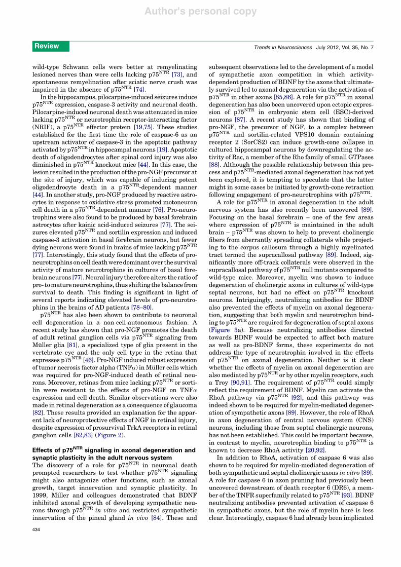
p75NTR has also been shown to contribute to neuronalcell degeneration in a non-cell-autonomous fashion. Arecent study has shown that pro-NGF promotes the deathof adult retinal ganglion cells via p75NTR signaling fromMu ller glia [81], a specialized type of glia present in thevertebrate eye and the only cell type in the retina thatexpresses p75NTR [46]. Pro-NGF induced robust expressionof tumor necrosis factor alpha (TNFa) in Mu ller cells whichwas required for pro-NGF-induced death of retinal neu-rons. Moreover, retinas from mice lacking p75NTR or sorti-lin were resistant to the effects of pro-NGF on TNFaexpression and cell death. Similar observations were alsomade in retinal degeneration as a consequence of glaucoma[82]. These results provided an explanation for the appar-ent lack of neuroprotective effects of NGF in retinal injury,despite expression of prosurvival TrkA receptors in retinalganglion cells [82,83] (Figure 2).
Effects of p75NTR signaling in axonal degeneration andsynaptic plasticity in the adult nervous systemThe discovery of a role for p75NTR in neuronal deathprompted researchers to test whether p75NTR signalingmight also antagonize other functions, such as axonalgrowth, target innervation and synaptic plasticity. In1999, Miller and colleagues demonstrated that BDNFinhibited axonal growth of developing sympathetic neu-rons through p75NTR in vitro and restricted sympatheticinnervation of the pineal gland in vivo [84]. These and
subsequent observations led to the development of a modelof sympathetic axon competition in which activity-dependent production of BDNF by the axons that ultimate-ly survived led to axonal degeneration via the activation ofp75NTR in other axons [85,86]. A role for p75NTR in axonaldegeneration has also been uncovered upon ectopic expres-sion of p75NTR in embryonic stem cell (ESC)-derivedneurons [87]. A recent study has shown that binding ofpro-NGF, the precursor of NGF, to a complex betweenp75NTR and sortilin-related VPS10 domain containingreceptor 2 (SorCS2) can induce growth-cone collapse incultured hippocampal neurons by downregulating the ac-tivity of Rac, a member of the Rho family of small GTPases[88]. Although the possible relationship between this pro-cess and p75NTR-mediated axonal degeneration has not yetbeen explored, it is tempting to speculate that the lattermight in some cases be initiated by growth-cone retractionfollowing engagement of pro-neurotrophins with p75NTR.
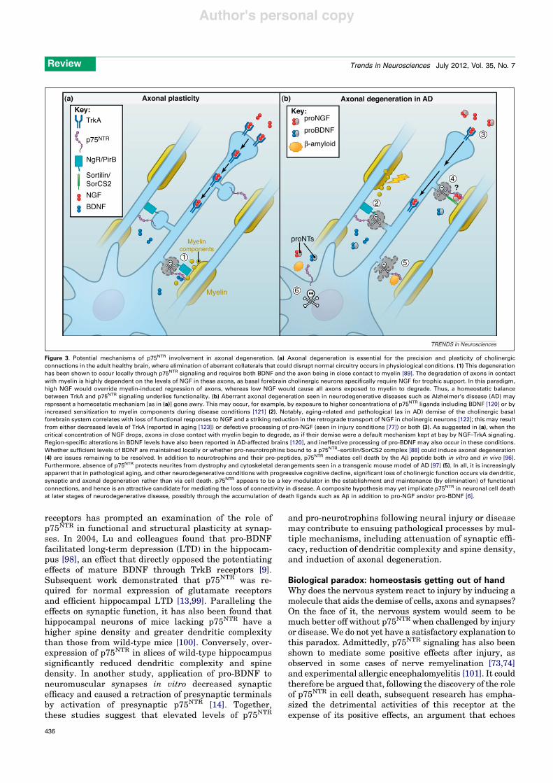
A role for p75NTR in axonal degeneration in the adultnervous system has also recently been uncovered [89].Focusing on the basal forebrain – one of the few areaswhere expression of p75NTR is maintained in the adultbrain – p75NTR was shown to help to prevent cholinergicfibers from aberrantly spreading collaterals while project-ing to the corpus callosum through a highly myelinatedtract termed the supracallosal pathway [89]. Indeed, sig-nificantly more off-track collaterals were observed in thesupracallosal pathway of p75NTR null mutants compared towild-type mice. Moreover, myelin was shown to inducedegeneration of cholinergic axons in cultures of wild-typeseptal neurons, but had no effect on p75NTR knockoutneurons. Intriguingly, neutralizing antibodies for BDNFalso prevented the effects of myelin on axonal degenera-tion, suggesting that both myelin and neurotrophin bind-ing to p75NTR are required for degeneration of septal axons(Figure 3a). Because neutralizing antibodies directedtowards BDNF would be expected to affect both matureas well as pro-BDNF forms, these experiments do notaddress the type of neurotrophin involved in the effectsof p75NTR on axonal degeneration. Neither is it clearwhether the effects of myelin on axonal degeneration arealso mediated by p75NTR or by other myelin receptors, sucha Troy [90,91]. The requirement of p75NTR could simplyreflect the requirement of BDNF. Myelin can activate theRhoA pathway via p75NTR [92], and this pathway wasindeed shown to be required for myelin-mediated degener-ation of sympathetic axons [89]. However, the role of RhoAin axon degeneration of central nervous system (CNS)neurons, including those from septal cholinergic neurons,has not been established. This could be important because,in contrast to myelin, neurotrophin binding to p75NTR isknown to decrease RhoA activity [20,92].
In addition to RhoA, activation of caspase 6 was alsoshown to be required for myelin-mediated degeneration ofboth sympathetic and septal cholinergic axons in vitro [89].A role for caspase 6 in axon pruning had previously beenuncovered downstream of death receptor 6 (DR6), a mem-ber of the TNFR superfamily related to p75NTR [93]. BDNFneutralizing antibodies prevented activation of caspase 6in sympathetic axons, but the role of myelin here is lessclear. Interestingly, caspase 6 had already been implicated
Review Trends in Neurosciences July 2012, Vol. 35, No. 7
434
Author's personal copy
in p75NTR-mediated death of hippocampal neurons by NGFor BDNF [19], suggesting that this may be a neurotrophin-specific effect. Whether myelin and neurotrophins operatevia parallel or overlapping pathways, as well as the rela-tionship between RhoA and caspase 6 in the induction ofaxonal degeneration, are questions that remain to beaddressed.
Although it is not yet clear how myelin and neurotro-phins cooperate to mediate axonal degeneration, it istempting to speculate that a similar process contributesto the failure of myelinated fibers to regenerate afterneural injury, leading to the release of myelin fragmentsand upregulation of p75NTR and pro-neurotrophins(Figure 3b). Additional mechanisms might operate in con-cert, as suggested by a recent study reporting thatSchwann cell-derived p75NTR prevented spontaneous rein-nervation of the adult spinal cord after dorsal root injury[94].
Axonal degeneration mediated by p75NTR may alsocontribute to the significant loss of synaptic connectivityassociated with AD (Figure 3b). The neurotoxic amyloid-b(Ab) peptide, which accumulates in AD, has been shown to
interact with the extracellular domain of p75NTR andinduce neuronal death [95,96] as well as neuritic dystrophyin a mouse model of AD [97]. In particular, this latter studyshowed that neurons from p75NTR knockout mice not onlyshowed reduced sensitivity to Ab-induced cell death butwere also protected from Ab-induced neuritic dystrophy.Intriguingly, degeneration of basal forebrain cholinergicneurites in the Thy1-hAPPLond/Swe mouse model of AD wassignificantly reduced when these mice were crossed withp75NTR knockout mice, despite the presence of the same Abload [97]. Although basal forebrain neuron death was notaddressed in vivo in these mice, in vitro data suggestedthat the effects of p75NTR in Ab-induced cell death mayoccur at higher Ab concentrations [95]. This is significantbecause degenerative changes resulting from cytoskeletalderangements and neuritic dystrophy precede neuronaldeath and manifest early in AD, contributing to the earlysymptoms of dementia. Together, these data suggest thatp75NTR plays a significant role in enabling Ab-inducedneurodegeneration.
The functional antagonism of p75NTR to many of thetrophic actions of mature neurotrophins through Trk
Retinal injury
proNGF
Sortilin
Glutamate
NRAGE
NFκB
TNF-α
TNFR
Retinal ganglionneuron
Müllerglial cell
GluRs
AMPAR
Ca++
Ca++
Ca++
p75NTR
TRENDS in Neurosciences
Figure 2. Indirect effects of p75NTR in neuronal injury mediated by glia. In retinal disease models, degeneration of retinal ganglion neurons (RGNs) is induced by variousmeans such as optic nerve axotomy or excitotoxic injury (acute), as well as chronic disease states such as glaucoma. Invariably, retinal injury results in the upregulation ofthe expression of p75NTR, which is, however, confined to the Muller glial cells and is not found in the dying neurons [81]. Despite this, p75NTR is believed to contribute to thedeath of RGNs via the upregulation of TNFa (another death receptor ligand) in Muller glial cells [81]. Increased expression of TNFa by a variety of stimuli, includingexcitotoxic injury by NMDA or glutamate, largely depends on NF-kB, a pathway also known to be activated by p75NTR. Release of TNFa by Muller glia after activation ofNMDA receptors or direct activation of p75NTR by pro-NGF leads to the subsequent death of RGNs [81,119]. Specific blockade or gene deletion of p75NTR, NF-kB inhibition orNRAGE (neurotrophin receptor interacting mage homolog) gene knockout, all decrease TNFa levels and protect RGNs from death in various models of retinal injury[81,82,119]. On the other hand, none of the above manipulations afford RGN protection when TNFa is directly administered (i.e. bypassing Muller glia) [81]. The mechanismby which TNFa in turn kills RGNs appears also indirect and does not involve the direct activation of caspases by TNFR. Instead, TNFR-mediated gene expression issuggested to increase cell surface expression of AMPA receptors (AMPAR), thus rendering RGNs susceptible to Ca++ overload and excitotoxic cell death [119]. In summary,death receptors such as p75NTR or TNFR may not function by inducing a simplistic death cascade in cells that express them, but rather integrate a variety of signals frommultiple cell types and the environment before committing a neuron that is injured beyond repair to death.
Review Trends in Neurosciences July 2012, Vol. 35, No. 7
435
Author's personal copy
receptors has prompted an examination of the role ofp75NTR in functional and structural plasticity at synap-ses. In 2004, Lu and colleagues found that pro-BDNFfacilitated long-term depression (LTD) in the hippocam-pus [98], an effect that directly opposed the potentiatingeffects of mature BDNF through TrkB receptors [9].Subsequent work demonstrated that p75NTR was re-quired for normal expression of glutamate receptorsand efficient hippocampal LTD [13,99]. Paralleling theeffects on synaptic function, it has also been found thathippocampal neurons of mice lacking p75NTR have ahigher spine density and greater dendritic complexitythan those from wild-type mice [100]. Conversely, over-expression of p75NTR in slices of wild-type hippocampussignificantly reduced dendritic complexity and spinedensity. In another study, application of pro-BDNF toneuromuscular synapses in vitro decreased synapticefficacy and caused a retraction of presynaptic terminalsby activation of presynaptic p75NTR [14]. Together,these studies suggest that elevated levels of p75NTR
and pro-neurotrophins following neural injury or diseasemay contribute to ensuing pathological processes by mul-tiple mechanisms, including attenuation of synaptic effi-cacy, reduction of dendritic complexity and spine density,and induction of axonal degeneration.
Biological paradox: homeostasis getting out of handWhy does the nervous system react to injury by inducing amolecule that aids the demise of cells, axons and synapses?On the face of it, the nervous system would seem to bemuch better off without p75NTR when challenged by injuryor disease. We do not yet have a satisfactory explanation tothis paradox. Admittedly, p75NTR signaling has also beenshown to mediate some positive effects after injury, asobserved in some cases of nerve remyelination [73,74]and experimental allergic encephalomyelitis [101]. It couldtherefore be argued that, following the discovery of the roleof p75NTR in cell death, subsequent research has empha-sized the detrimental activities of this receptor at theexpense of its positive effects, an argument that echoes
(a) Axonal plasticityKey: Key:
Axonal degeneration in AD
TrkA
NgR/PirB
proNTs
proNGFproBDNFβ-amyloid
Sortilin/SorCS2NGFBDNF
1
6
5
2
4
3
?
Myelincomponents
Myelin
p75NTR
(b)
TRENDS in Neurosciences
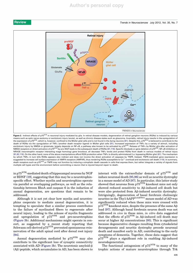
Figure 3. Potential mechanisms of p75NTR involvement in axonal degeneration. (a) Axonal degeneration is essential for the precision and plasticity of cholinergicconnections in the adult healthy brain, where elimination of aberrant collaterals that could disrupt normal circuitry occurs in physiological conditions. (1) This degenerationhas been shown to occur locally through p75NTR signaling and requires both BDNF and the axon being in close contact to myelin [89]. The degradation of axons in contactwith myelin is highly dependent on the levels of NGF in these axons, as basal forebrain cholinergic neurons specifically require NGF for trophic support. In this paradigm,high NGF would override myelin-induced regression of axons, whereas low NGF would cause all axons exposed to myelin to degrade. Thus, a homeostatic balancebetween TrkA and p75NTR signaling underlies functionality. (b) Aberrant axonal degeneration seen in neurodegenerative diseases such as Alzheimer’s disease (AD) mayrepresent a homeostatic mechanism [as in (a)] gone awry. This may occur, for example, by exposure to higher concentrations of p75NTR ligands including BDNF [120] or byincreased sensitization to myelin components during disease conditions [121] (2). Notably, aging-related and pathological (as in AD) demise of the cholinergic basalforebrain system correlates with loss of functional responses to NGF and a striking reduction in the retrograde transport of NGF in cholinergic neurons [122]; this may resultfrom either decreased levels of TrkA (reported in aging [123]) or defective processing of pro-NGF (seen in injury conditions [77]) or both (3). As suggested in (a), when thecritical concentration of NGF drops, axons in close contact with myelin begin to degrade, as if their demise were a default mechanism kept at bay by NGF–TrkA signaling.Region-specific alterations in BDNF levels have also been reported in AD-affected brains [120], and ineffective processing of pro-BDNF may also occur in these conditions.Whether sufficient levels of BDNF are maintained locally or whether pro-neurotrophins bound to a p75NTR–sortilin/SorCS2 complex [88] could induce axonal degeneration(4) are issues remaining to be resolved. In addition to neurotrophins and their pro-peptides, p75NTR mediates cell death by the Ab peptide both in vitro and in vivo [96].Furthermore, absence of p75NTR protects neurites from dystrophy and cytoskeletal derangements seen in a transgenic mouse model of AD [97] (5). In all, it is increasinglyapparent that in pathological aging, and other neurodegenerative conditions with progressive cognitive decline, significant loss of cholinergic function occurs via dendritic,synaptic and axonal degeneration rather than via cell death. p75NTR appears to be a key modulator in the establishment and maintenance (by elimination) of functionalconnections, and hence is an attractive candidate for mediating the loss of connectivity in disease. A composite hypothesis may yet implicate p75NTR in neuronal cell deathat later stages of neurodegenerative disease, possibly through the accumulation of death ligands such as Ab in addition to pro-NGF and/or pro-BDNF [6].
Review Trends in Neurosciences July 2012, Vol. 35, No. 7
436
Author's personal copy
a recent debate in the study of the related Fas receptor[also known as cluster of differentiation 95 (CD95)] [102].Aside from this, it could be useful to consider for a momentthe methodologies used to reveal the proapoptotic andprodegenerative effects of p75NTR. Nearly all injury para-digms utilized in this research involve quite severe lesionsto the nervous system, the majority of which would proba-bly be life-threatening to an organism in the wild. It isdifficult to see how a mere reduction in p75NTR expressionor activity could make much of a difference to an organismseverely compromised by a massive seizure, stroke ornerve damage. In such circumstances, mechanisms limit-ing p75NTR expression would simply fail to evolve. Howev-er, seizures, stroke and axotomy may also occur at muchsmaller scales, involving perhaps a few cells or axons, toofew to be useful as an experimental paradigm. Under thoseconditions, elimination of a few malfunctioning neurons,glial cells, or their connections may be beneficial for thenervous system and the longer-term survival of the organ-ism. Here p75NTR finds itself in a situation akin to that ofmediators of inflammatory responses. An inflammatoryresponse involves cells infiltrating a wound and cleaningup damage. Nevertheless, there can be ‘too much of a goodthing’, and with a severe injury, more damage can be donethan good. Evolutionarily speaking, an animal with a tornknee would probably not survive very long in the wild, sohaving an aggressive inflammation process is not much ofan added problem in that case. Seen in this light, ratherthan an evolutionary glitch, the deleterious effects ofp75NTR signaling in neural injury and disease could bepart of an homeostatic mechanism that becomes out ofcontrol. Finding experimental support for this concept mayrequire the analysis of more limited lesion paradigms, aswell as novel genetic and imaging tools. The study by Parket al. [89] showing astray basal forebrain axons in whitematter of p75NTR knockout mice represents one of the firstexamples of the homeostatic function of p75NTR in theadult brain.
Outstanding questionsThe role of p75NTR signaling in neural injury and diseasebrings both challenges and opportunities (Box 1). Clearly,the signaling pathways underlying the effects of p75NTR inneural injury need to be better understood. How p75NTR
couples to different signaling pathways and how thesecontribute to p75NTR function remain key challenges in
the field. With regard to cell death pathways, it is stillunclear how p75NTR signals to JNK and caspases in differ-ent cell types, and also how this pathway is balancedagainst the effects mediated by increased NF-kB activity.As the NF-kB pathway can mediate pro-survival as well aspro-inflammatory responses (which can lead to cell death,see e.g. Figure 2), it will also be important to elucidate thecontribution of NF-kB signaling to p75NTR-mediated celldamage in different injury paradigms. Answers to thesequestions will require the development of an accuratestructure–function map linking motifs within the p75NTR
intracellular domain to specific interactors and signalingevents. This would in turn allow the generation of genetictools for probing the physiological importance of the differ-ent signaling pathways engaged by p75NTR. It should alsobe noted that, in many of the disease paradigms studied sofar, it is still unclear whether p75NTR kills cells by cell-autonomous or non-cell-autonomous mechanisms. Resolv-ing this question will require studies in animal modelswith conditional ablation of p75NTR signaling in specificclasses of neurons and glial cells.
p75NTR is not always bad news for neurons: although itexacerbates the death of axotomized sensory and moto-neurons, p75NTR contributes to their survival duringdevelopment [68,69,103,104]. This duality is not uniqueto p75NTR and can be found in other members of the TNFRsuperfamily [64,102]. It may result from intrinsic differ-ences in the complement of intracellular mediators andcoreceptors between developing and adult cells. In linewith this, NGF was found to increase survival of youngcultures of Schwann cells through p75NTR signaling viaRIP2, but to induce cell death in older cultures – in whichRIP2 is downregulated [70]. On the other hand, differentextracellular environments, such as the balance betweenneurotrophins and pro-neurotrophins, may also contributeto different, or even opposed, p75NTR activities. Under-standing how different p75NTR ligands can elicit differentresponses through the same receptor is another key chal-lenge in the field.
In the case of the effects of p75NTR on axons, it is unclearwhether inhibition of axonal growth (as observed in re-sponse to myelin components) and axonal degenerationproceed through different or similar pathways. Becausethe latter requires both myelin and neurotrophins, it mayconceivably engage a different combination of intracellularmediators. It is also possible that different mechanisms ofreceptor activation are at play. This possibility is sup-ported by recent results demonstrating the importanceof transmembrane residue Cys257 in the formation of dis-ulphide-linked p75NTR dimers, and in receptor activationby neurotrophins – but not by myelin-derived moleculessuch as Nogo or myelin-associated glycoprotein (MAG)(Figure 1b) [105]. These and other results [106] suggestthat different p75NTR activation mechanisms, perhapsrelated to different receptor complexes, may operate inresponse to different p75NTR ligands.
The mechanisms that induce p75NTR expression duringinjury and neurodegeneration remain ill-defined. Onestudy found that hypo-osmotic stress can regulatep75NTR by increasing cellular levels of Sp1 in primarycortical neurons [55]. More recently, the pro-inflammatory
Box 1. Outstanding questions
! How does p75NTR couple to different downstream signalingpathways? How do these distinct pathways contribute to p75NTR
function?! Does p75NTR kill neurons after injury by cell-autonomous or non-
cell-autonomous mechanisms?! What are the similarities and differences between p75NTR func-
tions during development and after injury/insult in the adult CNS?! How do the different p75NTR ligands elicit distinct responses
through the same receptor?! Is there complete overlap between the p75NTR signaling pathways
mediating axon growth inhibition and axonal degeneration or arethere important distinctions?
! What are the specific molecular mechanisms that induce adultp75NTR expression during neural injury and neurodegeneration?
Review Trends in Neurosciences July 2012, Vol. 35, No. 7
437
Author's personal copy
cytokines IL-1b and TNF-a, which are highly expressed inthe injured brain [107], were shown to contribute to p75NTR
upregulation in both astrocytes and neurons [108]. IL-1binduced p75NTR via p38 mitogen-activated protein kinase(MAPK) in neurons, and via both p38 MAPK and NF-kB inastrocytes. TNF-a, on the other hand, induced p75NTR viaNF-kB both in neurons and astrocytes, showing that themechanisms governing this regulation are both cytokine-and cell-type specific. It would be interesting to determinewhether these mechanisms also contribute to p75NTR in-duction during development or are exclusive to inflamma-tion-mediated changes in the adult.
Therapeutic opportunitiesp75NTR signaling can induce neuronal death, reduce axo-nal growth and decrease synaptic function; hence, there isa strong rationale for inhibiting p75NTR in neural injuryand neurodegeneration. As for any transmembrane recep-tor, the most obvious process to target is ligand binding.Functional neurotrophin epitopes important for p75NTR
binding have been mapped by site-directed mutagenesis[109–111], and later confirmed by X-ray crystallography[112,113]. This information has been used to design smallp75NTR-binding peptides [114] and organic compounds[115]. However, there is no clear consensus at present asto whether these have agonistic or antagonistic functionson p75NTR signaling and bioactivity [82,115–117]. One setof p75NTR antagonists has been reported to show neuro-protective activity in retinal degeneration followingglaucoma or optic nerve transection [82]. Glaucoma andaxotomy represent both chronic and acute models of neu-rodegeneration, and p75NTR expression is upregulated inboth cases. The fact that p75NTR antagonism can protectretinal ganglion cells after both acute and chronic lesions,suggests these injury paradigms may be promising modelsfor developing p75NTR-based therapies.
Approaches to block ligand-binding are limited by therelatively high affinity of neurotrophins for p75NTR andcannot distinguish between prosurvival and proapoptoticeffects of the receptor. Other processes that would beamenable for targeting by inhibitors are receptor activa-tion and downstream signal propagation. Compared toligand binding, however, the development of strategiesto block these processes will require greater mechanisticunderstanding. The mechanism of p75NTR activation byneurotrophin ligands has recently begun to be clarified[105], opening the door to strategies directed at specificallyinhibiting this process. On the other hand, targeting down-stream signal propagation will require development of astructure–function map of the intracellular region ofp75NTR and the identification of specific structural motifsresponsible for the activation of different pathways.
Concluding remarksIn summary, work over recent years has indicated that theadult induction of p75NTR may be part of a homeostaticprogram that removes defective neurons, axons and synap-ses upon limited injury and degeneration. A downside of thisfunction is the exacerbated damage that p75NTR signalingproduces following severe lesions and in pronounced neu-rodegeneration. Many such circumstances are encountered
in neural injury, stroke and neurodegenerative diseases inhumans, and thus there is a strong rationale for inhibitingp75NTR signaling as a therapeutic approach for theseconditions. Efficient ways of inhibiting this receptor willrequire a greater mechanistic understanding of its activa-tion by different classes of ligands and its communicationwith downstream signaling pathways.
AcknowledgmentsThe authors apologize to all the colleagues whose work could not be citeddue to space constraints. Research in the authors’ laboratory is funded bygrants from the Swedish Research Council, Swedish Cancer Society,Swedish Foundation for Strategic Research and the European ResearchCouncil.
References1 Chao, M.V. (2003) Neurotrophins and their receptors: a convergence
point for many signalling pathways. Nat. Rev. Neurosci. 4, 299–3092 Aron, L. and Klein, R. (2011) Repairing the parkinsonian brain with
neurotrophic factors. Trends Neurosci. 34, 88–1003 Dechant, G. and Barde, Y-A. (2002) The neurotrophin receptor
p75(NTR): novel functions and implications for diseases of thenervous system. Nat. Neurosci. 5, 1131–1136
4 Roux, P.P. and Barker, P.A. (2002) Neurotrophin signaling throughthe p75 neurotrophin receptor. Prog. Neurobiol. 67, 203–233
5 Gentry, J.J. et al. (2004) The p75 neurotrophin receptor: multipleinteractors and numerous functions. Prog. Brain Res. 146, 25–39
6 Underwood, C.K. and Coulson, E.J. (2008) The p75 neurotrophinreceptor. Int. J. Biochem. Cell Biol. 40, 1664–1668
7 Reichardt, L.F. (2006) Neurotrophin-regulated signalling pathways.Philos. Trans. R. Soc. Lond. B: Biol. Sci. 361, 1545–1564
8 Kaplan, D.R. and Miller, F.D. (1997) Signal transduction by theneurotrophin receptors. Curr. Opin. Cell Biol. 9, 213–221
9 Lu, B. et al. (2005) The yin and yang of neurotrophin action. Nat. Rev.Neurosci. 6, 603–614
10 Lee, R. et al. (2001) Regulation of cell survival by secretedproneurotrophins. Science 294, 1945–1948
11 Nykjaer, A. et al. (2004) Sortilin is essential for proNGF-inducedneuronal cell death. Nature 427, 843–848
12 Nykjaer, A. et al. (2005) p75NTR – live or let die. Curr. Opin.Neurobiol. 15, 49–57
13 Woo, N.H. et al. (2005) Activation of p75NTR by proBDNF facilitateshippocampal long-term depression. Nat. Neurosci. 8, 1069–1077
14 Yang, F. et al. (2009) Pro-BDNF-induced synaptic depression andretraction at developing neuromuscular synapses. J. Cell Biol. 185,727–741
15 Liepinsh, E. et al. (1997) NMR structure of the death domain of thep75 neurotrophin receptor. EMBO J. 16, 4999–5005
16 Carter, B.D. et al. (1996) Selective activation of NF-kappa B by nervegrowth factor through the neurotrophin receptor p75. Science 272,542–545
17 Friedman, W.J. (2000) Neurotrophins induce death of hippocampalneurons via the p75 receptor. J. Neurosci. 20, 6340–6346
18 Yoon, S.O. et al. (1998) Competitive signaling between TrkA and p75nerve growth factor receptors determines cell survival. J. Neurosci.18, 3273–3281
19 Troy, C.M. et al. (2002) Mechanisms of p75-mediated death ofhippocampal neurons. Role of caspases. J. Biol. Chem. 277, 34295–34302
20 Yamashita, T. et al. (1999) Neurotrophin binding to the p75 receptormodulates Rho activity and axonal outgrowth. Neuron 24, 585–593
21 Wang, K.C. et al. (2002) P75 interacts with the Nogo receptor as a co-receptor for Nogo, MAG and OMgp. Nature 420, 74–78
22 Wong, S.T. et al. (2002) A p75(NTR) and Nogo receptor complexmediates repulsive signaling by myelin-associated glycoprotein.Nat. Neurosci. 5, 1302–1308
23 Yamashita, T. et al. (2002) The p75 receptor transduces the signalfrom myelin-associated glycoprotein to Rho. J. Cell Biol. 157, 565–570
24 Fournier, A.E. et al. (2001) Identification of a receptor mediatingNogo-66 inhibition of axonal regeneration. Nature 409, 341–346
25 Mi, S. et al. (2004) LINGO-1 is a component of the Nogo-66 receptor/p75 signaling complex. Nat. Neurosci. 7, 221–228
Review Trends in Neurosciences July 2012, Vol. 35, No. 7
438
Author's personal copy
26 He, Z. and Koprivica, V. (2004) The Nogo signaling pathway forregeneration block. Annu. Rev. Neurosci. 27, 341–368
27 Yamashita, T. et al. (2003) Enhanced insulin-sensitivity in mice lackingganglioside GM3. Proc. Natl. Acad. Sci. U.S.A. 100, 3445–3449
28 Friedman, W.J. et al. (1991) Temporal and spatial expression of NGFreceptor mRNA during postnatal rat brain development analyzed byin situ hybridization. Brain Res. Dev. Brain Res. 63, 43–51
29 Wyatt, S. et al. (1990) Expression of the NGF receptor gene in sensoryneurons and their cutaneous targets prior to and during innervation.Neuron 4, 421–427
30 Ernfors, P. et al. (1989) Expression of nerve growth factor receptormRNA is developmentally regulated and increased after axotomy inrat spinal cord motoneurons. Neuron 2, 1605–1613
31 Mobley, W.C. et al. (1989) Developmental regulation of nerve growthfactor and its receptor in the rat caudate-putamen. Neuron 3, 655–664
32 Cragnolini, A.B. et al. (2009) Nerve growth factor attenuatesproliferation of astrocytes via the p75 neurotrophin receptor. Glia57, 1386–1392
33 Ernfors, P. et al. (1988) Developmental and regional expression ofbeta-nerve growth factor receptor mRNA in the chick and rat. Neuron1, 983–996
34 Verge, V.M. et al. (1992) Colocalization of NGF binding sites, trkmRNA, and low-affinity NGF receptor mRNA in primary sensoryneurons: responses to injury and infusion of NGF. J. Neurosci. 12,4011–4022
35 Saika, T. et al. (1991) Effects of nerve crush and transection on mRNAlevels for nerve growth factor receptor in the rat facial motoneurons.Brain Res. Mol. Brain Res. 9, 157–160
36 Koliatsos, V.E. et al. (1991) Axotomy induces nerve growth factorreceptor immunoreactivity in spinal motor neurons. Brain Res. 549,297–304
37 Lowry, K.S. et al. (2001) A potential role for the p75 low-affinityneurotrophin receptor in spinal motor neuron degeneration in murineand human amyotrophic lateral sclerosis. Amyotroph. Lateral Scler.Other Motor Neuron Disord. 2, 127–134
38 Taniuchi, M. et al. (1986) Induction of nerve growth factor receptor inSchwann cells after axotomy. Proc. Natl. Acad. Sci. U.S.A. 83,4094–4098
39 Zhou, X.F. et al. (1996) Differential expression of the p75 nerve growthfactor receptor in glia and neurons of the rat dorsal root ganglia afterperipheral nerve transection. J. Neurosci. 16, 2901–2911
40 Obata, K. et al. (2006) Suppression of the p75 neurotrophin receptor inuninjured sensory neurons reduces neuropathic pain after nerveinjury. J. Neurosci. 26, 11974–11986
41 Syroid, D.E. et al. (2000) Induction of postnatal schwann cell death bythe low-affinity neurotrophin receptor in vitro and after axotomy. J.Neurosci. 20, 5741–5747
42 Petratos, S. et al. (2003) Schwann cell apoptosis in the postnatalaxotomized sciatic nerve is mediated via NGF through the low-affinityneurotrophin receptor. J. Neuropathol. Exp. Neurol. 62, 398–411
43 Hall, S.M. et al. (1997) Schwann cells responding to primarydemyelination in vivo express p75NTR and c-erbB receptors: alight and electron immunohistochemical study. J. Neurocytol. 26,679–690
44 Beattie, M.S. et al. (2002) ProNGF induces p75-mediated death ofoligodendrocytes following spinal cord injury. Neuron 36, 375–386
45 Dowling, P. et al. (1999) Up-regulated p75NTR neurotrophin receptoron glial cells in MS plaques. Neurology 53, 1676–1682
46 Hu, B. et al. (1998) Localization of p75 neurotrophin receptor in theretina of the adult SD rat: an immunocytochemical study at light andelectron microscopic levels. Glia 24, 187–197
47 Rudzinski, M. et al. (2004) Changes in retinal expression ofneurotrophins and neurotrophin receptors induced by ocularhypertension. J. Neurobiol. 58, 341–354
48 Lonngren, U. et al. (2006) The growth factor response in ischemic ratretina and superior colliculus after brimonidine pre-treatment. BrainRes. Bull. 71, 208–218
49 Giehl, K. et al. (2001) Endogenous brain-derived neurotrophic factorand neurotrophin-3 antagonistically regulate survival of axotomizedcorticospinal neurons in vivo. J. Neurosci. 21, 3492–3502
50 Martınez-Murillo, R. et al. (1993) Lesion-induced expression of low-affinity nerve growth factor receptor-immunoreactive protein inPurkinje cells of the adult rat. Neuroscience 52, 587–593
51 Roux, P.P. et al. (1999) p75 neurotrophin receptor expression isinduced in apoptotic neurons after seizure. J. Neurosci. 19,6887–6896
52 Oh, J.D. et al. (2000) Overexpression of neurotrophin receptor p75contributes to the excitotoxin-induced cholinergic neuronal death inrat basal forebrain. Brain Res. 853, 174–185
53 Kokaia, Z. et al. (1998) Focal cerebral ischemia in rats inducesexpression of P75 neurotrophin receptor in resistant striatalcholinergic neurons. Neuroscience 84, 1113–1125
54 Kommaddi, R.P. et al. (2011) Trk-dependent ADAM17 activationfacilitates neurotrophin survival signaling. FASEB J. 25, 2061–2070
55 Ramos, A. et al. (2007) Hypo-osmolar stress induces p75NTRexpression by activating Sp1-dependent transcription. J. Neurosci.27, 1498–1506
56 Mufson, E.J. and Kordower, J.H. (1992) Cortical neurons expressnerve growth factor receptors in advanced age and Alzheimerdisease. Proc. Natl. Acad. Sci. U.S.A. 89, 569–573
57 Ernfors, P. et al. (1990) Cholinergic neurons of the nucleus basalisexpress elevated levels of nerve growth factor receptor mRNA insenile dementia of the Alzheimer type. Dementia 1, 138–145
58 Rabizadeh, S. et al. (1993) Induction of apoptosis by the low-affinityNGF receptor. Science 261, 345–348
59 Barrett, G.L. and Bartlett, P.F. (1994) The p75 nerve growth factorreceptor mediates survival or death depending on the stage ofsensory neuron development. Proc. Natl. Acad. Sci. U.S.A. 91,6501–6505
60 Bartheld and von, C.S. et al. (1994) Positive and negative effects ofneurotrophins on the isthmo-optic nucleus in chick embryos. Neuron12, 639–654
61 Frade, J.M. et al. (1996) Induction of cell death by endogenous nervegrowth factor through its p75 receptor. Nature 383, 166–168
62 Casaccia-Bonnefil, P. et al. (1996) Death of oligodendrocytes mediatedby the interaction of nerve growth factor with its receptor p75. Nature383, 716–719
63 Bamji, S.X. et al. (1998) The p75 neurotrophin receptor mediatesneuronal apoptosis and is essential for naturally occurringsympathetic neuron death. J. Cell Biol. 140, 911–923
64 Haase, G. et al. (2008) Signaling by death receptors in the nervoussystem. Curr. Opin. Neurobiol. 18, 284–291
65 Raoul, C. et al. (2000) Active killing of neurons during developmentand following stress: a role for p75(NTR) and Fas? Curr. Opin.Neurobiol. 10, 111–117
66 Cheema, S.S. et al. (1996) Reducing p75 nerve growth factor receptorlevels using antisense oligonucleotides prevents the loss ofaxotomized sensory neurons in the dorsal root ganglia of newbornrats. J. Neurosci. Res. 46, 239–245
67 Jansen, P. et al. (2007) Roles for the pro-neurotrophin receptor sortilinin neuronal development, aging and brain injury. Nat. Neurosci. 10,1449–1457
68 Ferri, C. et al. (1998) Effects of facial nerve injury on mousemotoneurons lacking the p75 low-affinity neurotrophin receptor. J.Neurobiol. 34, 1–9
69 Wiese, S. et al. (1999) The role of p75(NTR) in modulatingneurotrophin survival effects in developing motoneurons. Eur. J.Neurosci. 11, 1668–1676
70 Khursigara, G. et al. (2001) A prosurvival function for the p75 receptordeath domain mediated via the caspase recruitment domain receptor-interacting protein 2. J. Neurosci. 21, 5854–5863
71 Cragnolini, A.B. and Friedman, W.J. (2008) The function of p75(NTR)in glia. Trends Neurosci. 31, 99–104
72 Cosgaya, J.M. et al. (2002) The neurotrophin receptor p75NTR as apositive modulator of myelination. Science 298, 1245–1248
73 Tomita, K. et al. (2007) The neurotrophin receptor p75NTR inSchwann cells is implicated in remyelination and motor recoveryafter peripheral nerve injury. Glia 55, 1199–1208
74 Song, X-Y. et al. (2006) Knockout of p75(NTR) impairs re-myelinationof injured sciatic nerve in mice. J. Neurochem. 96, 833–842
75 Volosin, M. et al. (2008) Induction of proneurotrophins and activation ofp75(NTR)-mediated apoptosis via neurotrophin receptor-interactingfactor in hippocampal neurons after seizures. J. Neurosci. 28,9870–9879
76 Domeniconi, M. et al. (2007) Pro-NGF secreted by astrocytes promotesmotor neuron cell death. Mol. Cell. Neurosci. 34, 271–279
Review Trends in Neurosciences July 2012, Vol. 35, No. 7
439
Author's personal copy
77 Volosin, M. et al. (2006) Interaction of survival and death signaling inbasal forebrain neurons: roles of neurotrophins and proneurotrophins.J. Neurosci. 26, 7756–7766
78 Fahnestock, M. et al. (2001) The precursor pro-nerve growth factor isthe predominant form of nerve growth factor in brain and is increasedin Alzheimer’s disease. Mol. Cell. Neurosci. 18, 210–220
79 Podlesniy, P. et al. (2006) Pro-NGF from Alzheimer’s disease andnormal human brain displays distinctive abilities to induceprocessing and nuclear translocation of intracellular domain ofp75NTR and apoptosis. Am. J. Pathol. 169, 119–131
80 Pedraza, C.E. et al. (2005) Pro-NGF isolated from the human brainaffected by Alzheimer’s disease induces neuronal apoptosis mediatedby p75NTR. Am. J. Pathol. 166, 533–543
81 Lebrun-Julien, F. et al. (2010) ProNGF induces TNFalpha-dependentdeath of retinal ganglion cells through a p75NTR non-cell-autonomoussignaling pathway. Proc. Natl. Acad. Sci. U.S.A. 107, 3817–3822
82 Bai, Y. et al. (2010) Chronic and acute models of retinalneurodegeneration TrkA activity are neuroprotective whereasp75NTR activity is neurotoxic through a paracrine mechanism. J.Biol. Chem. 285, 39392–39400
83 Shi, Z. et al. (2007) Neurotrophic rationale in glaucoma: a TrkAagonist, but not NGF or a p75 antagonist, protects retinal ganglioncells in vivo. Dev. Neurobiol. 67, 884–894
84 Kohn, J. et al. (1999) Functionally antagonistic interactions betweenthe TrkA and p75 neurotrophin receptors regulate sympatheticneuron growth and target innervation. J. Neurosci. 19, 5393–5408
85 Singh, K.K. and Miller, F.D. (2005) Activity regulates positive andnegative neurotrophin-derived signals to determine axoncompetition. Neuron 45, 837–845
86 Singh, K.K. et al. (2008) Developmental axon pruning mediated byBDNF–p75NTR-dependent axon degeneration. Nat. Neurosci. 11,649–658
87 Plachta, N. et al. (2007) Identification of a lectin causing thedegeneration of neuronal processes using engineered embryonicstem cells. Nat. Neurosci. 10, 712–719
88 Deinhardt, K. et al. (2011) Neuronal growth cone retraction relies onproneurotrophin receptor signaling through rac. Sci. Signal. 4, ra82
89 Park, K.J. et al. (2010) p75NTR-dependent, myelin-mediated axonaldegeneration regulates neural connectivity in the adult brain. Nat.Neurosci. 13, 559–566
90 Park, J.B. et al. (2005) A TNF receptor family member, TROY, is acoreceptor with Nogo receptor in mediating the inhibitory activity ofmyelin inhibitors. Neuron 45, 345–351
91 Shao, Z. et al. (2005) TAJ/TROY, an orphan TNF receptor familymember, binds Nogo-66 receptor 1 and regulates axonal regeneration.Neuron 45, 353–359
92 Yamashita, T. and Tohyama, M. (2003) The p75 receptor acts as adisplacement factor that releases Rho from Rho-GDI. Nat. Neurosci. 6,461–467
93 Nikolaev, A. et al. (2009) APP binds DR6 to trigger axon pruning andneuron death via distinct caspases. Nature 457, 981–989
94 Scott, A.L.M. and Ramer, M.S. (2010) Schwann cell p75NTR preventsspontaneous sensory reinnervation of the adult spinal cord. Brain133, 421–432
95 Perini, G. et al. (2002) Role of p75 neurotrophin receptor in theneurotoxicity by beta-amyloid peptides and synergistic effect ofinflammatory cytokines. J. Exp. Med. 195, 907–918
96 Sotthibundhu, A. et al. (2008) Beta-amyloid(1-42) induces neuronaldeath through the p75 neurotrophin receptor. J. Neurosci. 28,3941–3946
97 Knowles, J.K. et al. (2009) The p75 neurotrophin receptor promotesamyloid-beta(1-42)-induced neuritic dystrophy in vitro and in vivo. J.Neurosci. 29, 10627–10637
98 Pang, P.T. et al. (2004) Cleavage of proBDNF by tPA/plasmin isessential for long-term hippocampal plasticity. Science 306, 487–491
99 Rosch, H. et al. (2005) The neurotrophin receptor p75NTR modulateslong-term depression and regulates the expression of AMPA receptorsubunits in the hippocampus. Proc. Natl. Acad. Sci. U.S.A. 102,7362–7367
100 Zagrebelsky, M. et al. (2005) The p75 neurotrophin receptornegatively modulates dendrite complexity and spine density inhippocampal neurons. J. Neurosci. 25, 9989–9999
101 Copray, S. et al. (2004) Deficient p75 low-affinity neurotrophin receptorexpression exacerbates experimental allergic encephalomyelitis inC57/BL6 mice. J. Neuroimmunol. 148, 41–53
102 Peter, M.E. et al. (2007) The CD95 receptor: apoptosis revisited. Cell129, 447–450
103 Lee, K. et al. (1992) Targeted mutation of the gene encoding the lowaffinity NGF receptor P75 leads to deficits in the peripheral sensorynervous-system. Cell 69, 737–749
104 Lee, K-F. et al. (1994) p75-deficient embryonic dorsal root sensory andneonatal sympathetic neurons display a decreased sensitivity to NGF.Development 120, 1027–1033
105 Vilar, M. et al. (2009) Activation of the p75 neurotrophin receptorthrough conformational rearrangement of disulphide-linked receptordimers. Neuron 62, 72–83
106 Vilar, M. et al. (2009) Ligand-independent signaling by disulfide-crosslinked dimers of the p75 neurotrophin receptor. J. Cell Sci.122, 3351–3357
107 Eriksson, C. et al. (2000) Increased expression of mRNA encodinginterleukin-1 beta and caspase-1, and the secreted isoformof interleukin-1 receptor antagonist in the rat brain followingsystemic kainic acid administration. J. Neurosci. Res. 60, 266–279
108 Choi, S. and Friedman, W.J. (2009) Inflammatory cytokines IL-1b andTNF-a regulate p75NTR expression in CNS neurons and astrocytesby distinct cell-type-specific signalling mechanisms. ASN Neuro 1,e00010
109 Ibanez, C.F. et al. (1992) Disruption of the low affinity receptor-binding site in NGF allows neuronal survival and differentiationby binding to the Trk gene product. Cell 69, 329–341
110 Ryden, M. and Ibanez, C.F. (1997) A second determinant of binding tothe p75 neurotrophin receptor revealed by alanine-scanningmutagenesis of a conserved loop in nerve growth factor. J. Biol.Chem. 272, 33085–33091
111 Ryden, M. et al. (1995) Functional analysis of mutant neurotrophinsdeficient in low-affinity binding reveals a role for p75LNGFR in NT-4signalling. EMBO J. 14, 1979–1990
112 He, X-L. and Garcia, K.C. (2004) Structure of nerve growth factorcomplexed with the shared neurotrophin receptor p75. Science 304,870–875
113 Gong, Y. et al. (2008) Crystal structure of the neurotrophin-3 andp75NTR symmetrical complex. Nature 454, 789–793
114 Longo, F. et al. (1997) Synthetic NGF peptide derivatives preventneuronal death via a p75 receptor-dependent mechanism. J. Neurosci.Res. 48, 1–17
115 Massa, S.M. et al. (2006) Small, nonpeptide p75NTR ligands inducesurvival signaling and inhibit proNGF-induced death. J. Neurosci. 26,5288–5300
116 Lebrun-Julien, F. et al. (2009) Inhibition of p75(NTR) in gliapotentiates TrkA-mediated survival of injured retinal ganglioncells. Mol. Cell. Neurosci. 40, 410–420
117 Longo, F.M. and Massa, S.M. (2008) Small molecule modulation ofp75 neurotrophin receptor functions. CNS Neurol. Disord. DrugTargets 7, 63–70
118 Esposito, D. et al. (2001) The cytoplasmic and transmembranedomains of the p75 and TrkA receptors regulate high affinitybinding to nerve growth factor. J. Biol. Chem. 276, 32687–32695
119 Lebrun-Julien, F. et al. (2009) Excitotoxic death of retinal neurons invivo occurs via a non-cell-autonomous mechanism. J. Neurosci. 29,5536–5545
120 Durany, N. et al. (2000) Brain-derived neurotrophic factor andneurotrophin-3 levels in Alzheimer’s disease brains. Int. J. Dev.Neurosci. 18, 807–813
121 Gold, B. et al. (2012) White matter integrity and vulnerability toAlzheimer’s disease: preliminary findings and future directions.Biochim. Biophys. Acta 1822, 416–422
122 DeLacalle, S. et al. (1996) Reduced retrograde labelling withfluorescent tracer accompanies neuronal atrophy of basal forebraincholinergic neurons in aged rats. Neuroscience 75, 19–27
123 Cooper, J. et al. (1994) Reduced transport of [I-125] nerve growth-factor by cholinergic neurons and down-regulated Trka expression inthe medial septum of aged rats. Neuroscience 62, 625–629
Review Trends in Neurosciences July 2012, Vol. 35, No. 7
440