Embed Size (px)
Citation preview
KEY KNOWLEDGE
This chapter is designed to enable students to:■ gain understanding of the importance of the mitotic cell cycle in cell production
for growth and repair in eukaryotes■ recognise that the cell cycle produces daughter cells that are identical to each
other and are clones of the parent cell■ become familiar with the stages of the cell cycle and the chromosomal events
that occur at each stage.
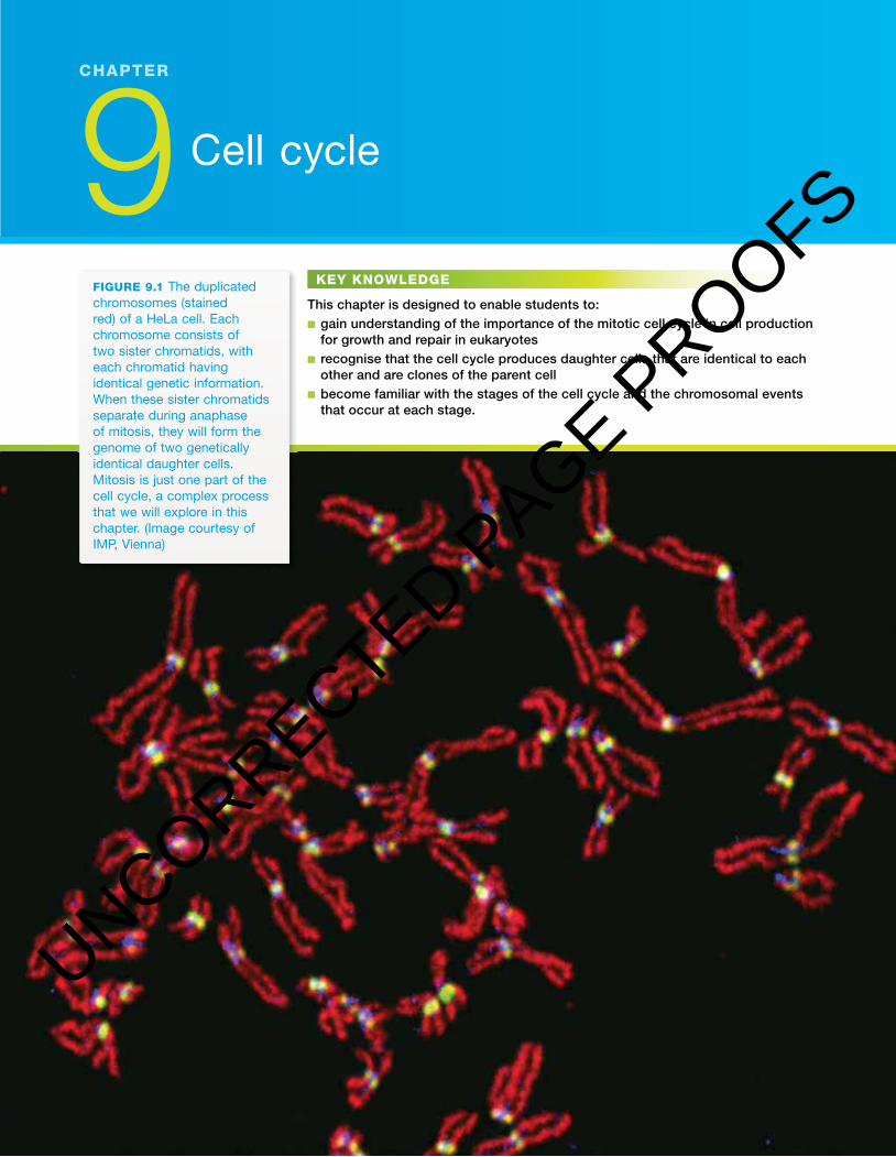
FIGURE 9.1 The duplicated chromosomes (stained red) of a hela cell. each chromosome consists of two sister chromatids, with each chromatid having identical genetic information. When these sister chromatids separate during anaphase of mitosis, they will form the genome of two genetically identical daughter cells. Mitosis is just one part of the cell cycle, a complex process that we will explore in this chapter. (image courtesy of iMP, Vienna)
9 Cell cycle
CHAPTERCHAPTER9Cell cycleSaving burns victims�e cell cycleNobel Laureate in Physiology or MedicineBiochallengeChapter review
UNCORRECTED PAGE P
ROOFS

NATURE OF BIOLOGY 1390
Saving burns victimsWhen Professor Fiona Wood (see � gure 9.2) of the Royal Perth Hospital was made 2005 Australian of the Year, it was in recognition of her work related to the treatment of people with severe burns. For about 10 years prior to March 2003 Professor Wood had been developing improved methods for growing replace-ment skin. When 28 Australians were badly wounded and burnt as a result of a terrorist bombing in Bali, Indonesia, it was decided that they should be returned to Australia as soon as possible for treatment. � ey were sent to Professor Wood and Australians followed their progress through the daily press. ‘Spray-on skin’, known commercially as CellSpray, and Professor Wood became famous.
Skin: the outer layerNormal intact skin provides a covering for the human body. � e skin is com-posed of an outer epidermis and an underlying dermis. � e epidermis and the dermis are held together by a non-cellular basement membrane.
� e epidermis consists of several cell layers (see � gure 9.3): • an outermost region consisting of layers of � attened dead cells, called the
stratum corneum (strata = layer, coat; corneum = horny). � ese cells are constantly being shed from the skin surface. � is layer is thickest on the soles of the feet and the palms of the hands.
• several layers of living cells called keratinocytes that are gradually pushed upwards, becoming � attened and eventually forming part of the outermost region of dead cells. � e keratinocytes form the bulk of the epidermis.
• a basal layer which includes stem cells that are constantly dividing. For each two cells produced by division of a stem cell, one becomes a new keratino-cyte and the other replaces the stem cell. � e continual division of stem cells in the basal layer pushes the overlying keratinocytes towards the skin surface. Another group of cells present in the basal layer are the pigment-producing cells, or melanocytes. � e dermis lies below the epidermis. � e dermis contains blood vessels, hair
follicles, sweat glands, touch-sensitive and pain-sensitive cells, muscle � bres and collagen � bres.
� e severity of burn damage to human skin may vary from � rst-degree burns, such as sunburn, that involve the epidermis only, to second-degree burns, such as scalds, that involve the epidermis and the upper section of the dermis, to severe third-degree or full thickness burns that destroy the epidermis and all or part of the dermis. Burns of this type were those seen in the seriously burnt victims of the Bali bombing and those who are badly burnt in bush� res in this country.
FIGURE 9.2 Professor Fiona Wood aM was awarded 2005 australian of the Year for her work on developing an improved method of skin-cell regeneration, leading to improved and more rapid treatment for people with skin burns.
Basement membranes occur throughout the human body. � ey consist of glycoproteins and provide structural support for tissues.
ODD FACT
it has been estimated that each person replaces, on average, about 18 kg of skin cells during a lifetime. Dandruff, skin cells from our scalps, represents just a fraction of the skin cells we must replace.
UNCORRECTED PAGE P
ROOFS

391CHAPTER 9 Cell cycle
FIGURE 9.3 Section through human skin (a) Diagram showing the epidermis that overlays the dermis. The basal layer of epidermal cells includes stem cells that are capable of cell division. as keratinocytes are pushed towards the skin surface, they fl atten and eventually become part of the dead outermost layers of skin cells. (b) Photomicrograph of stained epidermis of human skin. note the change in shape of the keratinocytes as they become older and move closer to the outer surface of the skin. What is the origin of the keratinocytes that form the bulk of the epidermal tissue?
Basal layer
Young
Old
Dermis
Dead outer layers
Layers ofkeratinocytes
(a)(b)
When areas of skin are severely damaged by � re, acid or some other trauma, the challenge is to get new skin to grow over the damaged area. In the past, the treatments available for persons su� ering third-degree burns included the use of skin grafts taken from an uninjured part of the victim’s body. Such a graft is called an autograft (auto = self ) because it is a transplant of healthy skin from one area of a person to a damaged area of the same person. How-ever, a problem with autografts is that the area of the graft must be as large as the area of the burned skin. So, for patients with severe burns over a large area of their skin surface, say 50 per cent or more, there is insu� cient unburned skin to be used for grafting. In some urgent cases, skin from another person may be grafted onto the burned area of the victim; such a graft is an allograft. A skin allograft is a temporary measure because this graft will be rejected.
Another treatment involved covering a burn area with a thin sheet of skin cells grown in plastic dishes in a laboratory. � is procedure used cells har-vested from a small area of skin from the patient. A problem with skin grown in plastic dishes is that this procedure takes considerable time, with up to 21 days being needed to produce sheets of skin cells su� ciently large to cover severely burned areas. In addition, the sheets of skin cells are thin and very fragile. Another problem is that the sheets begin to act like skin and the surface cells form keratin and die so that they are less active growers when the transplant is carried out. Scarring tends to be more severe the longer the patient waits to be treated and the longer wait also increases the chance of infection and other complications with the wounds.
Alternatively, synthetic skin can be used for skin grafts. One example is Integra Template. � e outer layer of this arti� cial skin is a thin � lm of sil-icone, and the second layer is made of cross-linked � brous proteins (col-lagen) and a complex carbohydrate (glycosaminoglycan). Synthetic skin is used to cover the burnt area where it acts as a sca� old that enables the patient’s own dermal cells to regenerate the skin dermis. � en the silicone � lm is removed and covered with a thin epidermal skin graft, thus replacing the skin epidermis.
UNCORRECTED PAGE P
ROOFS
NATURE OF BIOLOGY 1392
Spray treatments for burnsProfessor Wood’s research � rst led to the development of a spray-on solution of skin cells, or CellSpray, that contained a suspension of various skin cells. � e cells came from skin harvested from unburnt areas of a patient’s skin. � ese cells were � rst cultured in the laboratory for a period of about 5 days, during which their numbers increased by cell division. Later, when sprayed over a burn area, the cells spread and continued to divide forming a layer of skin.
A further development of this technology is ReCell Spray on Skin (see � gure 9.4), which is marketed as a self-contained kit (see � gure 9.5a). � e time interval from taking cells from a patient to applying these cells to a burn area on that patient is about 30 minutes. A small area of healthy skin — about 2 cm by 2 cm, about 0.2 mm thick and close to the area of the burn — is taken from a burns patient. � e skin sample includes basal stem cells, pigment-producing cells, keratinocytes and � broblast cells from the epidermis, as well as some cells from the dermis. � e skin tissue is treated through a series of steps (see � gure 9.5b) that includes treatment with an enzyme which removes the extracellular matrix that holds the skin cells together. � e � nal suspension of skin cells, plus growth factors to stimulate cell division, is delivered directly to the burn site with a special spray applicator.
ReCell Spray on Skin technology is used in conjunction with skin grafts for deep or third-degree burns. In cases of limited thickness or second-degree burns, the technology is used alone and can cover burn areas up to about 1900 cm2. Once the cell suspension is applied to a burn area, the basal stem cells will multiply through repeated cell cycles and, over time, the skin lost by the burn damage will be replaced.
As well as being used in the treatment of acute burns where donor skin grafts cannot be taken, the ReCell technology can be used to treat other conditions, such as chronic skin ulcers.
� e science involved in growing new skin cells is possible because living skin cells are able to regenerate. We continually shed our old skin cells and so we continually need to replace them. Skin cells are continually being replaced by the cell cycle, a process that results in the production of two new cells, each identical to the parent cell that gave rise to them. Mitosis is an important part of that cycle and involves the replication of the genetic material in the cell. � e cytoplasm of a cell is shared between the two new cells at cytokinesis.
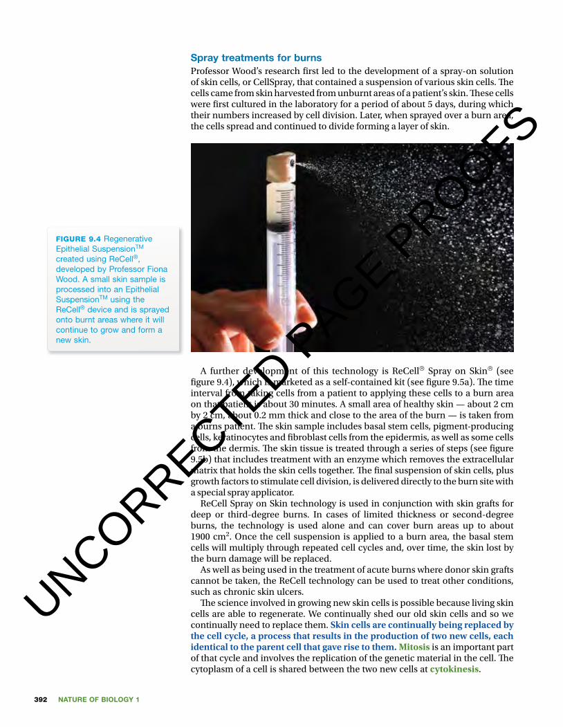
FIGURE 9.4 Regenerative epithelial SuspensionTM created using ReCell®, developed by Professor Fiona Wood. a small skin sample is processed into an epithelial SuspensionTM using the ReCell® device and is sprayed onto burnt areas where it will continue to grow and form a new skin.
UNCORRECTED PAGE P
ROOFS

393CHAPTER 9 Cell cycle
FIGURE 9.5 (a) The self-contained ReCell® Spray on Skin® kit. The sterile chambers and the tray are used for the various procedures involved in preparing a small sample of skin tissue for application to a burn site. What is the size of the patient’s skin sample used in this procedure? (image courtesy of avita Medical ltd) (b) Diagram showing the procedure that cells pass through with the ReCell kit. each square centimetre of skin taken from a patient converts to 1 ml of cell suspension, and this can cover 80 cm2 of burn area. (image adapted from www.avitamedical.com/wp-content/uploads/2015/03/avita-Corporate-Presentation-March-2015.pdf)
(a) Buffer
Scrape
Skinsample
Skinsample
Enzymesoak
Rinse
Autologoussuspension
Each cm2 of skin sample can produce up to a mLof suspension.1 mL of suspension covers 80 cm2 (a 1:80 expansionfrom donor area to treatment area).
(b)
In this chapter, we consider in some depth the importance of mitosis and cytokinesis. We also explore where these processes occur in a range of animals and plants.
The cell cycleCells have evolved complex and exact mechanisms to ensure that genetic information can be passed without error from one cell to two daughter cells of the next generation. It is through the mechanisms of the cell cycle that somatic cells of eukaryotes can divide, producing two daughter cells from one parent cell. � ese daughter cells are genetically identical to each other and genetically identical to the parent cell: a process of natural cloning. To achieve this, eukaryotic cells must � rst replicate their DNA, then orient their chromosomes in a very precise way, and then separate the sister chromatids.
Key events in the cell cycle� e key events that occur during a cell cycle are summarised in simple terms in table 9.1. � ese events occur in three distinct phases of the cell cycle: inter-phase, mitosis and cytokinesis.
TABLE 9.1 A simpli� ed summary of key events during the cell cycle
Cell cycle What happens Phase of cell cycle
step 1 replication of DNA of parent cell interphase
step 2 organisation of chromosomes, followed by their separation into two identical groups at di� erent poles of the parent cell
mitosis
step 3 division of parent cell into two cells cytokinesis
Let’s explore each of these steps in some detail.
UNCORRECTED PAGE P
ROOFS

NATURE OF BIOLOGY 1394
Interphase: period of DNA replicationAn essential process in the cell cycle is the replication of DNA, the genetic material. DNA replication occurs during a stage of the cell cycle known as the interphase. (� is stage was once called the ‘resting phase’, but the cells are far from resting during interphase.) If you looked through a light microscope at cells during interphase, you would see the cell nucleus, but you would not see any discrete chromosomes. In interphase, the chromosomes are decondensed and distributed through the nucleus. However, if you could watch the uptake of the nucleic acids that are the building blocks of DNA, you would see that the cells were busily copying their DNA and performing many other biochemical activities.
In a mammalian cell, a complete cell cycle takes about 24 hours. � e time spent by a cell in interphase is far longer than that spent in any other stage of the cell cycle. For example, in mammalian cells about 90 per cent of the time of a complete cell cycle is spent in interphase (see � gure 9.6), that is, about 22 hours. � is highlights the importance of the activities occurring during interphase.
M stage
G1 stageG2 stage
G0
S stage
Quiescence (not dividing)
Interphase is subdivided into three stages:1. � e G1 or Gap 1 stage. During the G1 stage of interphase, a cell under-
goes growth, increasing the amount of cell cytosol. � e cell also synthesises proteins that are needed for DNA replication. � e mitochondria of the cell divide and, in the cases of photosynthetic plant cells, their chloroplasts also divide. It is near the end of this stage that the cell will either commit to con-tinuing the cell cycle or will drop out and not divide. If the latter occurs, the cell enters a non-dividing quiescent G0 stage.
2. � e S or synthesis stage. During the S stage of interphase, the parent cell syn-thesises or replicates its DNA, the genetic material of the cell. At the end of the S stage, the parent cell contains two identical copies of its original DNA.
3. � e G2 or Gap 2 stage. During the G2 stage of interphase, further growth of the cell occurs in preparation for division. In addition, the synthesis of pro-teins occurs, including those that form the microtubules of the spindle. By the end of interphase, the cell has doubled its size.
For a typical human cell that requires 24 hours to complete one cell cycle, the time spent in the various stages might be: G1 stage about 11 hours, S stage about 8 hours, G2 stage about 4 hours and the remainder (mitosis and cytokinesis) about 1 hour. � is is in contrast to the rapid process of binary � ssion in prokar-yotes that produces two daughter cells within a period of 20 to 40 minutes.
Mitosis: organising and separating chromosomes� e appearance of chromosomes, initially thin and long, and the disappear-ance of the nuclear membrane mark the start of the part of the cell cycle known as mitosis, the M stage.
Unit 2 Eukaryotic cell cycle:InterphaseConcept summary and practice questions
AOS 1
Topic 1
Concept 2
FIGURE 9.6 Stages of the cell cycle. Most of the cell cycle is taken up by the three stages of interphase (g1, S and g2). The M stage is the division stage that includes the division of the nucleus (mitosis) and the division of the remainder of the cell (cytokinesis). What key event takes place during the S stage of interphase?
Unit 2 Eukaryotic cell cycle: mitosisConcept summary and practice questions
AOS 1
Topic 1
Concept 3
UNCORRECTED PAGE P
ROOFS
395CHAPTER 9 Cell cycle
Mitosis includes a number of di� erent stages:• Prophase. Chromosomes gradually condense — becoming shorter and
thicker — and become visible as double-stranded structures (see � gure 9.7). � e spindle forms and the nuclear membrane breaks down.
• Metaphase. � e double-stranded chromosomes, also called dyads, line up around the equator of the cell.
• Anaphase. � e sister chromatids separate and are pulled to opposite ends of the spindle by the contraction of spindle � bres (see � gure 9.8).
• Telophase. A nuclear membrane forms around each separate group of single-stranded chromosomes and the chromosomes gradually decondense. Mitosis completes the division of the nucleus.Figure 9.9 provides details of the di� erent stages of mitosis.
InteractivityMitosiseles-3027
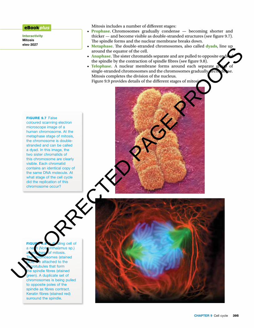
FIGURE 9.7 False coloured scanning electron microscope image of a human chromosome. At the metaphase stage of mitosis, the chromosome is double-stranded and can be called a dyad. In this image, the two sister chromatids of this chromosome are clearly visible. Each chromatid contains an identical copy of the same DNA molecule. At what stage of the cell cycle did the replication of this chromosome occur?
FIGURE 9.8 A dividing cell of a newt (Notophthalamus sp.) at anaphase of mitosis. The chromosomes (stained blue) are attached to the microtubules that form the spindle � bres (stained green). A duplicate set of chromosomes is being pulled to opposite poles of the spindle as � bres contract. Keratin � bres (stained red) surround the spindle.
UNCORRECTED PAGE P
ROOFS
NATURE OF BIOLOGY 1396
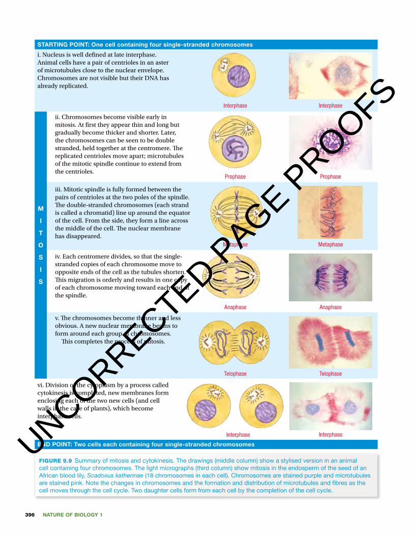
STARTING POINT: One cell containing four single-stranded chromosomes
i. Nucleus is well de� ned at late interphase. Animal cells have a pair of centrioles in an aster of microtubules close to the nuclear envelope. Chromosomes are not visible but their DNA has already replicated.
ii. Chromosomes become visible early in mitosis. At � rst they appear thin and long but gradually become thicker and shorter. Later, the chromosomes can be seen to be double stranded, held together at the centromere. � e replicated centrioles move apart; microtubules of the mitotic spindle continue to extend from the centrioles.
iii. Mitotic spindle is fully formed between the pairs of centrioles at the two poles of the spindle. � e double-stranded chromosomes (each strand is called a chromatid) line up around the equator of the cell. From the side, they form a line across the middle of the cell. � e nuclear membrane has disappeared.
iv. Each centromere divides, so that the single-stranded copies of each chromosome move to opposite ends of the cell as the tubules shorten. � is migration is orderly and results in one copy of each chromosome moving toward each end of the spindle.
v. � e chromosomes become thinner and less obvious. A new nuclear membrane begins to form around each group of chromosomes. � is completes the process of mitosis.
vi. Division of the cytoplasm by a process called cytokinesis is completed, new membranes form enclosing each of the two new cells (and cell walls in the case of plants), which become interphase cells.
END POINT: Two cells each containing four single-stranded chromosomes
Interphase
Prophase
Metaphase
Anaphase
Telophase
Interphase
M
I
T
O
S
I
S
Interphase
Prophase
Metaphase
Anaphase
Telophase
Interphase
FIGURE 9.9 Summary of mitosis and cytokinesis. The drawings (middle column) show a stylised version in an animal cell containing four chromosomes. The light micrographs (third column) show mitosis in the endosperm of the seed of an african blood lily, Scadoxus katherinae (18 chromosomes in each cell). Chromosomes are stained purple and microtubules are stained pink. note the changes in chromosomes and the formation and distribution of microtubules and fi bres as the cell moves through the cell cycle. Two daughter cells form from each cell by the completion of the cell cycle.
UNCORRECTED PAGE P
ROOFS
397CHAPTER 9 Cell cycle
Individual chromosomes � rst become visible as double, thread-like structures held together in a constricted region. Each of these threads is called a chromatid and the position where they are held together is called a centromere. � e fact that the chromosomes are double-stranded and there-fore contain two molecules of DNA indicates that the genetic material in the parent cell has already been replicated.
� e chromosomes continue to shorten and thicken and the nuclear mem-brane disintegrates. At the same time, the very � ne protein � bres or micro-tubules in the cytosol move towards the nucleus. � e function of the � bres is to guide the movement of the chromosomes in the cell. � e � bres become arranged in the cell, rather like the lines of longitude on a globe, to form a structure called a spindle. � e chromosomes become attached by their cen-tromeres around the ‘equator’ of the spindle.
Two things then happen. � e centromeres split, so that there are pairs of chromosomes, and the spindle � bres contract. � e contraction of the spindle � bres is responsible for the movement of the chromosomes towards the poles of the spindle. � e movement of the new chromosomes is very ordered. One of the new chromosomes from each pair moves to one end of the spindle; its identical pair moves towards the opposite pole. � e end result is a set of chromosomes at each end of the spindle. Because the new chromosomes behave in an orderly way, the set of chromosomes at one end of the spindle is identical to the set of chromosomes at the other end of the spindle.
� e chromosomes at each end of the spindle begin to lengthen and become less visible as distinct structures. At the same time, the protein � bres disperse back into the cytosol and a nuclear membrane develops around each group.
Remember that mitosis is a continuous process. � e stages of mitosis identify key changes in the appearance and the position of chromosomes. Remember also that chromosomes are not routinely visible when viewing cells through a light microscope. Only cells that are capable of division will ever show chromosomes and this will be for only a short period during the cell cycle. � e disappearance of discrete chromosomes does not mean that the genetic material has disappeared; rather, the DNA is present as chromatin granules dispersed throughout the nucleus.
Walther Flemming (1843–1905) was the German cytol-ogist who discovered chromosomes and their role in cell division. Flemming used newly developed ani-line dyes and improved microscopes to study nuclei in cells and found that scattered fragments in the nucleus became highly coloured. He named these fragments ‘chromatin’. He found that, during cell div-ision, the chromatin granules coalesced to form thread-like structures that were later called chromo-somes (chroma = colour; soma = body). He showed that, during cell division, chromosomes split length-wise and separate so that each daughter cell has as much genetic information as the original chromo-some. Flemming called this process mitosis. � e term mitosis comes from the Greek mitos = thread and osis = process.
WALTHER FLEMMING AND MITOSIS
A protein called cohesin holds sister chromatids together. (It appears as a blue stained region in � gure 9.1). Cohesin is removed at the metaphase–anaphase transition.
UNCORRECTED PAGE P
ROOFS
NATURE OF BIOLOGY 1398
Cytokinesis: one cell to two cellsAt the end of mitosis, the division of the nucleus into two new identical nuclei is complete. However, the cell cycle is completed only after the cytosol and organelles in the cytosol distribute around the new nuclei and become enclosed within an entire plasma membrane. � is � nal process of the cell cycle is called cytokinesis.
In January 2005, the journal Trends in Cell Biology (� gure 9.10) announced a series of special articles on research into cytokinesis under the title ‘ Cyto kinesis: the great divide’. In the � rst of these articles, Professor Jeremy Hyams of Massey University wrote:
Cytokinesis brings the curtain down on the cell cycle; it is the � nal dramatic act in which one cell becomes two.
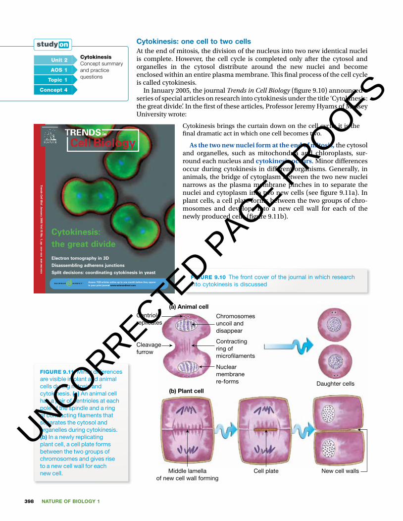
As the two new nuclei form at the end of mitosis, the cytosol and organelles, such as mitochondria and chloroplasts, sur-round each nucleus and cytokinesis occurs. Minor di� erences occur during cytokinesis in di� erent organisms. Generally, in animals, the bridge of cytoplasm between the two new nuclei narrows as the plasma membrane pinches in to separate the nuclei and cytoplasm into two new cells (see � gure 9.11a). In plant cells, a cell plate forms between the two groups of chro-mosomes and develops into a new cell wall for each of the newly produced cells (� gure 9.11b).
(a) Animal cellCentriolereplicates
Cleavagefurrow
Nuclearmembranere-forms
Contractingring ofmicro�laments
Chromosomesuncoil anddisappear
Daughter cells
New cell wallsCell plateMiddle lamellaof new cell wall forming
(b) Plant cell
FIGURE 9.11 Minor differences are visible in plant and animal cells during mitosis and cytokinesis. (a) an animal cell has a pair of centrioles at each pole of the spindle and a ring of contracting fi laments that separates the cytosol and organelles during cytokinesis. (b) in a newly replicating plant cell, a cell plate forms between the two groups of chromosomes and gives rise to a new cell wall for each new cell.
Unit 2 CytokinesisConcept summary and practice questions
AOS 1
Topic 1
Concept 4
FIGURE 9.10 The front cover of the journal in which research into cytokinesis is discussed
UNCORRECTED PAGE P
ROOFS

399CHAPTER 9 Cell cycle
Mitosis is essentially the same in plant and animal cells. � e small di� er-ences that do exist are not related to the genetic material, nor do they have an impact on the biological signi� cance of the process.
Cell cycle in prokaryotesProkaryotes, such as bacteria and archaea, also have a cell cycle. � is is a far less complex process than the cell cycle in eukaryotic cells. Note that bac-teria and other microbes have a single circular DNA molecule in contrast to the many chromosomes of eukaryotic cells. � e process in microbes is called binary � ssion and its essential components are shown in � gure 9.12.
� e process of asexual reproduction by binary � ssion in bacteria is simpler and faster than asexual reproduction in eukaryotic organisms. Asexual repro-
duction in eukaryotes involves the more complex process of mitosis followed by division of the cyto-plasm (cytokinesis). � is process typically takes many hours to complete. Binary � ssion in bacte-rial cells can be completed in about 20 minutes at room temperature. � is means that, if resources are available, one bacterial cell, through successive binary � ssions over an 8-hour period, could pro-duce 16 million descendants! � is is an example of exponential growth (discussed further in chapter 8) and it reminds us why a bacterial infec-tion, if not treated, can have serious outcomes. Figure 9.13 shows a cell of the bacterial species Escherichia coli dividing by binary � ssion.
FIGURE 9.13 Cells of the bacterial species Listeria sp., one of which is dividing by binary fi ssion. The circular outlines are cross-sections through bacterial cells. The Dna of the bacterial circular chromosome appears as darkly stained material.
DNA replicationand cell elongation
Division into two
DNA
FIGURE 9.12 Diagram showing the essence of the cell cycle in a bacterial cell. The cell cycle in prokaryotes, such as the one shown, is far less complex and much faster than the cell cycle of eukaryotes. What elements of the prokaryotic cell cycle are also present in the cell cycle of eukaryotes?
Unit 2 Prokaryotic cell divisionConcept summary and practice questions
AOS 1
Topic 1
Concept 1
UNCORRECTED PAGE P
ROOFS
NATURE OF BIOLOGY 1400
KEY IDEAS
■ Eukaryotic cells divide during the cell cycle giving rise to genetically identical daughter cells.
■ An essential early process in the cell cycle is the replication of DNA. ■ Carefully governed separation of sister chromatids is another essential step in cell division.
■ Mitosis is followed by cytokinesis. ■ Cell division in prokaryotes involves a relatively simple and rapid process of binary � ssion.
QUICK CHECK
1 What are the stages of interphase?2 What is the key event of the S stage of interphase?3 What is an average time for:
a a complete cell cycle in a mammal b a complete cell cycle by binary fi ssion in a microbe?
4 identify whether each of the following statements is true or false.a Sister chromatids separate at metaphase.b During interphase, double-stranded chromosomes are visible.c Cytokinesis is the last step in a cell cycle.d in a cell cycle, more time is spent in interphase than any other stage.e Binary fi ssion does not involve Dna replication.f The sequence of stages in interphase is g1 then g2 then S.
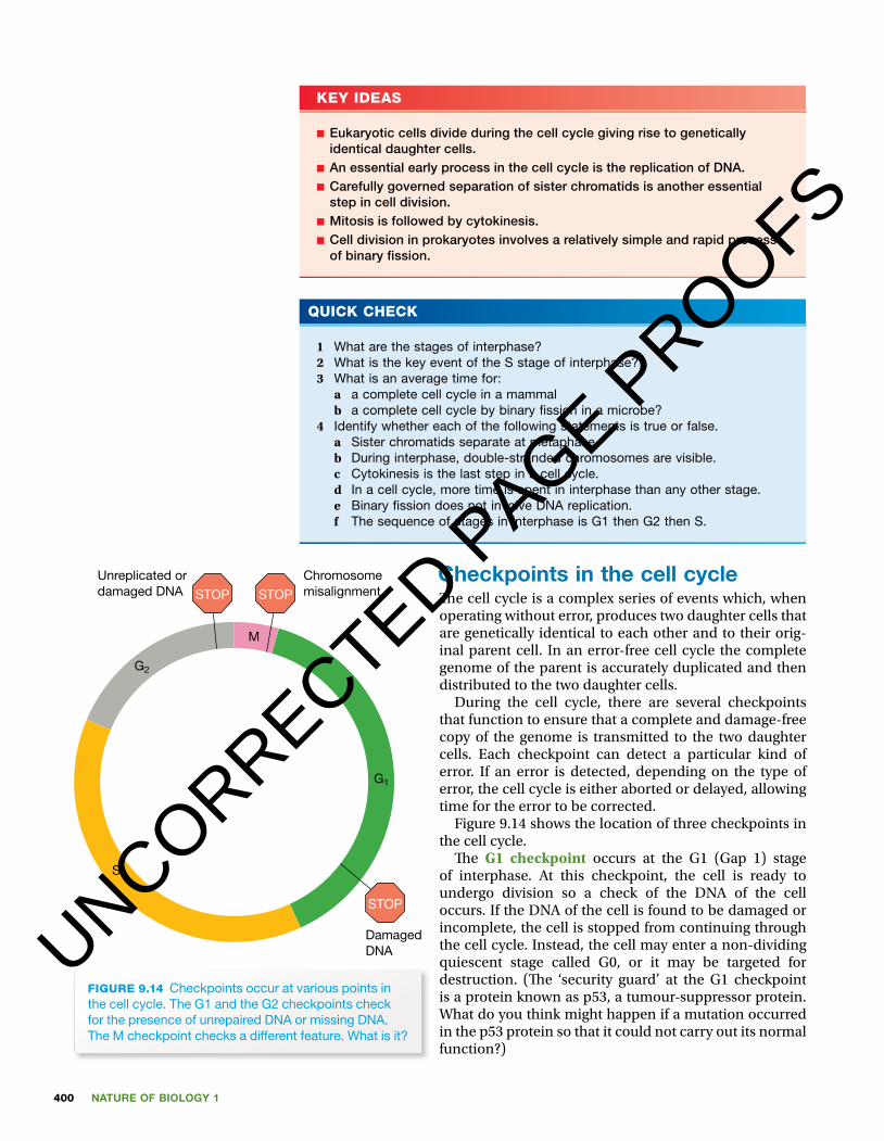
Checkpoints in the cell cycle� e cell cycle is a complex series of events which, when operating without error, produces two daughter cells that are genetically identical to each other and to their orig-inal parent cell. In an error-free cell cycle the complete genome of the parent is accurately duplicated and then distributed to the two daughter cells.
During the cell cycle, there are several checkpoints that function to ensure that a complete and damage-free copy of the genome is transmitted to the two daughter cells. Each checkpoint can detect a particular kind of error. If an error is detected, depending on the type of error, the cell cycle is either aborted or delayed, allowing time for the error to be corrected.
Figure 9.14 shows the location of three checkpoints in the cell cycle.
� e G1 checkpoint occurs at the G1 (Gap 1) stage of interphase. At this checkpoint, the cell is ready to undergo division so a check of the DNA of the cell occurs. If the DNA of the cell is found to be damaged or incomplete, the cell is stopped from continuing through the cell cycle. Instead, the cell may enter a non-dividing quiescent stage called G0, or it may be targeted for destruction. (� e ‘security guard’ at the G1 checkpoint is a protein known as p53, a tumour-suppressor protein. What do you think might happen if a mutation occurred in the p53 protein so that it could not carry out its normal function?)
Unreplicated ordamaged DNA
Chromosomemisalignment
DamagedDNA
G2
G1
M
S
STOP STOP
STOP
FIGURE 9.14 Checkpoints occur at various points in the cell cycle. The g1 and the g2 checkpoints check for the presence of unrepaired Dna or missing Dna. The M checkpoint checks a different feature. What is it?
UNCORRECTED PAGE P
ROOFS

401CHAPTER 9 Cell cycle
If the cell passes the G1 checkpoint, it proceeds into the cell cycle and enters the S stage of interphase. During the S stage, the cell replicates its DNA so that, by the end of the S stage, the cell should have double the amount of DNA and this DNA should be two complete and accurate copies of its genome. � e cell now moves to the G2 stage of interphase where it must pass the G2 checkpoint.
At the G2 checkpoint, the replicated DNA of the cell is checked for complete-ness and lack of damage. If the cell passes this checkpoint, it can then advance to the mitosis stage of the cell cycle.
� e M checkpoint (or spindle assembly checkpoint) occurs at the meta-phase stage of mitosis. A check is carried out to ensure that the sister chro-matids (i.e. the two strands of each double-stranded chromosome) are attached to the correct microtubules of the spindle. � is check is to ensure that the sister chromatids are pulled in opposite directions to di� erent poles of the spindle. If an error is detected, the cell cycle is delayed until the error is � xed.
The mitotic spindle� e focus in mitosis is typically on chromosomes. However, the positioning and the movement of the chromosomes depend on the presence of a micro tubule framework, the spindle.
In animal cells, once mitosis starts, the paired centrioles move to opposite ends of the cell where they form the poles of the spindle. Clusters of microtubules grow out from the centrioles towards the middle of the cell. � ese microtubule clusters are called spindle � bres. At metaphase, these � bres anchor the dou-ble-stranded chromosomes around the equator of the cell. Each chromatid has a special attachment site called a kinetochore by which it links to a spindle � bre (see � gure 9.15).
Spindle � bres from one pole attach to one sister chromatid and � bres from the opposite pole attach to its partner chro-matid. (What would happen if the two sister chromatids of one chromosome became linked to � bres from the same pole of the spindle?) At the M checkpoint, the connection between chro-matid and spindle � bres is checked and, if it is not correct, the cell cycle is delayed until the arrangement is corrected.
Spindle � bres are composed of actin, a contractile protein. At anaphase, the orderly migration of each pair of sister chromatids is achieved by contractions of the � bres that pull these now single-stranded chromosomes to the opposite poles of the spindle.
Mitochondria and chloroplasts also replicateWe have seen that mitosis is followed by cytokinesis. � is is essential so that the two new nuclei formed can each be combined with cytosol to give two new cells. Obviously the organelles such as mitochondria and chloroplasts within the cytosol must also be replicated during the cell cycle, otherwise cells would contain an ever-decreasing number of these structures.
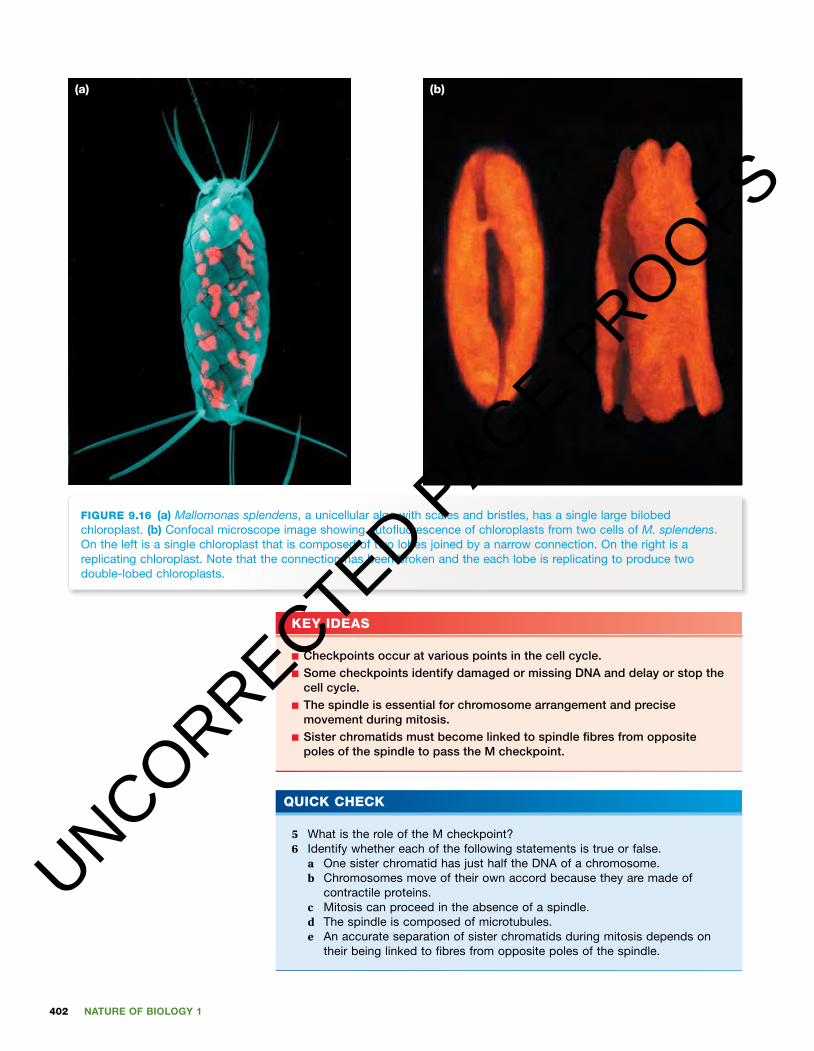
Just as a nucleus contains DNA that must replicate before two new nuclei are formed, mitochondria and chloroplasts contain DNA that must rep-licate before the organelles divide. � e alga Mallomonas splendens (see � gure 9.16a) has a single chloroplast composed of two lobes joined by a narrow connection. As a cell of M. splendens replicates, its chloroplast must also replicate. During replication of the chloroplast, the narrow connection breaks and each of the two lobes grows and constricts to give two, two-lobed chloro plasts (see � gure 9.16b). Organelles such as chloroplasts and mito-chondria can arise only from pre-existing organelles. Cells can arise only from pre-existing cells.
Wait! I’ll check tosee if your DNA
is in order.
FIGURE 9.15 DeltaVision Widefi eld microscope image of a hela cell undergoing mitosis and treated with various stains. The pericentrin stain shows the centrioles (orange). The aCa stain shows the kinetochores (purple) that are protein complexes located at the centromere and bind each chromosome to the microtubules of the spindle. The a-tubulin stain shows the microtubules of the spindle (green). (image courtesy a loynton-Ferrand, iMCF, University of Basel)UNCORRECTED P
AGE PROOFS
NATURE OF BIOLOGY 1402
FIGURE 9.16 (a) Mallomonas splendens, a unicellular alga with scales and bristles, has a single large bilobed chloroplast. (b) Confocal microscope image showing autofl uorescence of chloroplasts from two cells of M. splendens. on the left is a single chloroplast that is composed of two lobes joined by a narrow connection. on the right is a replicating chloroplast. note that the connection has been broken and the each lobe is replicating to produce two double-lobed chloroplasts.
(b)(a)
KEY IDEAS
■ Checkpoints occur at various points in the cell cycle. ■ Some checkpoints identify damaged or missing DNA and delay or stop the cell cycle.
■ The spindle is essential for chromosome arrangement and precise movement during mitosis.
■ Sister chromatids must become linked to spindle � bres from opposite poles of the spindle to pass the M checkpoint.
QUICK CHECK
5 What is the role of the M checkpoint?6 identify whether each of the following statements is true or false.
a one sister chromatid has just half the Dna of a chromosome.b Chromosomes move of their own accord because they are made of
contractile proteins.c Mitosis can proceed in the absence of a spindle.d The spindle is composed of microtubules.e an accurate separation of sister chromatids during mitosis depends on
their being linked to fi bres from opposite poles of the spindle.
UNCORRECTED PAGE P
ROOFS
403CHAPTER 9 Cell cycle
Cell cycle in action� e cell cycle is a critical process in:• growth, where the cell cycle produces new cells, resulting in an increase in cell
number. � is is most prominent during embryonic development of a multi-cellular organism. Early embryonic cell division is exponential, with one cell dividing to form two cells, these two form four cells, these four form eight cells, and so on. � e power of the cell cycle is seen in the fact that a typical human is composed of 37 trillion cells that came originally from a single fertilised egg cell.
• repair and maintenance (regeneration), where the cell cycle produces new cells to replace dead or damaged cells
• reproduction, where the cell cycle produces identical cells, such as spores, that give rise to the next generation.Not all cells of a multicellular organism are capable of dividing. For example,
in the human body, nerve cells generally cannot regenerate. Cells from other organs, such as kidney and liver, may divide occasionally to replace cells lost through injury or death, but, for most of the time, these cells are in the G0 qui-escent stage. However, cells of some human tissues produce new cells at a staggering rate. Let’s meet some of them.
Cell cycle in mammalsIn mammals, such as a human adult, actively dividing cells are found in sev-eral tissues, such as the epidermis of the skin, the epithelial lining of the gut and the bone marrow. Cell division normally occurs at a tightly regulated rate, so that the production of new cells matches or balances the rate of cell loss. Tissues with a population of actively dividing stem cells are tissues that have a high and continual level of cell loss or cell death.
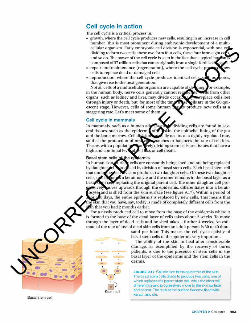
Basal stem cells of the epidermisIn human skin, surface cells are constantly being shed and are being replaced by daughter cells produced by division of basal stem cells. Each basal stem cell that undergoes cell division produces two daughter cells. Of these two daughter cells, one becomes a keratinocyte and the other remains in the basal layer as a basal stem cell, replacing the original parent cell. � e other daughter cell pro-gressively moves upwards through the epidermis, di� erentiates into a kerati-nocyte, and is shed from the skin surface (see � gure 9.17). Within a period of about 48 days, the entire epidermis is replaced by new cells. � is means that the skin that you have, say, today is made of completely di� erent cells from the skin that you had 2 months earlier.
For a newly produced cell to move from the base of the epidermis where it is formed to the base of the dead layer of cells takes about 2 weeks. To move through the layer of dead cells and be shed takes a further 4 weeks. An esti-mate of the rate of loss of dead skin cells from an adult person is 30 to 40 thou-
sand per hour. � is makes the cell cycle activity of basal stem cells of the epidermis very important.
� e ability of the skin to heal after considerable damage, as exempli� ed by the recovery of burns patients, is due to the presence of stem cells in the basal layer of the epidermis and the stem cells in the dermis.
Basal stem cell
Stem cell
FIGURE 9.17 Cell division in the epidermis of the skin. The basal stem cells divide to produce two cells, one of which replaces the parent stem cell, while the other will differentiate and progressively move to the skin surface and be lost. The cells at the surface become fi lled with keratin and die.
UNCORRECTED PAGE P
ROOFS
NATURE OF BIOLOGY 1404
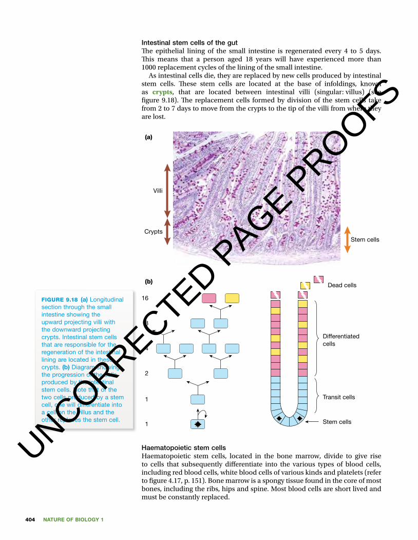
Intestinal stem cells of the gut � e epithelial lining of the small intestine is regenerated every 4 to 5 days. � is means that a person aged 18 years will have experienced more than 1000 replacement cycles of the lining of the small intestine.
As intestinal cells die, they are replaced by new cells produced by intestinal stem cells. � ese stem cells are located at the base of infoldings, known as crypts, that are located between intestinal villi (singular: villus) (see � gure 9.18). � e replacement cells formed by division of the stem cells take from 2 to 7 days to move from the crypts to the tip of the villi from where they are lost.
FIGURE 9.18 (a) longitudinal section through the small intestine showing the upward projecting villi with the downward projecting crypts. intestinal stem cells that are responsible for the regeneration of the intestinal lining are located in these crypts. (b) Diagram showing the progression of the cells produced by the intestinal stem cells. note that of the two cells produced by a stem cell, one will differentiate into a cell on the villus and the other replaces the stem cell.
Stem cells
Villi
Crypts
(a)
16
Dead cells
Differentiatedcells
Transit cells
Stem cells
8
4
2
1
1
(b)
Haematopoietic stem cellsHaematopoietic stem cells, located in the bone marrow, divide to give rise to cells that subsequently di� erentiate into the various types of blood cells, including red blood cells, white blood cells of various kinds and platelets (refer to � gure 4.17, p. 151). Bone marrow is a spongy tissue found in the core of most bones, including the ribs, hips and spine. Most blood cells are short lived and must be constantly replaced.
UNCORRECTED PAGE P
ROOFS

405CHAPTER 9 Cell cycle
NOBEL LAUREATE IN PHYSIOLOGY OR MEDICINE
Dr Elizabeth H BlackburnDr Elizabeth Blackburn received her Nobel Prize from His Majesty King Carl XVI Gustaf of Sweden in Stock-holm in December 2009 (� gure 9.19) for her work on telomeres (� gure 9.20) and telomerase. Telomeres are found at the ends of all eukaryotic chromosomes and comprise repetitive DNA strands that get shorter each time a cell divides. � is shortening of the telomeres eventually leads to the death of a cell. However, tel-omerase is an enzyme that prevents shortening in some cells and so extends the life of those cells.
FIGURE 9.19 Dr elizabeth Blackburn receiving her nobel Prize from his Majesty King Carl XVi gustaf of Sweden at the Stockholm Concert hall, 10 December 2009
Elizabeth Blackburn was born in Tasmania in 1948, one of seven children. After moving to Melbourne, she completed secondary school at the University High School. � is was followed by Honours (1971) and Masters degrees (1972) in Biochemistry from the University of Melbourne. Elizabeth then travelled to Cambridge University in England where she was admitted as a PhD student in the Medical Research Council’s Laboratory of Molecular Biology. After completing her PhD, Dr Blackburn did postdoctoral training at Yale in the USA, and then worked in the Department of Molecular Biology at the University of California, Berkeley. In 1990, she moved to the
Department of Microbiology and Immunology at the University of California, San Francisco (UCSF) (� gure 9.21). She is currently the Morris Herzstein Professor of Biology and Physiology at UCSF and a non-resident fellow of the Salk Institute.
FIGURE 9.20 Mitotic chromosomes (blue) with telomeres (yellow) at the tips of each chromatid
FIGURE 9.21 Dr elizabeth Blackburn in her lab at the University of California, San Francisco
Dr Blackburn has received many notable awards and honours. � ey include California Scientist of the Year (1999), American Cancer Society Medal of Honor (2000), L’Oreal–UNESCO Award for Women in Science (2008) and, of course, a Nobel Prize (2009). In 2007, Dr Blackburn was listed by Time magazine as one of the ‘100 most in� uential people in the world’.
UNCORRECTED PAGE P
ROOFS
NATURE OF BIOLOGY 1406
Cell cycle in other animalsPlanaria, phylum Platyhelminthes, are � atworms that live in water. � ey are one of the few animals that can reproduce asexually by regeneration. � e parent breaks into two or more pieces and each piece grows into a new pla-narian. � e new parts are produced by mitosis of cells and each new planarian is an exact copy of the parent.
If a star� sh loses some of its ‘arms’, new ones are regenerated by mitosis (see � gure 9.23).
FIGURE 9.23 if a starfi sh loses some of its ‘arms’, they regrow. here you can see six new ‘arms’ on a damaged starfi sh.
KEY IDEAS
■ The cell cycle is important for the growth, repair and maintenance of eukaryotes.
■ In some organisms, the cell cycle plays a role in producing cells involved in reproduction.
■ Actively dividing human tissues include the epidermis of the skin, the epithelium of the gut and the bone marrow.
■ Stem cells carry out the cell divisions that are responsible for tissue regeneration.
QUICK CHECK
7 identify where you would fi nd the following.a Skin stem cells b a red blood cell precursorc Keratinocytesd haematopoietic stem cells
8 identify whether each of the following statements is true or false.a all human cells regularly undergo cell division.b haematopoietic stem cells are responsible for skin regeneration.c The intestinal crypts are where intestinal stem cells are located.d Tissues composed of short-lived cells would be expected to show a high
rate of cell division.
FIGURE 9.22 if a starfi sh is cut into two, each half can regenerate into a whole.
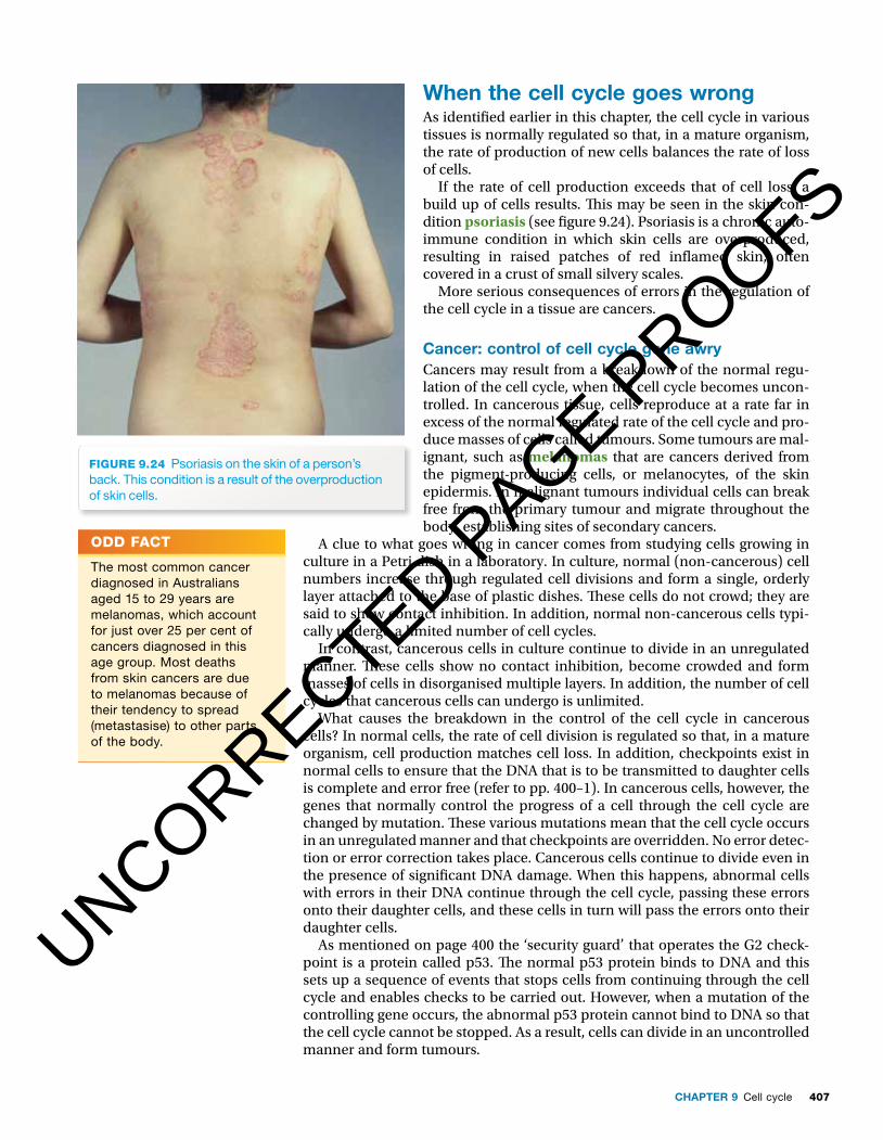
FIGURE 9. 24 Psoriasis on the skin of a person’s back. This condition is a result of the overproduction of skin cells.
UNCORRECTED PAGE P
ROOFS
407CHAPTER 9 Cell cycle
Cell cycle in other animalsPlanaria, phylum Platyhelminthes, are � atworms that live in water. � ey are one of the few animals that can reproduce asexually by regeneration. � e parent breaks into two or more pieces and each piece grows into a new pla-narian. � e new parts are produced by mitosis of cells and each new planarian is an exact copy of the parent.
If a star� sh loses some of its ‘arms’, new ones are regenerated by mitosis (see � gure 9.23).
FIGURE 9.23 if a starfi sh loses some of its ‘arms’, they regrow. here you can see six new ‘arms’ on a damaged starfi sh.
KEY IDEAS
■ The cell cycle is important for the growth, repair and maintenance of eukaryotes.
■ In some organisms, the cell cycle plays a role in producing cells involved in reproduction.
■ Actively dividing human tissues include the epidermis of the skin, the epithelium of the gut and the bone marrow.
■ Stem cells carry out the cell divisions that are responsible for tissue regeneration.
QUICK CHECK
7 identify where you would fi nd the following.a Skin stem cells b a red blood cell precursorc Keratinocytesd haematopoietic stem cells
8 identify whether each of the following statements is true or false.a all human cells regularly undergo cell division.b haematopoietic stem cells are responsible for skin regeneration.c The intestinal crypts are where intestinal stem cells are located.d Tissues composed of short-lived cells would be expected to show a high
rate of cell division.
FIGURE 9.22 if a starfi sh is cut into two, each half can regenerate into a whole.
FIGURE 9. 24 Psoriasis on the skin of a person’s back. This condition is a result of the overproduction of skin cells.
When the cell cycle goes wrongAs identi� ed earlier in this chapter, the cell cycle in various tissues is normally regulated so that, in a mature organism, the rate of production of new cells balances the rate of loss of cells.
If the rate of cell production exceeds that of cell loss, a build up of cells results. � is may be seen in the skin con-dition psoriasis (see � gure 9.24). Psoriasis is a chronic auto-immune condition in which skin cells are overproduced, resulting in raised patches of red in� amed skin, often covered in a crust of small silvery scales.
More serious consequences of errors in the regulation of the cell cycle in a tissue are cancers.
Cancer: control of cell cycle gone awryCancers may result from a breakdown of the normal regu-lation of the cell cycle, when the cell cycle becomes uncon-trolled. In cancerous tissue, cells reproduce at a rate far in excess of the normal regulated rate of the cell cycle and pro-duce masses of cells called tumours. Some tumours are mal-ignant, such as melanomas that are cancers derived from the pigment-producing cells, or melanocytes, of the skin epidermis. In malignant tumours individual cells can break free from the primary tumour and migrate throughout the body, establishing sites of secondary cancers.
A clue to what goes wrong in cancer comes from studying cells growing in culture in a Petri dish in a laboratory. In culture, normal (non-cancerous) cell numbers increase through regulated cell divisions and form a single, orderly layer attached to the base of plastic dishes. � ese cells do not crowd; they are said to show contact inhibition. In addition, normal non-cancerous cells typi-cally undergo a limited number of cell cycles.
In contrast, cancerous cells in culture continue to divide in an unregulated manner. � ese cells show no contact inhibition, become crowded and form masses of cells in disorganised multiple layers. In addition, the number of cell cycles that cancerous cells can undergo is unlimited.
What causes the breakdown in the control of the cell cycle in cancerous cells? In normal cells, the rate of cell division is regulated so that, in a mature organism, cell production matches cell loss. In addition, checkpoints exist in normal cells to ensure that the DNA that is to be transmitted to daughter cells is complete and error free (refer to pp. 400–1). In cancerous cells, however, the genes that normally control the progress of a cell through the cell cycle are changed by mutation. � ese various mutations mean that the cell cycle occurs in an unregulated manner and that checkpoints are overridden. No error detec-tion or error correction takes place. Cancerous cells continue to divide even in the presence of signi� cant DNA damage. When this happens, abnormal cells with errors in their DNA continue through the cell cycle, passing these errors onto their daughter cells, and these cells in turn will pass the errors onto their daughter cells.
As mentioned on page 400 the ‘security guard’ that operates the G2 check-point is a protein called p53. � e normal p53 protein binds to DNA and this sets up a sequence of events that stops cells from continuing through the cell cycle and enables checks to be carried out. However, when a mutation of the controlling gene occurs, the abnormal p53 protein cannot bind to DNA so that the cell cycle cannot be stopped. As a result, cells can divide in an uncontrolled manner and form tumours.
ODD FACT
The most common cancer diagnosed in australians aged 15 to 29 years are melanomas, which account for just over 25 per cent of cancers diagnosed in this age group. Most deaths from skin cancers are due to melanomas because of their tendency to spread (metastasise) to other parts of the body.
UNCORRECTED PAGE P
ROOFS

NATURE OF BIOLOGY 1408
KEY IDEAS
■ The cell cycle is normally regulated so that, in a mature organism, the rate of production of new cells balances the rate of loss of cells.
■ Cancers may result from the breakdown of the normal control of the cell cycle.
■ Cancerous cells are characterised by unregulated rates of cell division.
QUICK CHECK
9 identify whether each of the following statements is true or false.a Cancerous cells divide at rates in excess of the normal regulated rate.b Cell production and cell loss are kept in balance by genes that
regulate the cell cycle.c normal non-cancerous cells in culture show contact inhibition.
Cell cycle in plantsIn vascular plants, only the cells in meristematic tissues can complete cell cycles and divide to produce identical daughter cells. � e cells in perma-nent plant tissues cannot divide (refer to chapter 4, p. 180). Meristematic tissue is present in several locations including root tips (see � gure 9.25) and stems.
Other examples of the cell cycle in plants include those discussed below.
Epicormic shoots after a bushfi reBush� res are common in many areas of Australia. Although trees may appear to be burnt to a point that one might think they are dead, a picture such as the one in � gure 9.26 (taken just 6 weeks after the area was devastated by bush� re) shows this is not the case. It is clear from the photograph that the � re has completely destroyed the undergrowth of grasses, shrubs and
FIGURE 9.25 light microscope image of a longitudinal section through the meristematic tissue of a root tip. This is a region of active cell division and many rows of cells can be seen. examine this image and see if you can identify some cells that are in the mitosis stage of the cell cycle.
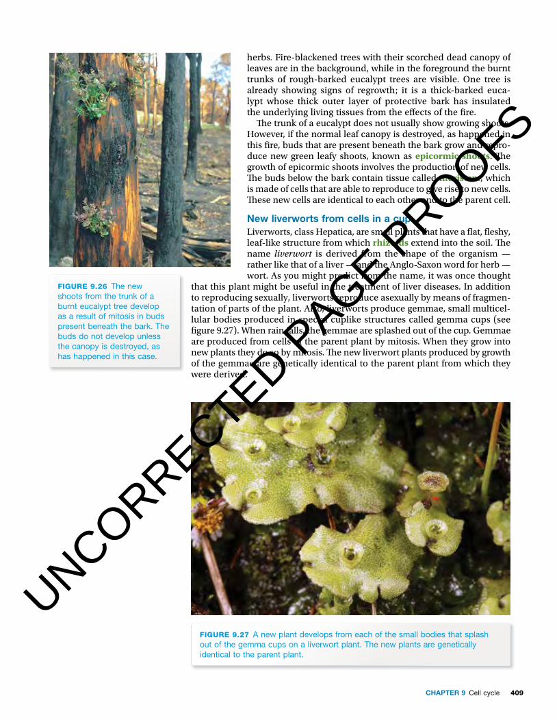
FIGURE 9.26 The new shoots from the trunk of a burnt eucalypt tree develop as a result of mitosis in buds present beneath the bark. The buds do not develop unless the canopy is destroyed, as has happened in this case.
UNCORRECTED PAGE P
ROOFS
409CHAPTER 9 Cell cycle
KEY IDEAS
■ The cell cycle is normally regulated so that, in a mature organism, the rate of production of new cells balances the rate of loss of cells.
■ Cancers may result from the breakdown of the normal control of the cell cycle.
■ Cancerous cells are characterised by unregulated rates of cell division.
QUICK CHECK
9 identify whether each of the following statements is true or false.a Cancerous cells divide at rates in excess of the normal regulated rate.b Cell production and cell loss are kept in balance by genes that
regulate the cell cycle.c normal non-cancerous cells in culture show contact inhibition.
Cell cycle in plantsIn vascular plants, only the cells in meristematic tissues can complete cell cycles and divide to produce identical daughter cells. � e cells in perma-nent plant tissues cannot divide (refer to chapter 4, p. 180). Meristematic tissue is present in several locations including root tips (see � gure 9.25) and stems.
Other examples of the cell cycle in plants include those discussed below.
Epicormic shoots after a bushfi reBush� res are common in many areas of Australia. Although trees may appear to be burnt to a point that one might think they are dead, a picture such as the one in � gure 9.26 (taken just 6 weeks after the area was devastated by bush� re) shows this is not the case. It is clear from the photograph that the � re has completely destroyed the undergrowth of grasses, shrubs and
FIGURE 9.25 light microscope image of a longitudinal section through the meristematic tissue of a root tip. This is a region of active cell division and many rows of cells can be seen. examine this image and see if you can identify some cells that are in the mitosis stage of the cell cycle.
FIGURE 9.26 The new shoots from the trunk of a burnt eucalypt tree develop as a result of mitosis in buds present beneath the bark. The buds do not develop unless the canopy is destroyed, as has happened in this case.
herbs. Fire-blackened trees with their scorched dead canopy of leaves are in the background, while in the foreground the burnt trunks of rough-barked eucalypt trees are visible. One tree is already showing signs of regrowth; it is a thick-barked euca-lypt whose thick outer layer of protective bark has insulated the underlying living tissues from the e� ects of the � re.
� e trunk of a eucalypt does not usually show growing shoots. However, if the normal leaf canopy is destroyed, as happened in this � re, buds that are present beneath the bark grow and repro-duce new green leafy shoots, known as epicormic shoots. � e growth of epicormic shoots involves the production of new cells. � e buds below the bark contain tissue called meristem, which is made of cells that are able to reproduce to give rise to new cells. � ese new cells are identical to each other and to the parent cell.
New liverworts from cells in a cupLiverworts, class Hepatica, are small plants that have a � at, � eshy, leaf-like structure from which rhizoids extend into the soil. � e name liverwort is derived from the shape of the organism — rather like that of a liver — and the Anglo-Saxon word for herb — wort. As you might predict from the name, it was once thought
that this plant might be useful in the treatment of liver diseases. In addition to reproducing sexually, liverworts reproduce asexually by means of fragmen-tation of parts of the plant. Also, liverworts produce gemmae, small multicel-lular bodies produced in special cuplike structures called gemma cups (see � gure 9.27). When rain falls, the gemmae are splashed out of the cup. Gemmae are produced from cells of the parent plant by mitosis. When they grow into new plants they do so by mitosis. � e new liverwort plants produced by growth of the gemmae are genetically identical to the parent plant from which they were derived.
FIGURE 9.27 a new plant develops from each of the small bodies that splash out of the gemma cups on a liverwort plant. The new plants are genetically identical to the parent plant.
UNCORRECTED PAGE P
ROOFS

NATURE OF BIOLOGY 1410
Cell cycle in fungi� e cell cycle plays an important role in the reproduction of fungi.
� e fungus or mould you see on bread or fruit grows by mitosis. A single cell, a fungal spore, lands on food and grows into a mass of threads called hyphae. Specialised stalks — each with a spore case at its tip — grow up from the mass of hyphae (see � gure 9.28). Mitosis occurs within the spore case and thou-sands of black spores are formed. On maturing, the spore case splits open and the tiny, light spores are scattered. When conditions are favourable, each spore germinates and grows into a new hyphal mass.
FIGURE 9.28 The fungus on a rotting tomato (a) comprises a mass of white threads or hyphae. asexual reproduction occurs at the tips of some hyphae and (b) large numbers of black spores are formed, each genetically identical with the parent.
Hyphaeof the
mycelium
(b)
Spores
(a)
KEY IDEAS
■ The meristematic tissue of plants contains cells that can complete the cell cycle and produce identical daughter cells.
■ In vascular plants, meristematic tissue is present in root tips, shoots and stems.
■ Cell division in epicormic shoots is important in the recovery of trees damaged by bush� re.
■ Some cells produced by the cell cycle have a reproductive function, but offspring from this process are genetically identical.
QUICK CHECK
10 in which plant tissues would you expect to fi nd dividing cells?11 Consider the gemmae of liverworts. Would the next generation of plants
that are derived from the gemmae of one liverwort be genetically identical or genetically dissimilar?
12 how do epicormic shoots contribute to the survival of fi re-damaged trees in the australian bush.
UNCORRECTED PAGE P
ROOFS
411CHAPTER 9 Cell cycle
BioChallenge
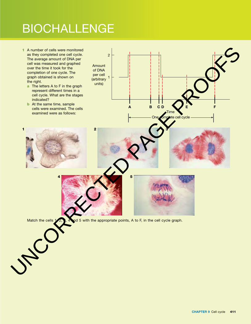
1 a number of cells were monitored as they completed one cell cycle. The average amount of Dna per cell was measured and graphed over the time it took for the completion of one cycle. The graph obtained is shown on the right. a The letters a to F in the graph
represent different times in a cell cycle. What are the stages indicated?
b at the same time, sample cells were examined. The cells examined were as follows:
1 32
4 5
Match the cells 1, 2, 3, 4 and 5 with the appropriate points, a to F, in the cell cycle graph.
Amountof DNAper cell
(arbitraryunits)
A B C D E F
One complete cell cycleTime
1
2
UNCORRECTED PAGE P
ROOFS
NATURE OF BIOLOGY 1412
Unit 2 The cell cycle
Sit topic test
AOS 1
Topic 1Chapter review
Key wordsallograft anaphase autograft binary � ssion cell cyclecentrioles centromere chromatid crypts
cytokinesis dermisdyad epicormic shoots epidermisexponential growth G1 checkpoint G1 stage of interphaseG2 checkpoint
G2 stage of interphaseinterphase keratinocyteskinetochoreM checkpoint melanocytesmelanomas meristem metaphase
mitosisprophase psoriasis rhizoids S stage of interphase spindle spindle � bres telophase
Questions 1 Making connections ➜ Use at least eight of the
chapter key words to draw a concept map. You may use other words in drawing your map.
2 Applying understanding ➜ A cell containing 24 chromosomes reproduced by mitosis. A genetic accident occurred and one of the resulting cells had only 23 chromosomes. a How many chromosomes would you expect in
the other cell produced? Explain why. b At what stage of cell reproduction do you think
the genetic accident occurred? 3 Interpreting and applying understanding of
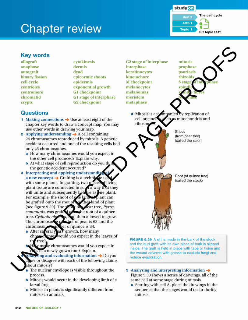
a new concept ➜ Grafting is a technique used with some plants. In grafting, two pieces of living plant tissue are connected in such a way that they will unite and subsequently behave as one plant. For example, the shoot of one kind of plant can be grafted onto the root of another kind of plant (see � gure 9.29). � e shoot of a pear tree, Pyrus communis, was grafted onto the root of a quince tree, Cydonia oblonga, and then allowed to grow. � e chromosome number of pear is 68 and the chromosome number of quince is 34. a After several years’ growth, how many
chromosomes would you expect in the leaves of the tree?
b How many chromosomes would you expect in cells of a newly grown root? Explain.
4 Analysing and evaluating information ➜ Do you agree or disagree with each of the following claims about mitosis? a � e nuclear envelope is visible throughout the
process. b Mitosis would occur in the developing limb of a
larval frog. c Mitosis in plants is signi� cantly di� erent from
mitosis in animals.
d Mitosis is accompanied by replication of cell organelles such as mitochondria and ribosomes.
Shoot(from pear tree)(called the scion)
Root (of quince tree)(called the stock)
FIGURE 9.29 a slit is made in the bark of the stock and the bud graft with its own piece of bark is slipped inside. The graft is held in place with tape or twine and the wound covered with grease to exclude fungi and reduce evaporation.
5 Analysing and interpreting information ➜ Figure 9.30 shows a series of drawings, all of the same cell at some stage during mitosis.a Starting with cell A, place the drawings in the
sequence that the stages would occur during mitosis.
UNCORRECTED PAGE P
ROOFS

413CHAPTER 9 Cell cycle
b Draw what you would expect to see next in the sequence.
FIGURE 9.30
A
B
C
D
E
6 Applying understanding to new concepts ➜ Some drugs used in the treatment of some cancers act on microtubules. � ey act by interfering with the normal contraction and extension capabilities of microtubules. Explain the e� ect you would expect such drugs to have on mitosis and cell replication.
7 Applying knowledge ➜ Arrange the following events in animal cell replication in the correct order. a Alignment of chromosomes on the spindle
equator b Attachment of microtubules to centromere
region c Breakdown of nuclear envelope d Condensation of chromosomes e Decondensation of chromosomes f Duplication of centromere g Elongation of the spindle h Pinching of cell into two i Re-formation of nuclear envelope j Separation of centromeres k Separation of sister chromatids
8 Demonstrating skills of understanding and communication ➜ You are shown a video sequence of the entire cell cycle for one cell, but you are not told whether this cell is from an animal or a plant.a Is it possible to decide the identity of the cell as
either animal or plant from the video?b If so, on what evidence would you base
your decision? If not, give a reason for your choice.
9 Developing an explanation ➜ After a cell with 10 chromosomes completed the cell cycle, its daughter cells were examined. One daughter cell was found to contain 11 chromosomes and the other daughter cell had only 9 chromosomes. Suggest a possible explanation in biological terms for this observation.
10 Interpreting data and demonstrating understanding ➜ Cell A has four pairs of chromosomes with a total DNA content of 12 units. Cell A undergoes one cell cycle. a List, in order, the stages that cell A would
proceed through, starting from the earliest.b At the end of this cell cycle, how many cells
would be present: one, two or three? c How many units of DNA would be present in
Cell A at the following point in the cell cycle? i G2 stage of interphase ii Anaphase of mitosisiii G1 stage of interphase
d How many units of DNA would be present in one daughter cell of cell A?
e How many chromosomes would be present in this daughter cell?
11 Making predictions based on given information ➜ A particular gene mutation a� ects a protein that is a key part of the special attachment site, the kinetochore, that allows a chromatid to be linked to spindle � bres. � is
UNCORRECTED PAGE P
ROOFS
414 NATURE OF BIOLOGY 1
mutation is present in cell B and it disables the function of the kinetochore. a Would this mutation be expected to a� ect the
progress of cell B through the cell cycle?b If so, what e� ect would you predict? If not, give
a reason for your decision. 12 Applying skills of analysis and communication ➜
Using a light microscope, you examine a longitudinal section of the meristematic tissue of a plant root. Consider each of the following statements in turn and identify whether or not you would expect to observe each in the particular cells you are viewing. Brie� y explain your choice.a � e majority of the cells would be in one of the
stages of mitosis.b A cleavage furrow would be present in cells at
telophase of mitosis.c Sister chromatids at anaphase of mitosis would
be moving away from the equator and towards opposite ends of the spindles.
d Double-stranded chromosomes would be visible in cells at the S stage of interphase.
13 Demonstrating understanding ➜ � e cell cycle in eukaryotes is highly regulated so that cell production in a tissue occurs at a rate that balances cell loss.a What is a possible outcome if a breakdown in
the regulation of the cell cycle occurs?b A disorder known as polycythemia vera is a
result of the overactivity of the bone marrow, resulting in the production of too many red blood cells. � is condition results in a thickening of the blood and the common treatment is the regular removal of a � xed amount of blood. � e cause of polycythemia vera is a mutation in the JAK2 gene. What is the probable function of the normal JAK2 gene?
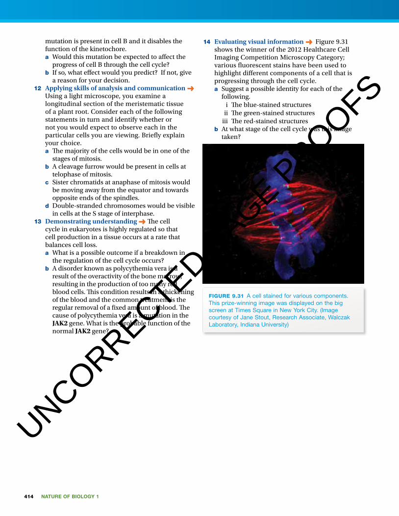
14 Evaluating visual information ➜ Figure 9.31 shows the winner of the 2012 Healthcare Cell Imaging Competition Microscopy Category; various � uorescent stains have been used to highlight di� erent components of a cell that is progressing through the cell cycle.a Suggest a possible identity for each of the
following. i � e blue-stained structures ii � e green-stained structuresiii � e red-stained structures
b At what stage of the cell cycle was this image taken?
FIGURE 9.31 a cell stained for various components. This prize-winning image was displayed on the big screen at Times Square in new York City. (image courtesy of Jane Stout, Research associate, Walczak laboratory, indiana University)
UNCORRECTED PAGE P
ROOFS