Embed Size (px)
Citation preview
PALESTRICA OF THE THIRD MILLENNIUM -
CIVILIZATION AND SPORT
A quarterly of multidisciplinary study and research
© Published by The “Iuliu Haţieganu” University of Medicine and Pharmacy of Cluj-Napocaand
The Romanian Medical Society of Physical Education and Sportsin collaboration with
The Cluj County School Inspectorate
A journal rated B+ by CNCS (Romanian National Research Council) since 2007, certified by CMR (Romanian College of Physicians) since 2003
and CFR (Romanian College of Pharmacists) since 2015
A journal with a multidisciplinary approach in the fields of biomedical science, health, medical rehabilitation, physical exercise, social sciences
applied to physical education and sports activities
A journal indexed in international databases:EBSCO, Academic Search Complete, USA;
Index Copernicus, Journals Master List, Poland;DOAJ (Directory of Open Access Journals), Sweden
CiteFactor, Canada/USA
3Vol. 18, No. 3, July-September 2017
Editorial BoardChief Editor Traian Bocu (Cluj-Napoca, Romania)
Deputy Chief Editors Simona Tache (Cluj-Napoca, Romania) Ioan Onac (Cluj-Napoca, Romania)Dan Riga (Bucureşti, Romania) Adriana Filip (Cluj-Napoca, Romania)
Bio-Medical, Health and Exercise Department
Cezar Login (Cluj-Napoca, Romania) Adriana Albu (Cluj-Napoca, Romania)Adrian Aron (Radford, VA, USA)Taina Avramescu (Craiova, Romania)Cristian Bârsu (Cluj-Napoca, Romania)Gheorghe Benga (Cluj-Napoca, Romania)Simion Bran (Cluj-Napoca, Romania)Consuela Monica Brăilescu (Bucureşti, Romania)Roxana Carare (Southampton, UK)Irina Chiș (Cluj-Napoca, Romania)Simona Clichici (Cluj-Napoca, Romania)Victor Cristea (Cluj-Napoca, Romania)Anne-Marie Constantin (Cluj-Napoca, Romania)Daniel Courteix (Clermont Ferrand, France)Gheorghe Dumitru (Constanţa, Romania)Lorena Filip (Cluj-Napoca, Romania) Mira Florea (Cluj-Napoca, Romania)Satoro Goto (Chiba, Japan)Smaranda Rodica Goţia (Timişoara, Romania)Nicolae Hâncu (Cluj-Napoca, Romania)Anca Ionescu (Bucureşti, Romania)Lászlo Irsay (Cluj-Napoca, Romania)Wolf Kirsten (Berlin, Germany)Gulshan Lal Khanna (Faridabad, India)Valeria Laza (Cluj-Napoca, Romania) Jordi Mañes (Valencia, Portugal)Daniela Motoc (Arad, Romania)Radu Oprean (Cluj-Napoca, Romania)Alina Pârvu (Cluj-Napoca, Romania)Liviu Pop (Cluj-Napoca, RomaniaZsolt Radak (Budapesta, Hungary)Suresh Rattan (Aarhus, Denmark)Sorin Riga (Bucureşti, Romania)Aurel Saulea (Chişinău, Republic of Moldavia)Francisc Schneider (Arad, Romania)Şoimiţa Suciu (Cluj-Napoca, Romania)Robert M. Tanguay (Quebec, Canada)Gheorghe Tomoaia (Cluj-Napoca, Romania)Rodica Ungur (Cluj-Napoca, Romania) Mirela Vasilescu (Craiova, Romania)
Social sciences and Physical Activities Department
Dana Bădău (Tg. Mureş, Romania)Daniela Aducovschi (București, Romania)Dorin Almăşan (Cluj-Napoca, Romania)Maria Aluaş (Cluj-Napoca, Romania)Robert Balazsi (Cluj-Napoca, Romania)Lorand Balint (Braşov, Romania)Vasile Bogdan (Cluj-Napoca, Romania)Marius Crăciun (Cluj-Napoca, Romania)Mihai Cucu (Cluj-Napoca, Romania)Remus Dumitrescu (Bucureşti, Romania)Ioan Virgil Ganea (Cluj-Napoca, Romania)Leon Gomboş (Cluj-Napoca, Romania)Emilia Florina Grosu (Cluj-Napoca, Romania)Vasile Guragata (Chişinău, Republic of Moldavia)Iacob Hanţiu (Oradea, Romania)Mihai Kiss (Cluj-Napoca, Romania)Eunice Lebre (Porto, Portugal)Sabina Macovei (Bucureşti, Romania)Ştefan Maroti (Oradea, Romania)Ion Măcelaru (Cluj-Napoca, Romania)Bela Mihaly (Cluj-Napoca, Romania)Alexandru Mureşan (Cluj-Napoca, Romania)Ioan Mureşan (Cluj-Napoca, Romania)Cătălin Nache (Nancy, France)Enrique Navarro (Madrid, Spain)Nicolae Neagu (Tg. Mureş, Romania)Ioan Paşcan (Cluj-Napoca, Romania)Constantin Pehoiu (Târgovişte, Romania)Nicolae Horaţiu Pop (Cluj-Napoca, Romania)Cornelia Popovici (Cluj-Napoca, Romania)Voichiţa Rus (Cluj-Napoca, Romania)Monica Stănescu (București, Romania)Demostene Şofron (Cluj-Napoca, Romania)Octavian Vidu (Cluj-Napoca, Romania)Alexandru V. Voicu (Cluj-Napoca, Romania)Ioan Zanc (Cluj-Napoca, Romania)
Honorary MembersUniv. Prof. MD. Marius Bojiţă (”Iuliu Haţieganu” University of Medicine and Pharmacy, Cluj-Napoca, Romania) Univ. Prof. MD. Mircea Grigorescu (”Iuliu Haţieganu” University of Medicine and Pharmacy, Cluj-Napoca, Romania)Univ. Prof. PhD. Radu Munteanu (Technical University, Cluj-Napoca, Romania)Univ. Prof. MD. Liviu Vlad (”Iuliu Haţieganu” University of Medicine and Pharmacy, Cluj-Napoca, Romania)
Editorial Office of the Journal of „Palestrica of the Third Millennium” Civilization and SportStreet: Clinicilor no. 1 400006, Cluj-Napoca Telephone: 0264-598575 E-mail: [email protected]
pISSN 1582-1943 eISSN 2247-7322 ISSN-L 1582-1943www.pm3.ro
Editors for English LanguageSally Wood-Lamont [email protected] Marineanu [email protected]
Marketing, PRCristian Potora [email protected]
International relationsTudor Mîrza [email protected] Chiș [email protected] Kiss [email protected] Popovici [email protected]
Website maintenanceTransmondo
Palestrica of the third millennium ‒ Civilization and sportVol. 18, no. 3, July-September 2017
123
Palestrica of the third millennium Civilization and sportVol. 18, no. 3, July-September 2017
Contents
ORIGINAL STUDIES Influence of the observation method as a selection procedure in the performance of Algerian goalkeepers
Zerf Mohammed, Besultan Hadje, Attouti Norddine, Touati Blidi, Mokkedes Moulay Idriss .................................................................................................. 125
Respiratory physical therapy influence on the stress level in hypertensive persons in the age group 40-60 years
Anca Jianu ........................................................................................................................................... 130Is there a relationship between flexibility ability and the selected long jump performance components of teenage athletes?
Emre Bağcı, Işık Bayraktar ................................................................................................................. 135Improving balance by experimenting through animal-assisted therapies
Dana Bădău, Papp Enikö, Flaviu Stelian Duşa, Iulia Macovei, Patricia-Maria Mălăncrăvean, Mircea Ion-Ene, Adriana Neofit, Ramona Natalia Ungur, Adela Bădău ..................... 139
The experimental effect of Lycium barbarum on redox homeostasis in physical exercise Camelia Manuela Mîrza, Diana Topârcean, Tudor-Valentin Mîrza .................................................. 144
REVIEWSImplications of rheumatology in sports medicine
Blanca Szolga, Teodora Alexescu ........................................................................................................ 151Features of the surgical management of inguinal hernias developed in professional athletes
Alexandra Bolocan, Dan Nicolae Păduraru, Oana Adelina Ionescu, Octavian Andronic ................. 158Mirror therapy in neurological rehabilitation
Adela-Raluca Nistor, Ioan Onac, Lăcrămioara Perju-Dumbravă, Ileana Monica Borda, Viorela Ciortea, Laszlo Irsay, Nicoleta Tohănean, Istvan Ver, Rodica Ungur .................................... 163
RECENT PUBLICATIONS Book reviews Ben Jackson, James Dimmock, Josh Compton (editors). Persuasion and Communication in Sport, Exercise, and Physical Activity
Gheorghe Dumitru ............................................................................................................................... 169
EVENTS The personality of Professor Dr. Iuliu Hațieganu commemorated at Dârja-Cluj (2)
Mihai Cucu ........................................................................................................................................... 170Xavier Pascual Fuertes, Professor Honoris Causa of the Babeș-Bolyai University
Leon Gomboș ...................................................................................................................................... 171The annual meeting of veteran athletes of the „U” Cluj Club (23)
Traian Bocu ......................................................................................................................................... 173
FOR THE ATTENTION OF CONTRIBUTORSThe editors ......................................................................................................................................................... 174
Palestrica mileniului III Civilizaţie şi sportVol. 18, no. 3, Iulie-Septembrie 2017
124
Cuprins
ARTICOLE ORIGINALEInfluența metodei observației ca procedură de selecție în performanța portarilor algerieni
Zerf Mohammed, Besultan Hadje, Attouti Norddine, Touati Blidi, Mokkedes Moulay Idriss .................................................................................................. 125
Influența kinetoterapiei respiratorii asupra nivelului de stres la persoanele hipertensive din grupa de vârstă 40-60 de ani
Anca Jianu ........................................................................................................................................... 130Există o relație între capacitatea de flexibilitate și componentele de performanță selectate ale săriturii in lungime la tinerii sportivi?
Emre Bağcı, Işık Bayraktar ................................................................................................................. 135Îmbunătățirea echilibrului prin experimentarea unor terapii asistate de animale
Dana Bădău, Papp Enikö, Flaviu Stelian Duşa, Iulia Macovei, Patricia-Maria Mălăncrăvean, Mircea Ion-Ene, Adriana Neofit, Ramona Natalia Ungur, Adela Bădău ..................... 139
Efectul experimental al Lycium barbarum asupra homeostaziei redox în efort fizic Camelia Manuela Mîrza, Diana Topârcean, Tudor-Valentin Mîrza .................................................. 144
ARTICOLE DE SINTEZĂ Implicații ale reumatologiei ȋn medicina sportivă
Blanca Szolga, Teodora Alexescu ........................................................................................................ 151Particularitățile managementului chirurgical al herniilor inghinale apărute la sportivii de performanță
Alexandra Bolocan, Dan Nicolae Păduraru, Oana Adelina Ionescu, Octavian Andronic ................. 158Terapia în oglindă în recuperarea neurologică
Adela-Raluca Nistor, Ioan Onac, Lăcrămioara Perju-Dumbravă, Ileana Monica Borda, Viorela Ciortea, Laszlo Irsay, Nicoleta Tohănean, Istvan Ver, Rodica Ungur .................................... 163
ACTUALITĂŢI EDITORIALERecenzii cărţi Ben Jackson, James Dimmock, Josh Compton (editori). Persuasiune și comunicare în activitatea fizică și sport
Gheorghe Dumitru ............................................................................................................................... 169
EVENIMENTE Personalitatea Profesorului Dr. Iuliu Hațieganu evocată la Dârja-Cluj (2)
Mihai Cucu ........................................................................................................................................... 170Xavier Pascual Fuertes, Profesor Honoris Causa al Universității Babeș-Bolyai
Leon Gomboș ....................................................................................................................................... 171Întâlnirea anuală a atleţilor veterani de la „U” Cluj (23)
Traian Bocu ......................................................................................................................................... 173
ÎN ATENŢIA COLABORATORILORRedacţia ............................................................................................................................................................. 177
125
Palestrica of the third millennium – Civilization and SportVol. 18, no. 3, July-September 2017, 125–129
ORIGINAL STUDIES
Influence of the observation method as a selection procedure in the performance of Algerian goalkeepers
Influența metodei observației ca procedură de selecție în performanța portarilor algerieni
Zerf Mohammed, Besultan Hadje, Attouti Norddine, Touati Blidi, Mokkedes Moulay Idriss Physical and Sports Education Institute Mostaganem, Sports Training Department Laboratory OPAPS, University Abdel Hamid Ibn Badis Mostaganem, Algeria
AbstractBackground. Many East European countries have revealed the weaknesses of the traditional method and have attempted to
develop identification methods underpinned by scientific theory and evidence.Aims. The present study was intended to determine the strengths and weaknesses of naked-eye appreciation practice by our
coaches as a method for the selection of potential goalkeepers.Methods. To achieve this objective, this comparative study tested 28 goalkeepers at the end of the first half of the season
by the Penalty Kick Test, ‘T’ Drill Test, Ruler Drop Test (TR) and anthropometric parameters (BH, BMI, WC, and BW). The subjects were distributed into three groups depending on their success in the penalty test (PK%: GP1≈50% - GP2≈60% -GP3≈70% success).
Results. Based on the applied statistical methods and success in the penalty kick test, our results confirm the weakness of traditional methods recognized by scientists through their subjectivity in assessing the amount of body fat and its effect on physical performance. The disadvantages and the subjectivity of the observation method used to detect the errors of the GK body shape and their correction by training was concluded in the present study.
Conclusions. To forecast the success of talented goalkeepers in adult elite competition, anthropometric and physical characteristics are actually crucial to discriminate talented from non-talented soccer players. For this purpose, we recommend our coaches to support their observations. Setting up predisposing tests is required to enhance the credibility and objectivity of decisions in selecting/detecting or evaluating the progress of players in the long term or in the short term.
Keywords: observation method, selection, performance of Algerian goalkeepers.
RezumatPremize. Numeroase țări est-europene au evidențiat slăbiciunile metodei tradiționale și au încercat să elaboreze metode de
identificare susținute de teorii și dovezi științifice.Obiective. Scopul acestui studiu a fost determinarea punctelor forte și slabe ale practicii de apreciere cu ochiul liber de către
antrenorii noștri ca metodă de selecție a potențialilor portari.Metode. Pentru realizarea acestui obiectiv, acest studiu comparativ testează 28 de portari la sfârșitul primei jumătăți a
sezonului, pe baza testului loviturilor de penalty, testului T de agilitate, testului timpului de reacție (TR) și a parametrilor an-tropometrici (H, IMC, CT și G). Subiecții au fost distribuiți în trei grupe în funcție de succesul la testul loviturilor de penalty (PK%: GP1≈50% - GP2≈60% -GP3≈70%).
Rezultate. Pe baza metodelor statistice aplicate și a succesului la testul loviturilor de penalty, rezultatele noastre confirmă slăbiciunile metodelor tradiționale recunoscute de cercetători prin subiectivitatea aprecierii cantității de grăsime corporală și a efectului acesteia asupra performanței fizice. Acest studiu concluzionează cu privire la dezavantajele și subiectivitatea metodei observației utilizate pentru detectarea erorilor de morfologie a portarilor și la corecția acestora prin antrenament.
Concluzii. Pentru predicția succesului portarilor la nivel competițional de elită la categoria adulți, caracteristicile antropo-metrice și fizice sunt de importanță crucială pentru a diferenția între jucătorii de fotbal talentați și cei netalentați. În acest scop, recomandăm antrenorilor noștri să-și susțină observațiile. Este necesară utilizarea unor teste de predispoziție pentru creșterea credibilității și obiectivității deciziilor în selecția/depistarea sau evaluarea progresului jucătorilor pe termen lung sau scurt.
Cuvinte cheie: metoda observației, selecție, performanța portarilor algerieni.
Copyright © 2010 by “Iuliu Haţieganu” University of Medicine and Pharmacy Publishing
Received: 2017, May 20; Accepted for publication: 2017, May 30 Address for correspondence: University Abdel Hamid Ibn Badis, Sports Education Institute, Mostaganem 27000, AlgeriaE-mail: [email protected] Corresponding author: Zerf Mohammed; [email protected]://doi.org/10.26659/pm3.2017.18.3.125
126
Zerf Mohammed et al.
IntroductionThe process of player selection and team formation in
multi-player sports is a complicated multi-criteria problem. As confirmed by scientists, our national football team never reached its cruising speed as long as the traditional method was used as a selection means in Algerian football. Zerf et al. (2016a) criticized the identification of overweight by the naked eye. According to FIFA, top overweight goalkeepers (Cantor & Konin, 2006) are asked to work harder (Kindall & Winkin, 2000) due to their extra pounds. Similar studies regarding the impact of anthropometric parameters on physical performance, as well as the limitations of traditional methods putting our coaches at risk for injuries in selecting their goalkeepers have been conducted (Zerf, 2016). Studies in various Eastern European countries have reported the weakness of the traditional method, which must be supported by scientific theories of evidence, as its observations are based on the naked eye of coaches (Wolstencroft, 2002; Zerf et al., 2016b). Papaioannou & Hackfort (2014) evidenced the multitude of problems that occur when coaches base their decisions on this method to differentiate talents from non-talents.
In general, the selection of soccer players and the formation of a team are based on judgments formulated by coaches relying on the best available information (Tavana et al., 2013), which is also the case of our national football team (Zerf et al., 2016c). Ziv & Lidor (2011) recommend that coaches should adopt a judicious approach when selecting their test protocols and devices for the assessment of the physiological attributes of goalkeepers.Hypothesis
The present study aimed to examine the strengths and weaknesses of the observation method, which is the most used in selecting potential goalkeepers. A qualitative and quantitative analysis based on football demands was performed, supported by a literature review. While quantitative values give the general trend, qualitative values suggest specific training for positions, which are shown in similar studies to be guidelines in establishing an individualized training and evaluation program in the players’ career plan. The application of a scientific method to confirm the coaches’ observations is currently supported. Setting up predisposing tests is required to enhance the credibility and objectivity of decisions in selecting/detecting or evaluating the progress of players in the long term or in the short term.Material and methods
Research protocolBased on suggestions that football coaches working with
GKs need to know, professional adult GKs usually have a body mass under 5% (kg/m2) over the ideal weight related to height, more body fat requiring different physical and physiological training aspects (Sporis et al., 2009). In order to achieve this objective, our protocol was based on the study of relations between the selected players’ performance in penalty stops vs others variables, which allowed us to distribute the sample into three groups depending on their success in the penalty test (PK%: GP1≈50% - GP2≈60% -GP3≈70% of success).
a) Period and place of the researchAll goalkeepers who participated in the present study
were aged under 17 years, with the best ranking in the Oran Football League for 2015-2016.
b) Subjects and groupsHomogeneity and normality were calculated based
on age, training, and skills in the penalty kick test, the time of reaction (Ruler Drop Test) and Agility T-test, and anthropometric parameters (BH, BW, BMI, WC) at the end of the first half of the season used in the current study.
c) Tests applied- Anthropometry, body composition, and body fat
percentThe body height (BH-cm) and body mass (BW-kg) of
each player were measured, and the body mass index (BMI) (kg/m2) was calculated, as well as the body fat percentage based on waist circumference, which was highly correlated with the amount of intra-abdominal or visceral fat. To evaluate the results, we referred to the normative data of BMI by the World Health Organization according to Brown et al. (2006). For waistlines WC (cm), we agreed with the normative data provided by Medical Science (Zerf et al., 2016a).
- Ruler Drop Test (TR)The objective of this test is to monitor the athlete’s
reaction time. To undertake this test, a meter ruler and an assistant are required:
The assistant holds the ruler between the outstretched index finger and thumb of the athlete’s dominant hand so that the top of the athlete’s thumb is level with the zero centimeter line on the ruler.
The assistant releases the ruler and the athlete catches the ruler between their index finger and thumb as quickly as possible.
The assistant records the distance between the bottom of the ruler and the top of the athlete’s thumb, where the ruler has been caught.
The test is repeated 2 more times and the average value is used for the assessment.
To evaluate the results, we referred to normative data adapted by Davis (2000) for 16 to 19 years of age.
- ‘T’ Drill Test (TD)Subjects start from the standing point at cone A, and they
are asked to run in a straight line to go to B. Then, they move to cone C, which is on the left side. After touching cone C, they move to the right and touch cone D. Finally, they run again to the left, touch cone B, and run back to the starting position. Every subject performed three trials with the best score recorded for analysis.
- Test penalty kick skills (PK%)In the penalty kick scenario, the goalkeeper is the
primary threatening source in the environment. In this study, we recruited 5 senior players who framed their shots well. Each goalkeeper had to stop the 5 shots. All penalties went by turns. Non-framed penalties were not counted (Hoffman, 2006). Based on results, we calculated the penalty stops % as a protocol tested in the present study.
d) Statistical processingThe results were analyzed using SPSS software (version
20.0; SPSS, Inc., Chicago, IL). To assess the differences between the selected players, ANOVA followed by LSD was
127
Influence of the observation method as a selection procedure
performed using each variable. The results are described as mean and SD. The level of significance was established at p<0.05. Shapiro-Wilk and Levene’s tests were conducted to calculate normality and homogeneity. The correlation was calculated individually (Tables I and II).
ResultsThe present study was designed to examine the strengths
and weaknesses of processes practised by Algerian coaches in selecting potential goalkeepers, based on the penalty test, as a protocol to predict the weaknesses of our selected goalkeepers.
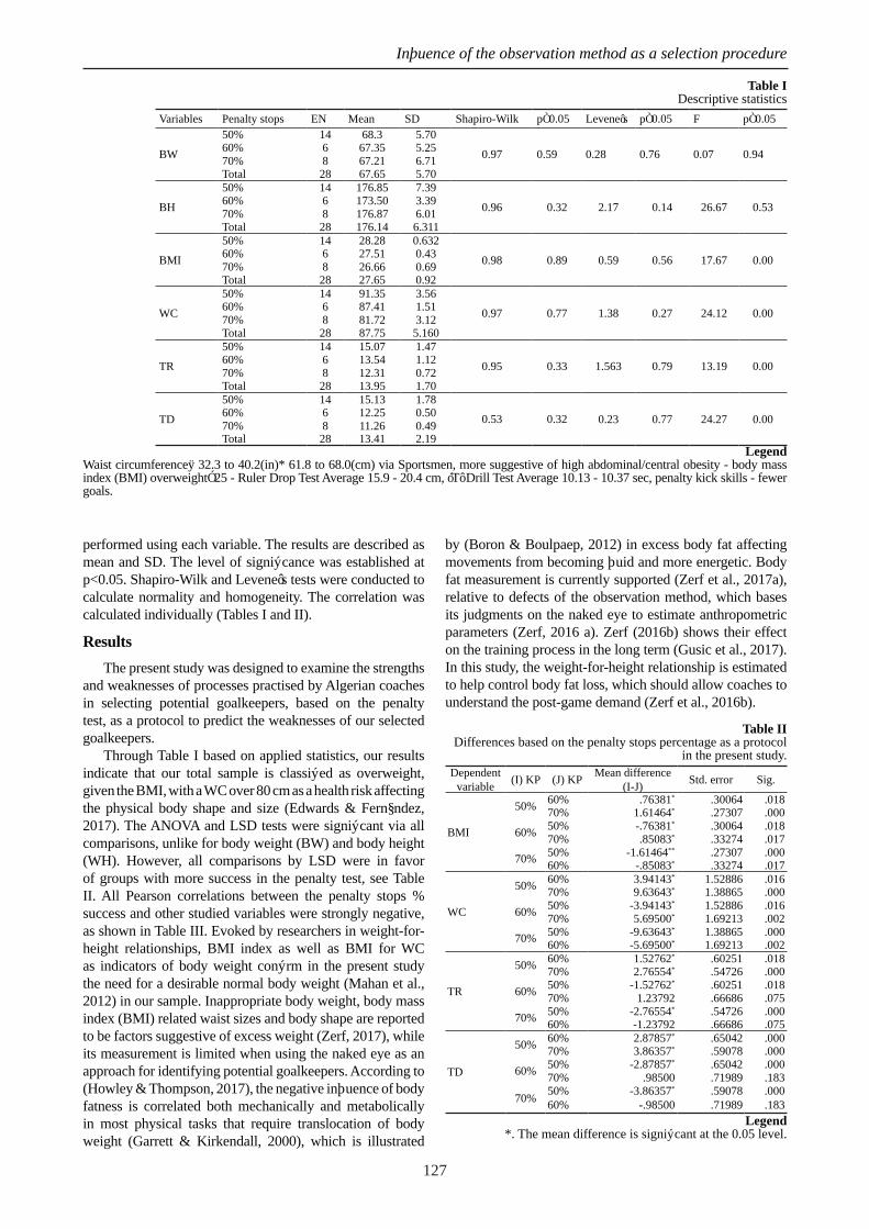
Through Table I based on applied statistics, our results indicate that our total sample is classified as overweight, given the BMI, with a WC over 80 cm as a health risk affecting the physical body shape and size (Edwards & Fernández, 2017). The ANOVA and LSD tests were significant via all comparisons, unlike for body weight (BW) and body height (WH). However, all comparisons by LSD were in favor of groups with more success in the penalty test, see Table II. All Pearson correlations between the penalty stops % success and other studied variables were strongly negative, as shown in Table III. Evoked by researchers in weight-for-height relationships, BMI index as well as BMI for WC as indicators of body weight confirm in the present study the need for a desirable normal body weight (Mahan et al., 2012) in our sample. Inappropriate body weight, body mass index (BMI) related waist sizes and body shape are reported to be factors suggestive of excess weight (Zerf, 2017), while its measurement is limited when using the naked eye as an approach for identifying potential goalkeepers. According to (Howley & Thompson, 2017), the negative influence of body fatness is correlated both mechanically and metabolically in most physical tasks that require translocation of body weight (Garrett & Kirkendall, 2000), which is illustrated
by (Boron & Boulpaep, 2012) in excess body fat affecting movements from becoming fluid and more energetic. Body fat measurement is currently supported (Zerf et al., 2017a), relative to defects of the observation method, which bases its judgments on the naked eye to estimate anthropometric parameters (Zerf, 2016 a). Zerf (2016b) shows their effect on the training process in the long term (Gusic et al., 2017). In this study, the weight-for-height relationship is estimated to help control body fat loss, which should allow coaches to understand the post-game demand (Zerf et al., 2016b).
Table II Differences based on the penalty stops percentage as a protocol
in the present study.Dependent
variable (I) KP (J) KP Mean difference (I-J) Std. error Sig.
BMI
50% 60% .76381* .30064 .01870% 1.61464* .27307 .000
60% 50% -.76381* .30064 .01870% .85083* .33274 .017
70% 50% -1.61464** .27307 .00060% -.85083* .33274 .017
WC
50% 60% 3.94143* 1.52886 .01670% 9.63643* 1.38865 .000
60% 50% -3.94143* 1.52886 .01670% 5.69500* 1.69213 .002
70% 50% -9.63643* 1.38865 .00060% -5.69500* 1.69213 .002
TR
50% 60% 1.52762* .60251 .01870% 2.76554* .54726 .000
60% 50% -1.52762* .60251 .01870% 1.23792 .66686 .075
70% 50% -2.76554* .54726 .00060% -1.23792 .66686 .075
TD
50% 60% 2.87857* .65042 .00070% 3.86357* .59078 .000
60% 50% -2.87857* .65042 .00070% .98500 .71989 .183
70% 50% -3.86357* .59078 .00060% -.98500 .71989 .183
Legend *. The mean difference is significant at the 0.05 level.
Table I Descriptive statistics
Variables Penalty stops EN Mean SD Shapiro-Wilk p≤0.05 Levene’s p≤0.05 F p≤0.05
BW
50% 14 68.3 5.70
0.97 0.59 0.28 0.76 0.07 0.9460% 6 67.35 5.2570% 8 67.21 6.71Total 28 67.65 5.70
BH
50% 14 176.85 7.39
0.96 0.32 2.17 0.14 26.67 0.5360% 6 173.50 3.3970% 8 176.87 6.01Total 28 176.14 6.311
BMI
50% 14 28.28 0.632
0.98 0.89 0.59 0.56 17.67 0.0060% 6 27.51 0.4370% 8 26.66 0.69Total 28 27.65 0.92
WC
50% 14 91.35 3.56
0.97 0.77 1.38 0.27 24.12 0.0060% 6 87.41 1.5170% 8 81.72 3.12Total 28 87.75 5.160
TR
50% 14 15.07 1.47
0.95 0.33 1.563 0.79 13.19 0.0060% 6 13.54 1.1270% 8 12.31 0.72Total 28 13.95 1.70
TD
50% 14 15.13 1.78
0.53 0.32 0.23 0.77 24.27 0.0060% 6 12.25 0.5070% 8 11.26 0.49Total 28 13.41 2.19
Legend Waist circumference‡ 32.3 to 40.2(in)* 61.8 to 68.0(cm) via Sportsmen, more suggestive of high abdominal/central obesity - body mass index (BMI) overweight≥25 - Ruler Drop Test Average 15.9 - 20.4 cm, ‘T’ Drill Test Average 10.13 - 10.37 sec, penalty kick skills - fewer goals.
128
Zerf Mohammed et al.
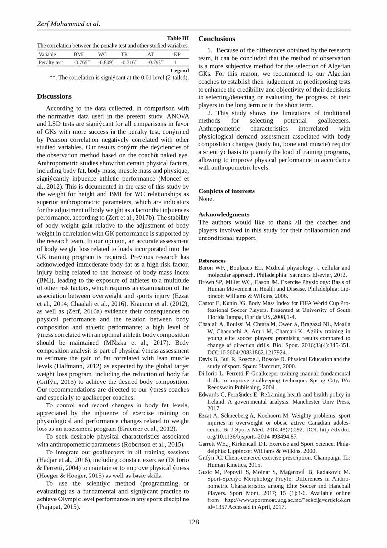
Table III The correlation between the penalty test and other studied variables. Variable BMI WC TR AT KPPenalty test -0.765** -0.809** -0.716** -0.793** 1
Legend**. The correlation is significant at the 0.01 level (2-tailed).
DiscussionsAccording to the data collected, in comparison with
the normative data used in the present study, ANOVA and LSD tests are significant for all comparisons in favor of GKs with more success in the penalty test, confirmed by Pearson correlation negatively correlated with other studied variables. Our results confirm the deficiencies of the observation method based on the coach’s naked eye. Anthropometric studies show that certain physical factors, including body fat, body mass, muscle mass and physique, significantly influence athletic performance (Moncef et al., 2012). This is documented in the case of this study by the weight for height and BMI for WC relationships as superior anthropometric parameters, which are indicators for the adjustment of body weight as a factor that influences performance, according to (Zerf et al., 2017b). The stability of body weight gain relative to the adjustment of body weight in correlation with GK performance is supported by the research team. In our opinion, an accurate assessment of body weight loss related to loads incorporated into the GK training program is required. Previous research has acknowledged immoderate body fat as a high-risk factor, injury being related to the increase of body mass index (BMI), leading to the exposure of athletes to a multitude of other risk factors, which requires an examination of the association between overweight and sports injury (Ezzat et al., 2014; Chaalali et al., 2016). Kraemer et al. (2012), as well as (Zerf, 2016a) evidence their consequences on physical performance and the relation between body composition and athletic performance; a high level of fitness correlated with an optimal athletic body composition should be maintained (Mączka et al., 2017). Body composition analysis is part of physical fitness assessment to estimate the gain of fat correlated with lean muscle levels (Halfmann, 2012) as expected by the global target weight loss program, including the reduction of body fat (Griffin, 2015) to achieve the desired body composition. Our recommendations are directed to our fitness coaches and especially to goalkeeper coaches:
To control and record changes in body fat levels, appreciated by the influence of exercise training on physiological and performance changes related to weight loss as an assessment program (Kraemer et al., 2012).
To seek desirable physical characteristics associated with anthropometric parameters (Robertson et al., 2015).
To integrate our goalkeepers in all training sessions (Hadjar et al., 2016), including constant exercise (Di Iorio & Ferretti, 2004) to maintain or to improve physical fitness (Hoeger & Hoeger, 2015) as well as basic skills.
To use the scientific method (programming or evaluating) as a fundamental and significant practice to achieve Olympic level performance in any sports discipline (Prajapat, 2015).
Conclusions1. Because of the differences obtained by the research
team, it can be concluded that the method of observation is a more subjective method for the selection of Algerian GKs. For this reason, we recommend to our Algerian coaches to establish their judgement on predisposing tests to enhance the credibility and objectivity of their decisions in selecting/detecting or evaluating the progress of their players in the long term or in the short term.
2. This study shows the limitations of traditional methods for selecting potential goalkeepers. Anthropometric characteristics interrelated with physiological demand assessment associated with body composition changes (body fat, bone and muscle) require a scientific basis to quantify the load of training programs, allowing to improve physical performance in accordance with anthropometric levels.
Conflicts of interestsNone.
AcknowledgmentsThe authors would like to thank all the coaches and players involved in this study for their collaboration and unconditional support.
ReferencesBoron WF, Boulpaep EL. Medical physiology: a cellular and
molecular approach. Philadelphia: Saunders Elsevier, 2012.Brown SP, Miller WC, Eason JM. Exercise Physiology: Basis of
Human Movement in Health and Disease. Philadelphia: Lip-pincott Williams & Wilkins, 2006.
Cantor E, Konin JG. Body Mass Index for FIFA World Cup Pro-fessional Soccer Players. Presented at University of South Florida Tampa, Florida US, 2008,1-4.
Chaalali A, Rouissi M, Chtara M, Owen A, Bragazzi NL, Moalla W, Chaouachi A, Amri M, Chamari K. Agility training in young elite soccer players: promising results compared to change of direction drills. Biol Sport. 2016;33(4):345-351. DOI:10.5604/20831862.1217924.
Davis B, Bull R, Roscoe J, Roscoe D. Physical Education and the study of sport. Spain: Harcourt, 2000.
Di Iorio L, Ferretti F. Goalkeeper training manual: fundamental drills to improve goalkeeping technique. Spring City, PA: Reedswain Publishing, 2004.
Edwards C, Fernández E. Reframing health and health policy in Ireland. A governmental analysis. Manchester Univ Press, 2017.
Ezzat A, Schneeberg A, Koehoorn M. Weighty problems: sport injuries in overweight or obese active Canadian adoles-cents. Br J Sports Med. 2014;48(7):592. DOI: http://dx.doi.org/10.1136/bjsports-2014-093494.87.
Garrett WE., Kirkendall DT. Exercise and Sport Science. Phila-delphia: Lippincott Williams & Wilkins, 2000.
Griffin JC. Client-centered exercise prescription. Champaign, IL: Human Kinetics, 2015.
Gusic M, Popović S, Molnar S, Mašanović B, Radakovic M. Sport-Specific Morphology Profile: Differences in Anthro-pometric Characteristics among Elite Soccer and Handball Players. Sport Mont, 2017; 15 (1):3-6. Available online from http://www.sportmont.ucg.ac.me/?sekcija=article&artid=1357 Accessed in April, 2017.
129
Influence of the observation method as a selection procedure
Hadjar KM, Koutchouk SM, Mime M, Zerf M, Zereg F. Which training improves the ability to control and manipulate the ball within the goalkeeper in football? Eur J Phys Ed Sport Sci., 2016;1(4): 58-52. doi:10.5281/zenodo.56005.
Halfmann P. Advanced Concepts of Strength & Conditioning for Tennis. E-book: IAAPH Gmbh, 1st ed.,2012.
Hoeger WWK, Hoeger SA. Principles and Labs for Physical Fit-ness. UK: Cengage Learning, 2015.
Hoffman J. Norms for Fitness, Performance, and Health. Cham-paign, IL: Human Kinetics, 2006.
Howley ET, Thompson DL. Fitness professional’s handbook. Champaign, IL: Human Kinetics, 2017.
Kindall J, Winkin J. The Baseball Coaching Bible. US:: Human Kinetics, 2000.
Kraemer WJ, Fleck SJ, Deschenes MR. Exercise Physiology: Integrating Theory and Application. Philadelphia: Wolters Kluwer/Lippincott Williams & Wilkins Health, 2012.
Mączka M, Sass A, Wojtyla A. Assessment of physical activity among pregnant women in context of weight gain in pregnan-cy. J Ed, Health and Sport. 2017;7(6):332-376. DOI:http://dx.doi.org/10.5281/zenodo.816311
Mahan LK, Escott-Stump S, Raymond JL, Krause MV. Krause’s Food & the Nutrition Care Process. St. Louis, Mo: Elsevier/Saunders, 2012.
Moncef C, Said M, Olfa N, Dagbaji G. Influence of Morphological Characteristics on Physical and Physiological Performances of Tunisian Elite Male Handball Players. Asian J Sports Med. 2012; 3(2):74-80. Available online from https://www.ncbi.nlm.nih.gov/pmc/articles/PMC3426725/#CIT0008 Accessed in April, 2017.
Papaioannou AG, Hackfort D. Routledge Companion to Sport and Exercise Psychology: Global perspectives and funda-mental concepts. London: Routledge, 2014.
Prajapat SK. Predicting excellence in field hockey. India: Laxmi Book Publication, 2015.
Robertson S, Woods C, Gastin P. Predicting higher selec-tion in elite junior Australian Rules football: The influence of physical performance and anthropometric attributes 2015;18(5):601-606. doi:10.1016/j.jsams.2014.07.019.
Sporis GJ, Ostojic SM, Milanovic D. Fitness profiling in soc-cer: physical and physiologic characteristics of elite players. J Strength Cond Res. 2009;23(7):1947-1953. DOI:10.1519/JSC.0b013e3181b3e141.
Tavana M, Azizi F, Azizi F Behzadian M. A fuzzy inference sys-tem with application to player selection and team formation in multi-player sports. Sport Manag Rev. 2013;16(1):97-110. Available online from http://econpapers.repec.org/article/eeespomar/v_3a16_3ay_3a2013_3ai_3a1_3ap_3a97-110.htm Accessed in April, 2017.
Wolstencroft E. Talent Identification and Development: An Aca-demic Review. The University of Edinburgh: sportscotland Caledonia House South Gyle Edinburgh, 2002.
Zerf M, Houar A, Mime M, Bengoua A. Height versus Weight which Cassel Parameter Determine Pulmonary Functions Fitness among the Algerians Soccer Players. J Pulm Respir Med, 2016a;6: 3. doi:10.4172/2161-105X.1000353
Zerf M, Houar A, Mime M, Bengoua A. Traditional versus scien-tific method: the differences exist between selecting players. JPES, 2016;16 Supp (1):673-678. DOI:10.7752/jpes.2016.s1108.
Zerf M, Mokkedes MI, Hamek B, Houar A, Bengoua A. Impact of Body Composition on Optimal Competitive Body and its Consequences on Athletic Performance in Healthy Young. Int J Women’s Health Wellness, 2016b;2(6). doi:10.23937/2474-1353/1510041.
Zerf M, Mokkedes MI, Lakhdar M, Hakim H, Bengoua A. The Benefits of Physical Activity for Health and Well-being Case Menopausal Women. Eur J Phys Ed Sport. 2016c;13(3):108-112. doi:10.13187/ejpe.2016.13.108.
Zerf M. Body composition versus body fat percentage as predic-tors of posture/balance control mobility and stability among football players under 21 years. Physical education of stu-dents, 2017;21(2): 96-102. doi:10.15561/20755279.2017.0208.
Zerf M. Impact of percent body fat on specific ability - Alge-rian soccer players (Vol. 1). Available online from http://sjss-sportsacademy.edu.rs/archive/details/impact-of-percent-body-fat-on-specific-ability-algerian-soccer-playersm-583.html, 2016a. Accessed in April, 2017.
Zerf M. Impact of theoretical courses on physical health per-formance. BLDE Univ J Health Sci. 2016b; 1(1):44-48. doi:10.4103/2456-1975.183285.
Ziv G, Lidor R. Physical characteristics, physiological attributes, and on-field performances of soccer goalkeepers. Int J Sports Physiol Perform. 2011;6(4): 509-524.
130
Palestrica of the third millennium – Civilization and SportVol. 18, no. 3, July-September 2017, 130–134
Respiratory physical therapy influence on the stress level in hypertensive persons in the age group 40-60 years
Influența kinetoterapiei respiratorii asupra nivelului de stres la persoanele hipertensive din grupa de vârstă 40-60 de ani
Anca Jianu Physical Education and Sports Faculty, Spiru Haret University, Bucharest
AbstractBackground. The introduction of a program of respiratory physical therapy in people with essential hypertension in the age
group of 40-60 years influences their stress level.Aims. Hypertension is known as a cardiovascular risk factor that promotes coronary and cerebral atherosclerosis. Another
cardiovascular risk factor, stress, is commonly associated with hypertension and may even be the generator of the latter. The increasing share of hypertensive people and the increasing presence of daily stress in human life have led us to study to what extent respiratory physical therapy, whose benefits are known, can help reduce these risk factors.
Methods. This study, type application, was completed between September 2012 – August 2013 on a number of 24 subjects, divided in two groups, one experimental and the other control, diagnosed with essential arterial hypertension. Information on the stress level of the subjects before and after the application of respiratory kinetotherapy was gathered through some ques-tions evaluating stress in 6 important areas. Each answer was given a number of points, and the overall score placed each subject in a certain stress level area (dangerously low, low, normal, high, dangerously high).
Results. The individual evolutions of the subjects of the experimental group show statistically significant differences between the tests, the mean of the experimental group being smaller, while the control group did not show any significant differences. In the comparative analysis between the groups, the effect size index (0.336) shows a small to moderate difference in favor of the experimental group (a lower stress level).
Conclusions. Reducing the level of stress in experimental research subjects gives respiratory physical therapy a psycho-therapeutic role on those with essential hypertension.
Keywords: essential arterial hypertension, respiratory physical therapy, stress level.
RezumatPremize. Introducerea unui program de kinetoterapie respiratorie la persoanele cu hipertensiune arterială esențială, aflate în
grupa de vârstă 40-60 de ani, influențează nivelul de stres al acestora.Obiective. Hipertensiunea arterială este cunoscută ca factor de risc cardiovascular ce intervine în favorizarea aterosclerozei
la nivel coronarian şi cerebral. Un alt factor de risc cardiovascular, stresul, se asociază frecvent hipertensiunii arteriale şi poate fi chiar generator al acesteia din urmă. Ponderea crescută a persoanelor hipertensive şi prezenţa în continuă creştere a stresului cotidian în viaţa omului ne-au determinat să studiem în ce măsură kinetoterapia respiratorie, ale cărei beneficii sunt cunoscute, poate contribui la reducerea factorilor de risc amintiţi.
Metodă. Studiul de faţă, de tip aplicativ, a fost efectuat în perioada septembrie 2012 - august 2013 pe un număr de 24 de subiecţi cu vârsta cuprinsă între 40-60 ani, împărțiți în două loturi, unul experimental și altul martor, diagnosticaţi cu hiperten-siune arterială esenţială. Informaţiile privind nivelul de stres al subiecţilor înainte şi după aplicarea kinetoterapiei respiratorii au fost colectate prin intermediul unor întrebări prin care a fost inventariat stresul pe 6 domenii importante. Fiecărui răspuns i s-a acordat un număr de puncte, iar punctajul general obţinut a încadrat fiecare subiect într-o anumită zonă a nivelului de stres (primejdios de scăzut, scăzut, normal, ridicat, primejdios de ridicat).
Rezultate. Evoluțiile individuale ale subiecților grupei experimentale arată existența unor diferențe semnificative statistic între testări, media grupei experimentale fiind mai mică, în timp ce la grupa de control nu prezintă diferențe semnificative. La analiza comparativă între loturi, indicele de mărime a efectului (0,336) arată existența unei diferențe mici spre mijlocie, în favoarea lotului experimental (un nivel de stres mai mic la acesta).
Concluzii. Reducerea nivelului de stres la subiecții cercetării experimentale conferă programelor de kinetoterapie respira-torie un rol psihoterapeutic asupra celor cu hipertensiune arterială esențială.
Cuvinte cheie: hipertensiune arterială esențială, kinetoterapie respiratorie, nivel de stres.
Copyright © 2010 by “Iuliu Haţieganu” University of Medicine and Pharmacy Publishing
Received: 2017, May 10; Accepted for publication: 2017, May 20 Address for correspondence: Physical Education and Sports Faculty, Spiru Haret University, 24, Berceni Str., sect. 4, zip code 041905,
BucharestE-mail: [email protected] Corresponding author: Anca Jianu; e-mail: [email protected]://doi.org/10.26659/pm3.2017.18.3.130
131
Respiratory physical therapy influence on the stress level
IntroductionHypertension is a condition, but also a cardiovascular
risk factor. Numerous specialty studies have identified the association between blood pressure variability and increased mortality and cerebrovascular and coronary events in hypertensive patients (Bădilă, E. et al., 2013).
Moreover, researchers such as Rothwell, PM. et al. (2010) or Fratolla, A. et al. (1993), who have been following hypertensive persons for several years, show the relationship between increased blood pressure and target organ damage such as left ventricular hypertrophy.
Also, studies by Bădilă, E. et al. (2012) and Volpe, M., Tocci, G. (2009) confirm the subclinical impairment of some organs (heart, kidneys, brain), explaining the need for a thorough evaluation of the hypertensive patient in terms of cardiovascular risk.
One of the cardiovascular risk factors commonly associated with high blood pressure, considered as a generator but also as a consequence of it, is stress that modern science defines as any type of change causing physical, emotional or psychological pressure, which can contribute to the development of psychic resistance, awareness, new perspectives, a sense of control if its time of action is short (Davidji, 2015).
Too much beneficial stress (eustress) can degenerate into distress, manifesting by different reactions: anger, anxiety, sadness, depression, negative emotions that affect cardiac consistency and cause chaos in the body physiology (Servan-Schreiber, D., 2007). Stress, associated directly with hypertensive disease, generates overall sympathetic tone (Cinteză, M., 2012).
In dealing with distress, breathing exercises play an important role (Arădăvoaice, G., 2010), which contributes to better oxygenation of body tissues, regulates cardiac activity, develops thoracic capacity, stimulates digestive organs and immunity, stabilizes the mental state and favors a state of emotional balance. Exercises of deep breathing “release us from stress” (Rodríguez, J., 2007) and produce a general relaxation state by inhibiting the sympathetic vegetative nervous system (Mantak, C., William, UW., 2017).
Specialists emphasize the significant relationship between cardiovascular complications and increased blood pressure during stress (Pickering, TG., 1982).
Hypertension is a risk factor for cardiovascular complications (Parati, G. et al., 2013), but difficult to control with conventional antihypertensive drug therapy (Parati, G. et al., 2008).
Thus, the treatment of this disease consists not only of a decrease of tension values, but also of cardiovascular risk (Mancia, G. et al., 2013). In this regard, specific antihypertensive medication should be associated with other forms of treatment to achieve these goals.
We consider that the introduction of respiratory physical therapy in the treatment of patients with essential hypertension belonging to the age group of 40-60 years influences their stress level and contributes to the reduction of the number of cardiovascular ischemic events and to the increase of the quality of their life.
ObjectivesThe respiratory physical therapy objectives were
adapted to the needs of the subjects of this study and consisted of the following:
- Balancing the nervous system and improving the mental state of the patients;
- Facilitating vasodilation in skeletal muscles in order to stimulate blood circulation
- Increasing vital capacity by increasing chest elasticity;
- Educating subjects and their families in order to adopt a rational lifestyle that will allow reducing as many cardiovascular risk factors as possible.
HypothesisThe introduction of a program of respiratory physical
therapy for hypertensive people in the age group of 40-60 years influences their stress level.
Material and methodsIn accordance with the Helsinki Declaration, the
Amsterdam Protocol and the Directive 86/609/EEC, the Ethics Committee’s approval was obtained from the Department of Physical Education and Sport of the National University of Physical Education and Sports for the conduct of the experimental research.
We note the written consent of the subjects regarding their participation in the research.
Research protocola) Period and place of the researchThe study was conducted between September 2012 and
August 2013 at the Spine Health Medical Recovery Center in Bucharest. Patients enrolled in the study were treated on an outpatient basis for 12 consecutive weeks with three respiratory gymnastics programs (the first program between weeks 1-3, the second program between weeks 4-8, and the third program between weeks 9-12).
b) SubjectsThe study was conducted on 24 subjects, 14 females
and 10 males, aged 40-60 years, diagnosed with essential hypertension.
Of the 24 subjects, 4 withdrew during the first 3 weeks for personal reasons, and 8 subjects did not want to be actively involved in this research, establishing the composition of two groups, an experimental (E) group and a control (C) group, with a total number of 12 subjects.
The subjects of the study were selected according to the following inclusion criteria:
- Resting cardiac frequency above 60 beats/minute,- Blood pressure below 180 mmHg,- Absence of lung diseases,- Absence of serious cardiovascular disease,- Absence of angina pectoris or other significant
symptoms during the exercise and effort test: vertigo, breathing difficulty (dyspnea), headache,
- Subjects accepting to cooperate in the research,- Conscious involvement of the subjects.c) Tests appliedThe subjects who met the conditions for inclusion in
the experiment were specifically evaluated to know their
132
Anca Jianu
stress levels and assess to what extent respiratory physical therapy in people with essential hypertension reduces mental tension.
In this regard, we applied for a category C stress test. The lack of a category C stress test, validated on the Romanian population, determined the use of the test designed by Melgosa, J. (2000) and mentioned by Arădăvoaice, D. (2010).
The stress test included questions related to 6 important areas (items), namely: lifestyle, environment, symptoms, job/occupation, relationships and personality.
The interpretation of the test consists of locating the level of stress through the score obtained in a certain area:
- Zone 1 (from 0 to 48 points) - The level of stress is dangerously low.
- Zone 2 (48-72 points) – The stress level is low.- Zone 3 (72-120 points) – The stress level is normal.- Zone 4 (120-144 points) – The stress level is con-
sidered high.- Zone 5 (≥ 144 points) - The stress level is dangerously
high.d) Studied momentsThe subjects in the two (experimental and control)
groups were evaluated at the beginning of the program (T1) and at the end of the 12 weeks (T2) from the start of the program.
The respiratory physical therapy program aimed at achieving the physiotherapy objectives recommended for subjects with essential hypertension, according to Mark, V., Dan, M. (2007), adapted to the needs of the subjects of this study.
The program was conducted under close supervision and the cardio-respiratory parameters (blood pressure, heart rate, respiratory rate, saturation of arterial blood in oxygen) were periodically measured. To avoid possible incidents, the subjects were individually worked. All subjects in the experimental group attended the 3 schedules.
Physical training was conducted in the heart area of low and moderate intensity exercise, using the value of 60-75% of the reserve heart rate. The progress of the intensity of the respiratory physical therapy session was gradual, individualized for each subject.
The central element of training was to establish synchronization of the slow movements of the body with the breathing rate. In all exercises breathing forced the rhythm, and the body followed it. The duration of each breath was increased progressively according to the ability of each subject to adapt to the effort and to focus on the movement and the ventilator process. Depending on the rate of respiration specific to each subject, in the first 3 weeks the duration of the program was 20-30 minutes, in weeks 4-8 it was 40-90 minutes, and in weeks 9-12 it lasted 60-75 minutes.
The program included aerobic exercises represented by ample and slow movements of the upper, lower limbs and torso to increase the myocardial contraction force, as well as exercises based on coordination, balancing, trunk twisting to rebalance the nervous system. The exercises were aimed at awareness of the respiratory act through the correct learning and exercising of diaphragmatic, thoracic and complete breathing. Account was taken of the ability
of each subject to expand the chest box during inspiration and to control the contraction of abdominal muscles during expiration. We emphasize the importance of achieving inspiration and expiration as slow, deep and prolonged as possible, and the execution of breathing phases at the nose, except for those exercises in which the expiration was sound and those requiring oral air removal due to the low tolerance of the subjects.
Of the following methodological aspects, we mention:- Correct body alignment was observed;- Exercises were performed from positions that
facilitated breathing. The following positions were used: sitting with abducted lower limbs on the seat and on the fitness ball, lying down, lying on the side, lying on the knee, standing on the knee and standing with abducted lower limbs;
- Exercises were used in which the holding of the hands during practice blocked the scapulohumeral girdle, knowing that on a fixed scapula the mobilizing thoracic muscles (serratus anterior muscle, minor pectoral muscle) take a better support point than the mobilization of the upper limbs ;
- Exercises were aimed at toning the respiratory muscles to increase pulmonary volumes, to control and coordinate the respiratory rhythm with influences on the rhythm of the heart. Thus, the main muscles were: inspiratory muscles (diaphragm, external intercostals), accessory muscles (scalene, serratus posterior, sternocleidomastoid, pectoral, trapezius and dorsal) and expiratory muscles (abdominal, intercostal, lumbar, the sternum triangle);
- Exercises were aimed at toning the postural and perineal muscle;
- Exercises were performed in the mirror to observe the movement and thus, to allow the mind and body to work together, the will being important in their practice.
e) Statistical processing For statistical characterization of the experimental
group E and the control group C we used statistical indicators of the central trend (arithmetic mean, median, quartile 1 (Q1), quartile 3 (Q3)), indicators of spreading (standard deviation, minimum and maximum values, coefficient of variation). The listed indicators were applied to the overall score obtained in the stress test.
The data of each subject were entered into the database. Analysis of statistical indicators and verification of statistical assumptions were carried out with the specific SPSS 17.0 software (Statistical Package for the Social Sciences).
To compare the initial and final tests for the C group or the initial and final tests for the E group, we used the non-parametric Wilcoxon signed ranks for two paired samples in the control group and the non-parametric Friedman tests for multiple samples, and Wilcoxon signed ranks as a post hoc test with the Bonferroni correction for the Friedman test. The Bonferroni correction considers the significance threshold equal to 0.05 divided by the number of comparisons that can be made. In this case, α = 0.017. The comparison of the final tests between the experimental group and the control group was performed with the non-parametric Mann-Whitney test for two independent samples.
133
Respiratory physical therapy influence on the stress level
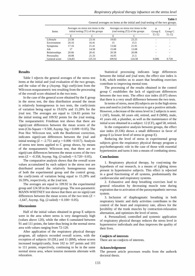
ResultsTable I reflects the general averages of the stress test
items at the initial and final evaluation of the two groups, and the value of the p (Asymp. Sig) coefficient from the Wilcoxon nonparametric test resulting from the processing of the overall score obtained in the two tests.
In the case of the general score obtained by the E group in the stress test, the data distribution around the mean is relatively homogeneous in two tests, the coefficients of variation being equal to 19.02% and 15.28% for the final test. The averages are equal to 124.83 points for the initial testing and 109.92 points for the final testing. The nonparametric Friedman test shows that there are significant differences between the mean scores of the tests (Chi-Square = 9.500, Asymp. Sig = 0.009 <0.05). The Post Hoc Wilcoxon test, with the Bonferroni correction, indicates significant differences between the final and initial testing (Z = -2.751 and p = 0.006 <0.017). Analysis of stress test items applied to C group shows, by means of the nonparametric Wilcoxon test, that there are no significant differences between the mean scores of the two tests (Z = -0.358, Asymp. Sig. (2-tailed) = 0.720> 0.05).
The comparative analysis shows that the overall score values accumulated by each subject in the stress test are distributed around the relatively homogeneous mean of both the experimental group and the control group, the coefficients of variation being equal to 15.28% and 16.59%, respectively, at the final test.
The averages are equal to 109.92 in the experimental group and 124.58 in the control group. The non-parametric MANN-WHITNEY test shows that there are no significant differences between the mean scores of the two tests (Z = -1,647, Asymp. Sig. (2-tailed) = 0.100> 0.05).
DiscussionsHalf of the initial values obtained in group E subjects
were in the area where stress is very dangerously high (values above 120), while the other 6 cumulated between 91 and 111 points, the latter thus being in the normal stress area with values ranging from 72-120.
After application of the respiratory physical therapy program, all subjects recorded overall scores, with the exception of subjects 10 (DL) and 11 (BM), whose scores increased insignificantly, from 102 to 107 points and 103 to 111 points, respectively, continuing to be in the same normal stress area, where tension moments alternate with relaxation.
Statistical processing indicates large differences between the initial and final tests; the effect size index is 0.46, which entitles us to assert that breathing exercises contribute to improving mental tone.
The processing of the results obtained in the control group C establishes the lack of significant differences between the two tests. The effect size index (0.07) shows that there is a very small difference between the two tests.
In terms of stress, most (8) subjects are in the high stress area and need to find the resources to get a positive attitude. However, a decrease of the stress level in 2 subjects is seen: 1 (AF), female, 60 years old, retired, and 8 (MM), male, 41 years old, a plumber, as well as the maintenance of the initial score obtained in a subject: 12 (CF), aged 58, retired.
In the comparative analysis between groups, the effect size index (0.336) shows a small difference in favor of group E (a lower level of stress in group E).
Reducing the level of stress in experimental group subjects gives the respiratory physical therapy program a psychotherapeutic role in the case of those with essential hypertension and turns it into a means of combating stress.
Conclusions1. Respiratory physical therapy, by confirming the
hypothesis of our research, is a means of fighting stress present in hypertensive subjects. This effect is reflected in a good functioning of all systems, predominantly the cardiovascular and respiratory systems.
2. Exhaustive and deep breathing exercises lead to general relaxation by decreasing muscle tone during expiration due to activation of the parasympathetic nervous system.
3. Inclusion of diaphragmatic breathing in both respiratory kinetic and daily activities contributes to the control of the heart and respiratory rate, allows for the flexibility of the trunk muscles by contraction-relaxation alternation, and optimizes the level of stress.
4. Personalized, controlled and systemic application of respiratory physical therapy reduces the stress level in hypertensive individuals and thus improves the quality of their lives.
Conflicts of interestsThere are no conflicts of interests.
AcknowledgementThe present article processes results from the author’s doctoral thesis.
Table IGeneral averages on items at the initial and final testing of the two groups.
ItemsAverages on stress test items in the
initial testing (T1) of the groups Averages on stress test items in the
final testing (T2) of the groups p
Group ET1-T2
pGroup CT1-T2E C E C
Lifestyle 23.58 23.16 19.83 23.25 - -Environment 20.16 16.41 15 15.5 - -Symptoms 17.16 21.41 13.66 21.91 - -Job 17 14.58 15.08 13.08 - -Relationships 20.66 20.41 20.58 20.08 - -Personality 18.08 21.33 16.05 21.5 - -General score 124,83 125.33 109.92 124.58 0.006 0.720

134
Anca Jianu
ReferencesArădăvoaice, G. Stres, Eustres, Distres-Terapii antistres, Editura
AntetXXPress, Filipeștii de Târg, Prahova, 2010, 7, 158-164.Bădilă E, Ţintea E, Daraban AM, Frunză S, Bartoş D, Cinteză
M. Variabilitatea tensiunii arteriale-o nouă viziune asupra riscului cardiovascular la pacienţii hipertensivi. În Progrese în cardiologie. Ed. Med. Publicis., 2013, 456-469.
Bădilă E, Daraban AM, Bartoş D, Arsenescu GC. Hipertensiunea arterială şi riscul cardiovascular-elemente noi în evaluare. În Progrese în cardiologie. Ed. Med. Publicis., 2012, 403-426.
Cinteză M. Soluţii intervenţionale în tratamentul eficient al hipertensiunii arteriale rezistente la tratament. În Progrese în cardiologie. Ed. Med. Publicis., 2012, 427-436.
Davidji. Tehnici de relaxare. Ghid practic pentru creştere personală, împlinire durabilă şi pace sufletească. Traducere: Oana Moldoveanu, Ed. Niculescu, Bucureşti. 2015, 25.
Fratolla A, Parati G, Cuspidi C, et al. Prognostic value of 24-hour blood pressure variability. J Hypertens 1993; 11:1133-1137.
Mancia G, Fagard R, Narkiewicz K et al. ESH/ESC Guidelines for the management of arterial hypertension: the Task Force for the management of arterial hypertension of the European Society of Hypertension (ESH) and of the European Society of Cardiology (ESC). J Hypertens. 2013; 31(7): 1281-1357.
Marcu V, Dan M. Kinetoterapie/Physiotherapy. Ed. Univ Oradea, Oradea, 2017, 213.
Mantak C, William, UW. Vindecarea prin TAO. Ghid ilustrat cu exerciţii de bază. Ed. Polirom, Bucureşti, 2017, 201.
Melgosa J. Fără stres, Casa de Editură, Viață și sănătate, București, 2000, 156-159.
Parati G, Stergiou GS, Asmar R, Bilo G, de Leeuw P, Imai Y, Kario K, Lurbe E, Manolis A, Mengden T, O’Brien E, Ohkubo T, Padfield P, Palatini P, Pickering T, Redon J, Revera M, Ruilope LM, Shennan A, Staessen JA, Tisler A, Waeber B, Zanchetti A, Mancia G; ESH Working Group on Blood Pressure Monitoring. European Society of Hypertension guidelines for blood pressure monitoring at home: a summary report of the Second International Consensus Conference on Home Blood Pressure Monitoring. J Hypertens.2008, 26(8):1505-1526.
Parati G, Ochoa JE, Lombardi C, Bilo G. Nature Rev.Cardiol.10:143-155; published online February 12th 2013.
Pickering TG, Harshfield GA, Kleinert HD, et al. Blood pressure during normal daily activities, sleep, and exercise: comparison of values in normal and hypertensive subjects. JAMA 1982; 247:992-997.
Rodríguez J. Metoda Pilates. Ed. Teora, Bucureşti, 2007, 20.Rothwell PM, Howard SC, Dolan E, O’Brien E, Dobson JE,
Dahlöf B, Ssever PS, Poulter NR. Prognostic significance of visittovisit variability, maximum systolic blood pressure and episodic hypertension. Lancet, 2010; 375(9718):895-905.
Servan-Schreiber D. Vindecă stresul, anxietatea şi depresia fără medicamente şi fără psihanaliză. Ed.Elena Francisc, Bucureşti.2007, 62.
Volpe M, Tocci G. ESH/ESC Guidelines for the management of Hypertension from theory to practise: global cardiovascular risk concept. J of Hypertens. 2009; 27:S3-S11; doi:10.1097/01.hjh.0000356766.86388e5.
135
Palestrica of the third millennium – Civilization and SportVol. 18, no. 3, July-September 2017, 135–138
Is there a relationship between flexibility ability and the selected long jump performance components of teenage athletes?
Există o relație între capacitatea de flexibilitate și componentele de performanță selectate ale sariturii in lungime la tinerii sportivi?
Emre Bağcı 1, Işık Bayraktar 2 1 Sport Science Faculty, Gazi University, Ankara, Turkey2 Department of Sport Education, General Directorate of Sport, Ankara, Turkey
AbstractBackground. The long jump is technically divided into four stages: run-up, jump, flight and landing. The total measured
distance of the long jump consists of the sum of these three lengths: take-off distance (L1), flight distance (L2) and landing distance (L3). The parts of the long jump were examined to determine by analyzing the various kinematic variables such as the percentage of L1, L2 and L3 rates in previous studies.
Aims. The aim of this study was to determine whether there is a relationship between flexibility ability and selected vari-ables of long jump performance components, particularly the landing distance (L3) for teenage athletes.
Methods. The research group comprised 32 male athletes, age 16.2 ± 0.6 years, who participated in the qualification round in the Turkey Youth Indoor Championships. All trials of the athletes were recorded and a two-dimensional analysis of their best performances was made. Velocity values (V10) of the last 10 meters of the approach run of the athletes, contact time at take-off, take-off angle, and long jump performance component (L1, L2, L3) values were calculated.
Result. When the relationship between the flexibility ability and other performance components of the athletes was examined, only a weak statistically significant correlation was found between the approach run velocity and flexibility (r=0.45). Various studies have been conducted in different events, examining the effects of flexibility on sprint and jumping ability.
Conclusions. Due to the L3 similarity of the body position in landing with the sit & reach test, the anticipated relationships were not found except for V10.
Keywords: flexibility, long jump, L3, teenage athlete. RezumatPremize. Săritura in lungime este tehnic împărțită în patru etape: elanul, desprinderea, zborul și aterizarea. Distanța totală
măsurată a săriturii în lungime reprezintă suma a trei lungimi: distanța de elan (L1), distanța de zbor (L2) și distanța de aterizare (L3). În studii anterioare, fazele săriturii în lungime au fost examinate pentru a determina valorile procentuale ale ratelor L1, L2 și L3 prin analizarea diferitelor variabile cinematice.
Obiective. Scopul acestui studiu a fost de a determina dacă există o relație între capacitatea de flexibilitate și variabilele selectate ale componentelor de performanță ale săriturii în lungime, în special distanța de aterizare (L3) pentru sportivii tineri.
Metode. Grupul de cercetare a fost format din 32 de sportivi de sex masculin, cu vârsta 16,2 ± 0,6 ani, care au participat la runda de calificare a Campionatului Indoor de Tineret, Turcia. Toate încercările sportivilor au fost înregistrate și a fost făcută o analiză bidimensională a celor mai bune performanțe. Au fost calculate valorile vitezelor (V10) pe ultimii 10 metri de abordare a săriturii în lungime, timpul de contact la bătaie, unghiul de desprindere și valorile componentelor de performanță pentru săritura în lungime (L1, L2, L3).
Rezultate. Atunci când s-au examinat relațiile dintre capacitatea de flexibilitate și celelalte componente ale performanței sportivilor, s-a constatat o corelație semnificativă statistic între viteza de deplasare și flexibilitate (r = 0,45). Diverse studii au fost realizate cu ocazia diferitelor evenimente, examinând efectele flexibilității asupra abilităților de sprint și săritură.
Concluzii. Datorită asemănării L3 a poziției corpului în aterizare cu testul sit & reach, relațiile așteptate nu au fost găsite decât în cazul V10.
Cuvinte cheie: flexibilitate, săritura în lungime, L3, viteza de deplasare.
Copyright © 2010 by “Iuliu Haţieganu” University of Medicine and Pharmacy Publishing
Received: 2017, June 7; Accepted for publication: 2017, June 20 Address for correspondence: Gazi University, Faculty of Sports Sciences, Post Code 06560 Ankara, TurkeyE-mail: [email protected] Corresponding author: Emre Bagci; [email protected]://doi.org/10.26659/pm3.2017.18.3.135
136
Emre Bağcı & Işık Bayraktar
IntroductionThe long jump is one of the natural but technically
complex disciplines of athletics. It primarily consists of an approach run at maximum speed and a flight phase starting as close as possible to the jump line. To be successful in this event, the long jumper must have the ability of a sprinter so that it can reach a sufficient speed in the approach run. One of the most important factors affecting performance in the long jump is the speed of the approach run (Theodorou et al., 2017). Another important factor determining the jump distance in a long jump is the vertical movement of the horizontal velocity of the center of gravity of the body via the approach run applied at high speed, at the instant of touchdown at the take-off board (Koyama et al., 2006). After Bob Beamon’s record in Mexico in 1968, a lot of work has been done on the biomechanics of the long jump. These studies have shown that good long jump performance can be described as a fast approach run and a skill of vertical transfer of the horizontally achieved velocity. However, arm movements also play an important role in the effectiveness of the vertical jump (Pradon et al., 2014). There are different techniques for transferring horizontal velocity to the vertical axis (Bridgett & Linthorne, 2006).
The long jump is technically divided into 4 phases: 1) Approach Run, 2) Jump, 3) Flight and 4) Landing. In a study investigating the ratio of total distance performance to jump phases for the long jump, 5.4% for jump (L1), 92.9% for flight (L2) and 8.0% for landing (L3) were found (Hay et al., 1986). The landing phase of the long jump technique (L3) recalls the movement in the sit and reach test, which is often used for flexibility in athletic performance tests.
The sit and reach test is one of the most commonly used tests of flexibility in determining physical fitness parameters. It was designed in 1952 to measure back and lower extremity flexibility. In 2008, flexibility was defined as the ability to move smoothly in the range of motion of the joint. Studies have shown that the sit and reach test can be used to measure the strength of the agonist muscle group and to determine the flexibility resistance of antagonist muscles. In 1966, it was reported that the sit and reach test was the most valid test compared to others for determining the folding flexibility of the torso (Carrasco et al., 2013).
The relationship between the level of flexibility and performance has always been the subject of research. In general, flexibility exercises are evaluated as a very important exercise that is applied with warming movements before training, providing many benefits such as protection from injury and increase of performance by increasing body heat and nerve conduction. For this reason, it is necessary for athletes to perform flexibility exercises at the beginning of training. Although the effects of static and dynamic flexibility exercises have been discussed, many studies have demonstrated the positive contributions of flexibility exercises to performance (Paradisis et al., 2014). Dynamic flexibility exercises particularly have a great contribution to the development of power, speed and jumping abilities. In the development of the level of flexibility in general, static flexibility exercises were found to be more successful than dynamic flexibility exercises, based on the results of
the sit and reach test (Samso et al., 2012). The relationship between flexibility and performance
has been constantly researched and the positive impact of flexibility on performance, especially the positive impact on speed and jumping, has been explained. Technically, there is a similarity between the positions of long jumpers in the landing phase and the body position in the sit-and-reach flexibility test. The purpose of this study was to determine whether there is a relationship between the flexibility ability and selected variables of long jump performance components, particularly the landing distance (L3), for young athletes.
HypothesisIn the long jump, due to the L3 similarity of the body
position in landing with the sit & reach test (L3), there may be a relationship between the flexibility ability of athletes and L3 percentage. Also, there may be a relationship between flexibility and the other long jump components such as run-up, contact time at take-off, take-off angle and percentage of jump phases.
Material and methodsResearch protocolAll trials of the athletes in the research group were
recorded during the Turkish Indoor Youth Championship. All trials of the athletes were recorded and two-dimensional analysis of their best performances was done.
a) Period and place of the researchThe study included 32 male long jumpers competing
in the Turkish Indoor Youth Championship organized in January 2015.
b) Subjects and groupsThe subjects of the study, 32 male long jumpers,
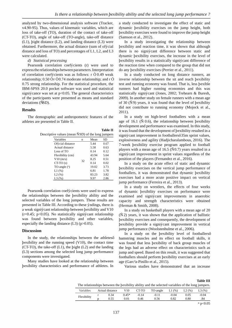
participated as volunteers in this study. They were informed in detail about the test procedures and benefits of the results. The study was performed in accordance with the Declaration of Helsinki. The data collection process started after the approval of the Turkish Athletic Federation was obtained. The general characteristics of the participants are shown in Table I.
Table I Athletes’ age, body mass, height, and sitting height variables
Variables n Mean SDAge (years)
32
16.2 0.6BM (kg) 62.8 7.2BH (cm) 173.4 6.3SH (cm) 90.41 3.60
LegendBM: body mass, BH: body height, SH: sitting height
c) Tests appliedAll trials of the athletes in the research group were
recorded by a camcorder at 100 fps (Panasonic HC-w850). The camera was placed perpendicular to the take-off board. The photocells, which were used to determine the running times of athletes, were placed at 1m and 11m distance from the take-off board (Smart Speed, Fusion Sport, Australia). Velocities for the 11m-1m sections 10m (V10) were calculated for each jump. The official jump distances were recorded. The best performances of the athletes were
137
Is there a relationship between flexibility ability and the selected long jump performance ?
analyzed by two-dimensional analysis software (Tracker, v4.90-95). Thus, values of kinematic variables, which are loss of take-off (TO), duration of the contact of take-off (CT-TO), angle of take-off (TO-angle), take-off distance (L1), flight distance (L2), and landing distance (L3) were obtained. Furthermore, the actual distance (sum of official distance and loss of TO) and percentages of L1, L2, and L3 were calculated.
d) Statistical processingPearson’s correlation coefficients (r) were used to
express the relationships between parameters. Interpretation of correlation coefficients was as follows: r ≤ 0.49 weak relationship; 0.50 ≤ r ≤ 0.74 moderate relationship; and r ≥ 0.75 strong relationship. For the statistical procedure, the IBM-SPSS 20.0 pocket software was used and statistical significance was set at p<0.05. The general characteristics of the participants were presented as means and standard deviations (±SD).
ResultsThe demographic and anthropometric features of the
athletes are presented in Table II.
Table II Descriptive values (mean ± SD) of the long jumpers
Variables n Mean SDOfficial distance
32
5.44 0.67Actual distance 5.58 0.63Loss of TO 0.14 0.12Flexibility (cm) 43.94 5.64V10 (m/s) 8.25 0.51CT-TO (s) 0.14 0.02TO-angle (o) 19.02 3.73L1 (%) 6.81 1.78L2 (%) 83.23 3.82L3 (%) 9.97 2.86
Pearson’s correlation coefficients were used to express the relationships between the flexibility ability and the selected variables of the long jumpers. These results are presented in Table III. According to these findings, there is a weak significant relationship between flexibility and V10 (r=0.45; p<0.05). No statistically significant relationship was found between flexibility and other variables, especially the landing distance (L3) (p>0.05).
DiscussionIn the study, the relationships between the athletes’
flexibility and the running speed (V10), the contact time (CT-TO), the take-off (L1), the flight (L2) and the landing (L3) sections among the selected long jump performance components were investigated.
Many studies have looked at the relationship between flexibility characteristics and performance of athletes. In
a study conducted to investigate the effect of static and dynamic flexibility exercises on the jump height, both flexibility exercises were found to improve the jump height (Samson et al., 2012).
In a study investigating the relationship between flexibility and reaction time, it was shown that although there is no significant difference between static and dynamic flexibility exercises, the increase in the level of flexibility results in a statistically significant difference of the reaction time when compared to the group that did not do any flexibility exercises (Perrier et al., 2011).
In a study conducted on long distance runners, an inverse relationship between the sit and reach flexibility test and running economy was found. That is, less flexible runners had higher running economies and this was statistically significant (Jones, 2002; Trehearn & Buresh, 2009). In another study on female runners with a mean age of 30 (± 9) years, it was found that the level of flexibility did not contribute to running economy (Mojock et al., 2011).
In a study on high-level footballers with a mean age of 16.1 (± 0.6), the relationship between flexibility development and performance was examined. In this study, it was found that the development of flexibility resulted in a significant improvement in footballers’ 35m sprint values, explosiveness and agility (Hadjicharalambous, 2016). The 7-week flexibility exercise program applied to football players with a mean age of 16.5 (± 0.7) years resulted in a significant improvement in sprint values regardless of the position of the players (Fernandez et al., 2016).
In a study on the acute effect of static and dynamic flexibility exercises on the vertical jump performance of footballers, it was demonstrated that dynamic flexibility exercises had a more acute positive impact on vertical jump performance (Ferreira et al., 2013).
In a study on wrestlers, the effects of four weeks of dynamic flexibility exercises on performance were examined and significant improvements in anaerobic capacity and strength characteristics were obtained (Herman & Smith, 2008).
In a study on basketball players with a mean age of 20 (± 2) years, it was shown that the application of ballistic flexibility exercises and consequently, the development of flexibility provide a significant improvement in vertical jump performance (Woolstenhulme et al., 2006).
In a study on the flexibility level of footballers’ hamstring muscles and its effect on football skills, it was found that less flexibility of back group muscles of the legs had an adverse effect on characteristics such as jump and speed. Based on this result, it was suggested that footballers should perform flexibility exercises at an early age (García-Pinillo et al., 2015).
Various studies have demonstrated that an increase
Table III The relationships between the flexibility ability and the selected variables of the long jumpers.Variables Actual distance V10 CT-TO TO-angle L1 (%) L2 (%) L3 (%)
Flexibility r 0.34 0.45* -0.14 -0.11 -0.04 0.05 -0.04p 0.55 0.01 0.46 0.56 0.82 0.80 .84
* p<0.05
138
Emre Bağcı & Işık Bayraktar
in the flexibility of athletes results in an improvement of jumping and speed values. In this study, a statistically significant relationship between the approach run up speed, which is one of the long jump performance components, and the flexibility of the athletes was found, while there was no statistically significant relationship between the landing stage (L3) values of the athletes (due to the L3 similarity of the body position in the sit and reach flexibility test) and flexibility.
Conclusions 1. Due to the L3 similarity of the body position
in landing with the sit & reach test, the anticipated relationships were not found except for V10.
2. A statistically significant weak positive correlation was found between the flexibility ability of the long jumpers in the study group and the approach run velocity (V10).
3. No statistically significant relationship was found between flexibility and other selected parameters. In the literature survey that was conducted, no other study examining the relationship between long jump performance components and flexibility was found.
Conflicts of interestsThe authors confirm that there are no known conflicts of interest associated with this publication.
AcknowledgmentsThanks to all subjects who participated in this study.
ReferencesBridgett LA, Linthorne NP. Changes in long jump take-off
technique with increasing run-up speed. J Sports Sci. 2006;24(8):889-897. doi:10.1080/02640410500298040.
Carrasco M, Sanz-Arribas I, Martinez-de-Haro V, Cid-Yague L, Martinez-Gonzalez-Moro I. Does the “Sit and Reach” Test Measures Flexibility? A Case Study. Rev Int Med Ci Activ Fis Dep. 2013;13(52):749-770.
Fernandez AR, Sanchez J, Rodriguez-Marroyo JA, Villa JG. Effects of seven weeks of static hamstring stretching on flexibility and sprint performance in young soccer players according to their playing position. J Sports Med Phys Fitness. 2016;56(4):345-351.
Ferreira VD, Muller BC, Achour A. Acute effects of static versus dynamic stretching on the vertical jump performance of soccer players. Motriz-Rev Ed Fis. 2013;19(2):450-459.
García-Pinillos F, Ruiz-Ariza A, Moreno del Castillo R, Latorre-Román PÁ. Impact of limited hamstring flexibility on vertical jump, kicking speed, sprint, and agility in young football players. J Sports Sci. 2015;33(12):1293-7. doi:
10.1080/02640414.2015.1022577. Epub 2015 Mar 12.Hadjicharalambous M. The effects of regular supplementary
flexibility training on physical fitness performance of young high-level soccer players. J Sports Med Phys Fitness. 2016;56(6): 699-708.
Hay JG, Miller JA, Canterna RW. The techniques of elite male long jumpers. J Biomech. 1986;19(10): 855-866.
Herman SL, Smith DT. Four-Week Dynamic Stretching Warm-up Intervention Elicits Longer-Term Performance Benefits. J Strength Cond Res. 2008;22(4):1286-1297. doi:10.1519/JSC.0b013e318173da50.
Jones AM. Running economy is negatively related to sit-and-reach test performance in international-standard distance runners. International J Sports Med. 2002;23(1):40-43. doi:DOI 10.1055/s-2002-19271.
Koyama H, Muraki Y, Ae M. Immediate effects of the use of modified take-off boards on the take-off motion of the long jump during training. Sports Biomech. 2006;5(2):139-153. doi:10.1080/14763140608522870.
Mojock CD, Kim JS, Eccles DW, Panton LB. The Effects of Static Stretching on Running Economy and Endurance Performance in Female Distance Runners during Treadmill Running. J Strength Cond Res. 2011;25(8):2170-2176. doi:10.1519/JSC.0b013e3181e859db.
Paradisis GP, Pappas PT, Theodorou AS, Zacharogiannis EG, Skordilis EK., Smirniotou AS. Effects of Static and Dynamic Stretching on Sprint and Jump Performance in Boys and Girls. J Strenght Cond Res. 2014;28(1):154-160. doi:10.1519/JSC.0b013e318295d2fb.
Perrier ET, Pavol M J, Hoffman MA. The Acute Effects of a Warm-up Including Static or Dynamic Stretching on Countermovement Jump Height, Reaction Time, and Flexibility. J Strength Cond Res. 2011;25(7):1925-1931. doi:10.1519/JSC.0b013e3181e73959.
Pradon D, Mazure-Bonnefoy A, Rabita G, Hutin E, Zory R, Slawinski J. The biomechanical effect of arm mass on long jump performance: A case study of a paralympic upper limb amputee. Prosthet Orthot Int.. 2014; 38(3):248-252. doi:10.1177/0309364613497392.
Samson M, Button DC, Chaouachi A, Behm DG. Effects of dynamic and static stretching within general and activity specific warm-up protocols. J Sports Sci Med. 2012;11(2):279-285.
Theodorou AS, Panoutsakopoulos V, Exell TA, Argeitaki P, Paradisis GP, Smirniotou A. Step characteristic interaction and asymmetry during the approach phase in long jump. J Sports Sci. 2017;35(4):346-354. doi:10.1080/02640414.2016.1164884.
Trehearn TL, Buresh RJ. Sit-and-Reach Flexibility and Running Economy of Men and Women Collegiate Distance Runners. J Strength Cond Res. 2009;23(1):158-162. doi:10.1519/JSC.0b013e31818eaf49.
Woolstenhulme MT, Griffiths CM, Woolstenhulme EM, Parcell AC. Ballistic stretching increases flexibility and acute vertical jump height when combined with basketball activity. J Strength Cond Res. 2006;20(4):799-803.
139
Palestrica of the third millennium – Civilization and SportVol. 18, no. 3, July-September 2017, 139–143
Improving balance by experimenting through animal-assisted therapies
Îmbunătățirea echilibrului prin experimentarea unor terapii asistate de animale
Dana Bădău 1, Papp Enikö 1, Flaviu Stelian Duşa 1, Iulia Macovei 1, Patricia-Maria Mălăncrăvean 1, Mircea Ion-Ene 2, Adriana Neofit 2, Ramona Natalia Ungur 3, Adela Bădău 3 1 Department of Human Movement Sciences, University of Medicine and Pharmacy Tîrgu-Mureș, Romania 2 Department of Individual Sports and Physiotherapy, “Dunărea de Jos” University Galați, Romania 3 Department of Physical Education, University of Medicine and Pharmacy Tîrgu-Mureș, Romania
AbstractBackground. By combining two types of assisted therapies with horses and dogs, we believe that greater progress will be
made in rehabilitating the balance compared to participating in a dog only- assisted therapy program.Aims. The aim of the research is to assess the efficiency of implementation of animal-assisted therapy programs in order to
improve the static and dynamic balance of children with neuromotor disabilities. Methods. A prospective study for a period of three months included two groups of 7 children each with neuromotor impair-
ments, age 5-7 years. The experimental group followed a dog and horse-assisted therapy program, and the control group a dog only-assisted program. Two sections of the Tinetti test were applied to assess the static and the standing balance. The main statistical indicators using SPSS 20. were: mean, standard deviation (SD), mean difference (MD), Z score. For the comparison of two groups we used: the t-test and the paired Wilcoxon test.
Results. The difference between the static balance tests in the Tinetti test: experimental group MD 1.23 ± 1.23, control group MD 0.61 ±1.12. Cohen’s effect size was d = 0.98 for the experimental group, which means a large effect, and d = 0.54 for the control group, meaning a medium effect size. When testing the standing balance in the Tinetti test: the experiment group MD 1.40 ± 1.19, the control group MD 0.06 ± 0.88; Cohen’s effect size was d = 1.17 for the experimental group, which means a very large effect and d = 0.07 for the control group, which means a very small effect size.
Conclusions. The animal-assisted therapy with dogs and horses increased the ability of children’s balance with neuromotor deficiencies because they tried to change their behavior and participate more actively in the treatment process. In both Tinetti static and standing tests, the experimental group made statistically significant improvements between the two tests, for p <0.05.
Keywords: balance, dog therapy, horse therapy, motor impairments, children. RezumatPremize. Prin combinarea a două tipuri de terapii asistate cu cai şi câini, considerăm că se va realiza un progres sporit în
ceea ce priveşte reabilitarea echilibrului comparativ cu participarea doar la programul de terapie asistată cu câini. Obiective. Scopul cercetării constă în evaluarea eficienţei implementării unor programe terapeutice asistate de animale în
vederea îmbunătăţirii echilibrului static și dinamic al copiilor cu deficienţe neuromotorii.Metode. Studiul prospectiv s-a desfăşurat pe o perioadă de 3 luni, incluzând două grupe cu câte 7 copii cu deficienţe neuro-
motorii, vârsta 5-7 ani. Grupa experiment a urmat un program de terapie asistată de câini și cai, iar grupa de control un program asistat doar de câini. Au fost aplicate două variante ale testului Tinetti pentru evaluarea echilibrului din aşezat şi ortostatism. Principalii indicatori statistici cu ajutorul programului SPSS 20. au fost: media, deviația standard (SD), diferența dintre medii (MD), scorul Z. Pentru compararea a două grupe, am utilizat: testul t și testul Wilcoxon pentru eșantioane pereche.
Rezultate. Diferenţa dintre testări privind echilibrul din aşezat la testul Tinetti: grupa experiment MD 1,23 ± 1,23, grupa de control MD 0,61 ± 1,12. Mărimea efectului Cohen a fost d = 0.98 pentru grupa experiment, o mărime mare a efectului, și d = 0,54 pentru grupa de control, o mărime medie a efectului. La testarea echilibrului din ortostatism prin testul Tinetti: grupa ex-periment MD 1,40 ± 1,19, grupa de control MD 0,06 ± 0,88; mărimea efectului Cohen a fost d = 1,17 pentru grupa experiment, o mărime foarte mare a efectului și d = 0,07 pentru grupa de control, o mărime foarte mică a efectului.
Concluzii. Terapia asistată de animale precum câinii și caii a determinat creșterea capacității de echilibru la copiii cu deficiențe neuromotorii, dat fiind că aceștia încearcă să-și modifice comportamentul și să participe mai activ la procesul tera-peutic. La ambele teste Tinetti din așezat și ortostatism, grupa experiment a înregistat progrese semnificative statistic între cele două testări, pentru p<0,05.
Cuvinte cheie: echilibru, terapie cu câini, terapie cu cai, deficiențe motorii, copii.
Copyright © 2010 by “Iuliu Haţieganu” University of Medicine and Pharmacy Publishing
Received: 2017, April 20; Accepted for publication: 2017, May 12 Address for correspondence: University of Medicine and Pharmacy, Faculty of Medicine, Department of Physical Education, Tirgu-
Mures, 38 Gheorghe Marinescu St., 540 139 RomaniaE-mail: [email protected] Corresponding author: Badau Adela, [email protected]://doi.org/10.26659/pm3.2017.18.3.139
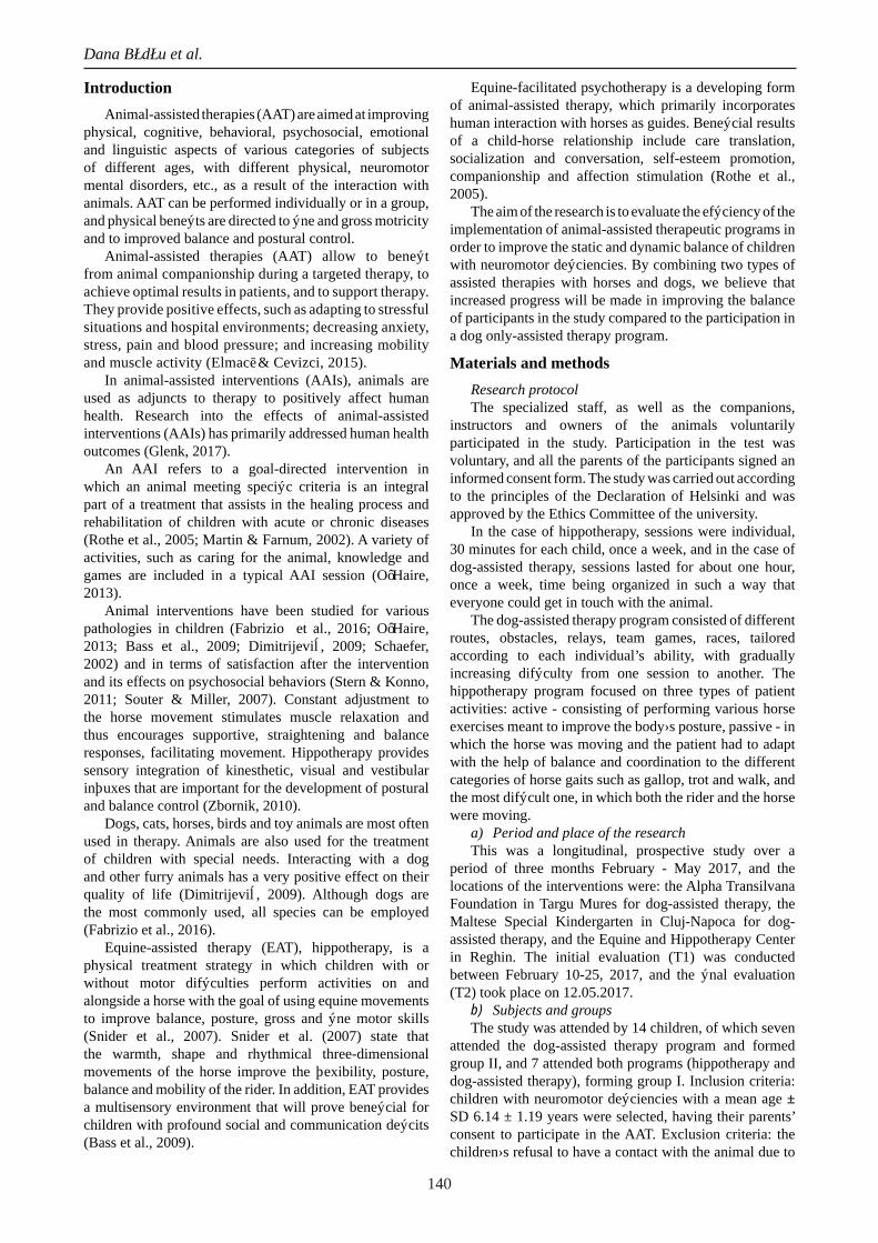
140
Dana Bădău et al.
IntroductionAnimal-assisted therapies (AAT) are aimed at improving
physical, cognitive, behavioral, psychosocial, emotional and linguistic aspects of various categories of subjects of different ages, with different physical, neuromotor mental disorders, etc., as a result of the interaction with animals. AAT can be performed individually or in a group, and physical benefits are directed to fine and gross motricity and to improved balance and postural control.
Animal-assisted therapies (AAT) allow to benefit from animal companionship during a targeted therapy, to achieve optimal results in patients, and to support therapy. They provide positive effects, such as adapting to stressful situations and hospital environments; decreasing anxiety, stress, pain and blood pressure; and increasing mobility and muscle activity (Elmacı & Cevizci, 2015).
In animal-assisted interventions (AAIs), animals are used as adjuncts to therapy to positively affect human health. Research into the effects of animal-assisted interventions (AAIs) has primarily addressed human health outcomes (Glenk, 2017).
An AAI refers to a goal-directed intervention in which an animal meeting specific criteria is an integral part of a treatment that assists in the healing process and rehabilitation of children with acute or chronic diseases (Rothe et al., 2005; Martin & Farnum, 2002). A variety of activities, such as caring for the animal, knowledge and games are included in a typical AAI session (O’Haire, 2013).
Animal interventions have been studied for various pathologies in children (Fabrizio et al., 2016; O’Haire, 2013; Bass et al., 2009; Dimitrijević, 2009; Schaefer, 2002) and in terms of satisfaction after the intervention and its effects on psychosocial behaviors (Stern & Konno, 2011; Souter & Miller, 2007). Constant adjustment to the horse movement stimulates muscle relaxation and thus encourages supportive, straightening and balance responses, facilitating movement. Hippotherapy provides sensory integration of kinesthetic, visual and vestibular influxes that are important for the development of postural and balance control (Zbornik, 2010).
Dogs, cats, horses, birds and toy animals are most often used in therapy. Animals are also used for the treatment of children with special needs. Interacting with a dog and other furry animals has a very positive effect on their quality of life (Dimitrijević, 2009). Although dogs are the most commonly used, all species can be employed (Fabrizio et al., 2016).
Equine-assisted therapy (EAT), hippotherapy, is a physical treatment strategy in which children with or without motor difficulties perform activities on and alongside a horse with the goal of using equine movements to improve balance, posture, gross and fine motor skills (Snider et al., 2007). Snider et al. (2007) state that the warmth, shape and rhythmical three-dimensional movements of the horse improve the flexibility, posture, balance and mobility of the rider. In addition, EAT provides a multisensory environment that will prove beneficial for children with profound social and communication deficits (Bass et al., 2009).
Equine-facilitated psychotherapy is a developing form of animal-assisted therapy, which primarily incorporates human interaction with horses as guides. Beneficial results of a child-horse relationship include care translation, socialization and conversation, self-esteem promotion, companionship and affection stimulation (Rothe et al., 2005).
The aim of the research is to evaluate the efficiency of the implementation of animal-assisted therapeutic programs in order to improve the static and dynamic balance of children with neuromotor deficiencies. By combining two types of assisted therapies with horses and dogs, we believe that increased progress will be made in improving the balance of participants in the study compared to the participation in a dog only-assisted therapy program.
Materials and methodsResearch protocolThe specialized staff, as well as the companions,
instructors and owners of the animals voluntarily participated in the study. Participation in the test was voluntary, and all the parents of the participants signed an informed consent form. The study was carried out according to the principles of the Declaration of Helsinki and was approved by the Ethics Committee of the university.
In the case of hippotherapy, sessions were individual, 30 minutes for each child, once a week, and in the case of dog-assisted therapy, sessions lasted for about one hour, once a week, time being organized in such a way that everyone could get in touch with the animal.
The dog-assisted therapy program consisted of different routes, obstacles, relays, team games, races, tailored according to each individual’s ability, with gradually increasing difficulty from one session to another. The hippotherapy program focused on three types of patient activities: active - consisting of performing various horse exercises meant to improve the body›s posture, passive - in which the horse was moving and the patient had to adapt with the help of balance and coordination to the different categories of horse gaits such as gallop, trot and walk, and the most difficult one, in which both the rider and the horse were moving.
a) Period and place of the researchThis was a longitudinal, prospective study over a
period of three months February - May 2017, and the locations of the interventions were: the Alpha Transilvana Foundation in Targu Mures for dog-assisted therapy, the Maltese Special Kindergarten in Cluj-Napoca for dog-assisted therapy, and the Equine and Hippotherapy Center in Reghin. The initial evaluation (T1) was conducted between February 10-25, 2017, and the final evaluation (T2) took place on 12.05.2017.
b) Subjects and groupsThe study was attended by 14 children, of which seven
attended the dog-assisted therapy program and formed group II, and 7 attended both programs (hippotherapy and dog-assisted therapy), forming group I. Inclusion criteria: children with neuromotor deficiencies with a mean age ± SD 6.14 ± 1.19 years were selected, having their parents’ consent to participate in the AAT. Exclusion criteria: the children›s refusal to have a contact with the animal due to
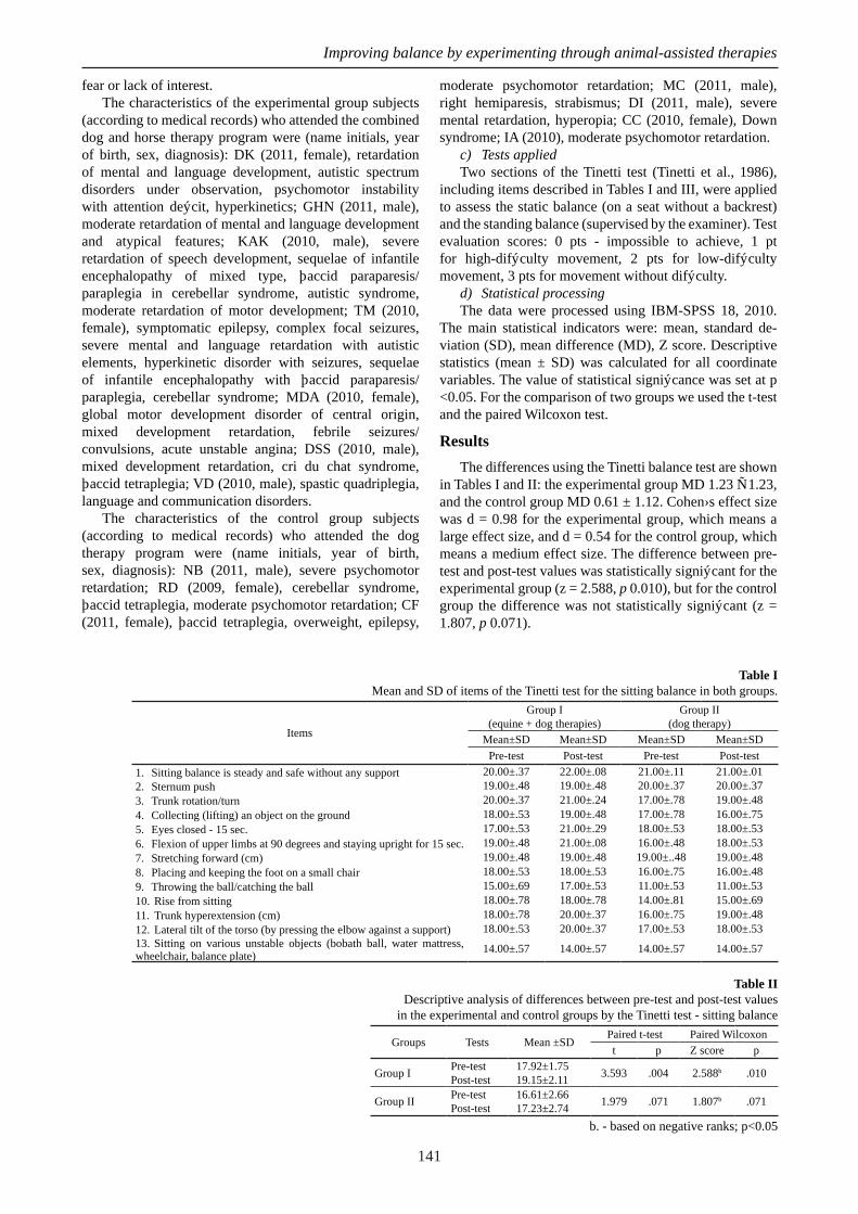
141
Improving balance by experimenting through animal-assisted therapies
fear or lack of interest. The characteristics of the experimental group subjects
(according to medical records) who attended the combined dog and horse therapy program were (name initials, year of birth, sex, diagnosis): DK (2011, female), retardation of mental and language development, autistic spectrum disorders under observation, psychomotor instability with attention deficit, hyperkinetics; GHN (2011, male), moderate retardation of mental and language development and atypical features; KAK (2010, male), severe retardation of speech development, sequelae of infantile encephalopathy of mixed type, flaccid paraparesis/paraplegia in cerebellar syndrome, autistic syndrome, moderate retardation of motor development; TM (2010, female), symptomatic epilepsy, complex focal seizures, severe mental and language retardation with autistic elements, hyperkinetic disorder with seizures, sequelae of infantile encephalopathy with flaccid paraparesis/paraplegia, cerebellar syndrome; MDA (2010, female), global motor development disorder of central origin, mixed development retardation, febrile seizures/convulsions, acute unstable angina; DSS (2010, male), mixed development retardation, cri du chat syndrome, flaccid tetraplegia; VD (2010, male), spastic quadriplegia, language and communication disorders.
The characteristics of the control group subjects (according to medical records) who attended the dog therapy program were (name initials, year of birth, sex, diagnosis): NB (2011, male), severe psychomotor retardation; RD (2009, female), cerebellar syndrome, flaccid tetraplegia, moderate psychomotor retardation; CF (2011, female), flaccid tetraplegia, overweight, epilepsy,
moderate psychomotor retardation; MC (2011, male), right hemiparesis, strabismus; DI (2011, male), severe mental retardation, hyperopia; CC (2010, female), Down syndrome; IA (2010), moderate psychomotor retardation.
c) Tests appliedTwo sections of the Tinetti test (Tinetti et al., 1986),
including items described in Tables I and III, were applied to assess the static balance (on a seat without a backrest) and the standing balance (supervised by the examiner). Test evaluation scores: 0 pts - impossible to achieve, 1 pt for high-difficulty movement, 2 pts for low-difficulty movement, 3 pts for movement without difficulty.
d) Statistical processingThe data were processed using IBM-SPSS 18, 2010.
The main statistical indicators were: mean, standard de-viation (SD), mean difference (MD), Z score. Descriptive statistics (mean ± SD) was calculated for all coordinate variables. The value of statistical significance was set at p <0.05. For the comparison of two groups we used the t-test and the paired Wilcoxon test.
ResultsThe differences using the Tinetti balance test are shown
in Tables I and II: the experimental group MD 1.23 ± 1.23, and the control group MD 0.61 ± 1.12. Cohen›s effect size was d = 0.98 for the experimental group, which means a large effect size, and d = 0.54 for the control group, which means a medium effect size. The difference between pre-test and post-test values was statistically significant for the experimental group (z = 2.588, p 0.010), but for the control group the difference was not statistically significant (z = 1.807, p 0.071).
Table I Mean and SD of items of the Tinetti test for the sitting balance in both groups.
Items
Group I(equine + dog therapies)
Group II(dog therapy)
Mean±SD Mean±SD Mean±SD Mean±SDPre-test Post-test Pre-test Post-test
1. Sitting balance is steady and safe without any support 20.00±.37 22.00±.08 21.00±.11 21.00±.012. Sternum push 19.00±.48 19.00±.48 20.00±.37 20.00±.373. Trunk rotation/turn 20.00±.37 21.00±.24 17.00±.78 19.00±.484. Collecting (lifting) an object on the ground 18.00±.53 19.00±.48 17.00±.78 16.00±.755. Eyes closed - 15 sec. 17.00±.53 21.00±.29 18.00±.53 18.00±.536. Flexion of upper limbs at 90 degrees and staying upright for 15 sec. 19.00±.48 21.00±.08 16.00±.48 18.00±.537. Stretching forward (cm) 19.00±.48 19.00±.48 19.00±..48 19.00±.488. Placing and keeping the foot on a small chair 18.00±.53 18.00±.53 16.00±.75 16.00±.489. Throwing the ball/catching the ball 15.00±.69 17.00±.53 11.00±.53 11.00±.5310. Rise from sitting 18.00±.78 18.00±.78 14.00±.81 15.00±.6911. Trunk hyperextension (cm) 18.00±.78 20.00±.37 16.00±.75 19.00±.4812. Lateral tilt of the torso (by pressing the elbow against a support) 18.00±.53 20.00±.37 17.00±.53 18.00±.5313. Sitting on various unstable objects (bobath ball, water mattress, wheelchair, balance plate) 14.00±.57 14.00±.57 14.00±.57 14.00±.57
Table II Descriptive analysis of differences between pre-test and post-test values
in the experimental and control groups by the Tinetti test - sitting balance
Groups Tests Mean ±SDPaired t-test Paired Wilcoxon t p Z score p
Group I Pre-test 17.92±1.75 3.593 .004 2.588b .010Post-test 19.15±2.11
Group II Pre-test 16.61±2.66 1.979 .071 1.807b .071Post-test 17.23±2.74b. - based on negative ranks; p<0.05

142
Dana Bădău et al.
Tables III and IV highlight the differences between the results obtained for the standing balance - Tinetti test, as follows: for the experimental group MD 1.40 ± 1.19, and for the control group MD 0.06 ± 0.88. Cohen›s effect size was d = 1.17 for the experimental group, a very large effect size, and d = 0.07 for the control group, a very small effect size. The difference between pre- and post-test values was statistically significant for the experimental group, which was revealed using the Wilcoxon signed rank test for two paired samples (z = 3.104, p 0.002), but for the control group the difference was not statistically significant (z = 0.302, p 0.763).
DiscussionsIn both Tinetti tests - sitting and standing, the experi-
mental group made statistically significant improvements between the two testings, but in the control group differ-ences were statistically insignificant between tests, p <0.05.
Research over the past 30 years indicates that therapy dogs may offer physiological, emotional, social, and physi-cal support to children (Friesen, 2010).
Various approaches exist for improving postural control and balance through AAT (Zadnikar & Kastrin, 2011; Shur-tleff, 2009; Hamill et al., 2007; Benda et al., 2003).
Hippotherapy - performing exercises on the back of an unsaddled horse (trunk straightening, rotation) helps im-prove numerous motor functions (Zadnikar, 2010).
The results of the study conducted in children aged 7-9 years with autism suggested that an EAT intervention may be beneficial to improve the balance, upper limb coordina-tion and possibly strength of children with ASD in a way
that is acceptable but also enjoyable to these children (De Milander et al., 2016).
A round therapy field 20 meters in diameter is recom-mended in horse therapy, where the patient, the thera-pist and the horse are in uninterrupted interaction. Such therapeutic sessions are practiced 2 to 3 times per week (Yeh, 2005).
Vidrine et al. (2002) summarize three studies using therapy with horses. One study in children with special educational needs showed an increase in positive behavior among the study participants.
During the past century, horses have been used in programs helping people with physical disabilities. Riders benefit from the gentle rocking motion that can help to relax muscles and improve balance. Some theorists contend that the horse power and size provide opportunities for riders to explore issues related to vulnerability, power, and control (Lentini & Knox, 2009).
Some studies concluded that equine-facilitated psy-chotherapy provides well-being and an improvement in the quality of life of children with mental health prob-lems; horses can put children therapeutically in touch with their own vitality by their large and gentle pres-ence (Rothe et al., 2005).
Conclusions1. Dog and horse-assisted therapy increased the abili-
ties of children with neuromotor deficiencies, because they tried to change their behavior and participate more actively in the treatment process.
2. AAT contributed to the improvement of the static
Table III Mean and SD of items of the Tinetti test for the standing balance in both groups.
Items
Group I(equine + dog therapies)
Group II(dog therapy)
Mean±SD Mean±SD Mean±SD Mean±SDPre-test Post-test Pre-test Post-test
1. Sitting balance is steady and safe without any support 19.00±.48 20.00±.37 19.00±.48 20.00±.652. Sternum push 17.00±.78 17.00±.78 17.00±.53 16.00±.483. Trunk rotation/turn 19.00±.48 18.00±.53 18.00±.53 17.00±.534. Collecting (lifting) an object on the ground 17.00±.78 18.00±.53 19.00±.48 14.00±.895. Eyes closed - 15 sec. 15.00±.37 17.00±.69 13.00±.69 14.00±.536. Flexion of upper limbs at 90 degrees and staying upright for 15 sec. 16.00±.48 19.00±..34 15.00±.89 16.00±.537. Stretching forward (cm) 18.00±.78 20.00±.48 14.00±.48 14.00±.728. Placing and keeping the foot on a small chair 15.00±.69 16.00±.75 15.00±.89 12.00±.759. Throwing the ball/catching the ball 11.00±.97 14.00±.57 16.00±.48 16.00±.4810. Standing on one foot 13.00±.37 15.00±.89 12.00±.49 12.00±.5311. Stepping over obstacles 14.00±.57 16.00±.57 15.00±.38 15.00±.3712. Walking on a drawn line 13.00±.37 14.00±.48 13.00±.37 12.00±.7513. Moving hands and trunk while walking 14.00±1.00 15.00±.57 14.00±.57 13.00±.7814. Climbing up and down stairs 17.00±.53 18.00±.53 16.00±.48 17.00±.5315. Running 15.00±.89 17.00±.75 15.00±.89 16.00±.48
Table IV Descriptive analysis of differences between pre-test and post-test values
in the experimental and control groups by the Tinetti test - standing balance.
Groups Tests Mean ±SDPaired t-test Paired Wilcoxon t p Z score p
Group I Pre-test 15.53±2.32 5.137 .000 3.104b .002Post-test 16.93±1.94
Group II Pre-test 15.40±2.13 .292 .774 .302b .763Post-test 15.46±2.19b. - based on negative ranks; p<0.05
143
Improving balance by experimenting through animal-assisted therapies
and dynamic balance, the results being statistically signifi-cant.
3. Regarding the improvement of the static and dyna-mic balance, the combined dog and horse-assisted therapy had superior effects compared to the horse only-assisted therapy.
Conflicts of interestThe authors have no conflict of interest.
Acknowledgments We wish to thank all the members of the Alpha Transilvana Foundation in Targu Mures, the Maltese Special Kindergarten in Cluj-Napoca and the Equine and Hippotherapy Center in Reghin.
ReferencesBass MM, Duchowny CA, Ilabre MM. The effect of therapeutic
horseback riding on social functioning in children with autism. J Autism Dev Disord. 2009;39(9):1261-1267. doi: 10.1007/s10803-009-0734-3
Benda W, McGibbon NH, Grant KL. Improvements in muscle symmetry in children with cerebral palsy after equine-assisted therapy (hippotherapy). J Altern Complement Med. 2003;9:817-825. DOI:10.1089/107555303771952163.
De Milander M, Bradley S, Fourie R. Equine-assisted therapy as intervention for motor proficiency in children with autism spectrum disorder: case studies. South African J Res Sport, Phys Educ Recreation. 2016;38(3): 37-49.
Dimitrijević, I. Animal-Assisted Therapy - A New Trend in the Treatment of Children and Addults. Psych Danubina. 2009;21(2):236-241.
Elmacı DT, Cevizci S. Dog-Assisted Therapies and Activities in Rehabilitation of Children with Cerebral Palsy and Physical and Mental Disabilities. Int. J. Environ. Res. Public Health. 2015;12(5):5046-5060. doi:10.3390/ijerph120505046.
Fabrizio B, Gualano M R, Camussi E, Pieve G, Voglino G, Siliquini R. Animal assisted intervention: A systematic review of benefits and risks. Eur J Integrative Med. 2016;8 (5):695-706. doi:10.1016/j.eujim.2016.05.0055.
Friesen L. Exploring Animal-Assisted Programs with Children in School and Therapeutic Contexts. Early Childhood Ed J. 2010;37(4):261-267. Doi: https://doi.org/10.1007/s10643-009-0349-5.
Glenk LM. Current Perspectives on Therapy Dog Welfare in Animal-Assisted Interventions. Animals. 2017;7(2):7; doi:10.3390/ani7020007.
Hamill D, Washington K, White OR. The effect of hippotherapy on postural control in sitting for children with cerebral
palsy. Phys Occup Ther Pediatr. 2007;27:23-42.Lentini JA, Knox MA. Qualitative and Quantitative Review of
Equine Facilitated Psychotherapy (EFP) with Children and Adolescents. The Open Complement Med J. 2009;1:51-57.
Martin F, Farnum J. Animal-assisted therapy for children with pervasive developmental disorders. West J Nurs Res. 2002;24(6):657-670. DOI: 10.2174/1876391X00901010051.
OꞌHaire ME. Animal-assisted intervention for autism spectrum disorder: A systematic literature review. J Autism Dev Disord. 2013;43(7):1606-1622. doi: 10.1007/s10803-012-1707-5.
Rothe EQ, Vega BJ, Torres RM, Soler SMC, Pazos RMM. From kids and horses: Equine facilitated psychotherapy for children. Int J Clin Health Psychol. 2005;5(2): 373-383.
Schaefer K. Human-animal interactions as a therapeutic intervention. Counsel Hum Develop. 2002;34(5):1-18.
Shurtleff TL, Standeven JW, Engsberg JR. Changes in dynamic trunk/head stability and functional reach after hippotherapy. Arch Phys Med Rehabil. 2009; 90(7):1185-1195. doi: 10.1016/j.apmr.2009.01.026.
Snider L, Korner-Bitensky N, Kammann C, Warner S, Saleh M. Horseback riding as therapy for children with cerebral palsy: Is there evidence of its effectiveness? Phys Occup Ther Paedi. 2007;27(2):5-23
Souter MA, Miller MD. Do Animal-Assisted Activities Effectively Treat Depression? A Meta-Analysis. Anthrozoös: A Multidiscip J Interac People Animals. 2007;20 (2):167-180. doi:10.2752/175303707X207954.
Stern C, Konno R. The effects of Canine-Assisted Interventions (CAIs) on the health and social care of older people residing in long term care: a systematic review. JBI Libr Syst Rev. 2011;9(6):146-206. doi:10.11124/01938924-201109060-00001.
Tinetti ME, Williams TF, Mayewski R. Fall Risk Index for elderly patients based on number of chronic disabilities. Am J Med. 1986;80:429-434
Vidrine M, Owen-Smith P, Faulkner P. Equine-facilitated group psychotherapy: applications for therapeutic vaulting. Issues in Ment Health Nurs. 2002;23(6):587-603. DOI:10.1080/01612840290052730.
Yeh ML. Lassie’s magic: animal-assisted therapy in Taiwan. Hu Li Za Zhi. J Nursing 2005;52:23-30.
Zadnikar M. Učinek hipoterapije in terapevtskega jahanja na kontrolo drže in ravnotežje pri otrocih s cerebralno paralizo. In M. Zadnikar (Ed.), Zbornik,“1. Kongres terapevtskega jahanja v Sloveniji. Konj kot terapevt - danes za jutri”. Kamnik: Zavod za usposabljanje invalidne mladine. 2010;33-44.
Zadnikar M, Kastrin A. Effects of hippotherapy and therapeutic horseback riding on postural control or balance in children with cerebral palsy: a meta-analysis. Develop Med & Child Neurol. 2011;53:684-691. doi:10.1111/j.1469-8749.2011.03951.x.
Zbornik E. Kongres terapevtskega jahanja v Sloveniji. Konj kot terapevt - danes za jutri. Kamnik: Zavod za usposabljanje invalidne mladine. 2010;11-22.
144
Palestrica of the third millennium – Civilization and SportVol. 18, no. 3, July-September 2017, 144–150
The experimental effect of Lycium barbarum on redox homeostasis in physical exercise
Efectul experimental al Lycium barbarum asupra homeostaziei redox în efort fizic
Camelia Manuela Mîrza 1, Diana Topârcean 2, Tudor-Valentin Mîrza 3
1 Pathophysiology Department, Faculty of Medicine, “Iuliu Haţieganu” University of Medicine and Pharmacy2 Student, Faculty of Medicine, “Iuliu Haţieganu” University of Medicine and Pharmacy3 National Institute of Public Health – Regional Center of Public Health Cluj
AbstractBackground. Goji has over 100 potentially beneficial constituents for the human body contained in all parts of the plant,
especially in fruit and roots. Data from the literature have suggested beneficial effects on the human body after long-term Goji juice intake.
Aims. We studied the antioxidant effect of Lycium barbarum (Goji) natural juice on the body subjected to physical exercise. Methods. The oxidant-antioxidant balance indicators were determined in 6 groups of male Wistar rats (n = 6 animals/group)
which performed physical exercise through the swimming test. Some groups only performed physical exercise, while others were subjected to physical exercise and received an additional daily intake of Lycium barbarum natural juice. Nitro-oxidative stress was measured by serum determination of total oxidant status (TOS), total antioxidant capacity (TAC), malondialdehyde (MDA), total thiols, nitrites and nitrates.
Results. Increased TAC and serum thiol levels confirmed the antioxidant potential of Lycium barbarum during physical exercise. The daily intake of Lycium barbarum natural juice led to the reduction of oxidative stress and, implicitly, of lipid peroxidation, as evidenced by the decrease of serum MDA values.
Conclusions. Under experimental conditions, the administration of natural Lycium barbarum juice was beneficial by increasing exercise capacity. Due to its energetic and antioxidant effect, Lycium barbarum could represent a natural, non-doping alternative for improving sports performance.
Keywords: Lycium barbarum (Goji), oxidant-antioxidant balance, physical exercise. RezumatPremize. Goji are peste 100 de constituenți cu potențial benefic pentru organismul uman conținuţi în toate părțile plantei,
mai ales la nivelul fructului şi al rădăcinii. Date din literatură au sugerat existenţa unor efecte benefice asupra organismului uman după consumul sucului de Goji pe termen lung.
Obiective. Am studiat efectul antioxidant al sucului natural de fructe Lycium barbarum (Goji) asupra organismului în efort fizic.
Metode. Au fost determinaţi indicatorii balanţei oxidanţi-antioxidanţi la 6 loturi de şobolani masculi rasa Wistar (n = 6 animale/lot) care au efectuat efort fizic prin proba de înot. Unele loturi au fost supuse doar efortului fizic prin proba de înot, iar celelalte au fost supuse efortului şi aportului suplimentar de Lycium barbarum. Stresul nitro-oxidativ a fost măsurat prin determinarea serică a statusului oxidativ total, a capacității antioxidante totale, a malondialdehidei şi a tiolilor totali.
Rezultate. Creşterea valorilor capacităţii antioxidante totale (TAC), a nivelului seric de tioli a confirmat potenţialul antioxi-dant al Lycium barbarum în condiţii de efort fizic. Suplimentarea cu suc natural de Lycium barbarum a condus la diminuarea stresului oxidativ şi, implicit, a peroxidării lipidice, dovedite prin scăderea valorilor malondialdehidei serice.
Concluzii. În condiţii experimentale, administrarea sucului natural de Lycium barbarum şi-a adus aportul benefic la creşterea capacităţii de efort. Datorită efectului energogen şi antioxidant în condiţii de efort, Lycium barbarum ar putea reprezenta o alternativă naturală, nedopantă, pentru îmbunătăţirea performanţelor sportive.
Cuvinte cheie: Lycium barbarum (Goji), balanţă oxidanţi/antioxidanţi, efort fizic.
Copyright © 2010 by “Iuliu Haţieganu” University of Medicine and Pharmacy Publishing
Received: 2017, August 5; Accepted for publication: 2017, September 2 Address for correspondence: Iuliu Haţieganu University of Medicine and Pharmacy, Faculty of Medicine, Pathophysiology Depart-
ment, 4 Victor Babeş Street, 400012, Cluj-Napoca, RomaniaE-mail: [email protected] Corresponding author: Camelia Manuela Mîrza, [email protected]://doi.org/10.26659/pm3.2017.18.3.144
145
The experimental effect of Lycium barbarum on redox homeostasis in physical exercise
IntroductionGoji was named Lycium barba rum in the botanical
nomenclature in 1753 (Carl Linnaeus) (Kulczyński & Gramza-Michałowska, 2016). It belongs to the Solanaceae family, Lycium genus (Ma et al., 2009). Although it is known in the literature under many names, the name Goji derived from the Chinese “gouqi” has been widely accepted (Hänsel et al., 1993).
Goji is a shrub with reddish-orange fruits, having a diameter of 1-2 cm2 and bitter-sweet taste (Zhang et al., 2001).
Goji has over 100 potentially beneficial constituents for the human body contained in all parts of the plant, especially in fruit and roots. Through these constituents, Lycium barbarum exhibits a powerful antioxidant and cytoprotective effect: hypoglycemic (Shaban et al., 1990; Wu et al., 2006), hypolipemic (Zhao et al., 2005; Luo et al., 2004), cardio- (Xu et al., 2005), neuro- (Lin et al., 2003; Yu et al., 2006; Yu et al., 2007), pancreato- and hepatoprotective (Kim et al., 1997; Chin et al, 2003; Slemmer et al., 2008), anti-aging (Zhao et al., 2005; Li et al., 2007b), anti-arterial hypertension (Jia et al., 1998; Chan et al., 2007); it contributes to increasing the body’s resistance to physical exercise, etc.
Goji fruits are consumed fresh or in the form of juice, tea (infusion) or wine (Zhufan, 2000). They can also be used in combination with conventional products such as yogurt or snacks with seeds, dehydrated fruits and oatmeal (Potterat, 2010) in various culinary preparations (Yin & Dang, 2008) or as seasonings (Jin et al., 2013). The recommended daily dose of dried Goji fruit is 5-12 g, calculated for an adult body (Bensky et al., 2004).
HypothesisThe beneficial effects of Goji as well as its contribution
to increased exercise capacity have led us to study the antioxidant effect of Lycium barbarum (Goji) natura l fruit juice on the body subjected to physical exercise through oxidative-antioxidant balance indicators.
Materials and methodsa) Research protocolThe experimental study was performed on 16 weeks old
male Wistar rats, with a mean weight of 200 grams, from the Biobase of the “Iuliu Hațieganu” University of Medicine and Pharmacy Cluj-Napoca. The study was approved by the Ethics Board, according to Good Practice Guidelines. The requirements of the Helsinki Declaration, Amsterdam Protocol, Directive 86/609/EEC and the regulations of the Bioethics Commission of the “Iuliu Hațieganu” University of Medicine and Pharmacy Cluj-Napoca were met. All conditions for reducing the suffering of experimental animals and the principles of biodiversity were ensured by observing the provisions of the Government Ordinance no. 37 on the protection of laboratory animals.
The research was carried out in the Experimental Research Laboratory of the Pathophysiology Department of the “Iuliu Hațieganu” University of Medicine and Pharmacy Cluj-Napoca. During the experiment, the animals were maintained under appropriate vivarium
conditions at a controlled temperature of 21-22 °C, 65% humidity, cycle light:dark 12:12 h, standardized feed and water ad libitum.
b) GroupsThe measurements were performed in 6 groups of
rats (n = 6 animals/group) that were subjected to physical exercise through the swimming test:
- group I - controls + exercise 1 day- group II - controls + supplement + exercise 1 day- group III - rats + exercise 7 days- group IV - rats + daily supplement + exercise 7 days- group V - rats + exercise 14 days- group VI - rats + daily supplement + exercise 14
days.The supplement was represented by 100% natural
Lycium barbarum fruit juice, imported and distributed by SC Goji Planet SRL. The daily dose of Goji juice of 0.3 ml/animal/day was given by oropharyngeal gavage for 1 day, 1 or 2 weeks.
c) Tests appliedOxidative stress was induced by physical exercise
for 14 days using the swimming test according to the standardized model in the literature (Nayanatara et al., 2005). The aerobic exercise capacity (AEC) was determined by measuring the time, expressed in seconds, from the moment the animals were introduced into the pool until their exhaustion (refusal to swim). The intensity of exercise was changed by loading the animals in groups III-VI with lead weights accounting for 15% of the animal’s weight.
The duration of the experiment was 14 days. The studied days were day 1 (T1), day 7 (T2) and day 14 (T3).
Biochemical determinations were performed in the Laboratory of the Pathophysiology Department of “Iuliu Hațieganu” University of Medicine and Pharmacy Cluj-Napoca. For the measurement of the indicators of the O/AO balance in the blood, venous blood samples were taken from the retro-orbital sinus, under general anesthesia with 2:1 10% ketamine and 2% xylazine. From the harvested blood, the serum was separated by centrifugation, in order to measure the indicators. Readings were made on the METERTEK SP-830 spectrophotometer. Animals were sacrificed under general anesthesia by cervical dislocation at the end of the experiment.
The serum indicators determined were total oxidant status (TOS), total antioxidant capacity (TAC), malondialdehyde (MDA) and total thiols. MDA was determined by using the microanalytical method of Pasha and Sadasivadu (Mitev et al., 2010). For the determination of the total oxidant status (TOS) a spectrophotometric method was used (Ozcan, 2005). Determination of thiols was performed by the Ellman method (Mitev et al., 2010) and total antioxidant capacity (TAC) was determined by using the Fenton reaction (Ozcan, 2004).
d) Statistical processingDescriptive statistics elements were calculated. Data
were presented using centrality, location and distribution indicators.
Normal distribution was tested with the Shapiro-Wilk test. ANOVA, Kruskal-Wallis, t (Student), Mann-Whitney (U) and Wilcoxon tests were used for statistical data
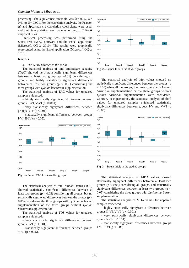
146
Camelia Manuela Mîrza et al.
processing. The significance threshold was α = 0.05, α = 0.01 or α = 0.001. For the correlation analysis, the Pearson (r) and Spearman (ρ) correlation coefficients were used, and their interpretation was made according to Colton’s empirical rules.
Statistical processing was performed using the StatsDirect v.2.7.2 software and the Excel application (Microsoft Office 2010). The results were graphically represented using the Excel application (Microsoft Office 2010).
Resultsa) The O/AO balance in the serumThe statistical analysis of total antioxidant capacity
(TAC) showed very statistically significant differences between at least two groups (p <0.01) considering all groups, and highly statistically significant differences between at least two groups (p <0.001) considering the three groups with Lycium barbarum supplementation.
The statistical analysis of TAC values for unpaired samples evidenced:
- highly statistically significant differences between groups II-VI, V-VI (p <0.001)
- very statistically significant differences between groups IV-V (p <0.01)
- statistically significant differences between groups I-VI, II-IV (p <0.05).
0,00
0,01
0,02
0,03
0,04
0,05
0,06
0,07
Group I Group II Group III Group IV Group V Group VI
mmol trolox eq/l Median 25% - 75% Min - MaxMean
Fig. 1 – Serum TAC in the studied groups.
The statistical analysis of total oxidant status (TOS) showed statistically significant differences between at least two groups (p < 0.05) considering all groups, but no statistically significant differences between the groups (p > 0.05) considering the three groups with Lycium barbarum supplementation or the three groups without Lycium barbarum supplementation.
The statistical analysis of TOS values for unpaired samples evidenced:
- very statistically significant differences between groups I-VI (p < 0.01)
- statistically significant differences between groups V-VI (p < 0.05).
0,00
0,05
0,10
0,15
0,20
0,25
0,30
0,35
0,40
0,45
Group I Group II Group III Group IV Group V Group VI
µmol H2O2/l Median 25% - 75% Min - MaxMean
Fig. 2 – Serum TOS in the studied groups
The statistical analysis of thiol values showed no statistically significant differences between the groups (p > 0.05) when all the groups, the three groups with Lycium barbarum supplementation or the three groups without Lycium barbarum supplementation were considered. Contrary to expectations, the statistical analysis of thiol values for unpaired samples evidenced statistically significant differences between groups I-V and V-VI (p <0.05).
0,00
0,05
0,10
0,15
0,20
0,25
0,30
0,35
0,40
0,45
Group I Group II Group III Group IV Group V Group VI
mmol/l Median 25% - 75% Min - MaxMean
Fig. 3 – Serum thiols in the studied groups
The statistical analysis of MDA values showed statistically significant differences between at least two groups (p < 0.05) considering all groups, and statistically significant differences between at least two groups (p < 0.05) considering the three groups with Lycium barbarum supplementation.
The statistical analysis of MDA values for unpaired samples evidenced:
- highly statistically significant differences between groups II-VI, V-VI (p < 0.001)
- very statistically significant differences between groups I-VI (p < 0.01)
- statistically significant differences between groups I-V, III-VI (p < 0.05).
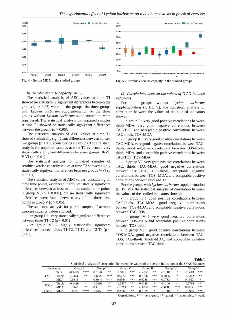
147
The experimental effect of Lycium barbarum on redox homeostasis in physical exercise
0,00
0,05
0,10
0,15
0,20
0,25
0,30
Group I Group II Group III Group IV Group V Group VI
nmol/l Median 25% - 75% Min - MaxMean
Fig. 4 – Serum MDA in the studied groups
b) Aerobic exercise capacity (AEC)The statistical analysis of AEC values at time T1
showed no statistically significant differences between the groups (p > 0.05) when all the groups, the three groups with Lycium barbarum supplementation or the three groups without Lycium barbarum supplementation were considered. The statistical analysis for unpaired samples at time T1 showed no statistically significant differences between the groups (p > 0.05).
The statistical analysis of AEC values at time T2 showed statistically significant differences between at least two groups (p < 0.05) considering all groups. The statistical analysis for unpaired samples at time T2 evidenced very statistically significant differences between groups III-VI, V-VI (p < 0.01).
The statistical analysis for unpaired samples of aerobic exercise capacity values at time T3 showed highly statistically significant differences between groups V-VI (p < 0.001).
The statistical analysis of AEC values, considering all three time points, evidenced highly statistically significant differences between at least two of the studied time points in group VI (p < 0.001), but no statistically significant differences were found between any of the three time points in group V (p > 0.05).
The statistical analysis for paired samples of aerobic exercise capacity values showed:
- in group III - very statistically significant differences between times T1-T2 (p < 0.01)
- in group VI - highly statistically significant differences between times T1-T2, T1-T3 and T2-T3 (p < 0.001).
0,00
10,00
20,00
30,00
40,00
50,00
60,00
70,00
80,00
Group I Group II Group III Group IV Group V Group VI Group III Group IV Group V Group VI Group V Group VI
T1 T2 T3
sec Median 25% - 75% Min - MaxMean
Fig. 5 – Aerobic exercise capacity in the studied groups
c) Correlations between the values of O/AO balance indicators
For the groups without Lycium barbarum supplementation (I, III, V), the statistical analysis of correlation between the values of the studied indicators showed:
- in group I – very good positive correlations between thiols-MDA, very good negative correlations between TAC-TOS, and acceptable positive correlations between TAC-thiols, TOS-MDA
- in group III – very good positive correlations between TAC-MDA, very good negative correlations between TAC-thiols, good negative correlations between TOS-thiols, thiols-MDA, and acceptable positive correlations between TAC-TOS, TOS-MDA
- in group V – very good positive correlations between TAC, thiols, TAC-MDA, good negative correlations between TAC-TOS, TOS-thiols, acceptable negative correlations between TOS- MDA, and acceptable positive correlations between thiols-MDA.
For the groups with Lycium barbarum supplementation (II, IV, VI), the statistical analysis of correlation between the values of the studied indicators showed:
- in group II – good positive correlations between TAC-thiols, TAC-MDA, good negative correlations between TOS-MDA, and acceptable negative correlations between TAC-TOS
- in group IV – very good negative correlations between TOS-MDA and acceptable positive correlations between TOS-thiols
- in group VI – good positive correlations between TOS-MDA, good negative correlations between TAC-TOS, TOS-thiols, thiols-MDA, and acceptable negative correlations between TAC-thiols.
Table I Statistical analysis of correlation between the values of the serum indicators of the O/AO balance.
Indicators Group I Group III Group V Group II Group IV Group VI
TAC - TOS -0.9429 **** 0.3198 ** -0.6821 *** -0.4838 ** -0.1804 * -0.5618 ***Thiols 0.3144 ** -0.8322 **** 0.6270 *** 0.7106 *** 0.2066 * -0.3362 **MDA 0.0315 * 0.8044 **** 0.5564 *** 0.6288 *** 0.0783 * 0.1352 *
TOS - Thiols -0.1429 * -0.5693 *** -0.5557 *** 0.0128 * 0.4140 ** -0.5786 ***MDA 0.3143 ** 0.4131 ** -0.3219 ** -0.6237 *** -0.8989 **** 0.5114 ***
Thiols - MDA 0.8690 **** -0.5453 *** 0.2896 ** 0.0139 * -0.1504 * -0.5966 ***Correlations: **** very good, *** good, ** acceptable, * weak
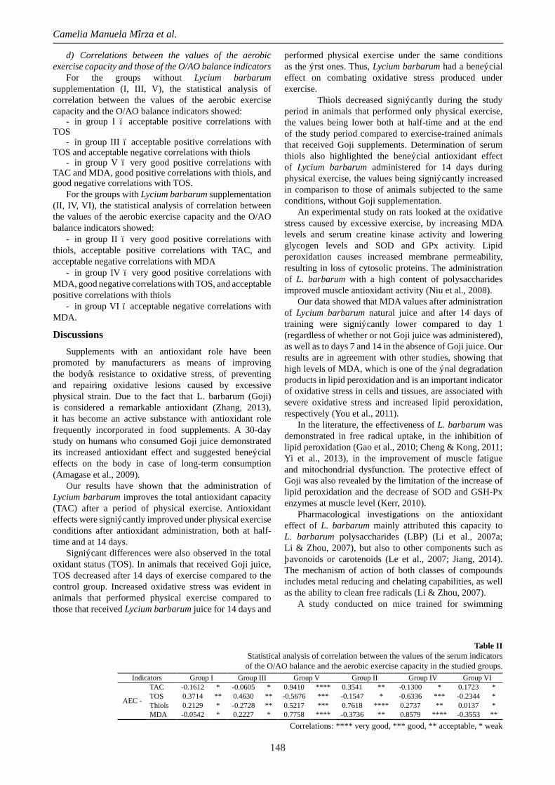
148
Camelia Manuela Mîrza et al.
d) Correlations between the values of the aerobic exercise capacity and those of the O/AO balance indicators
For the groups without Lycium barbarum supplementation (I, III, V), the statistical analysis of correlation between the values of the aerobic exercise capacity and the O/AO balance indicators showed:
- in group I – acceptable positive correlations with TOS
- in group III – acceptable positive correlations with TOS and acceptable negative correlations with thiols
- in group V – very good positive correlations with TAC and MDA, good positive correlations with thiols, and good negative correlations with TOS.
For the groups with Lycium barbarum supplementation (II, IV, VI), the statistical analysis of correlation between the values of the aerobic exercise capacity and the O/AO balance indicators showed:
- in group II – very good positive correlations with thiols, acceptable positive correlations with TAC, and acceptable negative correlations with MDA
- in group IV – very good positive correlations with MDA, good negative correlations with TOS, and acceptable positive correlations with thiols
- in group VI – acceptable negative correlations with MDA.
DiscussionsSupplements with an antioxidant role have been
promoted by manufacturers as means of improving the body’s resistance to oxidative stress, of preventing and repairing oxidative lesions caused by excessive physical strain. Due to the fact that L. barbarum (Goji) is considered a remarkable antioxidant (Zhang, 2013), it has become an active substance with antioxidant role frequently incorporated in food supplements. A 30-day study on humans who consumed Goji juice demonstrated its increased antioxidant effect and suggested beneficial effects on the body in case of long-term consumption (Amagase et al., 2009).
Our results have shown that the administration of Lycium barbarum improves the total antioxidant capacity (TAC) after a period of physical exercise. Antioxidant effects were significantly improved under physical exercise conditions after antioxidant administration, both at half-time and at 14 days.
Significant differences were also observed in the total oxidant status (TOS). In animals that received Goji juice, TOS decreased after 14 days of exercise compared to the control group. Increased oxidative stress was evident in animals that performed physical exercise compared to those that received Lycium barbarum juice for 14 days and
performed physical exercise under the same conditions as the first ones. Thus, Lycium barbarum had a beneficial effect on combating oxidative stress produced under exercise.
Thiols decreased significantly during the study period in animals that performed only physical exercise, the values being lower both at half-time and at the end of the study period compared to exercise-trained animals that received Goji supplements. Determination of serum thiols also highlighted the beneficial antioxidant effect of Lycium barbarum administered for 14 days during physical exercise, the values being significantly increased in comparison to those of animals subjected to the same conditions, without Goji supplementation.
An experimental study on rats looked at the oxidative stress caused by excessive exercise, by increasing MDA levels and serum creatine kinase activity and lowering glycogen levels and SOD and GPx activity. Lipid peroxidation causes increased membrane permeability, resulting in loss of cytosolic proteins. The administration of L. barbarum with a high content of polysaccharides improved muscle antioxidant activity (Niu et al., 2008).
Our data showed that MDA values after administration of Lycium barbarum natural juice and after 14 days of training were significantly lower compared to day 1 (regardless of whether or not Goji juice was administered), as well as to days 7 and 14 in the absence of Goji juice. Our results are in agreement with other studies, showing that high levels of MDA, which is one of the final degradation products in lipid peroxidation and is an important indicator of oxidative stress in cells and tissues, are associated with severe oxidative stress and increased lipid peroxidation, respectively (You et al., 2011).
In the literature, the effectiveness of L. barbarum was demonstrated in free radical uptake, in the inhibition of lipid peroxidation (Gao et al., 2010; Cheng & Kong, 2011; Yi et al., 2013), in the improvement of muscle fatigue and mitochondrial dysfunction. The protective effect of Goji was also revealed by the limitation of the increase of lipid peroxidation and the decrease of SOD and GSH-Px enzymes at muscle level (Kerr, 2010).
Pharmacological investigations on the antioxidant effect of L. barbarum mainly attributed this capacity to L. barbarum polysaccharides (LBP) (Li et al., 2007a; Li & Zhou, 2007), but also to other components such as flavonoids or carotenoids (Le et al., 2007; Jiang, 2014). The mechanism of action of both classes of compounds includes metal reducing and chelating capabilities, as well as the ability to clean free radicals (Li & Zhou, 2007).
A study conducted on mice trained for swimming
Table IIStatistical analysis of correlation between the values of the serum indicators of the O/AO balance and the aerobic exercise capacity in the studied groups.
Indicators Group I Group III Group V Group II Group IV Group VI
AEC -
TAC -0.1612 * -0.0605 * 0.9410 **** 0.3541 ** -0.1300 * 0.1723 *TOS 0.3714 ** 0.4630 ** -0.5676 *** -0.1547 * -0.6336 *** -0.2344 *Thiols 0.2129 * -0.2728 ** 0.5217 *** 0.7618 **** 0.2737 ** 0.0137 *MDA -0.0542 * 0.2227 * 0.7758 **** -0.3736 ** 0.8579 **** -0.3553 **
Correlations: **** very good, *** good, ** acceptable, * weak
149
The experimental effect of Lycium barbarum on redox homeostasis in physical exercise
revealed the beneficial effect of Lycium barbarum polysaccharide content on muscle fatigue, by lowering lactate concentration and increasing glycogen concentration (Yao & Li, 2010).
Our data on exercise capacity showed that this increased significantly on day 7 in animals that swam and received Goji compared to those that were only subjected to physical exercise by swimming. The same situation was maintained at the end of the study period. Exercise capacity decreased in animals that swam for 14 days without Goji supplementation, but the differences were not significant between the three studied time moments. In animals that swam for 14 days and received Goji supplements, the exercise capacity increased, the differences being significant between the studied times.
Finally, the results of the correlation analysis between the studied O/AO balance indicators suggest that Goji administration improves the ability to combat oxidative stress under exercise. Also, the correlation of the O/AO balance with the aerobic exercise capacity demonstrated that in the Goji supplemented group, with the increase of the exercise capacity, the values of oxidative stress indicators decreased at the end of the experimental study.
Conclusions1. The increase in the total antioxidant capacity (TAC)
confirmed the antioxidant potential of Lycium barbarum under physical exercise conditions.
2. The increase in serum thiol levels after supplementation with Lycium barbarum natural juice demonstrates the increase in the antioxidant capacity of the body under physical exercise conditions.
3. Supplementation with Lycium barbarum natural juice led to the reduction of oxidative stress and, implicitly, of lipid peroxidation, evidenced by the decrease of serum malondialdehyde values.
4. Administration of Lycium barbarum natural juice made a positive contribution to increasing exercise capacity.
5. Due to its energetic and antioxidant effect under physical exercise conditions, Lycium barbarum could be a natural, non-doping alternative for improving sports performance and post-exercise recovery.
Conflicts of interestNothing to declare.
ReferencesAmagase H, Sun B, Borek C. Lycium barbarum (goji) juice
improves antioxidant biomarkers in serum of healthy adults. Nutr Res. 2009;29(1):19-25.
Bensky D, Clavey S, Stöger E. Chinese herbal medicine, 3rd edition. Materia Medica. Seattle: Eastland Press, Inc.; 2004.
Chan HC, Chang RC, Koon-Ching IpA, Chiu K, YuenWH, Zee SY, So KF. Neuroprotective effects of Lycium barbarum Lynn on protecting retinal ganglion cells in an ocular hypertension model of glaucoma. Exp Neurol. 2007;203(1):269-273.
Cheng D, Kong H. The effect of Lycium barbarum polysaccharide on alcohol-induced oxidative stress in rats, Molecules. 2011;16(3):2542-2550.
Chin YW, Lim SW, Kim SH, Shin DY, Suh YG, Kim YB, Kim YC, Kim J. Hepatoprotective pyrrole derivatives of Lycium chinense fruits. Bioorg Med Chem Lett. 2003;13(1):79-81.
Gao K, Liu M, Cao J, Yao M, Lu Y, Li J, Zhu X, Yang Z, Wen A. Protective effects of Lycium barbarum polysaccharide on 6-OHDA-induced apoptosis in PC12 cells through the ROS-NO pathway, Molecules. 2014;20(1):293-308.
Hänsel R, Keller K, Rimpler H, Schneider G. Hagers Handbuch der pharmazeutischen Praxis. Vol 5: Drogen E–O. Berlin, Heidelberg, New York: Springer Verlag, 1993.
Jia YX, Dong JW, Wu XX, Ma TM, Shi AY. The effect of Lycium barbarum polysaccharide on vascular tension in two-kidney, one clip model of hypertension. Sheng Li Xue Bao. 1998;50(3):309-314.
Jiang L. Preparation and antioxidant activity of Lycium barbarum oligosaccharides. Carbohydr. Polym., 2014;99:646-648.
Jin M, Huang Q, Zhao, K, Shang P. Biological activities and potential health benefit effects of polysaccharides isolated from Lycium barbarum L. Int. J. Biol.Macromol. 2013;54:16-23.
Kerr DS. Treatment of mitochondrial electron transport chain disorders: a review of clinical trials over the past decade. Mol. Genet. Metab., 2010;99(3):246-255.
Kim SY, Choi YH, Huh H, Kim J, Kim YC, Lee HS. New antihepatotoxic cerebroside from Lycium chinense fruits. J Nat Prod. 1997;60(3):274-276.
Kulczyński B, Gramza-Michałowska A. Goji Berry (Lycium barbarum): Composition and Health Effects – a Review. Pol. J. Food Nutr. Sci. 2016;66(2):67-75.
Le K, Chiu F, Ng K. Identification and quantification of antioxidants in Fructus lycii. Food Chem 2007;105:353-363.
Li XL, Zhou AG. Evaluation of the antioxidant effects of polysaccharides extracted from Lycium barbarum. Med Chem Res 2007;15:471-482.
Li XM, Li XL, Zhou AG. Evaluation of antioxidant activity of the polysaccharides extracted from Lycium barbarum fruits in vitro. Eur Polymer J 2007a;43:488-497.
Li XM, Ma YL, Liu XJ. Effect of the Lycium barbarum polysaccharides on age-related oxidative stress in aged mice. J Ethnopharmacol, Mayo, 2007b;111(3):504-11.
Lin RD, Hou WC, Yen KY, Lee MH. Inhibition of monoamine oxidase B (MAO-B) by Chinese herbal medicines. Phytomed 2003;10(8):650-656.
Luo QL, Cai Y., Yan J., Sun M., Corke H. Hypoglycemic and hypolipidemic effects and antioxidant activity of fruit extracts from Lycium barbarum. Life Sci. 2004;76:137-149.
Ma M, Liu GH, Yu ZH, Chen G, Zhang X. Effect of the Lycium barbarum polysaccharides administration on blood lipid metabolism and oxidative stress of mice fed high-fat diet in vivo. Food Chem. 2009;113:872-877.
Mitev D, Gradeva H, Stoyanova Z, Petrova N, Karova N, Dimov D, Iliev V, Koychev A, Prakova G, Vlaykova T. Evaluation of thiol compounds and lipid peroxidative products in plasma of patients with COPD. Trakia J Sci. 2010;8(2):306-314.
Nayanatara AK, Nagaraja HS, Anupama BK. The effect of repetead swimming stress on organ weights and lipid peroxidation in rats. J Phys Biomed Sci. 2005;18(1):3-9.
Niu A-J, Wu J-M, Yu D-H, Wang R. Protective effect of Lycium barbarum polysaccharides on oxidative damage in skeletal muscle of exhaustive exercise rats. Inter. J. of Biol. Macromol., 2008;42:447-449.
Ozcan E. A new automated colorimetric method for measuring total oxidant status. Clinical Biochemistry. 2005;38(12):1103-1111.
Ozcan Erel. A novel automated method to measure total antioxidant response against potent free radical reactions. Clin Biochem. 2004;37(2):112-119.
150
Camelia Manuela Mîrza et al.
Potterat O. Phytochemistry, pharmacology and safety in the perspective of traditional uses and recent popularity. Planta Med, 2010;76:7-19.
Shabana MM, Mirhom YW, Genenah AA, Aboutabl EA, Amer H. A. Study into wild Egyptian plants of potential medicinal activity. Ninth communication: hypoglycaemic activity of some selected plants in normal fasting and alloxanised rats. Arch Exp Veterinarmed. 1990;44(3):389-394.
Slemmer JE, Shacka JJ, Sweeney MI, Weber JT. Antioxidants and free radical scavengers for the treatment of stroke, traumatic brain injury and aging. Curr. Med. Chem., 2008;15:404-414.
Wu H, Guo H, Zhao R. Effect of Lycium barbarum polysaccharide on the improvement of antioxidant ability and DNA damage in NIDDM rats. Yakugaku Zasshi. 2006;126(5):365-371.
Xu SL, Huang J, Tian GY. Effects of LbGp on the intracellular free calcium concentration of cardiomyocytes induced by hypoxia and KCl. Zhongguo Zhong Yao Za Zhi. 2005;30(7):534-538.
Yao LQ, Li FL. Lycium barbarum polysacchaides ameliorates physical fatigue. Afr J Agricult Res, 2010;5(16):2153-2157.
Yi R, Liu XM, Dong Q. A study of Lycium barbarum polysaccharides (LBP) extraction technology and its antiaging effect, Afr J Tradit., Complementary Altern. Med., 2013;10(4):171-174.
Yin G, Dang Y. Optimization of extraction technology of the. Lycium barbarum polysaccharides by Box–Behnken statistical
design. Carbohydrate Polymers, 2008;74(3):603-610. You LJ, Zhao MM, Regenstein JM, Ren J. In vitro antioxidant
activity and in vivo anti-fatigue effect of loach (Misgurnus anguillicaudatus) peptides prepared by papain digestion, Food Chem., 2011;124:188-194.
Yu MS, HoY S, So KF, YuenWH, Chang RC. Cytoprotective effects of Lycium barbarum against reducing stress on endoplasmic reticulum. Int J Mol Med. 2006;17(6):1157-1161.
Yu MS, Lai Cora SW, Ho YS, Zee SY, So KF, Yuen WH, Chang RCC. Characterization of the effects of anti-aging medicine Fructus Lycii on beta amyloid peptide neurotoxicity. Int J Mol Med 2007;20:261-268.
Zhang J. Antioxidant properties of Goji Beries. Wayne State University Theses, 2013,12-15.
Zhang KYB, Leung HW, Yeung HW, Wong RNS. Differentiation of Lycium barbarum from its related Lycium species using random amplified polymorphic DNA. Planta Med 2001;67:379-381.
Zhao H, Alexeev A, Chang E, Greenburg G, Bojanowski K. Lycium barbarum glycoconjugates: effect on human skin and cultured dermal fibroblasts, Phytomedicine. 2005;12(1-2):131-137.
Zhufan X. Practical traditional Chinese medicine. Beijing: Foreign Language Press, 2000.

151
Palestrica of the third millennium – Civilization and SportVol. 18, no. 3, July-September 2017, 151–157
REVIEWS
Implications of rheumatology in sports medicine
Implicații ale reumatologiei ȋn medicina sportivă
Blanca Szolga ¹, Teodora Alexescu ² 1 Diagnostic and Treatment Center Cluj-Napoca2 ”Iuliu Hațieganu” University of Medicine and Pharmacy, Cluj-Napoca, 4th Medical Clinic
AbstractRheumatology is the branch of medicine that studies, besides autoimmune diseases, musculoskeletal pathology as well.
These conditions may occur as a consequence of practicing sports or other recreational activities which involve physical over-load.
The ability of rheumatologists to recognize, investigate and treat musculoskeletal conditions makes rheumatology a dis-cipline with important applications in sports medicine. Musculoskeletal ultrasound, considered to be “the rheumatologist’s stethoscope”, has many uses in sports medicine.
The most frequent rheumatologic conditions in sports medicine are periarticular conditions of the shoulder, elbow, hip, knee and leg, as well as arthrosis. These include afflictions of the tendons, ligaments, bursae, joint capsule, cartilages, fasciae, muscles and peripheral nerves.
In most cases the diagnosis is clinical (+/- by ultrasound) and the treatment is conservative: pain control and rehabilitation treatment; sometimes periarticular infiltrations or surgical interventions are also required.
Most musculoskeletal conditions linked to physical overload can be prevented by observance of rest intervals, the use of correct training techniques, as well as the use of adequate sports equipment.
Keywords: rheumatology changes, athletes, sports medicine.
RezumatAfecţiunile musculoscheletale pot apărea ca urmare a practicării unui sport sau a unor activităţi recreacţionale care implică
suprasolicitare fizică. Abilitatea reumatologilor de a recunoaşte, a investiga şi a trata afecţiunile musculoscheletale face ca reumatologia să aibă
importante aplicaţii în medicina sportivă. Ecografia musculoscheletală, considerată “stetoscopul reumatologului”, are multe utilizări în medicina sportivă.
Cele mai frecvente afecţiuni reumatologice întâlnite în medicina sportivă sunt reprezentate atât de afecţiuni periarticulare ale umărului, cotului, şoldului, genunchiului şi piciorului, cât şi de boala artrozică. Acestea includ afecţiuni ale tendoanelor, ligamentelor, burselor, capsulei articulare, cartilajului, fasciilor, muşchilor şi nervilor periferici.
În majoritatea cazurilor diagnosticul este clinic (+/- ecografic), iar tratamentul este conservator: controlul durerii şi trata-ment de reabilitare; uneori este nevoie şi de infiltraţii periarticulare sau de intervenţie chirurgicală.
Majoritatea afecţiunilor musculoscheletale legate de suprasolicitare pot fi prevenite prin respectarea perioadelor de odihnă şi repaus, folosirea tehnicilor corecte de antrenament, precum şi folosirea de echipament sportiv adecvat.
Cuvinte cheie: modificări reumatologice, sportivi, medicină sportivă.
Copyright © 2010 by “Iuliu Haţieganu” University of Medicine and Pharmacy Publishing
Received: 2017, April 24; Accepted for publication: 2017, May 10 Address for correspondence: “Iuliu Hatieganu” University of Medicine and Pharmacy, Cluj-Napoca, 4th Medical Clinic, Republicii Str.
No.18, Postal Code 400015, Cluj-NapocaE-mail: [email protected] author: Teodora Alexescu, [email protected]://doi.org/10.26659/pm3.2017.18.3.151

152
Blanca Szolga & Teodora Alexescu
Upper limb conditionsThe shoulder is an extremely important joint judging by
the high degree of movements it is capable to perform; it is the most mobile joint of the body, hence its high proneness to injury (Bojinca, 2007; Speed, 2005). Sports shoulder injuries occur in case of repeated arm above the head movements, as well as through direct trauma in contact sports. The main sports where shoulder injuries occur are: baseball, tennis, swimming, throwing, gymnastics and weight lifting (1); (Birkelo et al., 2003; Burkhartt et al., 2003; Seroyer et al., 2009; Bennett, 2012).
The main symptoms of shoulder injuries are pain and functional impotence. Generally, shoulder pain is a term used by patients not only for the joint per se, but also for a wide range of upper thorax and arm conditions. Joint swelling is usually difficult to notice. Movement pain is one of the most important signs for this area (Bojinca, 2007); (2).
Rotator cuff lesions (tendons of the subscapularis, supraspinatus, infraspinatus and small round muscles) are the most common shoulder condition. These are tendinitis/tendinosis, partial or total tendon ruptures. They occur due to the inflammation and wear of the rotator cuff tendons, especially the supraspinatus tendon. They are often associated with subacromial bursitis (3); (Conway et al., 2001; Fleisig et al., 1998; Kuhn et al., 1998). Pain is felt in the deltoid region, is accentuated during the night and is associated with weakness in abduction and external rotation of the shoulder, with possible crepitus or even palpable defect in case of complete rupture. Rotator cuff tendinitis is characterized by the positive “painful arc sign”: The patient is asked to raise the arm laterally from 0° to 180°, pain occurring during movement within the 45° angle above and below the horizontal shoulder plane, but also during internal rotation and abduction. The “drop-arm” test shows the incapacity to actively sustain the shoulder at a 90º angle and indicates complete rupture. Incomplete rupture has similar symptomatology to that of tendinitis (Bojinca, 2007).
Radiologically, the upper migration of the humeral head and narrowing of the acromiohumeral interval in case of rotator cuff rupture are highlighted. MRI and ultrasound examinations can identify both tendinitis and tendon ruptures (Bojinca, 2007; Meller et al., 2007).
Treatment is conservative: rest, pain killer/anti-inflammatory medication, rehabilitation treatment; in case of tendon ruptures, surgical treatment is recommended. Injection of glucocorticoids in the subacromial bursa improves pain (Bojinca, 2007; Seroyer et al., 2009).
Subacromio-subdeltoid bursitis is usually associated with rotator cuff lesions, as a reactive process to it. Palpation of the deltoid area causes pain. Abduction of the arms at an over 90° angle, and especially abduction against resistance, causes intense pain (Bojinca, 2007). Ultrasound easily detects this condition. Treatment is conservative or local – infiltrations with glucocorticoids +/- anesthetics (Seroyer et al., 2009).
Bicipital tendinitis affects the passing of the biceps long head tendon through the bicipital groove. Clinically, it
is characterized by pain on the anterior side of the shoulder, irradiating to the biceps. The palpation of the tendon long head in the bicipital groove causes pain. It can occur at the same time as rotator cuff lesions. Shoulder flexion against resistance with extended elbow (Speed test), active supination of the forearm against resistance with the elbow bent at a 90º angle (Yergason test), lifting the arms over the head with interlaced fingers and active arm abduction (Ludlington test) all cause pain (Bojinca, 2007). Diagnosis is clinical +/- by ultrasound, and treatment is conservative. The local injection of glucocorticoids runs the risk of iatrogenic tendon rupture (Bojinca, 2007; Seroyer et al., 2009).
Other shoulder conditions linked to physical overload are calcific tendinitis, rupture of the articular labrum, glenohumeral instability (subluxation) (Seroyer et al., 2009).
Elbow lesions occur in sports that involve repetitive use of the forearm muscles, such as tennis, golf, and throwing moves. These are lateral epicondylitis, medial epicondylitis and olecranon bursitis (posttraumatic) (Luria & Chu, 2014; Bennett, 2012).
Lateral epicondylitis (“tennis elbow”/tennis player epicondylitis) is mostly due to the damage of the tendon of the extensor carpi radialis brevis muscle. It occurs in athletes who intensively use their forearms in racket sports or sports involving throwing movements with arms above the head. It causes pain in the lateral epicondyle at local pressure or during activities that involve the use of the forearm muscles. Pain can also be inflicted by elbow extension against resistance, with bent wrist (Opris, 2007).
Diagnosis is mainly clinical. Musculoskeletal ultrasound can show swelling, hypoechogenicity and sometimes neovascularization in the insertion areas of the tendon on the median epicondyle (Opris, 2007). In chronic cases, tendinous calcifications may occur; these calcifications can also be detected during radiological examination (Bojinca, 2007).
Treatment consists of rest, avoiding forearm muscle overuse, local cold compresses, painkillers, NSAIDs (preferably topical) (3). Local infiltrations with glucocorticoids are also effective (Bojinca, 2007). In the long run, muscle toning isometric exercises and adequate training technique are important (4).
Medial epicondylitis (“golfer’s elbow”/golfer’s epicondylitis) is rarer and is due to the tendon damage of the flexor carpi radialis muscle. It causes pain on pressure on the medial epicondyle and flexion of the fist against resistance (Bojinca, 2007). Diagnosis and treatment are similar to those for lateral epicondylitis.
Olecranon bursitis occurs following local trauma, most often by falling on hard surfaces (volleyball, contact sports). The bursa can be swollen and painful, and aspiration can sometimes highlight sanguineous fluid (Bojinca, 2007; Opris, 2007). Diagnosis is clinical +/- by ultrasound (hypo/isoechogenic collection in the olecranon bursa) (Bojinca, 2007; Opris, 2007). Treatment is conservative: local cold compresses, pain killers/anti-inflammatory drugs (preferably topical). In case of significant collection, liquid aspiration can be necessary (Bojinca, 2007).
153
Implications of rheumatology in sports medicine
The radiocarpal joint (fist) and the hand Fist and hand lesions frequently occur in sports and
may affect ligaments, tendons, bones, muscles and nerves (Carlson, 2012). In many cases it is acute conditions, injuries that cause sprains, fractures – a pathology that does not involve rheumatology. Repetitive moves of the fist and finger joints may cause subacute and chronic conditions such as tendinitis, tenosynovitis, synovial cyst and carpal tunnel syndrome. These occur in sports that intensively use the fist joint (baseball, volleyball, weight throw, tennis, gymnastics), but also following trauma by falling (5).
In the tendinitis and tenosynovitis of the hand flexors and extensors, the tendons of the finger flexors (superficial and deep), flexor carpi radialis and flexor carpi ulnaris muscles can be affected. Pain in the palm of the hand occurs, accentuated by finger flexion, more frequently in the middle and index fingers. The clinical exam is based on palpation and identification of sensitive and possibly swollen areas along the tendon sheaths. The most vulnerable of extensor tendons are: the extensor carpi ulnaris, the 5th finger extensor and the index finger extensor (Bojinca, 2007). Diagnosis is mainly clinical. The ultrasound examination shows a thickened and hypoechogenic tendon, with occasional collection in the tendon sheath (tenosynovitis) (5). Treatment is conservative: rest, splints/orthoses, cold local compresses, anti-inflammatory medication administered locally or orally. Glucocorticoids injected locally in the tendon sheath can sometimes be useful (Bojinca, 2007; Chung, 2016).
De Quervain’s tenosynovitis affects the thumb’s short extensor and long adductor muscle tendons. It results from repetitive activities that involve gripping with the thumb, along with fist movements: golf, squash, badminton (5). Clinically, there is pain with or without swelling above the radial styloid. Pain can be triggered through the Finkelstein test: thumb flexion in the palm, followed by the passively forced ulnar deviation of the fist (Bojinca, 2007); (5). Diagnosis, ultrasound examination and treatment are similar to those for hand flexor and extensor tenosynovitis.
The carpal tunnel syndrome is a frequent cause of hand paresthesia. The medial nerve and flexor tendons pass through a common tunnel determined by the carpal bone and transverse ligament. Any process that narrows the tunnel compresses the median nerve, which innervates the thenar muscles, the radial lumbrical muscles and the skin of the radial part of the palm (fingers 1, 2, 3 and the radial half of finger 4) (Bojinca, 2007). In the case of athletes, it is most often fist flexor tenosynovitis or repetitive movements of the fist joint that cause repeated compression of the median nerve (Bianchi et al., 2013). The carpal tunnel syndrome causes pain, paresthesia and sometimes anesthesia in the mentioned area, most frequently at night; they are generally alleviated by the movement of shaking of the hand. Symptomatology can be reproduced through Tinel’s sign (percussion of the transverse carpal ligament) and Phalen’s sign (maximum flexion of the hand on the forearm for 1 minute) (Bojinca, 2007). Diagnosis is mainly clinical. Musculoskeletal ultrasound is useful to measure the median nerve diameter (<12 mm) and to show the possible compression causes (Opris, 2007). Nerve conduction studies are sometimes necessary. Treatment
is mainly conservative: rest, use of orthoses, NSAIDs. Local cortisone infiltrations are also useful. Surgical decompression of the nerve might be necessary in chronic cases (Bojinca, 2007; Chung, 2016); (3).
Synovial cysts occur adjacently to joints or tendons. The most frequent location is the radiocarpal joint, both on the dorsal and palmar sides, followed by the base of the fingers on the palmar side. These are round or oval formations with clear liquid or gelatinous content. Their etiology is unclear, but it is possible they might be caused by repetitive local mechanical irritation. They occur in handball, tennis, racket sports players. They may grow in size or may spontaneously disappear, they can be symptomatic or not (6). Diagnosis is clinical. Musculoskeletal ultrasound shows a well-outlined hypoechogenic collection. Treatment is only necessary when these are symptomatic: NSAIDs, aspiration +/- injection of cortisone preparations; surgical excision is rarely necessary (Chung, 2016); (6).
Trigger finger The main cause is the overload of the finger, with
thickening of the fibro-connective anchorage system of the flexor tendons of the fingers. Other causes are tendinitis, tenosynovitis or local synovial cysts. The flexor tendons are stopped from properly sliding, especially during finger extensions, the tendons remaining blocked (Opris, 2007). It occurs in athletes practicing sports that involve repetitive grabbing movements, in basketball players (3). Diagnosis is clinical. Ultrasound examination highlights the cause.
The rupture of the fibro-connective anchorage system of the flexor tendons most frequently occurs during climbing. This will lead to the removal of tendons from the bone surface of the proximal or middle phalange, which can be highlighted by an ultrasound examination (Opris, 2007).
Lower limb conditionsSports traumas of the hip area usually cause tendon
ruptures, often with the avulsion of a bony fragment at the insertion level (Opris, 2007; Blankenbaker & De Smet, 2010; Smith et al., 2010). Overload in the hip area causes bursitis, tendinitis and sometimes a snapping hip (1); (8); (Seidenberg & Lynch, 2015; Anderson et al., 2001).
Bursitis is the inflammation of the bursae, either through direct trauma or through repetitive movements. Symptomatology mostly consists of spontaneous pain during local pressure or caused by certain movements (Bojinca, 2007; Blankenbaker & De Smet, 2010); (8). Diagnosis is clinical +/- by ultrasound. The ultrasound examination shows a hypoechogenic or transonic collection in the examined bursa. Through bursa tapping, serous or bloody fluid is extracted in case of direct trauma. Synovial proliferations and calcifications may occur in chronic bursitis (Opris, 2007). Treatment is conservative: rest, cold compresses, NSAIDs. In some cases, aspiration and glucocorticoid infiltrations are needed (Tammareddi et al., 2013).
Trochanteric bursitis is the most frequent hip bursitis. Pain is felt in the trochanteric and lateral area of the thigh. The sitting position exacerbates pain in the area concerned (Bojinca, 2007). It occurs following direct trauma, in cyclists and sports that involve running (1); (Bianchi et al., 2013).
154
Blanca Szolga & Teodora Alexescu
Ischiogluteal bursitis occurs following direct trauma, sitting on hard surfaces for long periods of time or repeated micro-trauma in the ischial area. Pain is exacerbated while sitting and at pressure on the ischial tuberosity (Bojinca, 2007); (8). It occurs in athletes practicing horseback riding, cycling (8).
Iliopsoas bursitis causes pain in the inguinal area and on the inside of the thigh, accentuated on passive hip hyperextension (Bojinca, 2007). It occurs during activities that involve repetitive hip flexion, in runners and swimmers (8); (Smith et al., 2010).
The snapping hip (coxa saltans) is characterized by the presence of a sound (snap) and sometimes pain during hip movements or walking. The causes can be intra-articular, internal and external. The intra-articular snap is due to certain lesions of the hip joint; the external snap is associated with a conflict between the thickened iliotibial band or the anterior side of the large gluteus and the greater trochanter. The internal snap occurs on the slip of the iliopsoas muscle tendon over the iliopectineal eminence. Ultrasound plays a very important role in the diagnosis of these changes. This condition occurs in athletes practicing gymnastics, ballet, karate (Opris, 2007); (8).
Hip adductor tendinitis causes subpubic pain that irradiates to the inner side of the thigh and occurs when one or more hip adductor tendons are damaged (8). Clinically, it can be observed through pain during palpation of the tendon insertions on the ischiopubic ramus, during the passive abduction and abduction of the thigh against resistance. It occurs in runners, gymnasts and horseback riders. Treatment is conservative: NSAIDs, pain killers, local cold compresses (Tammareddi, 2013); (3).
The knee is the largest and most complex joint of the body and, automatically, the most prone to injuries. The knee joint bears, in some effort situations, the body weight multiplied a few times over. Knee injuries occur by overloading of the joint during performance sports or intense physical activities, but also following a fall or a wrong movement (DeHaven & Lintner, 1986; Nicolini et al., 2014); (9). Among the most frequent knee injuries are those of the crossed ligaments, collateral ligaments, articular cartilage and meniscus (Nicolini et al., 2014); (9). The symptoms are pain and reduced mobility, but mostly feeling more or less intensely the knee joint instability (1); (Bianchi, 2013).
Meniscus lesions (ruptures) occur frequently, given the shallow location of the meniscus and the fact that very high functional pressure is put on it. These cause pain in the articular interlining, accentuated by knee movements and functional impotence, especially when climbing or descending stairs. In the case of recent lesions, hydrarthrosis can also be present. They occur in any sports activity that involves running and rotating movements of the knee (4); (Bianchi, 2013). Ultrasound is a suitable imaging method for the assessment of the meniscus, especially of the external part; the internal part can sometimes be difficult to examine. Meniscus ruptures have the appearance of a large hypoechogenic line from the surface of the meniscus and an irregular external outline (Opris, 2007). MRI is the examination of choice, providing a detailed image of
the lesion. Subtle or incomplete ruptures are treated in a conservative manner (orthoses/splints, NSAIDs/pain killers, cold compresses, rehabilitation treatment), while more severe lesions need surgical treatment (4); (10); (Bianchi, 2013).
The jumper’s knee is proximal patellar tendinosis that occurs by overbearing of the knee extensor mechanism. It develops following certain modifications caused by repeated micro-trauma or intratendinous cortisone injections. It occurs in athletes, especially volleyball and basketball players, and is one of the most important chronic conditions of the knee (9). Clinically, pain is the main symptom. Initially it occurs right after sports activities, but in advanced stages, pain is continuous and sports performance is reduced (Opris, 2007). Diagnosis is clinical and by ultrasound – focal or nodular hypoechogenic defects, hyperechogenic fibrosis areas or calcifications, neovascularization (Opris, 2007). Treatment is mainly conservative: cortisone infiltrations are not recommended, as there is a risk of tendon rupture (1).
The Pellegrini-Stieda syndrome is a calcification area of the medial collateral ligament, adjacent to the medial femoral epicondyle (11); (Kogon et al., 1987). Until now, it was linked to old trauma, but nowadays the lesion is considered to be a heterotopic ossification (Opris, 2007); (11). It sometimes causes pain in the medial side of the knee, sometimes with progressive movement limitation. Pain is usually self-limiting and improvement occurs within a few months, with the help of rest and NSAIDs. The lesion can be detected by ultrasound, as well as by radiological exam (Bojinca, 2007).
Knee bursitis occurs in sports with overload on the knee and high risk of falling and direct trauma, such as volleyball, football and wrestling. Diagnosis is clinical +/- by ultrasound and, in case of suspicion of lesions affecting other structures, radiological and MRI examinations may be necessary. Treatment is conservative; in some cases, bursa aspiration, injection of cortisone preparations or even surgical excision is needed. In case of bursa infection, antibiotic treatment and aspiration or drainage are required (Bojinca, 2007); (3).
The prepatellar bursa is frequently damaged in knee trauma. It occurs either through direct pressure on the walls, or through repetitive overload. Being situated on a rough plane with uneven outline, the prepatellar bursa is prone to develop friction bursitis. Clinically, it has the aspect of an inflammatory reaction, with painful and distended bursa (Opris, 2007).
Infrapatellar bursitis occurs between the patellar ligament and the tibia. It can also cause pain and swelling after trauma, and is sometimes associated with the jumper’s knee (Bojinca, 2007); (1).
Anserine bursitis causes pain in the medial region of the knee, approximately 5 cm under the articular interlining. Pain is exacerbated when climbing stairs and during pressure on the “pes anserinus” (the tendons of the sartorius, gracilis and semitendinosus muscles) (Bojinca, 2007).
The popliteal cyst (Baker’s cyst) is caused by the inflammation of the popliteal bursa. Any knee condition that causes accumulation of synovial liquid can be further complicated by the emergence of such a cyst (like a
155
Implications of rheumatology in sports medicine
hernia on the posterior side of the knee). Symptomatology depends on the cyst size. The cyst can be asymptomatic, it may cause discomfort during the complete extension and flexion of the leg or, in case of rupture, it can cause the pseudothrombophlebitis syndrome (Bojinca, 2007). Diagnosis is clinical and by ultrasound - confirmation and size of the cyst, possible associated lesions; MRI is useful in order to assess the associated pathology inside the knee. Treatment is based on the size and persistence of the cyst. Some cysts can spontaneously resorb, while others need aspiration and injection of glucocorticoids. In more severe cases with associated pathology, surgery may be necessary (1).
Iliotibial band syndrome is one of the most frequent overload lesions and is commonly found in runners, cyclists, weight lifters and footballers. It causes pain on the lateral side of the knee, through friction between the iliotibial band and the external femoral condyle, causing the band’s fibrillated structure to be thickened and effaced. Frequently, iliotibial bursitis is also associated. Initially, pain occurs only during physical activity and, as the lesion worsens, pain can irradiate to the external side of the thigh and sometimes the leg (Bianchi, 2013). Diagnosis is clinical, imaging being necessary only in few cases. Treatment consists of cold compresses, pain killers, stretching, local ultrasound; cortisone infiltrations run the risk of ligament and tendon lesions; surgical treatment is rarely necessary (1); (Bianchi, 2013).
The Osgood-Schlatter disease is one of the most common causes of anterior knee pain in children and teenagers (Nakase et al., 2015); (12). It is caused by traumatic rupture or overbearing of the ossification center from the tibial tuberosity (12). It occurs following actions that involve repetitive tractions of the patellar tendon (Opris, 2007; Nakase et al., 2015). Treatment is mainly conservative (Opris, 2007).
Other ligament and tendon lesions Crossed ligament lesions frequently occur during sports
activities and result from pivoting movements, single foot landing or direct trauma. They cause pain and instability in the knee. The anterior crossed ligament is more frequently affected than the posterior one. In order to assess these lesions, MRI examination is necessary. Surgery is also required (1); (9); (Bianchi, 2013).
Collateral ligament lesions usually involve a significant force, such as a fall during skiing, or direct impact on the lateral side of the knee or leg. If the trauma is very strong, the ligaments may rupture. Lesions cause pain, sometimes with swelling and hematomas. Treatment is based on the severity of the lesion: conservative, orthopedic or surgical (1); (9); (10).
The quadriceps tendon, formed by the tendinous ends of 4 muscles (rectus femoris, vastus medialis, lateralis and intermedius), may suffer partial ruptures (especially rectus femoris), or total ruptures, generally at the patellar insertion level. Sometimes the rupture is accompanied by the avulsion of a bone fragment at the base of the patella (Opris, 2007).
The patellar tendon may be totally or partially ruptured, more frequently at its proximal or distal insertion, it can be
accompanied by bone avulsion, sometimes with hematoma at the rupture site (Opris, 2007).
The ankle and the foot The ankle joint carries the entire body weight and
sometimes, during certain sports activities, it supports 20 times the body weight. The most frequent ankle and foot lesions occur following trauma, being acute lesions (1); (Hsu & Anderson, 2016). Ankle sprain is the most frequent lesion, occurring during physical activities and most of the time affecting the lateral structures of the ankle (Opris, 2007; Bianchi, 2013; Hsu & Anderson, 2016). Of all chronic lesions at this level, Achilles tendinopathy and plantar fasciitis must be considered.
Achilles tendinopathy Achilles tendon, the longest and most powerful of the
human body, is frequently damaged during sports activities that involve the sudden dorsiflexion of the foot, such as track and field, football, tennis, volleyball. Retrocalcaneal or superficial bursitis is frequently associated to tendon alterations (Opris, 2007). Partial or total ruptures may also occur. Achilles tendon lesions cause pain, swelling, sometimes motion crepitus and dorsiflexion pain (Bojinca, 2007). The use of the following terminology is recommended for Achilles tendon overload pathology: tendinopathy of the middle portion of the Achilles tendon, acute paratendinopathy, chronic paratendinopathy, insertional Achilles tendinopathy, retrocalcaneal bursitis, superficial calcaneal bursitis (1); (Opris, 2007). Diagnosis is clinical and by ultrasound (Hunt et al., 2013). Treatment is conservative and orthopedic, in some cases surgery being necessary (Chinn et al., 2010).
Peroneal tenosynovitis/tendinitis manifests through pain on the lateral side of the ankle, at lateral malleolus level. Peroneal tenosynovitis is frequent in athletes, being associated with trauma by inversion and chronic instability of the ankle. Peroneal tendinosis has a chronic lesion significance. Peroneal tendon dislocations (acute or chronic recurrent), especially short peroneal tendon dislocations, are caused by sudden dorsiflexion associated with ankle eversion and peroneal retinaculum rupture (Opris, 2007). Peroneal lesions occur in runners, basketball players, dancers and athletes practicing sports that involve jumping. Diagnosis is clinical and by ultrasound, and treatment is conservative, surgery being rarely necessary (1); (Opris, 2007; Hunt et al., 2013).
Posterior tibial tendinitis causes pain behind the medial malleolus, due to trauma or excessive pronation movements (Bojinca, 2007).
Plantar fasciitis is the most frequent cause of pain in the heel. Pain is more accentuated in the morning, when switching to orthostatism. It mostly occurs in athletes, dancers, basketball players and older persons practicing sports (Opris, 2007). Diagnosis is clinical +/- by ultrasound (Hunt et al., 2016). Treatment is conservative: cold compresses, pain killers, wearing plantar support bands, shoe inserts or shock absorbing shoes, physiotherapy. Sometimes cortisone infiltration can be useful (1).
Ligament lesions Most frequently, foot traumas result in ligament lesions.
156
Blanca Szolga & Teodora Alexescu
Lateral foot ligaments (talofibular and calcaneofibular) are the most frequently affected. In ankle inversion trauma, more than half of the cases show a single lesion of the anterior talofibular ligament. Diagnosis is clinical and by ultrasound, and treatment is conservative (Opris, 2007; Hsu & Anderson, 2016).
Arthrosis in athletesArthrosis is a multifactorial and heterogenic
degenerative arthropathy, characterized by the progressive degradation of the articular cartilage, accompanied by a hypertrophic reaction of the subchondral bone, which causes a neoformation of bone and cartilage, as well as various reactions from the other articular structures (synovia, capsule, meniscus) (Buckwalter & Lane, 1997). Arthrosis is by far the most frequent joint condition, its incidence increasing with age (Buckwalter, 2003). The most frequent location is in diarthrodial joints, such as the knee, the hip and the hand (Zeller & Sukenik, 2008; Vignon et al., 2006). The etiological factors responsible for arthrosis are: medical history, age, gender, obesity, articular hypermobility, mechanical stress and trauma (Buckwalter & Martin, 2004). Onset is insidious and evolution is slowly progressive (3). Clinically, it is characterized by mechanical pain. Diagnosis is clinical and radiological; in recent years, musculoskeletal ultrasound has been increasingly used to assess degenerative alterations, allowing extensive assessment of most articular alterations present in arthrosis. Treatment is conservative and symptomatic: painkillers, NSAIDs, chondroprotective drugs, physiotherapy, balneotherapy. Sometimes cortisone or viscoelastic solutions (hyaluronic acid) infiltrations are used. More advanced cases require surgical treatment (3).
It is well known that physical activity improves health and preserves articular function by fighting ankylosis. Also, physical activities are an important method to treat arthrosis but, at the same time, practicing sports can increase the risk of arthrosis by joint overload and high trauma risk (7).
Intense mechanical tensions on the joint surface cause micro-fractures of the subchondral bone. This process causes cartilage alterations. The absence of a recovery period may inhibit articular regeneration. Repeated accidents risk destabilizing the joint and increase friction, thus leading to the onset of arthrosis (Fodor, 2013). Traumas also contribute to it, either through a direct (rough impact) or indirect mechanism (chronic tendon and ligament lesions, articular instability, periarticular ossifications). The surgical treatment of pre-existing lesions (meniscectomy, osteosynthesis, ligament and tendon sutures/reconstructions) causes degenerative alterations of the joints (7); (Fodor, 2013).
Joint degeneration in performance sports is very common. Thus, gonarthrosis frequently occurs in football players (by affecting the peroneotibial joint), in weightlifting by flexion at 90 degrees with weights, and in any sports activity that involves running, jumping, trauma. Omarthrosis frequently affects rugby players through recurrent scapulo-humeral sprains, or tennis players through repetitive micro-traumas at this level. Due to insufficiency of the cotyloid cavity, coxarthrosis occurs
in dancers. Regarding the ankle, the most “arthrogenic” sports are: rugby, football, handball, basketball, track and field (Fodor, 2013).
Conclusions1. The risk of arthrosis in athletes seems to depend on
the practiced sports, their duration, and occurring injuries. 2. Contact sports increase the risk of arthrosis through
trauma and hyper-use. Lesions can be prevented by correct training techniques, use of adequate sports equipment and appropriate rest intervals.
ReferencesAnderson K, Strickland SM, Warren R. Hip and groin injuries in
athletes. Am J Sports Med. 2001; 29(4):521-33. DOI:10.1177/03635465010290042501.
Bennett GE. The Classic: Shoulder and Elbow Lesions Distinctive of Baseball Players. Clin Orthop Relat Res. 2012; 470(6):1531–1533.
Bianchi S, Almusa E, Chick G, Bianchi E. Ultrasound in wrist and hand sport injuries. Aspetar Sports Med. J. 2013; 2(3):374-383.
Birkelo JR, Padua DA, Guskiewicz KM, Karas SG. Prolonged overhead throwing alters scapular kinematics and scapular muscle strength. J Athl Train. 2003;38:S10–S11.
Blankenbaker DG, De Smet AA. Hip injuries in athletes. Radiol Clin North Am. 2010;48(6):1155-1178.
Bojinca V. Reumatismul abarticular. În: Esenţialul în reumatologie, Edt a 2-a , Ed Med Amaltea, 2007, 558-575.
Buckwalter JA. Sports, joint injury, and posttraumatic osteoarthritis. J Orthop Sports Phys Ther. 2003; 33(10):578-588. DOI:10.2519/jospt.2003.33.10.578.
Buckwalter JA, Lane NE. Athletics and osteoarthritis. Am J Sports Med. 1997; 25(6):873-881.
Buckwalter JA, Martin JA. Sports and osteoarthritis. Curr Opin Rheumatol. 2004;16(5):634-639.
Burkhart SS, Morgan CD, Kibler WB. The disabled throwing shoulder: spectrum of pathology. Part I: pathoanatomy and biomechanics. Arthroscopy. 2003;19(4):404-420.
Carlson M. Elite Athlete’s Hand and Wrist Injury, An Issue of Hand Clin. E-Book. 2012. Available online at: https://books.google.ro/books?isbn=1455744050.
Chinn L, Hertel J. Rehabilitation of Ankle and Foot Injuries in Athletes. Clin Sports Med. 2010; 29(1): 157-167. DOI: 10.1016/j.csm.2009.09.006.
Chung KC. Optimizing the Treatment of Upper Extremity Injuries in Athletes 2016. Available online at: https://books.google.ro/books?isbn=0323527930.
Conway JE. Arthroscopic repair of partial-thickness rotator cuff tears and SLAP lesions in professional baseball players. Orthop Clin North Am. 2001;32(3):443-456. https://doi.org/10.1016/S0030-5898(05)70213-3.
DeHaven KE, Lintner DM. Athletic injuries: Comparison by age, sport, and gender. The American J Sports Med. 1986;14(3): 218-224. DOI:10.1177/036354658601400307.
Fleisig GS, Escamilla RF, Barrentine SW. Biomechanics of pitching: mechanism and motion analysis. In: Injuries in Baseball, Andrews JR, Zarins B, Wilk KE eds. Philadelphia: Lippincott-Raven; 1998,3-22.
Fodor D. Ecografia musculoscheletală în medicina sportivă. Aplicaţii ale ecografiei musculoscheletale în reumatologie, Ed Med. Bucureşti, 2013,133-154.
Hsu AR, Anderson RB. Foot and Ankle Injuries in American Football. Am J Orthop. 2016;45(6):358-367.
Hunt KJ, Githens M, Riley GM, Kim M, Gold GE. Foot and
157
Implications of rheumatology in sports medicine
ankle injuries in sport: imaging correlation with arthroscopic and surgical findings. Clin Sports Med. 2013;32(3):525-557. DOI:10.1016/j.csm.2013.03.007.
Kogon PL, Tchoryk J, Fleming O. Pellegrini-stieda disease. J Can Chiropr Assoc.1987;31(4):191-192.
Kuhn JE, Plancher KD, Hawkins RJ. Symptomatic scapulothoracic crepitus and bursitis. J Am Acad Orthop Surg. 1998;6(5):267-273.
Luria A, Chu CR. Articular Cartilage Changes in Maturing Athletes. New Targets for Joint Rejuvenation. Sports Health. 2014; 6(1):18-30.
Meller R, Krettek C, Gösling T, Wähling K, Jagodzinski M, Zeichen J. Recurrent shoulder instability among athletes: changes in quality of life, sports activity, and muscle function following open repair. Knee Surg Sports Traumatol Arthrosc. 2007; 15(3): 295-304. https://doi.org/10.1007/s00167-006-0114-x.
Nakase J, Goshima K, Numata H, Oshima T, Takata Y, Tsuchiya H. Precise risk factors for Osgood-Schlatter disease. Arch Orthop Trauma Surg. 2015;135(9):1277-1281.DOI:10.1007/s00402-015-2270-2
Nicolini AP, de Carvalho RT, Matsuda MM, Sayum Filho J, Cohen M. Common injuries in athletes’ knee: experience of a specialized center. Acta Ortop Bras. 2014;22(3):127-131. DOI: 10.1590/1413-78522014220300475.
Opris D. Examenul pacientului reumatic. În: Esenţialul în reumatologie, Edt a 2-a. Ed. Amaltea, București, 2007,89-103.
Seidenberg PH, Lynch SA. Hip Pain in Athletes - When It is Not the Labrum. Curr Sports Med Rep. 2015;14(5):373-379. DOI:10.1249/JSR.0000000000000192.
Seroyer ST, Nho SJ, Bach, Jr BR, Bush-Joseph CA, Nicholson GP, Romeo AA. Shoulder Pain in the Overhead Throwing Athlete. Sports Health. 2009; 1(2):108-120.
Smith DV, Bernhardt DT. Hip Injuries in Young Athletes. Current Sports Medicine Reports. 2010;9(5): 278-283.
Speed CA. Sports and exercise medicine and rheumatology. Rheumatology 2005;44(2):143-144. https://doi.org/10.1093/rheumatology/keh501.
Tammareddi K, Morelli V, Reyes M Jr. The athlete’s hip and groin. Prim Care. 2013;40(2):313-333. DOI:10.1016/j.pop.2013.02.005.
Zeller L, Sukenik S. The association between sports activity and knee osteoarthritis. Harefuah. 2008;147(4):315-9,374.
Vignon E, Valat JP, Rossignol M, Avouac B, Rozenberg S, Thoumie P, Avouac J, Nordin M, Hilliquin P. Osteoarthritis of the knee and hip and activity: a systematic international review and synthesis (OASIS). Joint Bone Spine. 2006;73(4):442-455. DOI:10.1016/j.jbspin.2006.03.001.
Websites(1) Artroza în sportul de performanță. Available online from:
http://www.medicinasportiva.ro/recuperare/articole/Artroza_sportul_de_performanta.html. Access online 2017.
(2) Shoulderdoc; Available online from: https://www.shoulderdoc.co.uk/section/8. Access online 2017.
(3) Bogdan A. Medicina sportivă. Available online from: http://www.lectiadeortopedie.ro/medicina-sportiva/ Access online 2017.
(4) Sports Injuries/Sports-health. Available online from: http://www.sports-health.com/sports-injuries 2017. Access online 2017.
(5) Rheumatoid Sports Medicine. Available from: https://www.cedars-sinai.edu/Patients/Programs-and-Services/Rheumatology/Programs/Rheumatoid-Sports-Medicine-.aspx Access online: 2017
(6) Regional musculoskeletal pain syndromes, Eular On-line Course on Rheumatic Diseases - module no 33, 2007-2008. Available from: http://www.eular-onlinecourse.org/ . Access online 2017.
(7) Lindy Womack, Ganglion Cyst on Wrist. Available online from: http://www.sportsmd.com/wrist-hand-injuries/ganglion-cyst-on-wrist/ Access online 2017.
(8) The Most Common Causes of Hip Pain in Athletes. Available online at: https://www.verywell.com/common-causes-of-hip-pain-in-athletes-3120415. Access online 2017.
(9) Knee injuries - Athletic Injuries. Available online at: http://www.nsmi.org.uk/articles/athletic-injuries/knee-injuries-athletic.html. Access online 2017.
(10) Preventing Knee Injuries. Available online at: http://www.stopsportsinjuries.org/STOP/STOP/Prevent_Injuries/Knee_Injury_Prevention.aspx. Access online 2017.
(11) Pellegrini-Stieda Syndrome. Available online at: http://www.medic8.com/healthguide/sports-medicine/pellegrini-stieda-syndrome.html. Access online 2017.
(12) Osgood-Schlatter Disease. Available online at: http://orthoinfo.aaos.org/topic.cfm?topic=a00411. Access online 2017.
158
Palestrica of the third millennium – Civilization and SportVol. 18, no. 3, July-September 2017, 158–162
Features of the surgical management of inguinal hernias developed in professional athletes
Particularitățile managementului chirurgical al herniilor inghinale apărute la sportivii de performanță
Alexandra Bolocan 1,2, Dan Nicolae Păduraru 1,2, Oana Adelina Ionescu 2, Octavian Andronic 2
1 IIIrd Emergency General Surgery Department, Emergency University Hospital, Bucharest, Romania 2 “Carol Davila” University of Medicine and Pharmacy, Bucharest, Romania
AbstractInguinodynia occurs in 5-18% of athletes practicing football, hockey and other sports, a fact that can finish their career
prematurely. This is a controversial symptom in the clinical assessment of a patient in general and even more so in a patient practicing a performance sport. Inguinal pain can be attributed to a variety of osteoarticular, musculoaponeurotic, abdominal or uro-gynecological pathologies. Differential diagnosis involves multiple, complex investigations, not always available to the clinician. If the clinical appearance is not relevant, considering the physical profile of the patient, the physician may neglect a lesion easily identifiable in a normal patient. The therapeutic management of inguinal pain, until the identification of its etiology, may include physical rest, passive and active physical therapy, intravenously administered corticotherapy, and if the diagnosis of an inguinal parietal defect is confirmed, varied, open or laparoscopic surgery, tissue or alloplastic procedures will be performed.
Choosing the right therapeutic attitude and the right surgical procedure is sometimes difficult, and only a tailor-made, tar-geted therapy for each patient can lead to remarkable results. However, this paper aims to draw some general directions of the therapeutic attitude.
Keywords: inguinodynia, inguinal hernia, tissue reconstruction, alloplastic procedure.
RezumatInghinodinia apare la 5-18% dintre sportivii ce practică fotbal, hochei şi nu numai, determinând chiar încheierea precoce
a carierei sportive. Aceasta reprezintă un simptom controversat în evaluarea clinică a unui pacient în general şi cu atât mai mult la un pacient ce practică un sport de performanţă. Durerea inghinală poate fi atribuită unor variate patologii osteoarticu-lare, musculo-aponevrotice, abdominale sau uro-ginecologice. Diagnosticul diferenţial presupune investigaţii uneori multiple, complexe, nu întotdeauna la îndemâna clinicianului. Dacă aspectul clinic nu este unul relevant, având în vedere profilul fizic al pacientului, medicul poate omite un aspect lezional uşor identificabil la un pacient obişnuit. Managementul terapeutic al du-rerii inghinale, până la identificarea etiologiei acesteia, poate include repaus fizic, terapie fizică pasivă şi activă, corticoterapie administrată intravenous, iar în condiţiile în care se stabileşte diagnosticul de defect parietal inghinal, intervenţii chirurgicale variate, deschise sau laparoscopice, procedee tisulare sau aloplastice.
Alegerea atitudinii terapeutice corecte şi a procedeului chirurgical correct este uneori dificilă şi doar o terapie personalizată adresată ţintit fiecărui pacient poate conduce către rezultate remarcabile. Cu toate acestea, lucrarea de faţă îşi propune să traseze câteva direcţii generale de atitudine terapeutică.
Cuvinte cheie: durere inghinală, hernie inghinală, reconstrucție tisulară, procedură aloplastică.
Copyright © 2010 by “Iuliu Haţieganu” University of Medicine and Pharmacy Publishing
Received: 2017, June 16; Accepted for publication: 2017, June 28 Address for correspondence: “Carol Davila” University of Medicine and Pharmacy, Bucharest, Romania Faculty of Medicine, Eroilor
Sanitari Av. no.8E-mail: [email protected] author: Dan Nicolae Păduraru, [email protected]://doi.org/10.26659/pm3.2017.18.3.158
159
Features of the surgical management of inguinal hernias
IntroductionChronic inguinal pain is a symptom that occurs both
in athletes, with a frequency of 5-18% (Kachingwe & Grech), and in the general active population. It was inter-preted as athletic pubalgia, pubic osteitis, Gilmore hernia, enthesopathy of Ashby’s inguinal ligament, so it was con-sidered to be a pubic inguinal pain syndrome (Campanelli, 2010). This syndrome is often found in ball sports, when shear movements or those involving a sudden change of direction are performed, and may originate in any inguinal structure: muscular, tendinous, bone, fascial, nervous, ar-ticular (Jansen et al., 2008; Campanelli, 2010).
This condition has been drawing more and more atten-tion lately, given the risk of professional athletes of be-ing forced to give up successful careers. With regard to the term hernia, there are many discussions; in general terms, a real hernia exists when there is a hernia sac with internal content. In the field of sports medicine, the term “hernia” is not universally accepted; in the event of such symptomatology - inguinodynia, a rather reserved attitude towards a precise diagnosis is adopted, most often by the term pubalgia or painful inguinal syndrome. The British Hernia Society recommends the term “inguinal tear” that better describes the damage to the posterior wall of the in-guinal canal.
The inguinal canal is an interstitium located in the in-guinal region of the antero-lateral wall of the abdomen. It has a length of 4-5 cm, 2 rings, superficial and deep, and it is crossed by the spermatic cord in men, and the round ligament of uterus in women. The anterior wall is repre-sented by the aponeurosis of the external oblique muscle, covered by the superficial fascia. The posterior wall is formed by the transversalis fascia that coats the deep side of the transverse abdominal muscle. This fascia represents a weak point of the wall and is laterally strengthened by Hesselbach’s interfoveolar ligament, and medially, from surface to depth, by the reflex ligament, the conjoint ten-don and the Henle ligament. The upper wall in its lateral 2/3 is formed by the lower edges of the internal oblique muscle and the transverse abdominis, and in its medial 1/3 by the conjoint tendon, the latter being often used in tis-sue reconstruction of the posterior wall. The lower wall is represented by the inguinal ligament which is stretched between the antero-superior iliac spine and the pubic tu-bercle, it has the appearance of a concave ditch and its pos-terior slope is formed by the bandelette of Thompson or the iliopubic ligament, a condensation at this level of the transverse fascia. This bandelette is used in the anatomical reconstruction of the posterior wall of the inguinal canal, and the surgical risk of this maneuver is passing the thread through the femoral vein which is located under it (Ciomu & Bistran, 2008).
Inguinal hernia which develops in athletes is a pathol-ogy known for some time. In 1966, Cabot reported 202 cases in 42,000 Spanish soccer players who had been ex-amined over a period of 30 years. The London physician, O. Jeremy A Gilmore, describes this lesion for the first time and offers a surgical solution (Preskitt, 2011).
Athletic hernia is defined as a musculoskeletal lesion of the inguinal canal floor. It is not a typical hernia because
there is no protrusion of a hernia sac. Instead, its manage-ment is the same as for a usual inguinal hernia (Preskitt, 2011).
The most common cause of hernias in athletes is the weakness of the posterior wall of the inguinal canal due to the imbalance in strength between the stronger adductor muscles and the less developed lower abdominal muscles (Campanelli, 2010; Unverzagt, Schuemann and Mathisen, 2017; Van Der Donckt et al., 2003; Moeller, 2007) result-ing in transversalis fascia tear.
Material and methodsThe paper is a review of the specialized literature,
studied using platforms such as Pubmed, Google Scholar, NCBI, SpringerLink and other international databases.
In particular, the studies of 2004-2017 were taken into account, representing both original articles and guides of sports medical associations.
ResultsMost of the patients who took part in the studies that
we identified in the literature were males, aged between 20 to 50 years. The defining diagnostic element was insidious unilateral pain, sometimes with referred pain at the level of the pubic tubercle and the inner thigh. Symptomatology was aggravated by effort and relieved by rest. Sometimes patients had pain in tensioning the long adductor. The classic presentation of inguinal hernia in the form of a pseudotumoral mass in the inguinal region, with impulsion and cough expansion, was often absent (Campanelli, 2010; Fon & Spence, 2000; Ekberg et al., 1988). Clinical examination identified the dilation of the external inguinal ring, but without a pseudotumoral groin mass. Of a series of 250 athletes who underwent surgery for chronic inguinal pain, only 4% had clear clinical signs of inguinal hernia (Jansen et al., 2008; Campanelli, 2010). Usually, pain had an insidious onset, but it could become bilateral, with some patients also experiencing scrotum pain.
The imaging investigations that proved to be the most relevant in identifying the hernia lesion and implicitly dictated a surgical solution were ultrasound, computed tomography and magnetic resonance imaging. Magnetic resonance, although being an expensive examination, which is extremely rarely used in the public health system in Romania for establishing the diagnosis of hernia, was proven in the studies we analyzed in this research superior to tomography because it visualizes all structures, the inguinal canal, inferior epigastric vessels, the inguinal ligament, inguinal rings, the spermatic cord, and allows differential diagnosis of pubic inguinal pain syndrome (Ekberg et al., 1988; Deysine, Deysine and Reed, 2002). The European Hernia Society recommends a standard protocol that initially consists of ultrasonography, and if this is negative, MRI associated with the Valsalva maneuver will be performed; if this investigation is also negative, herniorrhaphy will be taken into consideration.
We managed to analyze the inguinal pain etiologies and tried to divide them into several major directions (Campanelli, 2010; Brunt, 2016):
- muscular and tendinous lesions, especially of the long adductor muscle and the rectus femoris muscle (Swan
160
Alexandra Bolocan et al.
& Wolcott, 2007; Lilly & Arregui, 2002);- osteitis pubis, more common in contact sports, in-
volves adductor and gracilis muscles (Kesek, Ekberg and Westlin, 2002; Heise, Sproat & Starling, 2002; Ziprin, Wil-liams and Foster, 1999);
- pelvic stress fractures, more common in athletes (van Veen et al., 2007);
- urological pathologies (prostatitis, epididymitis, ure-thritis, hydrocele) (Paajanen, Syvähuoko and Airo, 2004; Srinivasan & Schuricht, 2002);
- gynecological diseases (endometriosis, ovarian cyst, round ligament syndrome) (Jansen et al., 2008; Campan-elli, 2010);
- connective tissue pathologies (rheumatoid arthritis, ankylosing spondylitis, Reiter’s syndrome) (Swan & Wol-cott, 2007; Heise, Sproat and Starling, 2002; Hackney, 1993);
- spinal and hip pathology (hip osteoarthritis, femoro-acetabular impingement, acetabular labrum tear, femoral neck stress fracture, osteochondritis of vertebral bodies, L1 or L2 disc lesions).
Most authors whose papers we evaluated during the course of this work state that the initial therapeutic management of pubalgia should be physiotherapy for 6 to 8 weeks (Kachingwe & Grech, 2008; Jansen et al., 2008; McIntosh et al., 2000), but single physical therapy has not been proven to be effective.
A randomized study by Ekstand et al. comprised 66 football players, grouped into 4 categories: the first group had surgical treatment per primam, the second group had physiotherapy 3 times a week for 4 weeks, involving exercises for tightening the lower abdominal muscles plus non-steroidal anti-inflammatory therapy, the third group had daily physiotherapy including 3 types of exercises for lower abdominal muscles, and the fourth group was the control group.
Of the four groups, the third had a significant improvement in symptomatology after about 3 months, but only the first group had real long-term benefits from surgery, with the disappearance of symptoms after 6 months (Kachingwe & Grech, 2008). For patients who received conservative treatment and who did not show any improvement at the end of therapy, surgical treatment should be considered. As a conclusion of previous discussions, under the conditions of a clear diagnosis of inguinal hernia, surgery is indicated regardless of symptomatology.
Surgical treatment can be achieved by either the classic, open procedure, or laparoscopic approach, by tissue or prosthetic reconstruction; none of the surgical procedures cited in the literature was excluded from the therapeutic approach of inguinal hernia. Gilmore carried out a modified herniorrhaphy by reconstructing the aponeurosis of the external oblique muscle by strengthening the transversalis fascia and repairing the conjoint tendon. Other surgeons who approached the pathology that is the subject of this discussion and whose statements we identified in the literature used simple interrupted sutures at the ruptured edge of the external oblique fascia. The resumption of sports activity in these cases was in 5 weeks (Campanelli, 2010).
The surgical techniques currently used in athletic
pubalgia are (Brunt 2016):- primary tissue reconstruction: - Meyers - Muschaweck- open approach with tension-free mesh;- laparoscopic approach - totally extraperitoneal (TEP) - transabdominal preperitoneal (TAPP) - inguinal ligament release with TAPP (Lloyd)- adductor tenotomy (partial or complete).In Nam and Brody’s literature review, both methods
for repairing the posterior abdominal wall are included and analyzed: the open and laparoscopic approach. Over 90% of patients resumed their sports activity on average after 6 months. The open procedures used were Bassini and modified Bassini, modified Shouldice, Meyer pelvic floor repair with and without long adductor tenotomy. The open approach involves reattachment of the rectus abdominis, the conjoint tendon and transversalis fascia to the pubic tubercle and inguinal ligament. Often, the anatomical components are reinforced by a polypropylene mesh – the Lichtenstein procedure (Kachingwe & Grech, 2008). Particular attention should be paid to the ilioinguinal nerve, as it can be later embedded in the fibrosis around the mesh. If it is compromised anyway, its resection proximal to the internal inguinal ring may be considered (Brunt, 2016). Following alloplastic procedures, fewer recurrences occurred, and postoperative pain had a lower incidence. The laparoscopic approach is superior to the open one with respect to chronic pain and paresthesia. Surgeons who approach this type of pathology in sportsmen prefer the TEP technique, although the learning curve for TEP is longer than for Lichtenstein and even TAPP procedure. The convalescence period is clearly shorter in the case of laparoendoscopic interventions.
In general, in the surgical approach of parietal defects, alloplastic procedures are indicated for patients over 30 years of age, but in the case of athletes, this type of reconstruction may be considered for use in young men over the age of 18 years. However, young athletes in the period of growth will also be subjected to the tissue procedure (Brunt, 2016).
Young patients aged 18-30 years with unilateral hernia who underwent tissue surgery have a recurrence risk of at least 5% 5 years after the procedure was performed. In fact, patches less than 8x12 cm embedded laparoscopically are the cause of a higher rate of recurrence compared to those inserted using the Lichtenstein procedure (Simons et al., 2009).
The Baylor University Medical Center in Dallas tracked 100 athletes with inguinal hernia, operated over 12 years using the alloplastic procedure, the modified Lichtenstein technique. Patients were analyzed every 2 weeks and it was observed that already in the 3rd to 4th week of analysis, most patients no longer had postoperative pain.
They attended a cardiovascular exercise program to increase resistance. From the 6th week they could run. 98% of patients resumed their sports activity and were able to compete again after about three months (Preskitt, 2011).
Meyers and colleagues (Philadelphia) have the greatest surgical experience in this pathology that is the subject
161
Features of the surgical management of inguinal hernias
of this article. 5200 athletes were operated between 1986 and 2008, of which 95.3% resumed their activity after 3 months (Bittner, 2016).
Muschaweck and Berger in Munich, Germany, present the postoperative evolution of 129 athletes operated using a minimal reconstruction technique - tissue process, between 2008-2009. Patients were able to lift up to 20 kg on the first postoperative day, and two days later they could pedal the bicycle. The mean resumption of activity for these patients was 18.5 (Brunt, 2016).
Over the course of 18 years, a team in Montreal has operated on 98 professional hockey players using the Lichtenstein procedure. Patients had a mesh of polytetrafluoroethylene (PTFE) inserted in the inguinal canal. The intervention also involved ilioinguinal neurectomy. 97 of the 98 patients returned to competition, only 3 of the patients had recurrent symptoms (Brunt, 2016).
DisscusionsThe European Hernia Society describes a lower rate
of recurrence for alloplastic procedures versus tissue procedures, and we agree with their perspective. In our clinic, a general emergency surgery clinic, there are no surgeries performed using tissue procedures. For this kind of treatment, the same society recommends the Shouldice technique. Although endoscopic techniques have a lower rate of postoperative complications regarding wound infections and hematoma than the Lichtenstein technique, they have a higher incidence of seroma, a longer learning curve and a longer duration of intervention than the open technique.
Concerning this discussion, for Romania, an impediment to practicing laparoendoscopic techniques is represented by the costs associated with these interventions; usually the prosthesis is not covered by the health insurance system (Simons et al., 2009).
From a cost-efficacy point of view, in patients with unilateral hernia, the alloplastic open procedure is preferred, but in patients who have physically demanding jobs, such as athletes, especially in bilateral disease, the laparoscopic approach is desirable. In terms of cost-utility, the TEP technique is preferred.
The benefits of TEP versus TAPP take into consideration several aspects. In the studies whose results we evaluated, following TAPP, the pain score at 6 hours, 24 hours, 1 week and 6 weeks, respectively, was higher than in the case of TEP, and the need for parenterally administered analgesics was higher in the case of TAPP. Scrotal edema was more common after TAPP. The duration of surgery and hospitalization was shorter for TEP than for TAPP. Bansal et al. observed a single recurrence in his study group after TAPP and no recurrence after TEP (Bittner, 2016).
The incidence of seromas was higher after TEP; an expectative attitude was adopted for them. Drainage is not indicated. The need for TEP conversion to TAPP occurred due to peritoneal adhesions and posterior wall injuries of the rectus abdominis.
There were no significant intraoperative visceral complications in any of the laparoscopic techniques (Simons et al., 2009; Bittner, 2016).
In terms of quality of life, there were no major differences between TEP and TAPP. A significant improvement approximately 3 months post-surgery was observed in both procedures. Unlike TEP, TAPP experienced an improvement in all aspects of life, with the exception of vitality and social functions (Bittner, 2016).
Ten articles in the period 1993-2011, which included 196 operated patients, concluded that laparoscopically operated patients resumed their sports activity after 4-8 weeks (Muhammad, 2012). At the same time, 90% of the patients who underwent open surgery had a good evolution and returned to sports competitions.
Even so, there is no consensus on the best surgical method. The British Hernia Society showed in 2014 that normal activity could be resumed faster following laparoscopic interventions, but there were no well-defined case-control studies to prove this. The choice of treatment depends on the patient’s choice and on the surgeon’s experience in the type of procedure chosen.
Conclusions1. Athletic pubalgia requires a multidisciplinary
approach, comprising a specialist in sports medicine, a physiotherapist, an orthopedist and a surgeon.
2. Conservative treatment consisting of rest, injectable, oral or topical anti-inflammatory therapy, physiotherapy, and physical exercise is recommended for all patients at the start of therapy. Physical recovery alone has not proven to be useful in the long run.
3. In the case of inguinal hernias in athletes, surgical treatment is the only one proven to be effective and offers the best chance of resuming sports activities at a level close to that before the occurrence of this pathology. In the case of unilateral or bilateral primary hernia, as well as recurrences, Lichtenstein or endoscopic alloplastic procedures are preferred.
4. Postoperative inguinodynia is rarely encountered after laparoscopic surgery and also after the use of polypropylene meshes regardless of the procedure used.
5. In the short and long term, the benefits of TEP demonstrate the superiority of this technique versus TAAP, especially with regard to inguinodynia in athletic patients.
6. The resumption of sports activities following surgical treatment has been revealed to be approximately 4 weeks in most studies.
7. Exercise of the abdominal-pelvic muscle and hip muscle has greatly improved recovery and leads to a faster resumption of sports activities, similar to that before the development of the disease.
Conflicts of interestsNo conflict of interests.
AcknowledgmentsAll authors contributed to this paper in equal proportions.
ReferencesBittner JG IV. Incarcerated/ Strangulated Hernia: Open or
Laparoscopic ? Advances in Surgery. E-Book Elsevier Inc.
162
Alexandra Bolocan et al.
2016,50.Bittner R, Montgomery MA, Arregui E, Bansal V, Bingener
J, Bisgaard T, Buhck H, Dudai M, Ferzli GS, Fitzgibbons RJ, Fortelny RH, Grimes KL, Klinge U, Köckerling F, Kumar S, Kukleta J, Lomanto D, Misra MC, Morales-Conde S, Reinpold W, Rosenberg J, Singh K, Timoney M, Weyhe D, Chowbey P. Update of guidelines on laparoscopic (TAPP) and endoscopic (TEP) treatment of inguinal hernia (International Endohernia Society). Surg Endosc. 2015;29(2): 289-321. DOI: 10.1007/s00464-014-3917-8.
Brunt L. Michael, Hernia Management in the Athlete. Advances in Surgery. 2016;50(1);187-202.
Campanelli G. Pubic inguinal pain syndrome: the so-called sports hernia. Hernia. 2010;14(1);1-4. DOI:10.1007/s10029-009-0610-2.
Ciomu N, Bistran C, Anatomia clinică a aparatului genital masculin și feminin. 2008,7-22.
Deysine M, Deysine GR, Reed WP Jr. Groin pain in the absence of hernia: a new syndrome. Hernia 2002; (6):64–67.
Ekberg O, Persson NH, Abrahamsson PA, Westlin NE, Lilja B. Longstanding groin pain in athletes. A multidisciplinary approach. Sports Med, 1988,6(1):56–61.
Fon LJ, Spence RA. 2000 Sportsman’s hernia. Br J Surg 87(5):545–552.
Hackney RG. The sports hernia: a cause of chronic groin pain. Br J Sports Med 1993;27(1):58–62
Heise CP, Sproat IA, Starling JR. Peritoneography (herniography) for detecting occult inguinal hernia in patients with inguinodynia. Ann Surg 2002;235(1):140–144.
Jansen JA, Mens JM, Backx FJ, Kolfschoten N, Stam HJ. Treatment of longstanding groin pain in athletes: a systematic review. Scand J Med Sci Sports. 2008;18(3):263-274. DOI:10.1111/j.1600-0838.2008.00790.x.
Kachingwe AF, Grech S. Proposed Algorithm for the Management of Athletes with Athletic Pubalgia (Sports Hernia): A case Series. J Orthop Sports Phys Ther. 2008;38(12):768-781. DOI:10.2519/jospt.2008.2846.
Kesek P, Ekberg O, Westlin N. Herniographic findings in athletes with unclear groin pain. Acta Radiol 2002;43:603–608.
Lilly MC, Arregui ME. Ultrasound of the inguinal floor for evaluation of hernias. Surg Endosc 2002; 16(4):659-662. DOI:10.1007/s00464-001-8145-3.
McIntosh A, Hutchinson A, Roberts A, Withers H. Evidence-
based management of groin hernia in primary care—a systematic review. Fam Pract 2000;17(5):442-447.
Moeller JL. Sportsman’s hernia. Curr Sports Med Rep 2007;6(2):111-114.
Paajanen H, Syvähuoko I, Airo I. Totally extraperitoneal endoscopic (TEP) treatment of sportsman’s hernia. Surg Laparosc Endosc Percutan Tech 2004;14(4):215-218.
Preskitt JT. Sports hernia: the experience of Baylor University Medical Center at Dallas. Proc (Bayl Univ Med Cent). 2011;24(2):89-91.
Siddiqui MR., Kovzel M, Brennan S, Priest OH, Preston SR, Soon Y. A Literature Review on the Role of Totally Extraperitoneal Repairs for Groin Pain in Athletes. Int Surg. 2012;97(4):327-334.
Simons MP, Aufenacker T, Bay-Nielsen M, Bouillot JL, Campanelli G, Conze J, de Lange D, Fortelny R, Heikkinen T, Kingsnorth A, Kukleta J, Morales-Conde S, Nordin P, Schumpelick V, Smedberg S, Smietanski M, Weber G, Miserez M. European Hernia Society guidelines on the treatment of inguinal hernia in adult patients. Hernia. 2009;13(4):343-403. DOI:10.1007/s10029-009-0529-7.
Srinivasan A, Schuricht A. Long-term follow-up of laparoscopic preperitoneal hernia repair in professional athletes. J Laparoendosc Adv Surg Tech A. 2002;12(2):101-106. DOI:10.1089/10926420252939600.
Swan KG Jr, Wolcott M. The athletic hernia: a systematic review. Clin Orthop Relat Res, 2007; 455:78-87. DOI:10.1097/BLO.0b013e31802eb3ea.
Unverzagt CA, Schuemann T, Mathisen J. Differential Diagnosis of a Sports Hernia in a High-School Athlete. J Orthop Sports Phys Ther. 2008;38(2):63-70. doi: 10.2519/jospt.2008.2626.
Van Der Donckt K, Steenbrugge F, Van Den Abbeele K, Verdonk R, Verhelst M. Bassini’s hernial repair and adductor longus tenotomy in the treatment of chronic groin pain in athletes. Acta Orthop Belg 2003;69(1):35-41.
van Veen RN, de Baat P, Heijboer MP, Kazemier G, Punt BJ, Dwarkasing RS, Bonjer HJ, van Eijck CH Successful endo-scopic treatment of chronic groin pain in athletes. Surg Endosc 2007;21(2):189-193. DOI:10.1007/s00464-005-0781-6.
Ziprin P, Williams P, Foster ME. External oblique aponeurosis nerve entrapment as a cause of groin pain in the athlete. Br J Surg 1999;86(4):566-568. DOI:10.1046/j.1365-2168.1999.01081.x.
163
Palestrica of the third millennium – Civilization and SportVol. 18, no. 3, July-September 2017, 163–168
Mirror therapy in neurological rehabilitation
Terapia în oglindă în recuperarea neurologică
Adela-Raluca Nistor 2, Ioan Onac 1,2, Lăcrămioara Perju-Dumbravă 1, Ileana Monica Borda 1,2, Viorela Ciortea 1,2, Laszlo Irsay 1,2, Nicoleta Tohănean 1, Istvan Ver 1, Rodica Ungur 1,2
1 “Iuliu Hațieganu” University of Medicine and Pharmacy, Cluj-Napoca, Romania2 Clinical Rehabilitation Hospital Cluj-Napoca, Romania
AbstractNeurological rehabilitation is a continuously developing area, involving patients with a degree of disability secondary to
a nervous system disorder. Recently, a neurological rehabilitation technique has been innovated, mirror therapy, designed to influence neuroplasticity using an optical illusion.
This study aims to illustrate the positive effect of this therapy in neurological patients, adults and children, and to promote this modern therapeutic concept.
Using neurological rehabilitation, mirror neurons, mirror therapy as key words, the PubMed database was searched for studies monitoring the effects of this therapy in neurological patients.
The analyzed studies demonstrated the role of mirror therapy in improving phantom limb pain, in reducing bradykinesia in Parkinson’s disease, as well as in accelerating motor recovery in stroke survivors. Hypotheses regarding the implication of mirror neurons in autism were also launched.
In conclusion, the mirror neuron network, the cellular substrate of this therapy, has an impressive potential to provide solu-tions for neurological rehabilitation.
Keywords: neurological rehabilitation, mirror neurons, mirror therapy.
RezumatReabilitarea neurologică este o disciplină în continuă dezvoltare, adresată pacienților cu un grad de invaliditate secundar
unei afecțiuni a sistemului nervos. Recent, a fost inovată o tehnică de reabilitare neurologică, terapia în oglindă, concepută să influențeze neuroplasticitatea apelând la o iluzie optică.
Lucrarea își propune să ilustreze efectul pozitiv al acestei terapii la pacienții neurologici, adulți și copii, și să promoveze acest concept terapeutic modern.
Cu cuvintele cheie - reabilitare neurologică, neuroni oglindă, terapia în oglindă - au fost căutate în baza de date PubMed studii care au urmărit efectele terapiei la pacienții neurologici.
Studiile analizate au demonstrat rolul terapiei în oglindă în ameliorarea durerii membrului fantomă, în reducerea bradi- kineziei din boala Parkinson, dar și în accelerarea recuperării motorii la pacienții supraviețuitori unui accident vascular ce- rebral. Au fost lansate, de asemenea, ipoteze cu privire la implicarea neuronilor oglindă în autism.
În concluzie, rețeaua neuronilor în oglindă, substratul celular al acestei terapii, prezintă un potențial impresionant de a oferi soluții în recuperarea neurologică.
Cuvinte cheie: reabilitare neurologică, neuroni oglindă, terapia în oglindă.
Copyright © 2010 by “Iuliu Haţieganu” University of Medicine and Pharmacy Publishing
Received: 2017, June 2; Accepted for publication: 2017, June 25 Address for correspondence: “Iuliu Hațieganu” University of Medicine and Pharmacy, Cluj-Napoca, the Department of Medical Reha-
bilitation within the Clinical Rehabilitation Hospital in Cluj-Napoca 46-50, Viilor Street, Cluj-Napoca PC 400437E-mail: [email protected] author: Ileana Monica Borda, [email protected]://doi.org/10.26659/pm3.2017.18.3.163
164
Adela-Raluca Nistor et al.
IntroductionNeurological rehabilitation is a continuously developing
area, with a major impact on the increase of the quality of life of patients with a degree of disability secondary to a nervous system disorder. With the revolutionary discovery of mirror neurons, researchers in this field have provided neurophysiological evidence of their role in the acquisition of knowledge (language, gestures), in empathy, as well as in the recovery of diminished or lost sensory and motor functions. Recently, a neurological rehabilitation technique termed mirror therapy has been innovated, which was designed to activate the mirror neuron network using an optical illusion, and thus to influence neuroplasticity in a non-invasive manner.
Researchers in the field of neurophysiology and neurological rehabilitation have focused their attention on the beneficial effects of mirror therapy in adult and pediatric patients with disabilities secondary to neurological disorders, promoting this modern therapeutic concept as an effective, inexpensive, well-tolerated tool that can be applied at home, with a scientifically proven clinical effect.
Patients with various neurological disorders – sequelae after stroke or trauma/surgery, Parkinson’s disease, phantom limb pain, complex regional pain syndrome – were included in numerous clinical trials, and changes occurring in symptomatology, the recovery of sensory and motor deficits, the increase of autonomy and the improvement of the quality of life were monitored following application of mirror therapy along with classical rehabilitation methods.
It has been demonstrated that this therapy had a positive impact in improving phantom limb pain, in reducing bradykinesia in Parkinson’s disease, in rehabilitating stroke survivors. For patients with stroke, studies performed so far have shown the effectiveness of mirror therapy in increasing muscle strength in paretic upper and lower limbs, improving joint mobility, ameliorating coordination, balance and gait, and increasing the degree of autonomy in daily activities. Literature data suggest that mirror neurons will probably prove to be the etiology, as well as the key of the treatment of disorders such as autism.Mirror neurons
Mirror neurons are, according to the neuroscientist Ramachandran, “neurons that shaped civilization”. Some authors consider that these neurons represent a genetic adaptation meant to fulfill a socio-cognitive function, with a decisive role in the development of interhuman relations (Cook et al., 2013).
Mirror neurons were discovered two decades ago in the brain of macaque monkeys, initially in area F4 of the premotor cortex and subsequently in the parietal lobe (Oztop et al., 2013). At that time, it was believed that their only role in monkeys was to recognize some observed actions. Later research has shown a much more complex function: their implication in understanding actions, intentions and in imitation has been demonstrated, hence their name of “mirror” neurons. Thus, it was concluded that these are visual and motor neurons at the same time (Rizzolatti, 2005; Cinteză, 2012).
Studies in this direction, being non-invasive, were also applied to humans; neuroscientists have shown that when a human subject observes an action performed by
another subject, their motor cortex becomes active in the absence of initiation of an effective motor activity (Rizzolatti & Craighero, 2004). This was possible to demonstrate by high-performance imaging (functional MRI) and neurophysiological (magnetoencephalography, electroencephalography) methods, which recorded the activation of brain regions (area 44 and area 6) possibly corresponding to area F5 of the monkey brain (Rizzolatti, 2005).
Thus, the hypothesis of the presence in the human brain of a neuronal observation and motor execution system similar to that initially discovered in primates was launched (Oztop et al., 2013). In the literature, the concept of brain regions with “mirror properties” appeared. Neuroimaging showed that in humans, these brain regions are the precentral gyrus that comprises the premotor ventral cortex, the posterior region of the inferior frontal gyrus and the lower parietal lobe (McGarry, 2015).
It was assumed that this neuron network facilitates a direct connection between the emitter and the receiver of a message. Thus, the actions performed by a subject become messages easy to understand by another subject who observes them, without the cognitive processing of information being necessary (Rizzolatti & Craighero, 2004), given that the mirror neuron system turns visual information into knowledge (Cinteză, 2012). At the same time, researchers have suggested the fact that in the case of humans, the neurophysiological functioning mechanism of mirror neurons might be the basis of the development of language (Rizzolatti, 2005), also having implications in the field of affectivity, through empathy (Rizzolatti & Craighero, 2004).
Starting from the premise that mirror neurons are also present in humans, fulfilling functions that go beyond those identified in primates, neuroscientists have demonstrated by the same imaging and neurophysiological methods that in reality, there is an extensive mirror neuron network in the human brain, which does not function in isolation, but has the property to receive and transmit impulses to different brain regions, including the sensory motor cortex.
According to the authors of a study published in 2015, this extensive mirror neuron network overlaps larger brain areas than those initially discovered, which are connected to the classic mirror neuron system, having a distinctive role in processing emotional information. Imaging has evidenced that this network termed by authors extra-mirroring regions is most probably located in the insula, the middle and superior temporal gyrus and the superior temporal sulcus. It has been shown that in women this network is more extensive than in men, and that stimuli creating a certain emotional context activate at different intensities the supposed “mirror” brain regions (McGarry, 2015).
The transfer of these scientific findings to medical practice was materialized by the innovation of a therapy meant to activate the extensive mirror neuron network, and thus, to modulate neuroplasticity: mirror therapy.Mirror therapy. Hypotheses, indications, clinical results
Mirror therapy has captured the interest of researchers and clinicians due to the fact that it is an easy-to-use,
165
Mirror therapy in neurological rehabilitation
economical, patient-oriented treatment method (Arya, 2016). Having the mirror neuron network as a substrate, this therapeutic approach was designed for the reactivation of brain plasticity, with the aim of restoring lost functions and preserving residual functions. The starting point was the hypothesis that the movements of a limb visualized in front of a mirror trigger neuronal connections in the motor cortex concerned (Gurbuz et al., 2016). Neurophysiological and imaging studies using functional MRI were conducted in healthy human subjects, which demonstrated that the excitability of the primary motor cortex increases when the image of one’s own moving hand in the mirror is watched (Kim et al., 2016), which supports the idea of neuroplasticity modulation by applying this therapy. Mirror therapy involves overlapping the reflection of the movement of the healthy limb with the affected limb, creating in this way an optical illusion. A mirror is placed in the patient’s midsagittal plane so that the image in the mirror of the healthy hemibody overlaps the projection of the affected hemibody. Under the kinesiotherapist’s guidance, the patient attempts to perform symmetrical movements with both limbs, and then focuses only on observing the reflected image of the healthy limb (Guo et al., 2016). In this way, the patient has the impression of seeing the affected limb as having normal kinetics (Garbuz et al., 2016).
Neuroscientist Ramachandran and coworkers introduced the mirror therapy method to treat phantom limb pain in patients with amputations in 1996 (Guo et al., 2016). They were also the first who suggested that this technique might improve the motor function of a paretic limb after stroke (Kim et al., 2016).
Phantom limb painIt was assumed that this particular type of pain, phantom
limb pain, is triggered by the conflict created between visual feedback and the proprioceptive representation of the amputated limb. It was considered that the reduction of pain using mirror therapy might be due to the activation of mirror neurons in the cerebral hemisphere contralateral to the affected limb. Starting from these hypotheses, the authors of a study published in 2016 showed that mirror therapy performed for 15 minutes/day in patients with phantom limb pain reduced the intensity of pain, the number and duration of pain episodes in 100% of subjects (In et al., 2016). A research team in Istanbul has recently shown that in 15 patients with limb amputations, the severity of pain improved considerably after a 4 week mirror therapy program, applied daily for 40 minutes (Yıldırım & Kanan 2016).
Other studies published over the past years suggested that mirror therapy can also be extended with beneficial results to the treatment of pain in fibromyalgia, complex regional pain syndrome, diabetic neuropathy, pain of musculoskeletal origin, pain secondary to spinal cord injury (Castelnuovo et al., 2016).
Parkinson’s diseaseBradykinesia, one of the cardinal signs of Parkinson’s
disease, is defined as a reduction in the execution speed of a movement, with the progressive diminution of the range of
motion, until complete blockage during simple repetitive movements. This slowness of movements is possible due to a hypoactivation of the primary motor cortex (M1). An asymmetry of motor involvement is present in the majority of the cases, being even proposed as a diagnostic criterion.
A team of neuroscientists started from the premise that mirror therapy in association with physical training improves the performance of both hands in patients with Parkinson’s disease, by increasing the excitability of both primary motor cortices. A study was carried out which was aimed at checking whether mirror therapy could be a solution for the treatment of bradykinesia in these patients, and the effects of therapy on the hand more affected by bradykinesia were monitored in particular. The study included 12 patients with Parkinson’s disease (Hoehn and Yahr stages 1-3, with correctly administered antiparkinsonian medication), and 12 healthy subjects; both groups attended a mirror therapy program for 10 minutes daily, with focus on the more affected hand. Motor cortex (M1) excitability was evaluated using transcranial magnetic stimulation. At the end of the study, it was shown that the speed of movement increased after mirror therapy for both hands, in both groups. It resulted that this technique applied to patients with Parkinson’s disease improves finger movement in the hand more affected by bradykinesia. Also, imaging demonstrated that mirror therapy induced an increase of M1 cortical excitability in both groups (Bonassi et al., 2016).
StrokeAltschuler and coworkers suggested that mirror therapy
provides a visual perception of the normal movement of the affected limb and through this, it might compensate for diminished or lost proprioceptive functions ( Kim et al., 2016). In 1999, mirror therapy was introduced in post-stroke rehabilitation (Guo et al., 2016).
Subsequent studies provided additional evidence regarding the effectiveness of mirror therapy in the rehabilitation of motor functions in patients with limb paralysis: an increase in the gripping strength, range of motion, speed of movement, and an improvement of dexterity (Guo et al., 2016).
Garbuz et al. (2016) demonstrated the effectiveness of mirror therapy associated with conventional rehabilitation methods in the recovery of upper limb motor function in stroke patients. Patients with hemiplegia attended a daily mirror therapy program consisting of flexion and extension of the radiocarpal joint and fingers of the unaffected hand, in front of a mirror. It was shown that, applied concomitantly with conventional rehabilitation treatment methods, this therapy is beneficial for the motor recovery of the upper limbs in hemiplegic patients and might represent a new stage in neurological rehabilitation by modulating neuronal plasticity.
The authors of a study recently performed in Korea used mirror therapy along with conventional therapy for 20 minutes daily, 5 days/week, for 4 weeks in patients with upper limb motor deficit after stroke. In front of the mirror, the subjects were instructed to perform gestures imitating daily gestures – starting with the flexion and extension of the fingers and wrist in the first weeks, and ending with

166
Adela-Raluca Nistor et al.
complex tasks such as writing and drawing. At the end of the study, an important improvement in the function of the affected upper limb was obtained, with an increase in the patients’ autonomy in the performance of daily activities (Lim et al., 2016). Also, studies have demonstrated that mirror therapy can also be used for lower limbs, improving plantar dorsiflexion, an important element in the recovery of walking in patients with sequelae after stroke (Guo et al., 2016).
Using a technically advanced version of the concept of mirror therapy – virtual reality reflection therapy, a team of scientists in Korea obtained an amelioration of walking and postural balance in patients with gait disorders secondary to stroke. Therapy was applied for 30 minutes, 5 days/week for 4 weeks, with a progressive increase in the complexity of the program. Initially, the subjects watched the flexion, abduction, adduction movements of the healthy limb joints, trying to imitate them with the affected limb; in the next stage, the tasks became more complex - pushing buttons with the toes concomitantly with the above mentioned movements, first with the healthy lower limb, then with the affected one. At the end of the study, the results showed a statistically significant increase in the functional capacity of the affected lower limb, an improvement of balance and gait (In et al., 2016).Pediatric neurological pathology
Mirror therapy has been applied in pediatric neurorehabilitation, with promising results. In children with infantile cerebral palsy, it was shown that this therapy had the potential to improve the motor function of the upper limbs. A rehabilitation program using a mirror at home was conducted in 28 children, with 7 exercise models applied 15 minutes/day, 5 days/week, for 5 weeks. An increase in the speed of execution with a reduction in the time required for completing the exercise program from the first to the last week was obtained; adherence to the program was 100% and the level of satisfaction was high (Pasquet et al., 2016).
Stroke in children is a rare pathology, which leads to long-term cognitive and motor deficits. The authors of a review aimed to present the most frequent pediatric neurological rehabilitation techniques and to examine which of these techniques can be applied and has demonstrated effects in the rehabilitation of children with stroke. Alongside methods such as occupational therapy, hand arm bimanual training (HABIT), botulinum toxin injections, robotics, electrical stimulation, studies have shown that mirror therapy may have beneficial effects in neurological rehabilitation, particularly for upper limb motor deficit, if the intervention takes place as early as possible after the disease onset (Papathanasiou et al., 2016).
Mirror therapy applied concomitantly with occupational therapy to an 8-year-old female patient with tetralogy of Fallot and operated brain abscess, with secondary left hemiparesis, resulted in an improvement of daily manual activities, with an increase in the gripping strength and the muscle strength of the affected upper limb (Hebreo & Dungca, 2016).Empathy and autism spectrum disorders
The origin of the word emotion is in the word motion. When they are happy, people gesticulate in a way that
expresses their emotions; when they are sad, they adopt a tense posture, and by structural laryngeal changes, the tone of voice changes and sobbing appears; when the emotions of another person are observed, people are “moved”, experiencing feelings of pity or compassion. All these remarks are considered by neuroscientists to be more than a metaphor: it is considered that when empathizing with a person, people experience this empathy through a process of simulation of a “movement” at neural level, which also involves the participation of mirror neurons (McGarry et al., 2014).
Autism represents a pervasive neurodevelopmental disorder, which is characterized by socialization and communication dysfunctions, stereotyped behavior, and the presence of atypias in perception – for example, deficient visual processing of neutral or emotional facial expressions. Some researchers consider that the development of autism is secondary to deficient neuronal connections, particularly in the prefrontal and parietal brain regions. Persons with autism are supposed to present an impairment in recognizing emotions by means of facial expressions, although there is little literature evidence in this respect. Empathic response by gestures and affective expressions is considerably more reduced in children with autism spectrum disorders compared to children with normal mental development, a difference at neural level being observed between the two categories of children (Silva et al., 2016).
According to a recent hypothesis, autism is characterized by two abnormalities from a neuropsychiatric point of view. The first abnormality refers to social cognitive deficiencies, described as mental aloneness, the lack of contact with the exterior world, and the absence of empathy. The second abnormality is represented by sensory-motor defects such as temper tantrums, head banging and some stereotyped movements. All these deficiencies and defects are currently considered to be secondary to the abnormal development of mirror neurons (Acharya & Shukla, 2012).
There are literature data suggesting that the inability of children with autism to normally interact with other people and manage ordinary life situations results from the absence of a normofunctional mirror neuron system (Palau-Baduell et al., 2011). Recently, this hypothesis of mirror neuron dysfunction in persons with autism has become the focus of attention of neuroscientists. Silva et al. (2016) conducted a study to assess the functional capacity of mirror neurons in children with autism. The aim of this study was to investigate the reactivity of the mirror neuron system to emotional stimuli in subjects with autism and in subjects with the same demographic characteristics without autism or mental disease, and to test the hypothesis that these subjects can be stimulated by biofeedback.
The study included 30 children, of which 10 with demonstrated autism, 10 with intellectual deficit and 10 neuropsychically healthy children. Electroencephalography was performed at two times, before and after cerebral stimulation, and changes occurring in the brain regions theoretically considered to be correlated with the presence of mirror neurons were monitored. The results showed that for all 3 groups, the brain stimulation process induced an additional activation of the studied brain areas. It was proved that mirror neurons in subjects with autism are
167
Mirror therapy in neurological rehabilitation
present in the studied cortical areas and are reactive to situations involving emotion processing. In other words, children with autism smile or are sad inside themselves (they have an emotional experience), but they do not show these emotions (they do not have emotional expressions). This behavior is probably due to the fact that children with autism do not have the cognitive ability to process environmental data and internal data adequately to generate a visible motor expression.
The authors of the study concluded that since the same brain activation was recorded in subjects with autism and healthy subjects, the difficulty of patients with autism in expressing their emotions is not secondary to the absence of mirror neurons. Also, the authors considered that mirror neurons are present in subjects with autism and tend to react when the subjects are exposed to emotional stimuli. Thus, the absence of an adequate emotional expression and the lack of harmony observed during interactions between subjects with autism and healthy subjects are more probably unrelated to the activation of these neurons or these are not located in the studied cortical area (Silva et al., 2016).
Another study (Dapretto et al., 2006) proved the contrary: neuroimaging showed that pediatric subjects with autism did not exhibit mirror neuron activity in the inferior frontal gyrus (pars opercularis) compared to healthy children in the control group, in an exercise of imitation of facial expressions.
These differences can be explained by studies involving functional MRI which suggest that a more extensive network of human brain areas has “mirror” properties than was initially thought (Acharya et al., 2012), but also by the finding that mirror neurons are integrated in an extensive neuronal network (amygdala-hippocampal circuit, caudate nuclei, the cerebellum, and frontal-temporal regions) whose functioning is deficient in autism (Lauvin et al., 2012).
If mirror neurons are really involved in the interpretation of complex situations, the interruption of the neuronal circuit in which they are involved might explain the classic symptoms of autism and the lack of social skills in these patients (Silva et al., 2016), and might open perspectives using neurofeedback training for individuals with autism (Saffin & Tohid, 2016).
Further research providing new data about the influence on the functionality of mirror neurons might be the key to the treatment of adaptation disorders such as autism.
Conclusions1. Mirror therapy was designed by neuroscientists
secondarily to the discovery in the human brain of a neuronal observation and motor execution system – the mirror neuron network.
2. Mirror therapy was introduced in medical practice for its potential, demonstrated by imaging and neurophysiological methods, to reactivate brain plasticity, with the aim of restoring lost functions and preserving residual functions.
3. Initially used for the treatment of phantom limb pain, mirror therapy is currently successfully applied as an effective, accessible, patient-oriented and adverse effect-
free neurorehabilitation method, associated with classical rehabilitation methods.
4. The main indications of mirror therapy are: improvement of phantom limb pain, reduction of bradykinesia in Parkinson’s disease, rehabilitation of stroke survivors. It is also indicated in pediatric neurorehabilitation, with clinical effects demonstrated by studies.
5. The neurophysiological substrate of the activation of mirror neurons in the presence of visual and auditory stimuli, as well as their impressive potential to be the solution in neurological rehabilitation remains an area open to research.
Conflicts of interestNothing to declare
ReferencesAcharya S, Shukla S. Mirror neurons: enigma of the metaphysical
modular brain. J Natural Sci Biol Med. 2012;3(2):118-24.doi:10.4103/0976-9668.101878.
Arya KN. Underlying neural mechanisms of mirrortherapy: Implications for motor rehabilitation in stroke. Neurol India. 2016;64(1):38-44. doi: 10.4103/0028-3886.173622.
Bonassi G, Pelosin E, Ogliastro C, Cerulli C, Abbruzzese G, Avanzino L. Mirror Visual Feedback to Improve Bradykinesia in Parkinson’s Disease. Neural Plast. 2016;2016:8764238. doi: 10.1155/2016/8764238.
Castelnuovo G, Giusti EM, Manzoni GM, Saviola D, Gatti A, Gabrielli S, Lacerenza M, Pietrabissa G, Cattivelli R, Spatola CAM, Corti S, Novelli M, Villa V, Cottini A, Lai C, Pagnini F, Castelli L, Tavola M, Torta R, Arreghini M, Zanini L, Brunani A, Capodaglio P, D’Aniello GE, Scarpina F, Brioschi A, Priano L, Mauro A, Riva G, Repetto C, Regalia C, Molinari E, Notaro P, Paolucci S, Sandrini G, Simpson SG, Wiederhold B, Tamburin S. Psychological Treatments and Psychotherapies in the Neurorehabilitation of Pain: Evidences and Recommendations from the Italian Consensus Conference on Pain in Neurorehabilitation. Front. Psychol. 2016;7:115. doi: 10.3389/fpsyg.2016.00115.
Cinteză D. Concepte moderne privind readaptarea-recuperarea în afecțiunile SNC (sistemul oglindă). Balneo-Res J. 2012;3(1):28-30.
Cook R, Bird G, Catmur C, Press C, Heyes C. Mirror neurons: from origin to function. Behav Brain Sci. 2014;37(2):177-192. doi: 10.1017/S0140525X13000903.
Dapretto M, Davies MS, Pfeifer JH, Scott AA, Sigman M, Bookheimer SY, Iacoboni M. Understanding emotions in others: mirror neuron dysfunction in children with autism spectrum disorders. Nat Neurosci. 2006;9(1):28-30. DOI:10.1038/nn1611.
Guo F, Xu Q, Abo Salem HM, Yao Y, Lou J, Huang X. The neuronal correlates of mirror therapy: A functional magnetic resonance imaging study on mirror-induced visual illusions of ankle movements. Brain Res. 2016;1639:186-93. doi: 10.1016/j.brainres.2016.03.002.
Gurbuz N, Afsar SI, Ayaş S, Cosar SN. Effect of mirror therapy on upper extremity motor function in stroke patients: a randomized controlled trial. J Phys Ther Sci. 2016;28(9):2501-2506. DOI:10.1589/jpts.28.2501.
Hebreo AR, Dungca ML. Mirror therapy in improving hand function of a hemiparetic child with brain abcess and tetralogy of Fallot: a case report. PM &R : the journal of injury, function, and rehabilitation. 2016;8/9S(S320):1934-1482.
168
Adela-Raluca Nistor et al.
In T, Lee K,Song C. Virtual Reality Reflection Therapy Improves Balance and Gait in Patients with Chronic Stroke: Randomized Controlled Trials. Med SciMonit. 2016;22:4046-4053.
Kim K, Lee S, Kim D, Lee K, Kim Y. Effects of mirror therapy combined with motor tasks on upper extremity function and activities daily living of stroke patients. J Phys Ther Sci. 2016;28(2):483-487. doi: 10.1589/jpts.28.483.
Lauvin MA, Martineau J, Destrieux C, Andersson F, Bonnet-Brilhault F, Gomot M, et al. Functional morphological imaging of autism spectrum disorders: current position and theories proposed. Diagn Interv Imaging. 2012;93(3):139-147. DOI:10.1016/j.diii.2012.01.007.
Lim KB, Lee HJ, Yoo J, Yun HJ, Hwang HJ. Efficacy of Mirror Therapy Containing Functional Tasks in Poststroke Patients.Ann Rehabil Med 2016;40(4):629-636.
McGarry LM, Pineda JA, Russo FA. The role of the extended MNS in emotional and nonemotional judgments of human song. Cogn Affect Behav Neurosci. 2015;15(1):32-44. doi: 10.3758/s13415-014-0311-x.
Oztop E, Kawato M, Arbib MA. Mirror neurons: functions, mechanisms and models. Neurosci Lett. 2013;540:43-55. doi: 10.1016/j.neulet.2012.10.005.
Palau-Baduell M, Valls-Santasusana A, Salvadó-Salvadó B. Trastornos del espectro autista y ritmo mu: una nueva perspective neurofisiológica. Rev Neurol. 2011; 52(Supl 1): S141-S146.
Papathanasiou E, Chevignard M, Vuillerot C, Tiberghien A, Godard I. Pediatric stroke rehabilitation: A review of techniques facilitating motor recovery. Ann Phys Rehabil Med. 2016;59S:e2.
Pasquet T, Gaillard F, Newman CJ, JequierGygax M, Le Cornec C, Bonan I, Rauscent H. Feasibility of a self-rehabilitation program by mirror therapy in children with hemiplegic cerebral palsy.Ann PhysRehabil Med. 2016;59S:e9. doi: 10.1016/j.rehab.2016.07.023.
Rizzolatti G, Craighero L. The mirror-neuron system. Ann Rev Neurosci. 2004;27:169-192.
Rizzolatti G. The mirror neuron system and its function in humans. Anat Embryol (Berl). 2005;210(5-6):419-421.
Saffin JM, Tohid H. Walk like me, talk like me. The connection between mirror neurons and autism spectrum disorder. Neurosciences (Riyadh). 2016;21(2):108-119. doi: 10.17712/nsj.2016.2.20150472.
Silva VF, Calomeni MR, Nunes RA, Pimentel CE, Martins GP, Oliveira Pda C, Silva PB, Silva AP. Brain stimulation used as biofeedback in neuronal activation of the temporal lobe area in autistic children. Arq Neuropsiquiatr. 2016;74(8):632-637. doi: 10.1590/0004-282X20160092.
Yıldırım M, Kanan N. The effect of mirror therapy on the management of phantom limb pain. Agri. 2016;28(3):127-134. doi: 10.5505/agri.2016.48343.
169
Palestrica of the third millennium – Civilization and SportVol. 18, no. 3, July-September 2017, 169
Persuasion and Communication in Sport, Exercise,and Physical Activity (Persuasiune și comunicare în activitatea fizică și sport) Editors: Ben Jackson, James Dimmock, Josh Compton Publishing House: Routledge, August 9, 2017302 pages; Price: £105,00
Within the series “Routledge Psychology of Sport, Exercise and Physical Activity”, which aims to promote academic debate and bridge the gap between theory and practice in the field, a new really welcomed book was released at the end of this summer. It is an awaited and very useful book for all those – educators, instructors, coaches, personal trainers, etc. – whose difficult mission is to make people more physically active for improving their sport and exercise experiences and benefits. In other words, a book which helps the mentioned professionals to become more persuasive in effectively communicating messages to their so diverse, frequently even reluctant target populations. A work which considers the applications of persuasion frameworks within physical activity-related contexts and has the merits to summarize and adapt the recent developments in the field to communication topics in the respective settings.
Taking into consideration the complexity of the problems to be approached, the project brings together internationally renowned personalities of social, but also of health, and sport and exercise psychology. This is proved by the fact that apart from the three editors – Ben Jackson, specialized in exercise, health and sport psychology, James
A. Dimmock, focused on interpersonal and impersonal factors that influence motivation for exercise, and Josh Compton, “assistant professor of speech” – 40 other specialists from different countries and universities have collaborated putting together their expertise, their efforts finally resulting in a text which fully covers the topics that must be included in a book intended to exhaust all that the professionals mentioned above have to acquire for persuasively communicating in sport, exercise and phyical activity.
This text consists of two parts and a total of 16 chapters. Shortly speaking, the first 9 of them – i.e. Part I – address the main issues related to “Persuasion and Messaging”, primarily emphasizing on the theoretical frameworks that might be used when communicating in sport, exercise, and physical activity. The various topics approached within this part are essential to learn from theory and research and to ensure that our communication efforts finally achieve the intended outcomes.
Part II consists of only 7 chapters. This time, emphasis is more closely directed at interpersonal communication, the authors speaking about the different ways through which individuals communicate with others and, in some cases, even with themselves. Briefly, the topics addressed within this second part are as follows: ● the benefits of need-supportive communication in physical activity and sport settings, ● communication skills and behaviors that leaders need to possess for inspiring others, increasing their confidence and stimulating them to follow their goals, ● principled negotiation methods and interaction in physical activity and sport, ● how and why our bodily responses and postures may influence our feelings and behaviors and stimulate exercise participation, ● the nature and implications of communication processes in the particular case of sport teams, ● successfully communicating information about physical activity when addressing people with physical disabilities, and ● verbal and non-verbal methods of self-presentation in communicating information about ourselves within our sport and exercise environment.
Having in mind all these characteristics and qualities, as well as other overlooked but not minor ones, it can be said that the book represents a real invaluable tool for both those involved in research and practitioners. As a consequence, its publication must be considered a special happy editorial event, while its purchase is a necessity.
Gheorghe [email protected]
Copyright © 2010 by “Iuliu Haţieganu” University of Medicine and Pharmacy Publishing
Book reviews
RECENT PUBLICATIONS
https://doi.org/10.26659/pm3.2017.18.3.169

170
Palestrica of the third millennium – Civilization and SportVol. 18, no. 3, July-September 2017, 170–173
EVENTS
The personality of Professor Dr. Iuliu Hațieganu commemorated at Dârja-Cluj (2)
Personalitatea Profesorului Dr. Iuliu Hațieganu evocată la Dârja-Cluj (2)
If at the first edition of the “Old Fair” held last year in Dârja, Panticeu commune, Cluj county, the wish to complete the event by commemorating the personality of Professor Dr. Iuliu Hațieganu appeared, at this year’s edition on Saturday 26 August 2017, this intention was fulfilled thanks to the organizers – the Mayor’s Office in Panticeu and the JCI (Junior Chamber International) Organization.
Iuliu Hațieganu was born to the family of a Greek Catholic priest, in Dîrja village, in 1885. He attended the Superior Middle School in Blaj, and then, the Faculty of Medicine of the Cluj and Vienna Universities. Among his remarkable activities and interests along his career, here are some that are also mentioned on his memorial plaque:
- He participated, as a delegate, in the Great National Assembly in Alba Iulia, on 1 December 1918.
- He conducted the first extensive studies on the health of the population in the rural environment.
- He founded the school of internal medicine in Cluj. - He is considered to be the promoter of physical
education in higher education; he founded the University Sports Park in Cluj – built to a great extent from his own savings.
Copyright © 2010 by “Iuliu Hațieganu” University of Medicine and Pharmacy Publishing
- He had many valuable disciples: Leon Daniello, Ion Gavrilă, Aurel Moga, Octavian Fodor, Ioan Macavei, Ştefan Hărăguş, etc.
- He was Magnificus Rector of the Cluj University, a titular member of the Romanian Academy, and a state secretary minister in the Iorga government.
- He distinguished himself by his intelligence, his teaching talent, by his humane attitude towards all his patients, regardless of age, social status or ethnic group.
On the occasion of the event organized in Darja, a village museum was set up in a class room, where the creation of a Iuliu Haţieganu area is intended.
This year’s event was attended by more than 100 local people and guests. The Mayor’s Office was represented by the Deputy Mayor Similian Tite, while Ștefan Fluieraș, a village delegate, distinguished himself among the organizers. The religious service was conducted by the priest Răzvan Fărăgău. On this occasion, the book Words from Dârja (Vorbe din Dârja) authored by the same Ştefan Fluieraş was also launched.
Mihai [email protected]
Professor Dr. Iuliu Haţieganu Dr. Iuliu Haţieganu after graduating from the faculty, at the age of 24 years (1909)
Prof. Dr. Iuliu Haţieganu, alongside Nicolae Iorga (1931)
The University Sport Exhibition, 1943-1944 The unveiling of the memorial plaque The Iuliu Haţieganu memorial plaque on the facade of the school in Dârja
https://doi.org/10.26659/pm3.2017.18.3.170
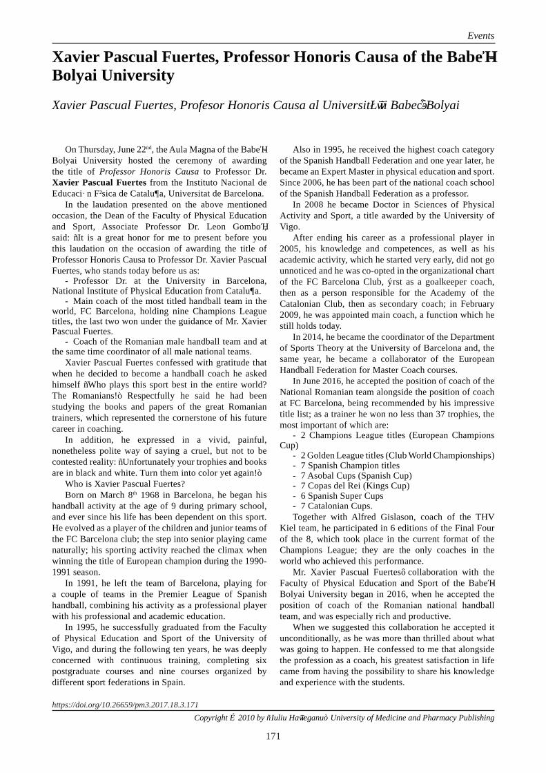
Events
171
On Thursday, June 22nd, the Aula Magna of the Babeș-Bolyai University hosted the ceremony of awarding the title of Professor Honoris Causa to Professor Dr. Xavier Pascual Fuertes from the Instituto Nacional de Educación Física de Cataluña, Universitat de Barcelona.
In the laudation presented on the above mentioned occasion, the Dean of the Faculty of Physical Education and Sport, Associate Professor Dr. Leon Gomboș, said: “It is a great honor for me to present before you this laudation on the occasion of awarding the title of Professor Honoris Causa to Professor Dr. Xavier Pascual Fuertes, who stands today before us as:
- Professor Dr. at the University in Barcelona, National Institute of Physical Education from Cataluña.
- Main coach of the most titled handball team in the world, FC Barcelona, holding nine Champions League titles, the last two won under the guidance of Mr. Xavier Pascual Fuertes.
- Coach of the Romanian male handball team and at the same time coordinator of all male national teams.
Xavier Pascual Fuertes confessed with gratitude that when he decided to become a handball coach he asked himself “Who plays this sport best in the entire world? The Romanians!” Respectfully he said he had been studying the books and papers of the great Romanian trainers, which represented the cornerstone of his future career in coaching.
In addition, he expressed in a vivid, painful, nonetheless polite way of saying a cruel, but not to be contested reality: “Unfortunately your trophies and books are in black and white. Turn them into color yet again!”
Who is Xavier Pascual Fuertes?Born on March 8th 1968 in Barcelona, he began his
handball activity at the age of 9 during primary school, and ever since his life has been dependent on this sport. He evolved as a player of the children and junior teams of the FC Barcelona club; the step into senior playing came naturally; his sporting activity reached the climax when winning the title of European champion during the 1990-1991 season.
In 1991, he left the team of Barcelona, playing for a couple of teams in the Premier League of Spanish handball, combining his activity as a professional player with his professional and academic education.
In 1995, he successfully graduated from the Faculty of Physical Education and Sport of the University of Vigo, and during the following ten years, he was deeply concerned with continuous training, completing six postgraduate courses and nine courses organized by different sport federations in Spain.
Also in 1995, he received the highest coach category of the Spanish Handball Federation and one year later, he became an Expert Master in physical education and sport. Since 2006, he has been part of the national coach school of the Spanish Handball Federation as a professor.
In 2008 he became Doctor in Sciences of Physical Activity and Sport, a title awarded by the University of Vigo.
After ending his career as a professional player in 2005, his knowledge and competences, as well as his academic activity, which he started very early, did not go unnoticed and he was co-opted in the organizational chart of the FC Barcelona Club, first as a goalkeeper coach, then as a person responsible for the Academy of the Catalonian Club, then as secondary coach; in February 2009, he was appointed main coach, a function which he still holds today.
In 2014, he became the coordinator of the Department of Sports Theory at the University of Barcelona and, the same year, he became a collaborator of the European Handball Federation for Master Coach courses.
In June 2016, he accepted the position of coach of the National Romanian team alongside the position of coach at FC Barcelona, being recommended by his impressive title list; as a trainer he won no less than 37 trophies, the most important of which are:
- 2 Champions League titles (European Champions Cup)
- 2 Golden League titles (Club World Championships)- 7 Spanish Champion titles - 7 Asobal Cups (Spanish Cup)- 7 Copas del Rei (Kings Cup)- 6 Spanish Super Cups- 7 Catalonian Cups.Together with Alfred Gislason, coach of the THV
Kiel team, he participated in 6 editions of the Final Four of the 8, which took place in the current format of the Champions League; they are the only coaches in the world who achieved this performance.
Mr. Xavier Pascual Fuertes’ collaboration with the Faculty of Physical Education and Sport of the Babeș-Bolyai University began in 2016, when he accepted the position of coach of the Romanian national handball team, and was especially rich and productive.
When we suggested this collaboration he accepted it unconditionally, as he was more than thrilled about what was going to happen. He confessed to me that alongside the profession as a coach, his greatest satisfaction in life came from having the possibility to share his knowledge and experience with the students.
Xavier Pascual Fuertes, Professor Honoris Causa of the Babeș-Bolyai University
Xavier Pascual Fuertes, Profesor Honoris Causa al Universității Babeș-Bolyai
https://doi.org/10.26659/pm3.2017.18.3.171Copyright © 2010 by “Iuliu Hațieganu” University of Medicine and Pharmacy Publishing
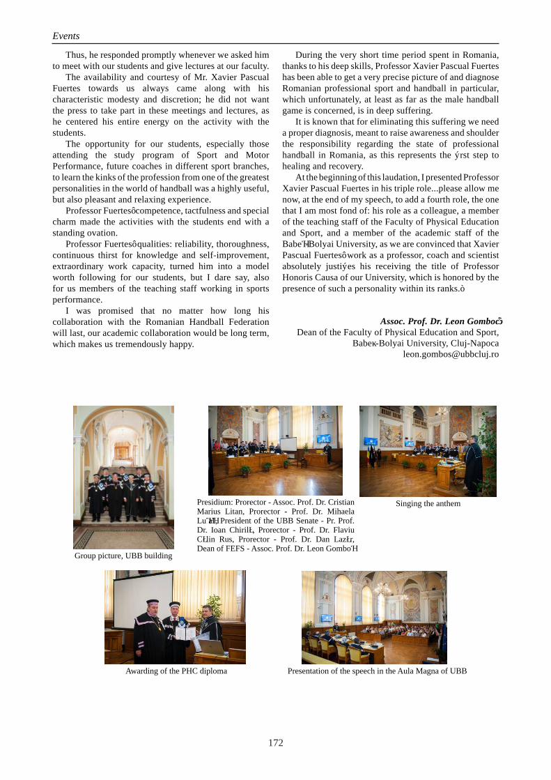
Events
172
Thus, he responded promptly whenever we asked him to meet with our students and give lectures at our faculty.
The availability and courtesy of Mr. Xavier Pascual Fuertes towards us always came along with his characteristic modesty and discretion; he did not want the press to take part in these meetings and lectures, as he centered his entire energy on the activity with the students.
The opportunity for our students, especially those attending the study program of Sport and Motor Performance, future coaches in different sport branches, to learn the kinks of the profession from one of the greatest personalities in the world of handball was a highly useful, but also pleasant and relaxing experience.
Professor Fuertes’ competence, tactfulness and special charm made the activities with the students end with a standing ovation.
Professor Fuertes’ qualities: reliability, thoroughness, continuous thirst for knowledge and self-improvement, extraordinary work capacity, turned him into a model worth following for our students, but I dare say, also for us members of the teaching staff working in sports performance.
I was promised that no matter how long his collaboration with the Romanian Handball Federation will last, our academic collaboration would be long term, which makes us tremendously happy.
During the very short time period spent in Romania, thanks to his deep skills, Professor Xavier Pascual Fuertes has been able to get a very precise picture of and diagnose Romanian professional sport and handball in particular, which unfortunately, at least as far as the male handball game is concerned, is in deep suffering.
It is known that for eliminating this suffering we need a proper diagnosis, meant to raise awareness and shoulder the responsibility regarding the state of professional handball in Romania, as this represents the first step to healing and recovery.
At the beginning of this laudation, I presented Professor Xavier Pascual Fuertes in his triple role...please allow me now, at the end of my speech, to add a fourth role, the one that I am most fond of: his role as a colleague, a member of the teaching staff of the Faculty of Physical Education and Sport, and a member of the academic staff of the Babeș-Bolyai University, as we are convinced that Xavier Pascual Fuertes’ work as a professor, coach and scientist absolutely justifies his receiving the title of Professor Honoris Causa of our University, which is honored by the presence of such a personality within its ranks.”
Assoc. Prof. Dr. Leon GomboșDean of the Faculty of Physical Education and Sport,
Babeş-Bolyai University, [email protected]
Group picture, UBB building
Presidium: Prorector - Assoc. Prof. Dr. Cristian Marius Litan, Prorector - Prof. Dr. Mihaela Luțaș, President of the UBB Senate - Pr. Prof. Dr. Ioan Chirilă, Prorector - Prof. Dr. Flaviu Călin Rus, Prorector - Prof. Dr. Dan Lazăr, Dean of FEFS - Assoc. Prof. Dr. Leon Gomboș
Singing the anthem
Awarding of the PHC diploma Presentation of the speech in the Aula Magna of UBB
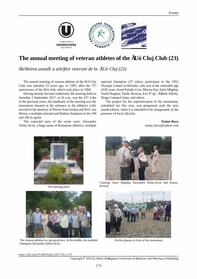
Events
173
The annual meeting of veteran athletes of the “U” Cluj Club was initiated 23 years ago, in 1995, after the 75th anniversary of the “U” club, which took place in 1994.
Having already become traditional, the meeting held on Saturday 2 September 2017, at 10 a.m., was the 23rd. Like in the previous years, the landmark of the meeting was the monument situated at the entrance to the athletics field, erected in the memory of Doctor Ioan Arnăut and Prof. Ion Moina, a multiple national and Balkan champion in the 100 and 200 m sprint.
The expected stars of the event were: Alexandra Taifas Sicoe, a huge name of Romanian athletics, multiple
national champion (27 titles), participant in the 1952 Olympic Games in Helsinki, who was at the venerable age of 85 years, Aurel Palade-Ursu, Mircea Pop, Ilarie Măgdaş, Vasile Bogdan, Vasile Sărucan, Eva Zörgö, Ráduly Károly, Draga Comşa-Crişan, and others.
The project for the superelevation of the monument, scheduled for this year, was postponed until the next year’s edition, when it is intended to be inaugurated, in the presence of local officials.
Traian [email protected]
The annual meeting of veteran athletes of the „U” Cluj Club (23)
Întâlnirea anuală a atleţilor veterani de la „U” Cluj (23)
The meeting pointChatting: Ilarie Magdaş, Alexandra Taifas-Sicoe and Emma Konrad
The veteran athletes in a group picture. In the middle, the multiple champion Alexandra Taifas-Sicoe.
On the plateau in front of the monument
https://doi.org/10.26659/pm3.2017.18.3.173Copyright © 2010 by “Iuliu Hațieganu” University of Medicine and Pharmacy Publishing
Palestrica of the third millennium – Civilization and sportVol. 18, no. 3, July-September 2017, 174‒176
174
FOR THE ATTENTION OF CONTRIBUTORS
The subject of the JournalThe journal has a multidisciplinary nature oriented toward biomedical, health, exercise, social sciences fields, applicable
in activities of physical training and sport, so that the dealt subjects and the authors belong to several disciplines in these fields. The main rubrics are: “Original studies” and “Reviews”.
Regarding “Reviews” the main subjects that are presented are: oxidative stress in physical effort; mental training; psycho-neuroendocrinology of sport effort; physical culture in the practice of the family doctor; extreme sports and risks; emotional determinatives of performance; the recovery of patients with spinal column disorders; stress syndromes and psychosomatics; olympic education, legal aspects of sport; physical effort in the elderly; psychomotricity disorders; high altitude sportive training; fitness; biomechanics of movements; EUROFIT tests and other evaluation methods of physical effort; adverse re-actions of physical effort; sport endocrinology; depression in sportsmen/women; classical and genetic drug usage; Olympic Games etc.
Among articles devoted to original studies and researches we are particularly interested in the following: the methodo-logy in physical education and sport; influence of some ions on effort capacity; psychological profiles of students regarding physical education; methodology in sport gymnastics; the selection of performance sportsmen.
Other articles approach particular subjects regarding different sports: swimming, rhythmic and artistic gymnastics, hand-ball, volleyball, basketball, athletics, ski, football, field and table tennis, wrestling, sumo.
The authors of the two rubrics are doctors, professors and educators, from universities and preuniversity education, trai-ners, scientific researchers etc.
Other rubrics of the journal are: the editorial, editorial news, reviews of the latest books in the field and others that are presented rarely (inventions and innovations, universitaria, preuniversitaria, forum, memories, competition calendar, por-traits, scientific events).
We highlight the rubric “The memory of the photographic eye”, where photos, some very rare, of sportsmen in the past and present are presented.
Articles signed by authors from the Republic of Moldova regarding the organization of sport education, variability of the cardiac rhythm, the stages of effort adaptability and articles by some authors from France, Portugal, Canada must also be mentioned.
The main objective of the journal is highlighting the results of research activities as well as the permanent and actual dissemination of information for specialists in the field. The journal assumes an important role regarding the achievement of necessary scores of the teaching staff in the university and preuniversity education as well as of doctors in the medical network (by recognizing the journal by the Romanian College of Physicians), regarding didactic and professional promotion.
Another merit of the journal is the obligatory publication of the table of contents and an English summary for all articles. Frequently articles are published in extenso in a language with international circulation (English, French).
The journal is published quarterly and the works are accepted for publication in the Romanian and English language. The journal is sent by e-mail or on a floppy disk (or CD-ROM) and printed, by mail at the address of the editorial staff. The works of contributors that are resident abroad and of Romanian authors must be mailed to the Editorial staff at the following address:
„Palestrica of the third millennium – Civilization and sport”Chief Editor: Prof. dr. Traian Bocu Contact address: [email protected] or [email protected] address: Clinicilor street no. 1 postal code 400006, Cluj-Napoca, RomâniaTelephone:0264-598575Website: www.pm3.ro
Objectives Our intention is that the journal continues to be a route to highlight the research results of its contributors, especially by
stimulating their participation in project competitions. Articles that are published in this journal are considered as part of the process of promotion in one’s university career (accreditation that is obtained after consultation with the National Council for Attestation of Universitary Titles and Diplomas).
We also intend to encourage the publication of studies and research, that include original relevant elements especially from young people. All articles must bring a minimum of personal contribution (theoretical or practical), that will be highli-ghted in the article.
In the future we propose to accomplish criteria that would allow the promotion of the journal to superior levels according international recognition.
THE STRUCTURE AND SUBMISSION OF ARTICLESThe manuscript must be prepared according to the stipulations of the International Committee of Medical Journal Editors
(http://www.icmjee.org). The number of words for the electronic format:
– 4000 words for original articles;
Copyright © 2010 by “Iuliu Haţieganu” University of Medicine and Pharmacy Publishing
175
For the attention of contributors
– 2000 words for case studies; – 5000-6000 words for review articles.
Format of the page: edited in WORD format, A4. Printed pages of the article will be numbered successively from 1 to the final page.
Font: Times New Roman, size 11 pt.; it should be edited on a full page, with diacritical marks, double spaced, respecting equal margins of 2 cm.
Illustrations:The images (graphics, photos etc.) should be numbered consecutively in the text, with arabic numbers. They should
be edited with EXCEL or SPSS programs, and sent as distinct files: „figure 1.tif”, „figure 2. jpg”, and at the editors de-manding in original also. Every graphic should have a legend, written under the image.
The tables should be numbered consecutively in the text, with roman numbers, and sent as distinct files, accompa-nied by a legend that will be put above the table.
PREPARATION OF THE ARTICLES1. Title page: – includes the title of article (maximum 45 characters), the name of authors followed by surname, work
place, mail address of the institute and mail adress and e-mail address of the first author. It will follow the name of article in the English language.
2. Summary: For original articles a summary structured like this is necessary: (Premize-Background, Obiective-Aims, Metode-Methods, Resultate-Results, Concluzii-Conclusions), in the Romanian language, of maximum 250 words, followed by 3-8 key words (if its possible from the list of established terms). All articles will have a summary in the English language. Within the summary (abstract) abbreviations, footnotes or bibliographic references should not be used.
Premises and objectives. Description of the importance of the study and explanation of premises and research objectives.Methods. Include the following aspects of the study:
Description of the basic category of the study: of orientation and applicative. Localization and the period of study. Description and size of groups, sex (gender), age and other socio-demographic variables should be given.Methods and instruments of investigation that are used.
Results. The descriptive and inferential statistical data (with specification of the used statistical tests): the differences between the initial and the final measurement, for the investigated parameters, the significance of correlation coefficients are necessary. The specification of the level of significance (the value p or the dimension of effect d) and the type of the used statistical test etc are obligatory.
Conclusions. Conclusions that have a direct link with the presented study should be given.Orientation articles and case studies should have an unstructured summary (without respecting the structure of experi-
mental articles) to a limit of 150 words.3. TextOriginal articles should include the following chapters which will not be identical with the summary titles: Introduction
(General considerations), Hypothesis, Materials and methods (including ethical and statistical informations), Results, Dis-cussing results, Conclusions and suggestions. Other type of articles, as orientation articles, case studies, Editorials, do not have an obligatory format. Excessive abbreviations are not recommended. The first abbreviation in the text is represented first in extenso, having its abbreviation in parenthesis, and thereafter the short form should be used.
Authors must undertake the responsibility for the correctness of published materials.4. BibliographyThe bibliography should include the following data:For articles from journals or other periodical publications the international Vancouver Reference Style should be used:
the name of all authors as initials and the surname, the year of publication, the title of the article in its original language, the title of the journal in its international abbreviation (italic characters), number of volume, pages.
Articles: Pop M, Albu VR, Vişan D et al. Probleme de pedagogie în sport. Educaţie Fizică şi Sport 2000; 25(4):2-8.Books: Drăgan I (coord.). Medicina sportivă, Editura Medicală, 2002, Bucureşti, 2002, 272-275.Chapters from books: Hăulică I, Bălţatu O. Fiziologia senescenţei. In: Hăulică I. (sub red.) Fiziologia umană, Ed. Medi-
cală, Bucureşti, 1996, 931-947.Starting with issue 4/2010, every article should include a minimum of 15 bibliographic references and a maximum of
100, mostly journals articles published in the last 10 years. Only a limited number of references (1-3) older than 10 years will be allowed. At least 20% of the cited resources should be from recent international literature (not older than 10 years).
Peer-review processIn the final stage all materials will be closely reviewed by at least two competent referees in the field (Professors, and
Docent doctors) so as to correspond in content and form with the requirements of an international journal. After this stage, the materials will be sent to the journal’s referees, according to their profiles. After receiving the observations from the refe-rees, the editorial staff shall inform the authors of necessary corrections and the publishing requirements of the journal. This process (from receiving the article to transmitting the observations) should last about 4 weeks. The author will be informed if the article was accepted for publication or not. If it is accepted, the period of correction by the author will follow in order to correspond to the publishing requirements.
176
For the attention of contributors
Conflict of interest The authors must mention all possible conflicts of interest including financial and other types. If you are sure that there is
no conflict of interest we ask you to mention this. The financing sources should be mentioned in your work too.
SpecificationsThe specifications must be made only linked to the people outside the study but which have had a substantial contribu-
tion, such as some statistical processing or review of the text in the English language. The authors have the responsibility to obtain the written permission from the mentioned persons with the name written within the respective chapter, in case the readers refer to the interpretation of results and conclusions of these persons. Also it should be specified if the article uses some partial results from certain projects or if these are based on master or doctoral theses sustained by the author.
Ethical criteriaThe Editors will notify authors in due time, whether their article is accepted or not or whether there is a need to modify
texts. Also the Editors reserve the right to edit articles accordingly. Papers that have been printed or sent for publication to other journals will not be accepted. All authors should send a separate letter containing a written statement proposing the article for submission, pledging to observe the ethics of citation of sources used (bibliographic references, figures, tables, questionnaires).
For original papers, according to the requirements of the Helsinki Declaration, the Amsterdam Protocol, Directive 86/609/EEC, and the regulations of the Bioethical Committees from the locations where the studies were performed, the authors must provide the following:
- the informed consent of the family, for studies in children and juniors;- the informed consent of adult subjects, patients and athletes, for their participation;- malpractice insurance certificate for doctors, for studies in human subjects;- certificate from the Bioethical Committees, for human study protocols;- certificate from the Bioethical Committees, for animal study protocols.The data will be mentioned in the paper, in the section Materials and Methods. The documents will be obtained before
the beginning of the study. Will be mentioned also the registration number of the certificate from the Bioethical Committees.Editorial submissions will be not returned to authors, whether published or not.
FOR THE ATTENTION OF THE SPONSORSRequests for advertising space should be sent to the Editors of the “Palestrica of the Third Millennium” journal, 1, Cli-
nicilor St., 400006, Cluj-Napoca, Romania. The price of an A4 full colour page of advertising for 2012 will be EUR 250 and EUR 800 for an advert in all 4 issues. The costs of publication of a logo on the cover will be determined according to its size. Payment should be made to the Romanian Medical Society of Physical Education and Sports, CIF 26198743. Banca Transilvania, Cluj branch, IBAN: RO32 BTRL 0130 1205 S623 12XX (RON).
SUBSCRIPTION COSTSThe “Palestrica of the Third Millennium” journal is printed quarterly. The subscription price is 100 EUR for institutions
abroad and 50 EUR for individual subscribers outside Romania. For Romanian institutions, the subscription price is 120 RON, and for individual subscribers the price is 100 RON. Note that distribution fees are included in the postal costs.
Payment of subscriptions should be made by bank transfer to the Romanian Medical Society of Physical Education and Sports, CIF 26198743. Banca Transilvania, Cluj branch, IBAN: RO32 BTRL 0130 1205 S623 12XX (RON), RO07 BTRL 01,304,205 S623 12XX (EUR), RO56 BTRL 01,302,205 S623 12XX (USD). SWIFT: BTRLRO 22
Please note that in 2010 a tax for each article submitted was introduced. Consequently, all authors of articles will pay the sum of 150 RON to the Romanian Medical Society of Physical Education and Sport published above. Authors who have paid the subscription fee will be exempt from this tax. Other information can be obtained online at www.pm3.ro “Instructions for Authors”, at our e-mail address [email protected] or at the postal address: 1, Clinicilor St., 400006, Cluj-Napoca, Romania, phone: +40264-598575.
INDEXING Title of the journal: Palestrica of the third millennium – Civilization and sportpISSN: 1582-1943; eISSN: 2247-7322; ISSN-L: 1582-1943 Profile: a Journal of Study and interdisciplinary researchEditor: “Iuliu Haţieganu” University of Medicine and Pharmacy of Cluj-Napoca and The Romanian Medical Society of
Physical Education and Sports in collaboration with the Cluj County School InspectorateThe level and attestation of the journal: a journal rated B+ by CNCSIS in the period 2007-2011 and certified by CMR
since 2003 Journal indexed into International Data Bases (IDB): EBSCO, Academic Search Complete, USA and Index Copernicus,
Journals Master List, Poland; DOAJ (Directory of Open Access Journals), Sweden.Year of first publication: 2000Issue: quarterlyThe table of contents, the summaries and the instructions for authors can be found on the internet page: http://www.pm3.
ro. Access to the table of contents and full text articles (in .pdf format) is free.
Palestrica Mileniului III ‒ Civilizaţie şi SportVol. 18, no. 3, Iulie-Septembrie 2017, 177‒179
177
ÎN ATENŢIA COLABORATORILOR
Tematica revisteiCa tematică, revista are un caracter multidisciplinar orientat pe domeniile biomedical, sănătate, efort fizic, ştiinţe soci-
ale, aplicate la activităţile de educaţie fizică şi sport, astfel încât subiectele tratate şi autorii aparţin mai multor specialităţi din aceste domenii. Principalele rubrici sunt: „Articole originale” şi „Articole de sinteză”.
Exemplificăm rubrica „Articole de sinteză” prin temele importante expuse: stresul oxidativ în efortul fizic; antrenamentul mintal; psihoneuroendocrinologia efortului sportiv; cultura fizică în practica medicului de familie; sporturi extreme şi riscuri; determinanţi emoţionali ai performanţei; recuperarea pacienţilor cu suferinţe ale coloanei vertebrale; sindroame de stres şi psihosomatică; educaţia olimpică, aspecte juridice ale sportului; efortul fizic la vârstnici; tulburări ale psihomotricităţii; pre-gătirea sportivă la altitudine; fitness; biomecanica mişcărilor; testele EUROFIT şi alte metode de evaluare a efortului fizic; reacţii adverse ale eforturilor; endocrinologie sportivă; depresia la sportivi; dopajul clasic şi genetic; Jocurile Olimpice etc.
Dintre articolele consacrate studiilor şi cercetărilor experimentale notăm pe cele care vizează: metodica educaţiei fizi-ce şi sportului; influenţa unor ioni asupra capacităţii de efort; profilul psihologic al studentului la educaţie fizică; metodica în gimnastica sportivă; selecţia sportivilor de performanţă.
Alte articole tratează teme particulare vizând diferite sporturi: înotul, gimnastica ritmică şi artistică, handbalul, vole-iul, baschetul, atletismul, schiul, fotbalul, tenisul de masă şi câmp, luptele libere, sumo.
Autorii celor două rubrici de mai sus sunt medici, profesori şi educatori din învăţământul universitar şi preuniversitar, antrenori, cercetători ştiinţifici etc.
Alte rubrici ale revistei sunt: editorialul, actualităţile editoriale, recenziile unor cărţi - ultimele publicate în domeniu, la care se adaugă şi altele prezentate mai rar (invenţii şi inovaţii, universitaria, preuniversitaria, forum, remember, calendar competiţional, portrete, evenimente ştiinţifice).
Subliniem rubrica “Memoria ochiului fotografic”, unde se prezintă fotografii, unele foarte rare, ale sportivilor din trecut şi prezent.
De menţionat articolele semnate de autori din Republica Moldova privind organizarea învăţământului sportiv, variabi-litatea ritmului cardiac, etapele adaptării la efort, articole ale unor autori din Franţa, Portugalia, Canada.
Scopul principal al revistei îl constituie valorificarea rezultatelor activităţilor de cercetare precum şi informarea per-manentă şi actuală a specialiştilor din domeniile amintite. Revista îşi asumă şi un rol important în îndeplinirea punctajelor necesare cadrelor didactice din învăţământul universitar şi preuniversitar precum şi medicilor din reţeaua medicală (prin recunoaşterea revistei de către Colegiul Medicilor din România), în avansarea didactică şi profesională.
Un alt merit al revistei este publicarea obligatorie a cuprinsului şi a câte unui rezumat în limba engleză, pentru toate articolele. Frecvent sunt publicate articole în extenso într-o limbă de circulaţie internaţională (engleză, franceză).
Revista este publicată trimestrial iar lucrările sunt acceptate pentru publicare în limba română şi engleză. Arti-colele vor fi redactate în format WORD (nu se acceptă articole în format PDF). Expedierea se face prin e-mail sau pe dischetă (sau CD-ROM) şi listate, prin poştă pe adresa redacţiei. Lucrările colaboratorilor rezidenţi în străinătate şi ale autorilor români trebuie expediate pe adresa redacţiei:
Revista «Palestrica Mileniului III»Redactor şef: Prof. dr. Traian Bocu Adresa de contact: [email protected] sau [email protected] poştală: Str. Clinicilor nr.1 cod 400006, Cluj-Napoca, RomâniaTelefon:0264-598575Website: www.pm3.ro
Obiective Ne propunem ca revista să continue a fi o formă de valorificare a rezultatelor activităţii de cercetare a colaboratorilor
săi, în special prin stimularea participării acestora la competiţii de proiecte. Menţionăm că articolele publicate în cadrul revistei sunt luate în considerare în procesul de promovare în cariera universitară (acreditare obţinută în urma consultării Consiliului Naţional de Atestare a Titlurilor şi Diplomelor Universitare).
Ne propunem de asemenea să încurajăm publicarea de studii şi cercetări, care să cuprindă elemente originale relevante mai ales de către tineri. Toate articolele vor trebui să aducă un minimum de contribuţie personală (teoretică sau practică), care să fie evidenţiată în cadrul articolului.
În perspectivă ne propunem îndeplinirea criteriilor care să permită promovarea revistei la niveluri superioare cu recu-noaştere internaţională.
STRUCTURA ŞI TRIMITEREA ARTICOLELORManuscrisul trebuie pregătit în acord cu prevederile Comitetului Internaţional al Editurilor Revistelor Medicale (http://
www.icmjee.org). Numărul cuvintelor pentru formatul electronic:
– 4000 cuvinte pentru articolele originale, – 2000 de cuvinte pentru studiile de caz, – 5000–6000 cuvinte pentru articolele de sinteză.
Copyright © 2010 by “Iuliu Haţieganu” University of Medicine and Pharmacy Publishing
178
În atenţia colaboratorilor
Format pagină: redactarea va fi realizată în format A4. Paginile listate ale articolului vor fi numerotate succesiv de la 1 până la pagina finală.
Font: Times New Roman, mărime 11 pt.; redactarea se va face pe pagina întreagă, cu diacritice, la două rânduri, res-pectând margini egale de 2 cm pe toate laturile.
Ilustraţiile:Figurile (grafice, fotografii etc.) vor fi numerotate consecutiv în text, cu cifre arabe. Vor fi editate cu programul
EXCEL sau SPSS, şi vor fi trimise ca fişiere separate: „figura 1.tif”, „figura 2. jpg”, iar la solicitarea redacţiei şi în original. Fiecare grafic va avea o legendă care se trece sub figura respectivă.
Tabelele vor fi numerotate consecutiv în text, cu cifre romane, şi vor fi trimise ca fişiere separate, însoţite de o legendă ce se plasează deasupra tabelului.
PREGĂTIREA ARTICOLELOR1. Pagina de titlu: – cuprinde titlul articolului (maxim 45 caractere), numele autorilor urmat de prenume, locul de
muncă, adresa postală a instituţiei, adresa poştală şi adresa e-mail a primului autor. Va fi urmat de titlul articolului în limba engleză.
2. Rezumatul: Pentru articolele experimentale este necesar un rezumat structurat (Premize-Background, Obiective-Aims, Metode-Methods, Rezultate-Results, Concluzii-Conclusions), în limba română, de maxim 250 cuvinte (20 de rânduri, font Times New Roman, font size 11), urmat de 3–5 cuvinte cheie (dacă este posibil din lista de termeni consa-craţi). Toate articolele vor avea un rezumat în limba engleză. Nu se vor folosi prescurtări, note de subsol sau referinţe.
Premize şi obiective: descrierea importanţei studiului şi precizarea premizelor şi obiectivelor cercetării.Metodele: includ următoarele aspecte ale studiului:
Descrierea categoriei de bază a studiului: de orientare sau aplicativ. Localizarea şi perioada de desfăşurare a studiului. Colaboratorii vor prezenta descrierea şi mărimea loturilor, sexul (genul), vârsta şi alte variabile socio-demografice.Metodele şi instrumentele de investigaţie folosite.
Rezultatele vor prezenta datele statistice descriptive şi inferenţiale obţinute (cu precizarea testelor statistice folosite): diferenţele dintre măsurătoarea iniţială şi cea finală, pentru parametri investigaţi, semnificaţia coeficienţilor de corelaţie. Este obligatorie precizarea nivelului de semnificaţie (valoarea p sau mărimea efectului d) şi a testului statistic folosit etc.
Concluziile care au directă legătură cu studiul prezentat.Articolele de orientare şi studiile de caz vor avea un rezumat nestructurat (fără a respecta structura articolelor experi-
mentale) în limita a 150 cuvinte (maxim 12 rânduri, font Times New Roman, font size 11).3. TextulArticolele experimentale vor cuprinde următoarele capitole: Introducere, Ipoteză, Materiale şi Metode (inclusiv infor-
maţiile etice şi statistice), Rezultate, Discutarea rezultatelor, Concluzii (şi propuneri). Celelalte tipuri de articole, cum ar fi articolele de orientare, studiile de caz, editorialele, nu au un format impus.
Răspunderea pentru corectitudinea materialelor publicate revine în întregime autorilor.4. BibliografiaBibliografia va cuprinde:Pentru articole din reviste sau alte periodice se va menţiona: numele tuturor autorilor şi iniţialele prenumelui, anul
apariţiei, titlul articolului în limba originală, titlul revistei în prescurtare internaţională (caractere italice), numărul volu-mului, paginile
Articole: Pop M, Albu VR, Vişan D et al. Probleme de pedagogie în sport. Educaţia Fizică şi Sportul 2000; 25(4):2-8.Cărţi: Drăgan I (coord.). Medicina sportivă aplicată. Ed. Editis, Bucureşti 1994, 372-375.Capitole din cărţi: Hăulică I, Bălţatu O. Fiziologia senescenţei. În: Hăulică I. (sub red.) Fiziologia umană. Ed. Medi-
cală, Bucureşti 1996, 931-947.Începând cu revista 4/2010, fiecare articol va trebui să se bazeze pe un minimum de 15 şi un maximum de 100 referinţe
bibliografice, în majoritate articole nu mai vechi de 10 ani. Sunt admise un număr limitat de cărţi şi articole de referinţă (1-3), cu o vechime mai mare de 10 ani. Un procent de 20% din referinţele bibliografice citate trebuie să menţioneze lite-ratură străină studiată, cu respectarea criteriului actualităţii acesteia (nu mai vechi de 10 ani).
Procesul de recenzare (peer-review)Într-o primă etapă toate materialele sunt revizuite riguros de cel puţin doi referenţi competenţi în domeniu respectiv
(profesori universitari doctori şi doctori docenţi) pentru ca textele să corespundă ca fond şi formă de prezentare cerinţelor unei reviste serioase. După această etapă materialele sunt expediate referenţilor revistei, în funcţie de profilul materialelor. În urma observaţiilor primite din partea referenţilor, redacţia comunică observaţiile autorilor în vederea corectării acestora şi încadrării în cerinţele de publicare impuse de revistă. Acest proces (de la primirea articolului până la transmiterea ob-servaţiilor) durează aproximativ 4 săptămâni. Cu această ocazie se comunică autorului daca articolul a fost acceptat spre publicare sau nu. În situaţia acceptării, urmează perioada de corectare a articolului de către autor în vederea încadrării în criteriile de publicare.
Conflicte de intereseSe cere autorilor să menţioneze toate posibilele conflicte de interese incluzând relaţiile financiare şi de alte tipuri. Dacă
sunteţi siguri că nu există nici un conflict de interese vă rugăm să menţionaţi acest lucru. Sursele de finanţare ar trebui să
179
În atenţia colaboratorilor
fie menţionate în lucrarea dumneavoastră.
PrecizăriPrecizările trebuie făcute doar în legătură cu persoanele din afara studiului, care au avut o contribuţie substanţială la
studiul respectiv, cum ar fi anumite prelucrări statistice sau revizuirea textului în limba engleză. Autorii au responsabilita-tea de a obţine permisiunea scrisă din partea persoanelor menţionate cu numele în cadrul acestui capitol, în caz că cititorii se referă la interpretarea rezultatelor şi concluziilor acestor persoane. De asemenea, la acest capitol se vor face precizări în cazul în care articolul valorifică rezultate parţiale din anumite proiecte sau dacă acesta se bazează pe teze de masterat sau doctorat susţinute de autor, alte precizări.
Criterii deontologiceRedacţia va răspunde în timp util autorilor privind acceptarea, neacceptarea sau necesitatea modificării textului şi îşi
rezervă dreptul de a opera modificări care vizează forma lucrărilor.Nu se acceptă lucrări care au mai fost tipărite sau trimise spre publicare la alte reviste. Autorii vor trimite redacţiei
odată cu articolul propus spre publicare, într-un fişier word separat, o declaraţie scrisă în acest sens, cu angajamentul respectării normelor deontologice referitoare la citarea surselor pentru materialele folosite (referinţe bibliografice, figuri, tabele, chestionare).
Pentru articolele originale, în conformitate cu îndeplinirea condiţiilor Declaraţiei de la Helsinki, a Protocolului de la Amsterdam, a Directivei 86/609/EEC şi a reglementărilor Comisiilor de Bioetică din locaţiile unde s-au efectuat studiile, autorii trebuie să prezinte:
- acordul informat din partea familiei, pentru studiile pe copii şi juniori;- acordul informat din partea subiecţilor adulţi, pacienţi şi sportivi, pentru participare;- adeverinţă de Malpraxis pentru medici, pentru cercetările/studiile pe subiecţi umani;- adeverinţă din partea Comisiilor de Etică, pentru protocolul de studiu pe subiecţi umani;- adeverinţă din partea Comisiilor de Bioetică, pentru protocolul de studiu pe animale.Datele vor fi menţionate în articol la secţiunea Material şi metodă. Documentele vot fi obţinute înainte de începerea
studiului. Se va menţiona şi numărul de înregistrare al adeverinţei din partea Comisiilor de Etică. Materialele trimise la redacţie nu se restituie autorilor, indiferent dacă sunt publicate sau nu.
ÎN ATENŢIA SPONSORILORSolicitările pentru spaţiile de reclamă, vor fi adresate redacţiei revistei “Palestrica Mileniului III”, Str. Clinicilor nr. 1,
cod 400006 Cluj-Napoca, România. Preţul unei pagini de reclamă full color A4 pentru anul 2012 va fi de 250 EURO pen-tru o apariţie şi 800 EURO pentru 4 apariţii. Costurile publicării unui Logo pe coperţile revistei, vor fi stabilite în funcţie de spaţiul ocupat. Plata se va face în contul Societăţii Medicale Române de Educaţie Fizică şi Sport, CIF 26198743. Banca Transilvania, sucursala Cluj Cod IBAN: RO32 BTRL 0130 1205 S623 12XX (LEI).
ÎN ATENŢIA ABONAŢILORRevista ”Palestrica Mileniului III” este tipărită trimestrial, preţul unui abonament fiind pentru străinătate de 100 Euro
pentru instituţii, şi 50 Euro individual. Pentru intern, preţul unui abonament instituţional este de 120 lei, al unui abona-ment individual de 100 lei. Menţionăm că taxele de difuzare poştală sunt incluse în costuri.
Plata abonamentelor se va face prin mandat poştal în contul Societăţii Medicale Române de Educaţie Fizică şi Sport, CIF 26198743. Banca Transilvania, sucursala Cluj Cod IBAN: RO32 BTRL 0130 1205 S623 12XX (LEI); RO07 BTRL 01304205 S623 12XX (EURO); RO56 BTRL 01302205 S623 12XX (USD). SWIFT: BTRLRO 22
Precizăm că începând cu anul 2010 a fost introdusă taxa de articol. Ca urmare, toţi autorii semnatari ai unui articol vor achita împreună suma de 150 Lei, în contul Societăţii Medicale Române de Educaţie Fizică şi Sport publicat mai sus.
Autorii care au abonament vor fi scutiţi de această taxă de articol.Alte informaţii se pot obţine online de pe www.pm3.ro „Pentru autori” sau pe adresa de mail a redacţiei palestrica@
gmail.com sau pe adresa poştală: Str. Clinicilor nr.1 cod 400006, Cluj-Napoca, România, Telefon:0264-598575.
INDEXAREA Titlul revistei: Palestrica Mileniului III – Civilizaţie şi sportpISSN: 1582-1943; eISSN: 2247-7322; ISSN-L: 1582-1943 Profil: revistă de studii şi cercetări interdisciplinareEditor: Universitatea de Medicină şi Farmacie „Iuliu Haţieganu” din Cluj-Napoca şi Societatea Medicală Română de
Educaţie Fizică şi Sport, în colaborare cu Inspectoratul Şcolar al Judeţului ClujNivelul de atestare al revistei: revistă acreditată în categoria B+ de CNCS în perioadele 2007-2011 şi atestată CMR
din anul 2003 şi în prezentRevistă indexată în Bazele de Date Internaţionale (BDI): EBSCO, Academic Search Complete, USA şi Index Coper-
nicus, Journals Master List, Polonia, DOAJ (Directory of Open Access Journals), SwedenAnul primei apariţii: 2000Periodicitate: trimestrialăCuprinsul, rezumatele şi instrucţiunile pentru autori se găsesc pe pagina de Internet: http://www.pm3.ro Accesul la
cuprins şi articole in extenso (în format .pdf) este gratuit.