Embed Size (px)
Citation preview
Original Article
INTRODUCTION
Since its development in the 1980’s, EUS hasdramatically expanded the reach of the gastrointestinalendoscope especially into the pancreaticobiliary system. Itslimitations have been overcome with the adjunctivecapabilities of EUS guided FNA and other interventional EUSprocedures. More than 2000 publications have shown EUSto be a safe and accurate method for diagnosing, staging andsampling a variety of benign and malignant lesions within theGI tract, NSCLC, mediastinal masses, celiac lymph nodes,Pancreas, Gall bladder and CBD lesions.
METHODS
All cases of endoscopic ultrasound were performedusing either the new electronic radial array or the electroniclinear array echoendoscopes manufactured by OlympusCorporation, Japan. We utilized the new EU-ME1ultrasound processor with a CLV-180 lightsource/processor.
Most diagnostic cases were conducted using the radialor linear echoendoscope and FNA cases were performedutilizing the linear echoendoscope. FNA was conducted inall cases using the Olympus EZ shot 22G needle or theWilson Cook 22/25G needle.
Most patients were given Propofol by consultantAnesthesiologists; however a few diagnostic cases wereperformed under Fortwin and Midazolam moderatesedation.
Cytopathology technicians were available onsite for eachof the cases and an average of 3-5 passes were made toobtain adequate material.
303 Apollo Medicine, Vol. 7, No. 4, December 2010
PANCREATICOBILIARY ENDOSCOPIC ULTRASOUND (EUS):CURRENT STATUS AT DELHI APOLLO HOSPITAL
Sandeep BhargavaSenior Consultant Gastroenterology, Hepatology and Endoscopic Ultrasound, Indraprastha Apollo Hospitals,
Sarita Vihar, New Delhi 110 076, India.
Objective: To evaluate the results of EUS in patients having pancreaticobiliary lesions. Methods: EUS wasperformed in 45 patients (CBD lesions in 20, pancreatic in 19 and periampullary in 6 patients using EUSequipment. Results: Final diagnosis was achieved by either diagnostic and or FNA in al patients. None hadany complications. Conclusions: Results are very similar to what has been reported from establishedcenters.
Key words: Endoscopic Ultrasound (EUS); Fine needle aspiration (FNA).
No procedure, sedation or FNA related complicationswere observed.
EUS was performed in 67 patients (March 2010 toNovember, 2010) which included Esophageal (7 patents),Gastric (4 patients), Mediastinal (3 patients), Duodenal 2patients), Colon (3 patients), CBD (20 patients), Pancreatic(19 patients), Periampullary (6 patients) and Miscellaneous(3 patients). This observational study details my initialexperience of EUS at Apollo Hospital, Delhi in pancreatico-biliary lesions. All referred patients with pancreatico-biliarylesions attending Indraprastha Apollo Hospital wereenrolled and diagnosis of different types was made basedon ultra-sonography (USG) or computed tomography(CT) or MRI/MRCP. These patients showed pancreaticmass lesions or cysts, pancreatic duct dilatation, presenceof stones or sludge in the CBD, CBD mass/stricture andperiampullary lesions.
RESULTS
Pancreas
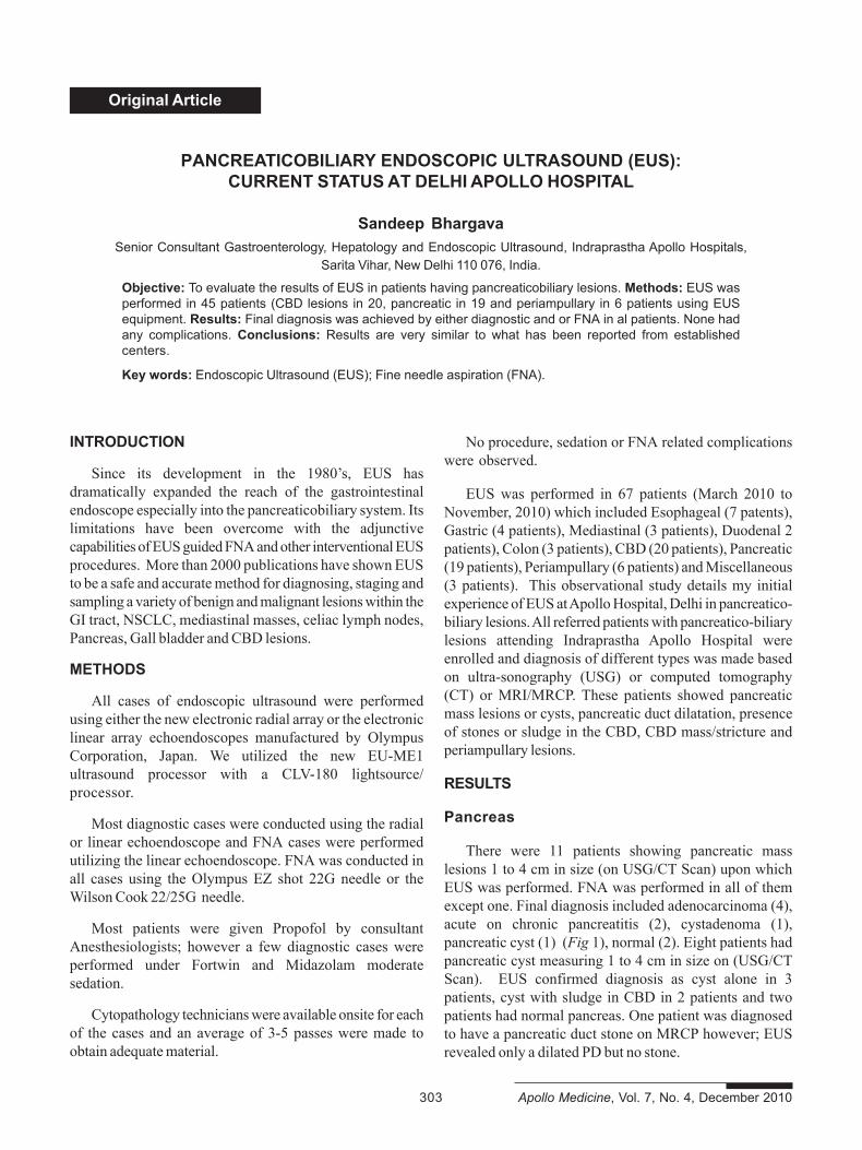
There were 11 patients showing pancreatic masslesions 1 to 4 cm in size (on USG/CT Scan) upon whichEUS was performed. FNA was performed in all of themexcept one. Final diagnosis included adenocarcinoma (4),acute on chronic pancreatitis (2), cystadenoma (1),pancreatic cyst (1) (Fig 1), normal (2). Eight patients hadpancreatic cyst measuring 1 to 4 cm in size on (USG/CTScan). EUS confirmed diagnosis as cyst alone in 3patients, cyst with sludge in CBD in 2 patients and twopatients had normal pancreas. One patient was diagnosedto have a pancreatic duct stone on MRCP however; EUSrevealed only a dilated PD but no stone.

Apollo Medicine, Vol. 7, No. 4, December 2010 304
Original Article
Fig 1. Pancreatic cyst.
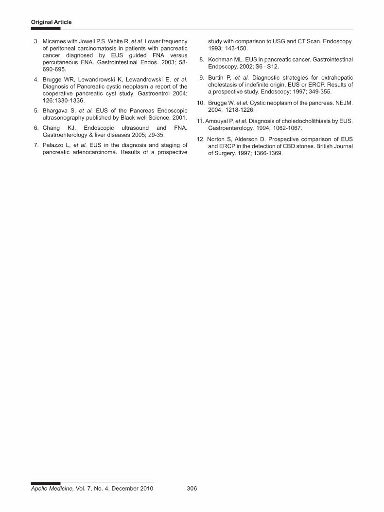
Fig 3. Common bile-duct stones.
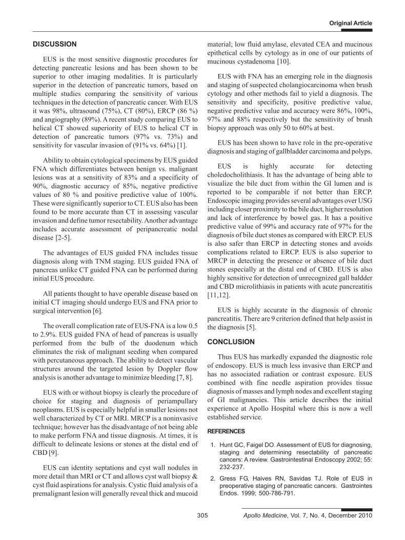
Fig 2. Periampullory lesions.(a) (b)
Fig 4. Cholelithiasis
Periampullary lesions
There were six patients in this group. The lesions weredetected on (USG, CT/MRCP). EUS with FNA revealedadenocarcinoma in 2, neuroendocrine tumor in 1,tuberculosis in 1, benign polyp in 1 and in one patient EUScould not detect any abnormality (Fig 2).
COMMON BILE DUCT LESIONS
Fifteen patients had suspected CBD stones (on USG/MRCP). EUS showed a normal CBD (9 patients). CBDstones and CBD sludge (3 patients). EUS was alsoevaluated in CBD mass/stricture in five patients. Finaldiagnosis on the basis of EUS & FNA revealedadenocarcinoma in two patients, two hadcholangiocarcinoma and 1 had CBD stone (Fig 3 & 4).
Original Article
305 Apollo Medicine, Vol. 7, No. 4, December 2010
DISCUSSION
EUS is the most sensitive diagnostic procedures fordetecting pancreatic lesions and has been shown to besuperior to other imaging modalities. It is particularlysuperior in the detection of pancreatic tumors, based onmultiple studies comparing the sensitivity of varioustechniques in the detection of pancreatic cancer. With EUSit was 98%, ultrasound (75%), CT (80%), ERCP (86 %)and angiography (89%). A recent study comparing EUS tohelical CT showed superiority of EUS to helical CT indetection of pancreatic tumors (97% vs. 73%) andsensitivity for vascular invasion of (91% vs. 64%) [1].
Ability to obtain cytological specimens by EUS guidedFNA which differentiates between benign vs. malignantlesions was at a sensitivity of 83% and a specificity of90%, diagnostic accuracy of 85%, negative predictivevalues of 80 % and positive predictive value of 100%.These were significantly superior to CT. EUS also has beenfound to be more accurate than CT in assessing vascularinvasion and define tumor resectability. Another advantageincludes accurate assessment of peripancreatic nodaldisease [2-5].
The advantages of EUS guided FNA includes tissuediagnosis along with TNM staging. EUS guided FNA ofpancreas unlike CT guided FNA can be performed duringinitial EUS procedure.
All patients thought to have operable disease based oninitial CT imaging should undergo EUS and FNA prior tosurgical intervention [6].
The overall complication rate of EUS-FNA is a low 0.5to 2.9%. EUS guided FNA of head of pancreas is usuallyperformed from the bulb of the duodenum whicheliminates the risk of malignant seeding when comparedwith percutaneous approach. The ability to detect vascularstructures around the targeted lesion by Doppler flowanalysis is another advantage to minimize bleeding [7, 8].
EUS with or without biopsy is clearly the procedure ofchoice for staging and diagnosis of periampullaryneoplasms. EUS is especially helpful in smaller lesions notwell characterized by CT or MRI. MRCP is a noninvasivetechnique; however has the disadvantage of not being ableto make perform FNA and tissue diagnosis. At times, it isdifficult to delineate lesions or stones at the distal end ofCBD [9].
EUS can identity septations and cyst wall nodules inmore detail than MRI or CT and allows cyst wall biopsy &cyst fluid aspirations for analysis. Cystic fluid analysis of apremalignant lesion will generally reveal thick and mucoid
material; low fluid amylase, elevated CEA and mucinousepithetical cells by cytology as in one of our patients ofmucinous cystadenoma [10].
EUS with FNA has an emerging role in the diagnosisand staging of suspected cholangiocarcinoma when brushcytology and other methods fail to yield a diagnosis. Thesensitivity and specificity, positive predictive value,negative predictive value and accuracy were 86%, 100%,97% and 88% respectively but the sensitivity of brushbiopsy approach was only 50 to 60% at best.
EUS has been shown to have role in the pre-operativediagnosis and staging of gallbladder carcinoma and polyps.
EUS is highly accurate for detectingcholedocholithiasis. It has the advantage of being able tovisualize the bile duct from within the GI lumen and isreported to be comparable if not better than ERCP.Endoscopic imaging provides several advantages over USGincluding closer proximity to the bile duct, higher resolutionand lack of interference by bowel gas. It has a positivepredictive value of 99% and accuracy rate of 97% for thediagnosis of bile duct stones as compared with ERCP. EUSis also safer than ERCP in detecting stones and avoidscomplications related to ERCP. EUS is also superior toMRCP in detecting the presence or absence of bile ductstones especially at the distal end of CBD. EUS is alsohighly sensitive for detection of unrecognized gall baldderand CBD microlithiasis in patients with acute pancreatitis[11,12].
EUS is highly accurate in the diagnosis of chronicpancreatitis. There are 9 criterion defined that help assist inthe diagnosis [5].
CONCLUSION
Thus EUS has markedly expanded the diagnostic roleof endoscopy. EUS is much less invasive than ERCP andhas no associated radiation or contrast exposure. EUScombined with fine needle aspiration provides tissuediagnosis of masses and lymph nodes and excellent stagingof GI malignancies. This article describes the initialexperience at Apollo Hospital where this is now a wellestablished service.
REFERENCES
1. Hunt GC, Faigel DO. Assessment of EUS for diagnosing,staging and determining resectability of pancreaticcancers: A review. Gastrointestinal Endoscopy 2002; 55:232-237.
2. Gress FG, Haives RN, Savidas TJ. Role of EUS inpreoperative staging of pancreatic cancers. GastrointesEndos. 1999; 500-786-791.
Apollo Medicine, Vol. 7, No. 4, December 2010 306
Original Article
3. Micames with Jowell P.S. White R, et al. Lower frequencyof peritoneal carcinomatosis in patients with pancreaticcancer diagnosed by EUS guided FNA versuspercutaneous FNA. Gastrointestinal Endos. 2003; 58-690-695.
4. Brugge WR, Lewandrowski K, Lewandrowski E, et al.Diagnosis of Pancreatic cystic neoplasm a report of thecooperative pancreatic cyst study. Gastroentrol 2004;126:1330-1336.
5. Bhargava S, et al. EUS of the Pancreas Endoscopicultrasonography published by Black well Science, 2001.
6. Chang KJ. Endoscopic ultrasound and FNA.Gastroenterology & liver diseases 2005; 29-35.
7. Palazzo L, et al. EUS in the diagnosis and staging ofpancreatic adenocarcinoma. Results of a prospective
study with comparison to USG and CT Scan. Endoscopy.1993; 143-150.
8. Kochman ML. EUS in pancreatic cancer. GastrointestinalEndoscopy. 2002; S6 - S12.
9. Burtin P, et al. Diagnostic strategies for extrahepaticcholestasis of indefinite origin, EUS or ERCP. Results ofa prospective study. Endoscopy: 1997; 349-355.
10. Brugge W. et al. Cystic neoplasm of the pancreas. NEJM.2004; 1218-1226.
11. Amouyal P, et al. Diagnosis of choledocholithiasis by EUS.Gastroenterology. 1994; 1062-1067.
12. Norton S, Alderson D. Prospective comparison of EUSand ERCP in the detection of CBD stones. British Journalof Surgery. 1997; 1366-1369.
![Endoscopic ultrasound-guided biopsy in chronic liver ...scopic ultrasound-guided liver biopsy (EUS-LB) is another method of acquiring liver tissue [8,9]. The feasibility of EUS-LB](https://img.pdfslide.net/doc/110x75/600c40491939a52c585d9ae9/endoscopic-ultrasound-guided-biopsy-in-chronic-liver-scopic-ultrasound-guided.jpg)


![EUS in the Management of Pancreaticobiliary ...1].4 Gress.EUS Mgmt.pdf · pancreatic cancer 120 patients with known pancreatic cancer EUS was: 98% sensitive for tumor detection (86%](https://img.pdfslide.net/doc/110x75/5f0250c77e708231d403a9a4/eus-in-the-management-of-pancreaticobiliary-14-gresseus-mgmtpdf-pancreatic.jpg)