Embed Size (px)
Citation preview
American Journal ofPathology, Vol. 150, No. 3, March 1997Copynight C) American Societyfor Investigative Pathology
Pannocytes: Distinctive Cells Found inRheumatoid Arthritis Articular Cartilage Erosions
Nathan J. Zvaifler,* Van Tsai,tSaifeddin Alsalameh,t Johannes von Kempis,*Gary S. Firestein,* and Martin Lotz*From the Department ofMedicine,* Division ofRheumatology, University of California, San Diego, andCytel Corporation,t San Diego, California; and theDepartment ofMedicine III and Institute of ClinicalImmunology and Rheumatology,* University ofErlangen-Nuremberg, Nuremberg, Germany
A distinctive ceUl was identified from sites ofrheumatoid arthritis cartilage injury. Similarcells are notfound in lesions of osteoarthritiscartilage. We have designated them as panno-cytes (PCs). Their rhomboid morphology dif-fers from the bipolar shape offibroblast-likesynoviocytes or the spherical configuration ofprimary human articular chondrocytes. Chon-drocytes are short-lived, whereas the originalPC line grew for 25 passages before becomingsenescent. Features in common with culturedprimary chondrocytes include maximal prolif-eration in response to transforming growthfactor-g3 a catabolic response to interleukin-1 1,collagenase production, and mRNA for the in-duced lymphocyte antigen and inducible nitricoxide synthase. Despite the presence of theinducible nitric oxide synthase message, PCsdo not produce NO either constitutively orwhen cytokine stimulated. Each of the mesen-chymal cells, fibroblast-like synoviocytes, pri-mary chondrocytes, and PCs have the genefortype I collagen, but the type II collagen gene isdetected only inprimary chondrocytes. PCs canbe distinguished from fibroblast-like synovio-cytes and primary chondrocytes by their mor-phology, bright VCAM-1 staining, and growthresponse to cytokines and growth factors.Theirprolonged life span in vitro suggests thatPCs might represent an earlier stage of mesen-chymal cell differentiation, and they couldhave a heretofore unrecognized role in rheu-matoid arthritis joint destruction. (Am JPathol 1997, 150:1125-1138)
Joint destruction brought about by disruption of lig-aments, tendons, articular cartilage, and bone is analmost invariable consequence of chronic rheuma-toid arthritis (RA).1,2 The responsible cells are be-lieved to have their origin in the synovial lining.These, under the influence of locally produced cyto-kines and growth factors, proliferate and express abroad array of proteinases, such as collagenase andstromelysin, that are thought to be directly responsi-ble for the tissue destruction.3 6 Simultaneously,chondrocytes digest the matrix in which they arenormally embedded and osteoclasts destroy bone.In vivo studies of the erosive, destructive granulationtissue in RA (called pannus) have usually been lim-ited to materials obtained at the time of joint replace-ment surgery and thus reflect long-standing, estab-lished lesions. Indeed, it is not absolutely certain thatthe pannus seen in conventional light and electronmicroscopy represents the cause of the observedtissue destruction or reflects a process of healing,because the lesions have many attributes of a scar.7To characterize the cellular elements in the pan-
nus, numerous studies have been performed usingenzymatically dispersed synovial membranes. Lym-phocytes and macrophages constitute approxi-mately one-third each of the isolated populations,abut a major interest has focused on what has looselybeen called synovial lining cells. A portion of thesehave the appearance of macrophages, are phago-cytic, and display surface HLA-DR antigens (type Asynoviocytes), IgG Fc receptors, and other markersassociated with cells of the monocyte lineage.9-11The remainder of the adherent cells are nonphago-cytic, fibroblast-like cells. In time, the monocytes, themacrophages, and the T lymphocytes die out with invitro culture, and only the fibroblast-like synoviocytes(FLSs) are perpetuated. These, also known as type B
Supported in part by funds from National Institutes of Health grantsAR40525, AG07996, and AR39799 and a grant to N. J. Zvaifler fromImmulogic Corporation.Accepted for publication November 4, 1996.
Address reprint requests to Dr. Nathan J. Zvaifler, University ofCalifornia, San Diego, 9500 Gilman Drive, La Jolla, CA 92093-0664.
1125
1126 Zvaifler et alAJP March 1997 Vol. 150, No. 3
synoviocytes, form the basis of many investigationson synoviocyte proliferation, proteinase production,and HLA-DR expression, both at rest or under theinfluence of cytokines and growth factors (reviewedin Ref. 2). Indeed, many investigators consider thecultured FLSs derived from digested synovial tissuesas in vitro surrogates for pannus. However, we haverecently isolated cells directly from erosions in RAcartilage at a distance from the synovial membraneor beneath the leading edge of the hyperplastic sy-
novium where it burrows into cartilage and adjacentbone. These are a homogeneous population withmorphological, phenotypic, genotypic, and func-tional characteristics that differ from chondrocytesand type B FLSs. A description of their distinctivefeatures forms the basis of this report and suggeststhat these cells, which we have called pannocytes(PCs), may have relevance to the tissue destructionobserved in RA.
Materials and Methods
Reagents
Human recombinant interleukin (IL)-1lB (specific ac-
tivity) (sp. act.), 5 x 10⁷ U/mg; purity, >95%; lipo-polysaccharide (LPS) content of the concentratestock, <0.015 ng/mg) was purchased from AmgenBiologicals (Thousand Oaks, CA). Human recombi-nant tumor necrosis factor (TNF)-a (sp. act., 5 x 10⁷U/mg; purity, >95%; <0.008 ng/mg) was providedby Genentech (South San Francisco, CA). Humanrecombinant interferon (IFN)-y (sp. act., 2 x 10⁷U/mg; purity >98%; LPS, <0.048 ng/mg) was a giftfrom Amgen Biologicals. Human recombinant IL-6(sp. act., 5 x 10⁹ U/mg; purity, >95%; LPS, <0.006ng/mg) and human recombinant transforminggrowth factor (TGF)-l31 (purity, >97%; LPS, <0.02ng/mg) were purchased from R&D Systems (Minne-apolis, MN). In each case, the final concentration ofLPS in culture after dilution of recombinant cytokineswas <0.005 ng/ml. Platelet-derived growth factor(PDGF)-a and basic fibroblast growth factor (bFGF)were obtained from R&D Systems. Actinomycin D,cycloheximide, and polymyxin B (7500 U/mg) were
purchased from Sigma Chemical Co. (St. Louis,MO). Culture medium and supplements were fromGIBCO BRL Laboratories (Grand Island, NY).
Patient Selection and Preparation of Tissues
Synovial tissues and cartilage are obtained routinelyin the Rheumatology Research Laboratory at theUniversity of California, San Diego, Medical Center
from patients undergoing joint replacement surgery.The majority are patients with osteoarthritis, but sam-ples are also obtained from patients with definite(American College of Rheumatology criteria) chronicdestructive RA. Samples from hip surgery includethe femoral head and neck with attached synovium.These are placed in petri dishes and washed threetimes with phosphate-buffered saline (PBS) to re-move blood. The pinkish-brown synovial tissue isseparated by scissors and scalpel and cut into smallpieces (1 cm3). Some are snap-frozen in 2-methylbutane and liquid nitrogen and stored at -70°C untilused for routine staining or immunohistology. Theremaining pieces are minced and digested as de-scribed below.
PCs were obtained from intact articular cartilagefrom hips and knees. The cartilages from smallerjoints came in bits and pieces. The cells were ob-tained by scraping erosive lesions in the cartilage ata distance from invading synovium into PBS in asterile petri dish. These isolated erosions were ob-served only in intact femoral heads. An alternativesite for PC isolation was from the cartilage-pannusjunction. The synovial tissue overlying erosive le-sions was removed with a scalpel blade down to theunderlying cartilage or bone. The eroded area wasscraped with a scalpel blade and the tissues ob-tained were combined with those from isolated ero-sions when available. The tissues were placed in a50-ml conical tube, spun, washed with PBS, andresuspended in a digestion solution of collagenase(type VIII, Sigma) at 0.5 mg/ml in RPMI. The digest-ing material was stirred at 370C for 90 minutes andfiltered through a 70-,tm cell strainer (Falcon, Fran-klin Lakes, NJ). The filtered cell mixture was col-lected into a 50-ml conical tube, centrifuged,washed once with 10% fetal calf serum (FCS), andplated at 100,000 cells/ml in 10% FCS in completeDulbecco's minimal essential medium (DMEM) in 24-well cell culture cluster plates (Costar, Cambridge,MA). The next day the nonadherent cells are re-moved; the adherent cells were washed and thencultured in complete DMEM/10% FCS until confluent.Thereafter, PCs and FLSs were handled identically.Six PC lines were established from RA samples,three from femoral head samples, two from knees,and one from a metatarsal head, removed at arthro-plasty. In two instances, simultaneous cultures ofPCs and FLSs were established from the cartilagelesions and synovial membrane of the same opera-tive samples. These are designated lines 2A and 2C,and RA FLS line 227 and PC line 1 (see Figures 10,5, and 8, respectively).
Pannocytes in RA Cartilage Injury 1127AJPMarch 1997, Vol. 150, No. 3
Cell Culture
Synovial cells (FLSs) were isolated by enzymaticdispersion of synovial tissues obtained from patientswith RA undergoing joint surgery as described.5Briefly, the tissues were minced and incubated with0.5 mg/ml collagenase (Sigma), in serum-free DMEMfor 2 hours at 370C, filtered through a nylon mesh,extensively washed, and cultured in DMEM supple-mented with 10% FCS (GIBCO BRL; endotoxin con-tent, <0.006 ng ml), penicillin, streptomycin, andL-glutamine (hereafter referred to as completeDMEM) in a humidified 5% CO2 atmosphere. Afterovernight culture, nonadherent cells were removedand adherent cells were cultivated in completeDMEM/10% FCS. At confluence, cells weretrypsinized, split at a 1:3 ratio, and recultured inmedium. Synoviocytes were used from passages 3through 9 in these experiments, during which timethey were a homogeneous population of FLS (<1%CD11b, <1% phagocytic, and <1% Fc-yRII recep-tor positive).
Chondrocytes were obtained at the time of au-topsy from donors without a history of joint disease.Cartilage slices removed from the femoral condylesand tibial plateau are washed in complete DMEM,minced with a scalpel, transferred into a digestionbuffer containing DMEM/5% FCS and 2 mg/ml clos-tridial collagenase type IV (Sigma), and incubatedon a gyratory shaker at 370C until the fragments weredigested (typically overnight). Residual multicellularaggregates were removed by sedimentation (1 x g)and the cells washed three times in completeDMEM/5% FCS before use in experiments. Unlessspecifically stated, only primary cultures were stud-ied.
Electron MicroscopyPCs and synoviocytes were dislodged from cultureplates with 1 mmol/L EDTA in cold Ca2+/Mg2+-freePBS. The cells were washed twice with RPMI at roomtemperature and fixed in 2% glutaraldehyde (Elec-tron Microscopy Science, Fort Washington, PA),RRMI with 20 mmol/L HEPES (GIBCO BRL), pH 7.4,for 1 hour in single-cell suspensions. The cells werewashed three times with PBS and postfixed in apellet form with 1% osmium tetroxide for 1 hour onice. The cell pellets were embedded in Spur fortransmission electron microscopy. An EM 300 (Phil-ips Electronic Instruments, Mahwah, NJ) was usedfor transmission electron microscopy analysis.
Immunoperoxidase StainingCells (PCs, FLSs, or primary chondrocytes) wereremoved from culture plates by treatment with 0.5%trypsin and 0.53 mmol/L EDTA, and washed inDMEM/5% FCS, followed by a wash of 0.1% bovineserum albumin (BSA)/PBS and cytocentrifuged (300rpm) onto poly-L-lysine-coated glass slides. The cellswere then fixed with 3.7% formaldehyde (10 min-utes), rinsed in PBS, and treated with methanol at-20°C (5 minutes) and acetone at -200C (3 min-utes). After three washes with PBS, the slides werestored at -70°C and used within 1 week. Beforestaining, the slides were left at room temperature toequilibrate and then blocked sequentially with 0.1%BSA/PBS (5 minutes), 1% horse serum/PBS (5 min-utes), and 2% human AB serum/PBS (5 minutes).The fixed and blocked cells were incubated with a 1to 5 ,ug/ml dilution of the appropriate monoclonalantibody for 60 minutes at room temperature. Con-trols were performed with an isotype-matchedmouse Ig at identical concentrations. After washingthe slides three times with PBS, biotinylated horseanti-mouse second antibody (Vector Laboratories,Burlingame, CA) was added for 30 minutes at 40C.Slides were washed three times with PBS, and en-dogenous peroxidase was depleted with 0.3% hy-drogen peroxide in PBS for 20 minutes. Cells werethen incubated with ABC horseradish peroxidasecomplex (Vector) for 30 minutes at 40C. The peroxi-dase-catalyzed reaction was developed with 0.5mg/ml diaminobenzidine and 0.02% hydrogen per-oxide for 3 to 5 minutes at room temperature. Cellswere considered positive when the specific stainingwas equal to or exceeded the controls.
FACS AnalysisFLSs or PCs (1 x 10⁵ to 2 x 10⁵) cultured in six-wellplastic dishes (Costar) in complete DMEM/10% FCSwere harvested with 1 mmol/L EDTA Ca2+/Mg2+-freePBS, washed once with cold DMEM at 40C, andstained with the corresponding monoclonal antibody(MAb) as described. 12 Phycoerythrin-conjugatedFab2 anti-mouse IgG (Tago, Burlingame, CA) wasused as a secondary antibody instead of a fluores-cein isothiocyanate conjugate to avoid problemswith the green autofluorescence exhibited by syno-viocytes. Pooled mouse IgG (Cappel Laboratories,Malvern, PA) or an isotype-matched MAb was usedas negative control. A total of 5000 cells were ana-lyzed from each sample adjusting the fluorescencegain so that the mean fluorescence channel of cellsstained with the negative control was 40 to 50. The
1128 Zvaifler et alAJP March 1997, Vol. 150, No. 3
cutoff for the positive cells was arbitrarily establishedin the channel that would include 98% of controlcells. The results are displayed in a logarithmic scaleof increasing fluorescence and presented as linearmean fluorescence.
Cell GrowthPCs and FLSs were removed from culture and re-suspended at 1 x 10⁴/ml in complete DMEM/10%FCS and incubated overnight in 24-well Falconplates at 1 ml/well. The supernatants of these cul-tures were then removed and the cells exposed tomedium without FCS for 24 hours. Thereafter thecells were cultured for 5 days in complete DMEM/10% FCS in the presence or absence of 5 ng/mlIL-1,B, 100 ng/ml TNF-a, or both. Each condition wasperformed in triplicate. On day 5, the cells wereremoved from each well with cold trypsin/EDTA,washed once, resuspended in 1 ml of DMEM, andcounted with a Coulter counter. Each sample wascounted twice. In a few experiments cells were enu-merated with both a hemocytometer and Coultercounter with good concordance.
ProliferationCells (FLSs, PCs, or chondrocytes) were distributedinto 96-well plates (1 x 10⁴/well) in a total volume of200 ,tl of complete DMEM/10% FCS. The cells wereallowed to adhere for 24 to 48 hours and thenwashed twice with DMEM. After the final wash, 200,tl of complete DMEM was left in the well for a 24-hour starvation period. At this point, the medium wasreplaced with 200 Al of cytokines or growth factors incomplete DMEM/1% FCS and cultured for an addi-tional 72 hours, and 16 hours before harvesting,[3H]thymidine ([3H]TdR; 1 ,uCi/well) was added andthe cells harvested on a Cambridge Technologiescell harvester. Total radioactivity was determined byliquid scintillation counting. All experiments wereperformed in triplicate.
Jurkat BindingConfluent FLS or PC monolayers were grown in 96-well plates. The T cell lymphoblastoid line Jurkat waslabeled with 51Cr (50 ,uCi/108 cells) for 2 hours andthen washed sequentially with RPMI 1640 plus 10%FCS, PBS plus 1 mmol/L EDTA, and RPMI 1640 plus1% BSA. 51Cr-Labeled Jurkat cells were resus-pended in RPMI plus 1% BSA and 5 x 10⁴ cells/welladded to FLS or PC monolayers that had been
washed with RPMI 1640 plus 1% BSA. For inhibitionexperiments, either labeled Jurkat cells or adherentFLSs or PCs were preincubated for 20 minutes at4°C with MAbs to adhesion molecules: LB3, 1 (con-trol anti-MHC class 11) or P4H9 (anti-f32), obtainedfrom T. Springer, Harvard Medical School (Boston,MA) or P3H12 (anti-VCAM-1). Jurkat cell adhesion toFLSs or PCs was allowed to proceed for 15 minutesat 37°C followed by the removal of unbound cells bywashing three times with RPMI plus 1% BSA. BoundJurkat cells were lysed with 0.1% sodium dodecylsulfate, and radioactivity in cell lysates was analyzedin a gamma counter.
Collagenase Production
PCs were cultured to near confluence in 6-well plateswith 1 ml of 1% FCS medium with or without IL-113 (1ng/ml) or TNF-a (100 ng/ml) and incubated at 37°Cin a humidified 5% CO2 atmosphere. Supernatantswere harvested after 72 hours and assayed by asolid phase enzyme-linked immunosorbent assay(ELISA) for collagenase employing antibodies pro-vided by Dr. David Taylor. The ELISA consists of arabbit capture antibody, sheep detection antibody,and signal amplification by biotin-avidin complex for-mation. The assay can detect as little as 1 ng/ml andwas performed as described.13 Briefly, a 96-wellplate was coated overnight at 4°C with 200 ,ul ofrabbit anti-proMMP-1 IgG in PBS followed by over-night blocking at 40C with 0 to 5% (w/v) BSA inPBS/0.05% Tween 20 (BSA/PBS/Tween). After thewells were emptied and washed in PBS/Tween, ei-ther 100 ,ul of pro matrix metalloproteinase-1(MMP-1) standard (0.4 to 25 ng/ml) in a 1:21 dilutionof horse serum in BSA/PBS/Tween or sample dilutedby 1:21 with BSA/PBS/Tween was added, followedby 100 Al of biotinylated anti-human proMMP-1 IgGin a 1:25 dilution of normal sheep serum in 300mmol/l NaCI, 5 mmol/l CaCI2, 20 mmol/l Tris, pH 7.5.After overnight incubation at 40C, each well waswashed three times in PBS/Tween and once in Tris-buffered saline, and then 200 ,ul of avidin-biotin com-plex (Dako) in Tris-buffered saline was added for 30minutes at room temperature. After three additionalwashings in Tris-buffered saline and one in 0.5mmol/l MgCI2 and 0.1 mol/l diethanolamine, pH 9.6,200 Al of 1 mg/ml p-nitrophenyl phosphate (Sigma)was added to each well and, after color develop-ment, absorbance was read at 405 nm at approxi-mately 1 hour.
Pannocytes in RA Cartilage Injury 1129A/P Marcb 1997, Vol. 150, No. 3
Nitric Oxide (NO) ProductionFor studies on NO production, chondrocytes (prima-ry culture), PCs, or FLSs are plated in 96-well platesat 50,000 cells per well. The cells were cultured inserum-free DMEM supplemented with L-glutamineand antibiotics for 24 hours in the presence or ab-sence of cytokines (IL-1,B and TNF-a) or LPS. Themedium was replaced, and 48 hours later, the con-centration of nitrites, the stable end products of cel-lular NO breakdown, in the conditioned media wasdetermined by the Griess reaction using NaNO2 asstandard.14 Briefly, 50 ptl of conditioned medium wasmixed with 50 ,ul of Griess reagent (50 ml of 1%sulfanilamide, 0.1% N-(1 -naphthyl)ethylene-diaminedihydrochloride in 25% H3PO4) and incubated atroom temperature for 5 minutes, and absorbancewas read at 550 nm using a Molecular DevicesELISA plate reader. Each pooled sample was as-sayed in triplicate. All results are expressed asnmoles of nitrates per 105 cells.
RNA IsolationRNA isolation was performed as described.15 Briefly,total RNA was isolated by a single-step guanidiniumthiocyanate-phenol-chloroform method from 500,000cells. Cells were lysed directly in the flasks usingRNA Stat-60 (Tel-Test B, Friendswood, TX) and thesamples were processed following the manufactur-er's protocol. Poly A+ RNA was obtained with thePolyATract mRNA isolation system (Promega, Mad-ison, WI).
Reverse TranscriptionTotal RNA (up to 5 ,ug) was reverse transcribed in a20-,ul volume containing 4 Al of 5X reverse transcrip-tion) buffer (GIBCO BRL), 10 mmol/L dithiothreitol,500 mmol/L dNTPs, 1 Al of random hexanucleotides,200 U of mouse Moloney leukemia virus reversetranscriptase (GIBCO BRL) and 20 U of RNasin (Pro-mega) for 30 to 120 min at 370C.
Polymerase Chain Reaction (PCR) for ILAPCR was performed with 2 Al of the reverse tran-scription reaction product in a 25- or 50-,ul volumewith 1 U of Taq DNA polymerase (Perkin Elmer Ce-tus, Emeryville, CA), 140 mmol/L dNTPs, and 10pmol/L each primer in 1OX incubation buffer (100mmol/L Tris/HCI, 15 mmol/L MgCI, 500 mmol/L KCI,1 mg/ml gelatin, pH 8.3, at 200C), 5 U/lul on anEricomp (San Diego, CA) or a Hybaid-Omnigenethermocycler. On the Ericomp cycler, after a 5-
minute denaturation step at 94°C, the reactionproceeded in 35 cycles of 30 seconds at 94°C, 30seconds at 550C, and 75 seconds at 720C, followedfinally by 1 cycle of 7 minutes at 72°C. On the Om-nigene cycler, a 1-minute denaturation step at 940Cwas followed by 35 cycles of 5 seconds at 94°C, 20seconds at 550C, and 50 seconds at 720C with onefinal cycle of 7 minutes at 720C.
The following PCR primers were used: includedlymphocyte antigen (ILA; 486-bp product) sense,CAT TCC CGG GTC CTT GTA GTA AC (nucleotides69 to 91), and antisense, CGG TGA TCA TCC TGGCTC TCT CGC AGG GGC (nucleotides 55 to 527);GAPDH (190-bp product) sense, TGG TAT CGTGGA AGG ACT CAT GAC, and antisense, ATG CCAGTG AGC TTC CCG TTC AGC.PCR for GAPDH was performed on the Omnigene
thermocycler using the following program: 1 cycle of2 minutes at 920C and 1 minute at 60°C, 25 cycles of10 seconds at 720C, 5 seconds at 920C, and 10seconds at 600C, and 1 cycle of 5 minutes at 720C.
iNOS PCRiNOS PCR was performed as described.16 A 2-,ulvolume of cDNA from the total of 20 gi of reversetranscription product in 20 p.l of PCR reaction wasmixed with 1 U of Taq DNA polymerase (BoehringerMannheim, Indianapolis, IN), 4 mmol/L dNTPs, 1.5mmol/L MgCI2, 10 mmol/L Tris, pH 8.3, 50 mmol/LKCI, and 20 pmol/L each primer. The following am-plification protocol was used: 25 cycles for 30 sec-onds at 940C, 30 seconds at 600C, and 60 secondsat 720C. The primer sequence of the sense strandwas nucleotides 2215 to 2147, ACA TTG ATG AGAAGC TGT CCC AC, and that of the antisense wasnucleotides 2360 to 2340, CAA AGG CTG TGA GTCCTG CAC. These primers resulted in the amplifica-tion of a 236-bp PCR product. Primers for iNOS andILA span an intron. Genomic DNA thus does notcontribute to the PCR signals obtained from cDNA.For the three genes examined here, 25 cycles wereused for GAPDH, 25 to 30 cycles for iNOS, and 35cycles for ILA. These cycle numbers were deter-mined in experiments in which chondrocyte cDNAswere amplified for varying cycle numbers. This cali-bration showed a linear range of amplification for theindicated cycle numbers.
Collagen PCRPoly A+-enriched RNA was prepared from culturedcells (at least 106 cells) according to a modifiedprotocol of Chomczynski et al.17 A 2-,ug amount of
1130 Zvaifler et alAJPMarch 1997, Vol. 150, No. 3
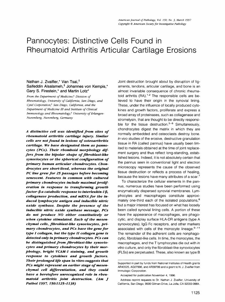
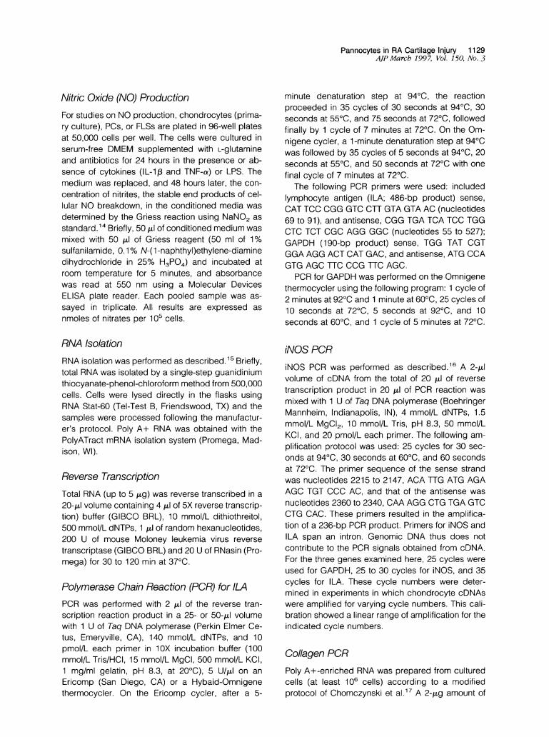
Figure 1. Phase-contrast microscopic appearance offibroblast-like synoviocytes and PCs in suibconfluent cuilture. A: RA synovialfibroblasts (passage5) have a thin bipolar configuration with occasional long processes, as compared with B. B: PC line 1 (passage 4) shows more homogeneous,rhomboid-appearing cells. The nuicleuis is small and hypochromic uith prominent nucleoli, often two to three in nuimber. Magnification, X-350.
RNA was used in the production of cDNA andoligo-dT was used to initiate the synthesis. Super-script reverse transcriptase (400 U; Promega) wasused because this enzyme has been engineered toeliminate RNAse H activity, thus resulting in greaterfull-length cDNA synthesis. A 1/10 volume (2 ,ul) ofthe cDNA product and 1 U of Taq DNA polymerase(Boehringer Mannheim) were used for the PCR.Thirty PCR cycles (1 minute at 950C, 1 minute at600C, and 1 minute at 720C) were run followed byextension for 10 minutes, and the amplification prod-ucts were visualized after electrophoresis on 1.4%agarose gel under ultraviolet illumination in the pres-ence of ethidium bromide.
The following primers were provided by Daniel Cohn(University of California, Los Angeles) (UCLA): colla-gen 1 primers, sense primer, CGAGAAAGGATC-CCCTGGTGCTGAT; antisense primer, ]TTACCG-GTCTCACCACGGTGA; PCR product size, 327 bp;collagen 2 primers, sense primer, GATGGACAGCCT-GGGGCCAA; antisense primer, GGACCTGGTGGAC-CTTCGGC; PCR product size, 395 bp.
Positive control PCR primers were those that am-plify a 450-bp segment of the glyceraldehyde3-phosphate dehydrogenase (G3PDH) gene.
StatisticsAnalysis was performed using the Student's t-test.Data are presented as mean + SEM.
Results
Morphology of Pannocytes and Fibroblast-Like SynoviocytesRA synovial tissues and cartilage were obtained atthe time of joint replacement surgery. The synovial
tissues were digested with collagenase, filtered, ex-tensively washed, and then cultured on sterile plasticin medium supplemented with 10% FCS. Nonadher-ing cells were removed and the adherent cells con-tinuously cultivated as FLSs. Tissue obtained fromeither the cartilage pannus junction or cells removeddirectly from a cartilage erosion at a distance fromthe synovium were treated in an identical manner tosynovial tissue. Cells obtained from these sites here-after are referred to as PCs. Six RA cartilage speci-mens were examined for PCs: three hips, two knees,and one metatarsal head. All yielded similar appear-ing cells. Despite extensive scraping of eroded ar-eas of five femoral heads removed for advancedosteoarthritis, similar cells were not obtained.
Six PC lines were established and maintained forvarious periods of time in long-term culture. One,PC-1, was grown continuously through 25 passagesbefore it became senescent; the others were storedafter various passages in liquid nitrogen for subse-quent study. In contrast to FLSs, which display a thinbipolar configuration with occasional long processes(Figure 1A), PCs have a more homogeneous rhom-boid appearance with hypochromic nuclei and twoor three prominent nucleoli (Figure 1B). Both PCsand FLSs exhibit contact inhibition and retain theirmorphology in confluent cultures. Cytospun Giemsa-stained confluent FLSs were heterogeneous in sizeand larger (12.9 + 4.2 ,m) than the more homoge-neous PCs (8.9 + 0.3 ,um; n = 10 fields; P < 0.006).Both the appearance and surface phenotype (seebelow) of PCs were retained through multiple (7 to15) passages.
Electron Microscopic AppearanceCells from a multipassaged FLS and the PC-1 linewere harvested, washed, pelleted by centrifugation,
Pannocytes in RA Cartilage Injury 1131AJP March 199 7, Vol. 150, No. 3
Pq
.I1
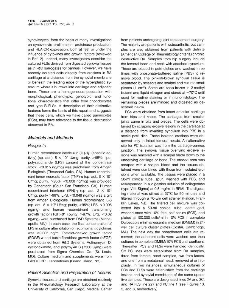
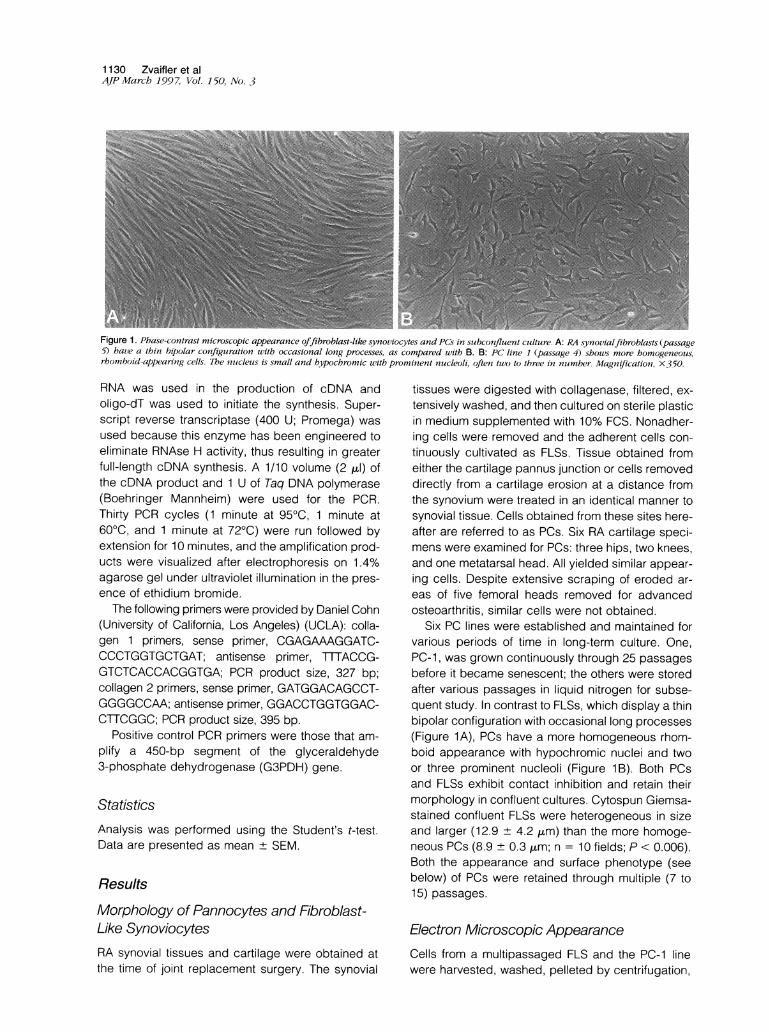
Figure 2. A: Low magnification transmission electron micrograph ofafibroblast-like synoviocyte culturedfrom RA synovium. The membrane isfoldedinto multiple well developedfilopodia ( F). The cytoplasm contains numerous organelles. There is a marked increase in rough endoplasmic reticulumwith dilated cisternae (C). Lysosomal bodies ( L) containing whorled membranes and electron-dense particles are a regularfeature. Magnification,X 7500. B: Low magnification transmission electron microscopic appearance ofa PC. PC line 1 (passage 4) has a smooth surface membrane withoutfilopodia and does not show the numerous cytoplasmic organellesfound in FLSs. The cytoplasm contains many lysosomes and residual bodies (seeinset; magnification X20,000). Magnification, x 7500.
and fixed for transmission electron microscopy. Theultrastructure of FLS prepared as single cell suspen-
sions looks similar to earlier descriptions of culturedsynovial fibroblasts and type B-fibroblast-like syno-
viocytes in tissue. 7,18 The FLSs had an irregularsurface with multiple, well developed filopodia (Fig-ure 2A). Their cytoplasm contains numerous or-
ganelles and a marked increase in endoplasmic re-
ticulum, and their cisternae are invariably dilated (notshown). The ultrastructural appearance of PCs was
different from FLSs. They had smooth surface mem-branes, lacked the numerous cytoplasmic or-
ganelles found in FLSs, and showed a lower nucleusto cytoplasm ratio. The nucleus was often irregularand easily distinguished. The cytoplasm of PCs isfilled with residual bodies (Figure 2B). Both cell typeshave lysosomal bodies that contain whorled mem-
branes and electron-dense particles.
Surface Phenotype of PCs
The constitutive expression of surface antigens on
PCs was determined on cytospun samples by immu-nohistology. PCs did not show surface antigens ofhematopoietic stem cells (CD34 and CD45), mono-
cytes (CD1lb and CD14), T lymphocytes (CD3), or
activated endothelial cells (E-selectin). PCs andFLSs were examined by FACS. They did not stainwith monoclonal antibodies to von Willebrand factor(factor VIII) or CD31 (PECAM); both were IgG1 anti-bodies and were compared with an isotype-specificmouse Ig (data not shown). Isolated PCs did notconsitutively express HLA-DR when analyzed byFACS, but class 11 antigens could be induced with a
72-hour exposure to 100 U of IFN-,y, similar to thatobserved with FLSs (data not shown).5 Granulocyte/macrophage colony-stimulating factor treatment hadno effect on HLA-DR expression by PCs. NeitherFLSs nor PCs showed the distinctive actin array thathas been described in myofibroblasts, a late-stagefibroblast thought to be responsible for wound con-
tracture and identified by large amounts of intracel-lular actin.19,20PCs constitutively express both ICAM-1 (CD54)
and VCAM-1. The latter is particularly conspicuouson PCs and present in significantly greater amountsthan seen on FLSs. Four separate lines of PCs were
64.75% to 80.3% VCAM-1 positive with linear meanfluorescence ranging from 16.9 to 27.9 (21.5 +
2.43). For comparison, thirteen different FLSs were
23.53 ± 4.2% VCAM-1 positive, as previously re-
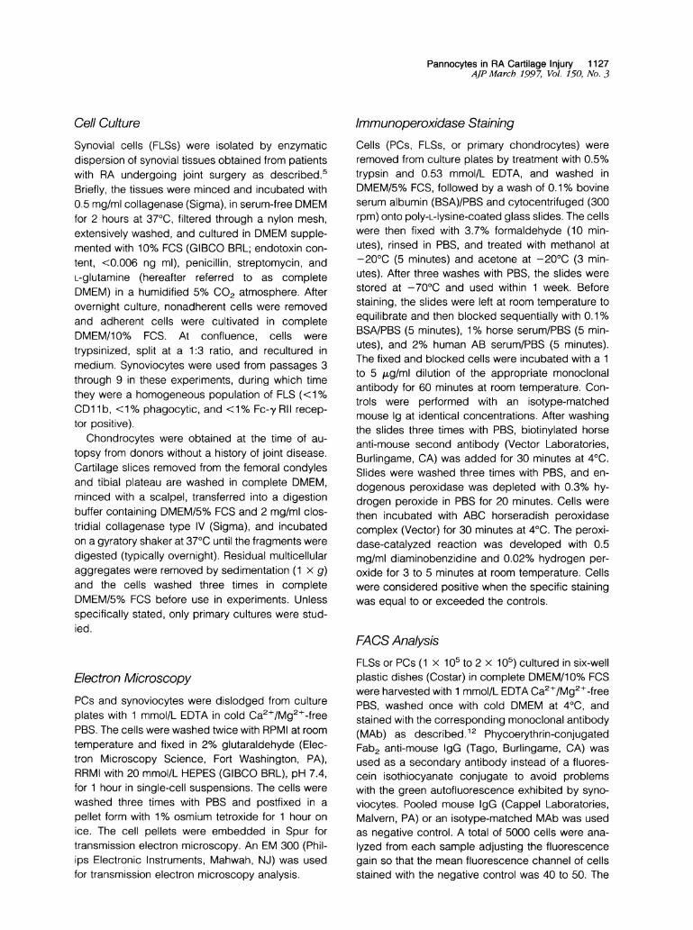
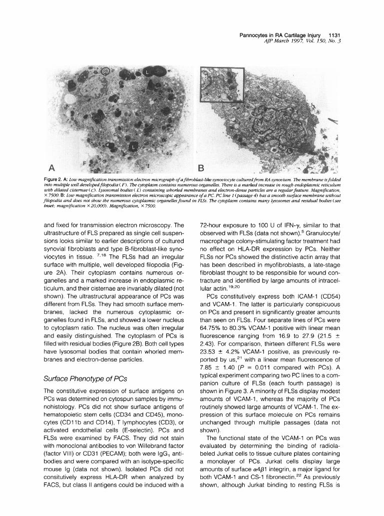
ported by us,21 with a linear mean fluorescence of7.85 ± 1.40 (P = 0.011 compared with PCs). Atypical experiment comparing two PC lines to a com-panion culture of FLSs (each fourth passage) isshown in Figure 3. A minority of FLSs display modestamounts of VCAM-1, whereas the majority of PCsroutinely showed large amounts of VCAM-1. The ex-
pression of this surface molecule on PCs remainsunchanged through multiple passages (data notshown).The functional state of the VCAM-1 on PCs was
evaluated by determining the binding of radiola-beled Jurkat cells to tissue culture plates containinga monolayer of PCs. Jurkat cells display largeamounts of surface a4f31 integrin, a major ligand forboth VCAM-1 and CS-1 fibronectin.22 As previouslyshown, although Jurkat binding to resting FLSs is
A
1132 Zvaifler et alAJPMarch 1997, Vol. 150, No. 3
#Qn _uV
CT FLS
PANNOCTEY1 PANNOCTE 3
Figure 3. FACS analysis. A comparison of VCAM-1 surface expressionon FLSs and PCs. The outlined histograms showv resting unstimulatedcells; the shaded areas are stained with either an irrelevant, isotype-matched IgG control (upper left) or the anti-VCAM-1 antibody(P3H1H2) on FLSs (upper right) and PC line 1 anzd PC lin7e 3 (lower left:and right, respectively). Cell nuimbers are displayed on the vertical axisand fluorescence intensity on a logarithmic scale on the horizonitalaxis.
mediated by a4 integrin, VCAM-1 plays only a minorrole (presumably binding is mediated through CS-1).In contrast, the VCAM-1 on resting PCs is responsi-ble for approximately one-half of the Jurkat cell bind-ing (see Figure 4).
Cell Growth
Chondrocytes from middle-aged adults seldompropagate beyond 6 to 8 passages in tissue culture.By this time they have assumed the appearance offibroblasts and enter senescence.2324 FLSs can beretained in culture through multiple passages, butafter 10 to 12 passages, even when supplementedwith growth factors, their doubling time gets progres-
FLS (OA) FLS (RA) Pannocyte
Figure 4. A representative experiment shoo,ing the effect of an anti-VCAM- I monoclotnal antibody (P13H12) on adhesion ofJurkat cells tomonolavers of FLSsfrom osteoarthritis (OA), (RA), or PC line 2 (pas-sage 5). Antibodies were used to block adherenice, as described inMaterials and Methods. The percentage of cells bound are shown on
the x axis. The control inicludes medium and an unrelated monoclonalIgG antibody to MHC class II (LB3. 1) or an anti-,82 integrin (P4H9).P < 0.05 compared with IgG control.
"it0C)x
a).0Ez
0
20
10
A
* Medium8 IL-1[11 TNFo IL-1 + TNF
I--
T
I[ I I II
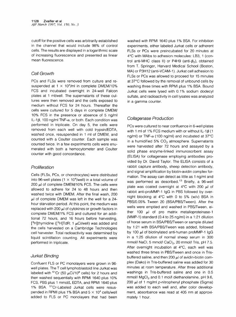
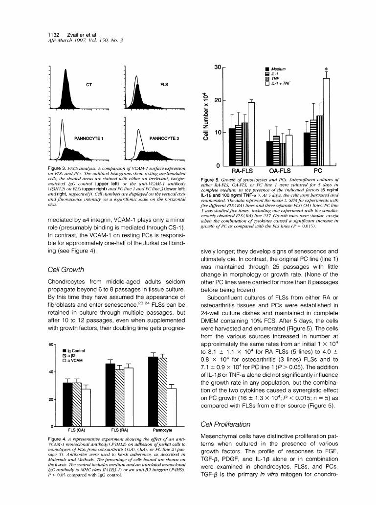
RA-FLS OA-FLS PCFigure 5. Growth of synoviocytes and PCs. Subconfluent cultures ofeither R4-FLS, OA-FLS, or PC line 1 wvere cultured for 5 days incomplete medium in the presence of the indicated factors (5 ng/mlIL-1,i and 100 ng/ml TNF-a ). At 5 days, the cells were harvested andenumerated. The data represent the mean ± SEMfor experiments uithfive different FLS (RA) lines and three separate FLS ( OA) lines. PC line1 was sttudiedfive times, including one experiment uith the simulta-neously obtained FLS (RA) line 227. Grouth rates wvere similar, exceptwheni the combination of cytokines caused a significanlt increase ingrowth ofPC as conmpared with the FLS lines (P = 0.015).
sively longer; they develop signs of senescence andultimately die. In contrast, the original PC line (line 1)was maintained through 25 passages with littlechange in morphology or growth rate. (None of theother PC lines were carried for more than 8 passagesbefore being frozen).
Subconfluent cultures of FLSs from either RA orosteoarthritis tissues and PCs were established in24-well culture dishes and maintained in completeDMEM containing 10% FCS. After 5 days, the cellswere harvested and enumerated (Figure 5). The cellsfrom the various sources increased in number atapproximately the same rates from an initial 1 x 10⁴to 8.1 ± 1.1 x 10for RA FLSs (5 lines) to 4.0±0.8 x 10⁴ for osteoarthritis (3 lines) FLSs and to7.1 + 0.9 x 10⁴ for PC line 1 (P > 0.05). The additionof IL-1 3 or TNF-a alone did not significantly influencethe growth rate in any population, but the combina-tion of the two cytokines caused a synergistic effecton PC growth (16 ± 1.3 x 10⁴; P < 0.015; n = 5) ascompared with FLSs from either source (Figure 5).
Cell ProliferationMesenchymal cells have distinctive proliferation pat-terns when cultured in the presence of variousgrowth factors. The profile of responses to FGF,TGF-13, PDGF, and IL-1,B alone or in combinationwere examined in chondrocytes, FLSs, and PCs.TGF-03 is the primary in vitro mitogen for chondro-
L- L-sss. s -mmbooI
r
_r-
Pannocytes in RA Cartilage Injury 1133AJP March 1997, Vol. 150, No. 3
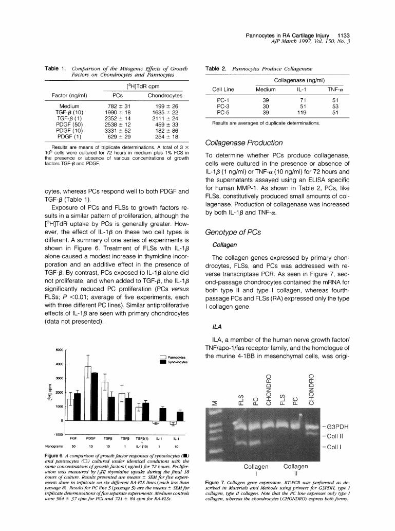
Table 1. Comparson of the Mitogenic Effects of GrowthFactors on Chondrocytes and Pannocytes
Factor (ng/ml) PCs
[3H]TdR cpm
Chondrocytes
Medium 782 ± 31 199 26TGF-p (10) 1990 18 1635 22TGF-, (1) 2352 14 2111 24PDGF (50) 2538 + 12 459 ± 33PDGF (10) 3331 + 52 182 + 86PDGF (1) 629 29 254 18
Results are means of triplicate determinations. A total of 3 x10⁵ cells were cultured for 72 hours in medium plus 1% FCS inthe presence or absence of various concentrations of growthfactors TGF-,B and PDGF.
cytes, whereas PCs respond well to both PDGF andTGF-f3 (Table 1).
Exposure of PCs and FLSs to growth factors re-sults in a similar pattern of proliferation, although the[3H]TdR uptake by PCs is generally greater. How-ever, the effect of IL-1,8 on these two cell types isdifferent. A summary of one series of experiments isshown in Figure 6. Treatment of FLSs with IL-1,3alone caused a modest increase in thymidine incor-poration and an additive effect in the presence ofTGF-,B. By contrast, PCs exposed to IL-113 alone didnot proliferate, and when added to TGF-3, the IL-1Bsignificantly reduced PC proliferation (PCs versusFLSs; P <0.01; average of five experiments, eachwith three different PC lines). Similar antiproliferativeeffects of IL-1B are seen with primary chondrocytes(data not presented).
Table 2. Pannocytes Produce Collagenase
Cell Line
PC-1PC-3PC-5
Mediur
393039
Collagenase (ng/ml)n IL-1
7151119
TNF-a
515351
Results are averages of duplicate determinations.
Collagenase ProductionTo determine whether PCs produce collagenase,cells were cultured in the presence or absence ofIL-1,B (1 ng/ml) or TNF-a (10 ng/ml) for 72 hours andthe supernatants assayed using an ELISA specificfor human MMP-1. As shown in Table 2, PCs, likeFLSs, constitutively produced small amounts of col-lagenase. Production of collagenase was increasedby both IL-1: and TNF-a.
Genotype of PCsCollagen
The collagen genes expressed by primary chon-drocytes, FLSs, and PCs was addressed with re-verse transcriptase PCR. As seen in Figure 7, sec-ond-passage chondrocytes contained the mRNA forboth type 11 and type collagen, whereas fourth-passage PCs and FLSs (RA) expressed only the typecollagen gene.
ILA
5000
_ Pannocytes4000 Synoviocytes
3000
E0.o2000
1000
0
.1000FGF PDGF TGFO TGFI3 TGF(1) IL-1 IL-1
Nanograms 50 10 10 1 IL-1(10) 1 10
Figure 6. A comparison ofgrowtbfactor responses ofsynoviocytes (U)and pannocytes (t) cultured under identical conditions with thesame concentrations ofgrowth factors ( ng/ml) for 72 hours. Prolifer-ation was measured by [JH] thymidine uptake during the final 18hours of culture. Results presented are means + SEMforfive experi-ments done in triplicate on six different RA-FLS lines (each less thanpassage 8). ResultsJbr PC line 5 (passage 5) are the means ± SEMfortriplicate determinations offive separate experiments. Medium controlswere 564 + 3 7 cpm for PCs and 721 ± 84 cpm for RA-FLSs.
ILA, a member of the human nerve growth factor/TNF/apo-1/fas receptor family, and the homologue ofthe murine 4-1 BB in mesenchymal cells, was origi-
0 0a: a:z z
CO t] I (n CN
- G3PDH-Coll 11
-Coll I
Collagen CollagenI 11
Figure 7. Collagen gene expression. RT-PCR was performed as de-scribed in Materials and Methods using primers for G3PDH, type Icollagen, type II collagen. Note that the PC line expresses only type Icollagen, whereas the chondrocytes (CHONDRO) express both forms.
1134 Zvaifler et alAJP March 1997, Vol. 150, No. 3
CHOZ LL
M'- F
SYN
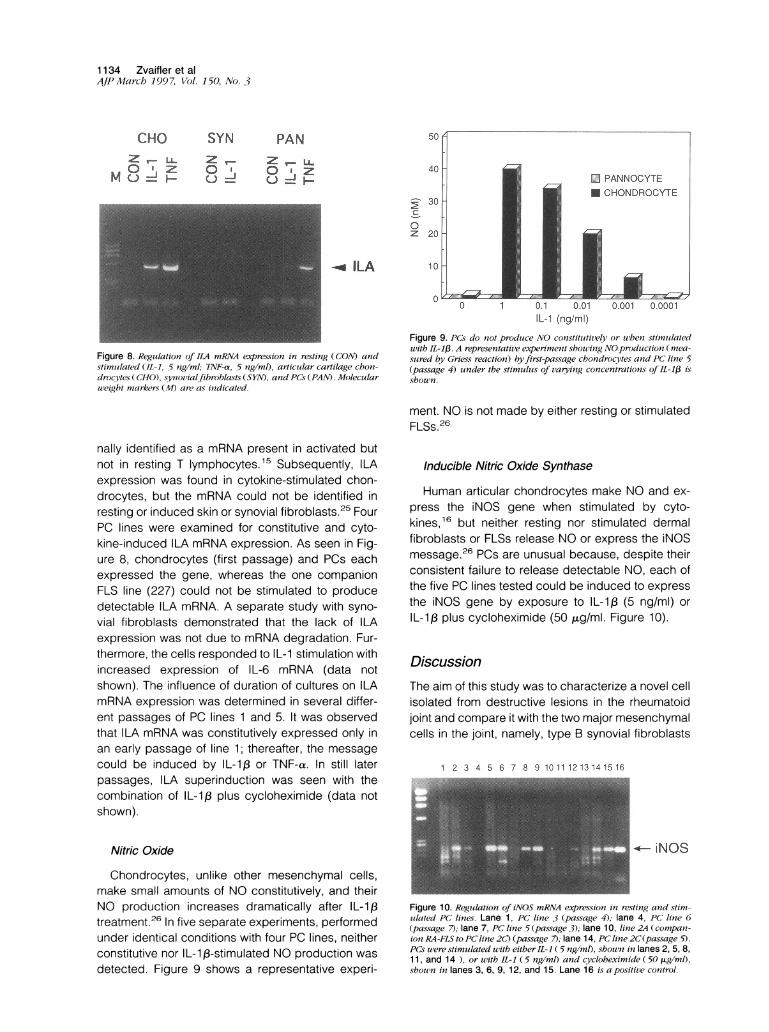
0 ''Figure 8. Regulation of ILA mRNA expression in resting (CON) andstimuilated (IL1, 5 ng/ml; TNF-a, 5 ng/ml), articular cartilage choni-drocytes (CHO), synovialfibroblasts (SYN), and PCs (PAN). Moleculariveigbt miarker.s (14 are as inidicated.
IL-1 (ng/ml)Figure 9. PCs do not produce NO constitutively or wthen stinmutlatedwith II-1,B. A representative experiment shou.'ing NOproduction (inea-sured by Griess reaction) byfirst-passage chondrocytes and PC line 5(passage 4) under the stimuluis of varying concentrations of IL-1,8 isshown.
ment. NO is not made by either resting or stimulatedFLSs.26
nally identified as a mRNA present in activated butnot in resting T lymphocytes.15 Subsequently, ILAexpression was found in cytokine-stimulated chon-drocytes, but the mRNA could not be identified inresting or induced skin or synovial fibroblasts.25 FourPC lines were examined for constitutive and cyto-kine-induced ILA mRNA expression. As seen in Fig-ure 8, chondrocytes (first passage) and PCs eachexpressed the gene, whereas the one companionFLS line (227) could not be stimulated to producedetectable ILA mRNA. A separate study with syno-
vial fibroblasts demonstrated that the lack of ILAexpression was not due to mRNA degradation. Fur-thermore, the cells responded to IL-1 stimulation withincreased expression of IL-6 mRNA (data notshown). The influence of duration of cultures on ILAmRNA expression was determined in several differ-ent passages of PC lines 1 and 5. It was observedthat ILA mRNA was constitutively expressed only inan early passage of line 1; thereafter, the message
could be induced by IL-1,B or TNF-a. In still laterpassages, ILA superinduction was seen with thecombination of IL-1,B plus cycloheximide (data notshown).
Nitric Oxide
Chondrocytes, unlike other mesenchymal cells,make small amounts of NO constitutively, and theirNO production increases dramatically after IL-1,3treatment.26 In five separate experiments, performedunder identical conditions with four PC lines, neitherconstitutive nor IL-1X3-stimulated NO production was
detected. Figure 9 shows a representative experi-
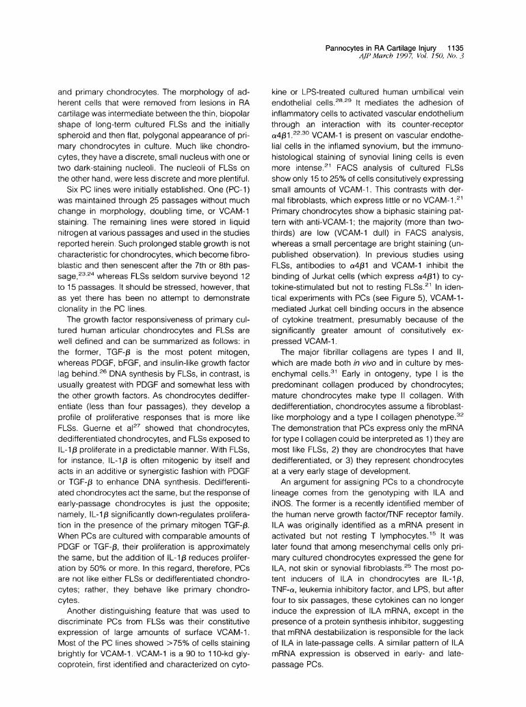
inducible Nitric Oxide Synthase
Human articular chondrocytes make NO and ex-
press the iNOS gene when stimulated by cyto-kines,16 but neither resting nor stimulated dermalfibroblasts or FLSs release NO or express the iNOSmessage.26 PCs are unusual because, despite theirconsistent failure to release detectable NO, each ofthe five PC lines tested could be induced to express
the iNOS gene by exposure to IL-1,3 (5 ng/ml) or
IL-1,8 plus cycloheximide (50 ,ug/ml. Figure 10).
Discussion
The aim of this study was to characterize a novel cellisolated from destructive lesions in the rheumatoidjoint and compare it with the two major mesenchymalcells in the joint, namely, type B synovial fibroblasts
1 2 3 4 5 6 7 8 9 10 11 1213 141516
iNOS
Figure 10. Regulation of iNOS mRNA expression in resting and stim-ulated PC lines. Lane 1, PC line 3 (pas.sage 4); lane 4, PC line 6(passage 7); lane 7, PC line 5 (passage 3); lane 10, linle 2A (compan-ionI RA-FLS to PC line 2C) (passage 7); lane 14, PC line 2C(passage 5).
PCs were stimulated with either IL-1 (5 ng/ml), shown in lanes 2, 5, 8,1 1, and 14 ), or with IL-1 (5 ng/ml) and cycloheximide (50 ,ug/ml),shown in lanes 3, 6, 9, 12, and 15. Lane 16 is a positive control.
PANZ _L
U ' z
1-
0z
_ ILA
Pannocytes in RA Cartilage Injury 1135AJPMarch 1997, Vol. 150, No. 3
and primary chondrocytes. The morphology of ad-herent cells that were removed from lesions in RAcartilage was intermediate between the thin, biopolarshape of long-term cultured FLSs and the initiallyspheroid and then flat, polygonal appearance of pri-mary chondrocytes in culture. Much like chondro-cytes, they have a discrete, small nucleus with one ortwo dark-staining nucleoli. The nucleoli of FLSs onthe other hand, were less discrete and more plentiful.
Six PC lines were initially established. One (PC-1)was maintained through 25 passages without muchchange in morphology, doubling time, or VCAM-1staining. The remaining lines were stored in liquidnitrogen at various passages and used in the studiesreported herein. Such prolonged stable growth is notcharacteristic for chondrocytes, which become fibro-blastic and then senescent after the 7th or 8th pas-
23,2sage, 24 whereas FLSs seldom survive beyond 12to 15 passages. It should be stressed, however, thatas yet there has been no attempt to demonstrateclonality in the PC lines.
The growth factor responsiveness of primary cul-tured human articular chondrocytes and FLSs arewell defined and can be summarized as follows: inthe former, TGF-,3 is the most potent mitogen,whereas PDGF, bFGF, and insulin-like growth factorlag behind.26 DNA synthesis by FLSs, in contrast, isusually greatest with PDGF and somewhat less withthe other growth factors. As chondrocytes dediffer-entiate (less than four passages), they develop aprofile of proliferative responses that is more likeFLSs. Guerne et al27 showed that chondrocytes,dedifferentiated chondrocytes, and FLSs exposed toIL-1/3 proliferate in a predictable manner. With FLSs,for instance, IL-1,B is often mitogenic by itself andacts in an additive or synergistic fashion with PDGFor TGF-13 to enhance DNA synthesis. Dedifferenti-ated chondrocytes act the same, but the response ofearly-passage chondrocytes is just the opposite;namely, IL-i ,3 significantly down-regulates prolifera-tion in the presence of the primary mitogen TGF-f.When PCs are cultured with comparable amounts ofPDGF or TGF-3, their proliferation is approximatelythe same, but the addition of IL-l3 reduces prolifer-ation by 50% or more. In this regard, therefore, PCsare not like either FLSs or dedifferentiated chondro-cytes; rather, they behave like primary chondro-cytes.
Another distinguishing feature that was used todiscriminate PCs from FLSs was their constitutiveexpression of large amounts of surface VCAM-1.Most of the PC lines showed >75% of cells stainingbrightly for VCAM-1. VCAM-1 is a 90 to 110-kd gly-coprotein, first identified and characterized on cyto-
kine or LPS-treated cultured human umbilical veinendothelial cells.2829 It mediates the adhesion ofinflammatory cells to activated vascular endotheliumthrough an interaction with its counter-receptora4f3i 22.30 VCAM-1 is present on vascular endothe-lial cells in the inflamed synovium, but the immuno-histological staining of synovial lining cells is evenmore intense.21 FACS analysis of cultured FLSsshow only 15 to 25% of cells consitutively expressingsmall amounts of VCAM-1. This contrasts with der-mal fibroblasts, which express little or no VCAM-i 21Primary chondrocytes show a biphasic staining pat-tern with anti-VCAM-1; the majority (more than two-thirds) are low (VCAM-1 dull) in FACS analysis,whereas a small percentage are bright staining (un-published observation). In previous studies usingFLSs, antibodies to a4j31 and VCAM-1 inhibit thebinding of Jurkat cells (which express a4l3) to cy-tokine-stimulated but not to resting FLSs.21 In iden-tical experiments with PCs (see Figure 5), VCAM-1-mediated Jurkat cell binding occurs in the absenceof cytokine treatment, presumably because of thesignificantly greater amount of consitutively ex-pressed VCAM-1.The major fibrillar collagens are types and 11,
which are made both in vivo and in culture by mes-enchymal cells.31 Early in ontogeny, type is thepredominant collagen produced by chondrocytes;mature chondrocytes make type 11 collagen. Withdedifferentiation, chondrocytes assume a fibroblast-like morphology and a type collagen phenotype.32The demonstration that PCs express only the mRNAfor type collagen could be interpreted as 1) they aremost like FLSs, 2) they are chondrocytes that havededifferentiated, or 3) they represent chondrocytesat a very early stage of development.An argument for assigning PCs to a chondrocyte
lineage comes from the genotyping with ILA andiNOS. The former is a recently identified member ofthe human nerve growth factor/TNF receptor family.ILA was originally identified as a mRNA present inactivated but not resting T lymphocytes.15 It waslater found that among mesenchymal cells only pri-mary cultured chondrocytes expressed the gene forILA, not skin or synovial fibroblasts.25 The most po-tent inducers of ILA in chondrocytes are IL-1i3,TNF-a, leukemia inhibitory factor, and LPS, but afterfour to six passages, these cytokines can no longerinduce the expression of ILA mRNA, except in thepresence of a protein synthesis inhibitor, suggestingthat mRNA destabilization is responsible for the lackof ILA in late-passage cells. A similar pattern of ILAmRNA expression is observed in early- and late-passage PCs.
1136 Zvaifler et alAJPMarch 1997, Vol. 150, No. 3
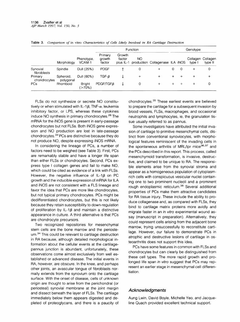
Table 3. Comparison of in vitro Characteristics of Cells Likely Involved in RA Cartilage Destruction
Function GenotypePrimary Growth
Phenotype, growth factor NO Collagen CollagenMorphology VCAM-1 factor plus IL-1 production Collagenase ILA iNOS type type II
Synovial Spindle Dull (25%) PDGFfibroblasts
Primary Spheroid, Dull (60%) TGF-,1chondrocytes polygonal
PCs Rhomboid Bright PDGF/TGFj3(>70%)
FLSs do not synthesize or secrete NO constitu-tively or when stimulated with IL-1,B, TNF-a, leukemiainhibitory factor, or LPS, whereas these cytokinesinduce NO synthesis in primary chondrocytes.26 ThemRNA for the iNOS gene is present in early-passagechondrocytes but not FLSs. Both iNOS gene expres-sion and NO production are lost in late-passagechondrocytes.33 PCs are distinctive because they donot produce NO, despite expressing iNOS mRNA.
In considering the lineage of PCs, a number offactors need to be weighed (see Table 3). First, PCsare remarkably stable and have a longer life spanthan either FLSs or chondrocytes. Second, PCs ex-press type collagen genes and fail to make NO,which could be cited as evidence of a link with FLSs.However, the negative influence of IL-1 , on PCgrowth and the inducible expression of mRNA for ILAand iNOS are not consistent with a FLS lineage andfavor the idea that PCs are more like chondrocytes,but not typical primary chondrocytes. PCs might bededifferentiated chondrocytes, but this is not likelybecause they retain susceptibility to down-regulationof proliferation by IL-13 and maintain a distinctiveappearance in culture. A third alternative is that PCsare chondrocyte precursors.Two recognized repositories for mesenchymal
stem cells are the bone marrow and the perioste-um.34 This could be relevant to cartilage destructionin RA because, although detailed morphological in-formation about the cellular events at the cartilage-pannus junction is abundant, unfortunately, theseobservations come almost exclusively from well es-tablished or advanced disease. The initial events inRA, however, are obscure. In the knee, and perhapsother joints, an avascular tongue of fibroblasts nor-mally extends from the synovium onto the cartilagesurface. With the onset of disease, cells of unknownorigin are thought to arise from the perichondral (orperiosteal) synovial membrane at the joint marginand dissect beneath the layer of FLSs. The cartilageimmediately below them appears digested and de-pleted of proteoglycans, and there is a paucity of
T 0 + 0 0 + 0
I
'I 0
+ + +
+ + +
+ +
+ 0
chondrocytes.35 These earliest events are believedto prepare the cartilage for a subsequent invasion byblood vessels, FLSs, macrophages, and occasionalneutrophils and lymphocytes, ie, the granulation tis-sue usually referred to as pannus.Some investigators have attributed the initial inva-
sion of cartilage to primitive mesenchymal cells, dis-tinct from conventional synoviocytes, with morpho-logical features reminiscent of the invading cells inthe spontaneous arthritis of MRL/lpr mice3637 andthe PCs described in this report. This process, calledmesenchymoid transformation, is invasive, destruc-tive, and claimed to be unique to RA. The responsi-ble elements arise from the synovial stroma andappear as a homogeneous population of cytoplasm-rich cells with conspicuous vesicular nuclei contain-ing one to two prominent nucleoli and a prominentrough endoplasmic reticulum.38 Several additionalproperties of PCs make them attractive candidatesfor RA tissue injury. These include the ability to pro-duce collagenase and, as compared with FLSs, theybind to cartilage matrix proteins more avidly andmigrate faster in an in vitro experimental wound as-say (manuscript in preparation). Alternatively, theycould represent cells arising from the adjacent bonemarrow, trying unsuccessfully to reconstitute carti-lage. However, our failure to demonstrate PCs inatrophic and destructive lesions of cartilage in os-teoarthritis does not support this idea.
PCs have some features in common with FLSs andchondrocytes but can clearly be distinguished fromthese cell types. The more rapid growth and pro-longed life span in vitro suggest that PCs may rep-resent an earlier stage in mesenchymal cell differen-tiation.
AcknowledgmentsAung Lwin, David Boyle, Michelle Yeo, and Jacque-line Quach provided excellent technical support.

Pannocytes in RA Cartilage Injury 1137AJP March 1997, Vol. 150, No. 3
References
1. Harris EDJ: Rheumatoid arthritis: pathophysiology andimplications for therapy. N Engi J Med 1990, 322:1277-1289
2. Krane SM, Conca W, Stephenson ML, Amento EP,Goldring MB: Mechanisms of matrix degradation inrheumatoid arthritis. Ann NY Acad Sci 1990, 580:340-354
3. Remmers EF, Sano H, Wilder RL: Platelet-derivedgrowth factors and heparin-binding (fibroblast) growthfactors in the synovial tissue pathology of rheumatoidarthritis. Semin Arthritis Rheum 1991, 21:191-199
4. Zvaifler NJ, Firestein GS: Pannus and pannocytes: al-ternative models of joint destruction in rheumatoid ar-thritis. Arthritis Rheum 1994, 37:783-789
5. Alvaro-Gracia JM, Zvaifler NJ, Firestein GS: Cytokinesin chronic inflammatory arthritis. V. Mutual antagonismbetween interferon-y and tumor necrosis factor-a onHLA-DR expression, proliferation, collagenase produc-tion, and granulocyte macrophage colony-stimulatingfactor production by rheumatoid arthritis synoviocytes.J Clin Invest 1990, 86:1790-1798
6. Firestein GS, Paine MM, Littman BH: Gene expression(collagenase, tissue inhibitor of metalloproteinases,complement, and HLA-DR) in rheumatoid arthritis andosteoarthritis synovium: quantitative analysis and effectof intraarticular corticosteroids. Arthritis Rheum 1991,34:1094-1105
7. Barland P, Novikoff AB, Hamerman D: Electron micros-copy of the human synovial membrane. J Cell Biol1962, 14:207-220
8. Firestein GS, Tsai V, Zvaifler NJ: Cellular immunity inthe joints of patients with rheumatoid arthritis and otherforms of chronic synovitis. Rheum Dis Clin North Am1987, 13:191-213
9. Theofilopoulos AN, Carson DA, Tavassoli M, Slovin SF,Speers WC, Jensen FB, Vaughan JH: Evidence for thepresence of receptors for C3 and IgG Fc on humansynovial cells. Arthritis Rheum 1980, 23:1-9
10. Burmester GR, Dimitriu-Bona A, Waters SJ, WinchesterRJ: Identification of three major synovial lining cell pop-ulations by monoclonal antibodies directed to la anti-gens and antigens associated with monocytes/macro-phages and fibroblasts. Scand J Immunol 1983, 17:69-82
11. Koch AE, Polverini PJ, Leibovich SJ: Functional heter-ogeneity of human rheumatoid synovial tissue macro-phages. J Rheumatol 1988, 15:1058-1063
12. Alvaro-Gracia JM, Zvaifler NJ, Firestein GS: Cytokinesin chronic inflammatory arthritis. IV. Granulocyte/mac-rophage colony-stimulating factor-mediated inductionof class 11 MHC antigen on human monocytes: a pos-sible role in rheumatoid arthritis. J Exp Med 1989,170:865-875
13. Taylor DJ, Cheung NT, Dawes PT: Increased serumproMMP-3 in inflammatory arthritides: a potential indi-
cator of synovial inflammatory monokine activity. AnnRheum Dis 1994, 53:768-772
14. Green LC, Wagner DA, Glogowski J, Skipper PL, Wish-nok JS, Tannenbaum SR: Analysis of nitrate, nitrite and[15N] nitrate in biological fluids. Anal Biochem 1982,126:131-138
15. Schwarz H, Tuckwell J, Lotz M: A receptor induced bylymphocyte activation (ILA): a new member of the hu-man nerve-growth-factor/tumor-necrosis-factor recep-tor family. Gene 1993, 134:295-298
16. Maier R, Bilbe G, Rediske J, Lotz M: Inducible nitricoxide synthase from human articular chondrocytes:cDNA cloning and analysis of mRNA expression. Bio-chim Biophys Acta 1994, 1208:145-150
17. Chomczynski P, Sacchi N: Single-step method of RNAisolation by acid-guanidinium-thiocyanate-phenol-chloroform extraction. Anal Biochem 1987, 162:156-159
18. Vuorio E: Rheumatoid disease in cultured human syno-vial cells. Scand J Clin Lab Invest 1977, 37(suppl 149):1-72
19. Gabbiani G, Hirschel BJ, Ryan GB, Statkov PR, MajnoG: Granulation tissue as a contractile organ: a study ofstructure and function. J Exp Med 1972, 135:719-734
20. Sappino AP, Schurch W, Gabbiani G: Differentiationrepertoire of fibroblastic cells: expression of cytoskel-etal proteins as marker of phenotypic modulations. LabInvest 1990, 63:144-161
21. Morales-Ducret J, Wayner E, Elices MJ, Alvaro-GraciaJM, Zvaifler NJ, Firestein GS: a4//31-lntegrin (VLA-4)ligands in arthritis: vascular cell adhesion molecule-1expression in synovium and on fibroblast-like synovio-cytes. J Immunol 1992, 149:1424-1431
22. Hemler ME, Elices MJ, Parker C, Takada Y: Structure ofthe integrin VLA-4 and its cell-cell and cell-matrix ad-hesion functions. Immunol Rev 1990, 114:45-65
23. Lotz M: Regulation of chondrocytes in aging. BiologicalRegulation of the Chondrocytes. Edited by M Adolphe.Boca Raton, FL, CRC Press, 1992, pp 237-273
24. Evans CH, Georgescu HI: Observations on the senes-cence of cells derived from articular cartilage. MechAgeing Dev 1983, 22:179-191
25. von Kempis H, Schwarz H, Lotz M: Differentiation-de-pendent expression of ILA, a new member of the hu-man NGF/TNF receptor family in mesenchymal cells.FASEB J 1994, 8:4:A221
26. Rediske JJ, Koehne CF, Zhang B, Lotz M: The induc-ible production of nitric oxide by articular cell types.Osteoarthritis Cartilage 1994, 2:199-206
27. Guerne PA, Sublet A, Lotz M: Growth factor respon-siveness of human articular chondrocytes. J CellPhysiol 1994, 158:476-484
28. Rice GE, Bevilacqua MP: An inducible endothelial cellsurface glycoprotein mediates melanoma adhesion.Science 1989, 246:1303-1306
29. Osborn L, Hesslon C, Tizard R, Vassallo C, LuhowskyjS, Chi-Rosso G, Lobb R: Direct expression cloning ofvascular cell adhesion molecule 1, a cytokine-induced
1138 Zvaifler et alAJP March 1997, Vol. 150, No. 3
endothelial protein that binds to lymphocytes. Cell1989, 59:1203-1211
30. Albelda SM, Buck CA: Integrins and other cell adhe-sion molecules. FASEB J 1990, 4:2868-2880
31. Postlewaithe AE, Kang A: Fibroblasts and matrix pro-teins. Inflammation: Basic Principles and Clinical Cor-relates. Edited by JI Gallin, IM Goldstein, R Snyder-man. New York, Raven Press, 1992
32. Burgeson RE: New collagens, new concepts. AnnuRev Cell Biol 1988, 4:551-577
33. Benya PD, Shaffer JD: Dedifferentiated chondrocytesreexpress the differentiated collagen phenotype whencultured in agarose gels. Cell 1982, 30:215-224
34. Blanco FJ, Geng Y, Lotz M: Differentiation-dependenteffects of IL-1 and TGF-,B on human articular chondro-cyte proliferation are related to inducible nitric oxidesynthase expression. J Immunol 1995,154:4018-4026
35. Caplan Al: Mesenchymal stem cells. J Orthop Res1991, 9:641-650
36. Allard SA, Bayliss M, Maini RN: The synovium-cartilagejunction of the normal human knee: implications forjoint destruction and repair. Arthritis Rheum 1990, 33:1170-1179
37. Fassbender HG: Histomorphological basis of articularcartilage destruction in rheumatoid arthritis. Coll RelatRes 1983, 3:141-155
38. O'Sullivan FX, Fassbender HG, Gay S, Koopman WJ:Etiopathogenesis of the rheumatoid arthritis-like dis-ease in MRL/I mice. Arthritis Rheum 1985, 28:529-536
39. Fassbender HG: Rheumatoid Arthritis. Current Trendsin Diagnostics, Conservative Treatment, and SurgicalReconstruction. Edited by H Baumgartner, J Dvorak, DGrob, U Munzinger, BR Simmen. New York, ThiemeMedical Publishers, 1995, pp 32-39