Embed Size (px)
Citation preview

D-amino acids enhanced biocide mitigation of field biofilm consortia in lab tests
Yingchao Li, Tingyue Gu ([email protected]) Dept. of Chemical & Biomolecular Engineering Institute for corrosion & multiphase technology
Ohio University Athens, OH 45701
Dake Xu
Institute of Metal Research Chinese Academy of Sciences
72 Wenhua Road, Shenyang 110016 China
Peiyu Zhang Dept. of Bioengineering
University of Missouri-Columbia Columbia, MO 65211
Congmin Xu School of Materials Science & Engineering
Xi’an Shiyou University 18 Dianzi 2nd Road, Xi’an 710065
China
ABSTRACT Microbiologically influenced corrosion (MIC) is a major problem in the oil and gas industry as well as many other industries. Current treatment methods rely mostly on pigging and biocide dosing. Because field systems are not sterile, microbes always recover, leading to repeated treatment cycles. It is anticipated that the application of the same biocide will selectively promote resistant microbes. Overtime, this can lead to the biocide dosage escalating, resulting in a cost increase and environmental concerns. Previously published work demonstrated that some D-amino acids are biocide enhancers. D-amino acids are naturally occurring. They occupy a significant fraction of amino acids in processed food because of heat conversion of L-amino acids to D-amino acids. It has been postulated that D-amino acids can replace the D-alanine terminus in bacterial cell walls. Under a biocide stress, these D-amino acids can disperse recalcitrant biofilms such as the Desulfovibrio vulgaris biofilm on carbon steel coupons. It is well known that planktonic cells are much easier to treat than sessile cells. Because D-amino acids are used as signal molecules, only relatively low concentrations are needed. They can reduce biocide dosage while achieving increased efficacy. The new data provided herein reveal that a mixture of D-amino acids enhanced biocide treatment of two recalcitrant biofilm consortia, thus paving the way for field trials. Key words: biocide, D-amino acid, biofilm consortia, microbiologically influenced corrosion
1
Paper No.
5522
©2015 by NACE International. Requests for permission to publish this manuscript in any form, in part or in whole, must be in writing to NACE International, Publications Division, 15835 Park Ten Place, Houston, Texas 77084. The material presented and the views expressed in this paper are solely those of the author(s) and are not necessarily endorsed by the Association.

INTRODUCTION
Microbiologically influenced corrosion (MIC) was first reported over 100 years ago.1 It has become a major problem in the oil and gas industry in recent years since water injection is practiced more frequently than ever to increase well pressure. In this process, microbes and nutrients are introduced into the reservoirs and subsequently showing up in the pipelines, leading to MIC. The pipeline leak from a 0.25” × 0.5” hole in the bottom of the Trans-Alaska pipeline† occurred in 2006, in which MIC was considered a major contributing factor.2 In a separate reported case, an 8” diameter pipeline carrying oil and produced water failed in only 8 months due to MIC.3 A recent pipeline failure case was published suggesting that MIC was likely the culprit in the leak of a 24” CO2 gathering line 2 years after commissioning, after ruling out other corrosion mechanisms.4 Skovhus and Eckert discussed several MIC cases and pointed out that MIC is becoming more prevalent nowadays due to aging equipment and increased awareness.5 Although MIC has been investigated for decades, its mechanisms are still not completely understood. One of the theories is the classical cathodic depolarization theory (CDT).6 In 2009, Gu et al. proposed the biocatalytic cathodic sulfate reduction (BCSR) theory to explain MIC by sulfate reducing bacteria (SRB) based on SRB bioenergetics.7 The team suggested that SRB attack on carbon steel is the result of the utilization of extracellular electrons for energy production during sulfate reduction in the cytoplasm of sulfate reducing bacteria (SRB), Anodic reaction: 4Fe 4Fe2+ + 4e- (Iron dissolution) −Eo'= +447 mV (1) Cathodic reaction: SO4
2- + 9H+ + 8e- HS- + 4H2O (BCSR) Eo'= −217 mV (2) Elemental iron (Fe0) in carbon steel serves as an electron donor and unlike organic carbon nutrients it is insoluble. Its oxidation occurs extracellularly, meaning that the extracellularly released electrons must be transported into the cytoplasm where sulfate reduction occurs. Reactions (1) and (2) produce a positive reaction cell potential (ΔEo') of 230 mV, corresponding to a negative Gibbs free energy (ΔGo') of −177 kJ/mol of sulfate, which means energy is produced by the redox reaction.8 Xu et al. designed an SRB starvation experiment to support their theory.9 For this study, they first obtained mature SRB biofilms on carbon steel coupons in the full-strength culture medium. These biofilms were subsequently cultured in fresh culture media with different concentrations of lactate as the carbon source. They found that under carbon source starvation, MIC pitting accelerated despite the fact that the biofilm was weakened due to the starvation. During this experiment, Fe0 served as a fuel (electron donor) for SRB. In this kind of SRB MIC, cross-cell wall electron transport is required, meaning that the biofilm must be electrogenic.10 Additional research by Zhang et al. suggested that electron transfer is a bottleneck in SRB MIC against 304 stainless steel.11 They demonstrated that adding an electron mediator to enhance electron transfer significantly increased MIC pitting and weight loss without increasing the biofilm density on the coupons. Inspired by the bioenergetics theory for SRB above, Xu et al. performed an experiment on carbon steel MIC by a nitrate reducing bacterium (NRB).8 They found that Bacillus licheniformis was more corrosive than typical SRB when it was grown as an NRB because it provided a larger thermodynamic driving force. The aforementioned cases of MIC are classified as Type I MIC due to the requirement of cross-cell wall electron transfer. MIC by acid producing bacteria (APB) belongs to Type II MIC because the oxidant (proton) is secreted and is reduced outside the cells on the steel surface rather than in the cytoplasm.10 In the preceding MIC mechanisms, biofilms play a critical role. For instance, in Type I MIC, a biofilm is needed to transport the electrons, since planktonic cells cannot accept electrons across a body of water because electrons cannot freely exist in water. Furthermore, in Type II MIC, oxidants (e.g., H+ and undissociated organic acids) are concentrated underneath a biofilm due to local secretion. In the field, microbes form synergistic biofilm consortia. It is well known that biofilm cells (sessile cells) are far more recalcitrant than planktonic cells. Through several different mechanisms, a biofilm protects
† Trade name
2
©2015 by NACE International. Requests for permission to publish this manuscript in any form, in part or in whole, must be in writing to NACE International, Publications Division, 15835 Park Ten Place, Houston, Texas 77084. The material presented and the views expressed in this paper are solely those of the author(s) and are not necessarily endorsed by the Association.
inner sessile cells from harmful factors. In one way, a biofilm can slow down the diffusion of antimicrobial agents.12, 13 Additionally, it can slow down the metabolic rate to reduce the intake of antimicrobial agents. Tuomanen et al. found that the increased resistance to antibacterial agents was accompanied by a low growth rate or no growth.14 Biofilms also preserve persister cells when they are under attack. These persister cells quickly rebound when the environment becomes less hostile.15 Sessile cells in biofilms use efflux pumps to prevent antimicrobial agents from entering the cells. They can upregulate resistant genes to break down antimicrobial agents.15 These mechanisms make the mitigation of biofilm consortia difficult. It is said that as a rule of thumb, 10X or higher biocide concentration may be required to treat biofilms compared with that needed for the treatment of planktonic cells.16–18 Pigging and biocides are two primary ways to mitigate problematic biofilms.19 Biocide applications can be performed during pigging by placing a biocide “plug” in a pipeline between two pigs. However, some pipelines are not piggable due to complicated elbows and other devices.20 Since pigging cannot completely remove biofilms, it is likely that the residual sessile cells can bounce back quickly. A biocide is needed to delay the recovery of the sessile community. Tetrakis hydroxymethyl phosphonium sulfate (THPS) is one of the widely used biocides in the oil and gas industry because it is biodegradable and effective against a broad spectrum of microorganisms. THPS is designated by the US Environmental Protection Agency (EPA) as a green chemical.21 It disrupts the disulphide bond in proteins and enzymes in microbes.22–25 In field operations, repeated use of the same biocide will inevitably cause dosage escalation because of selective promotion of resistant microbes over time. In some field operations, THPS dosage is so high such that the sulfate introduced by THPS precipitates with barium in the drilling fluid causing problematic scale formation at downhole. High doses of biocides also cause environmental concerns in addition to increased cost.
Despite continued research in new biocides, it is unlikely that a blockbuster biocide will replace THPS or glutaraldehyde any time soon. Thus, it is desirable to use biocide enhancers to make existing biocides more effective in the mitigation of field biofilm consortia. D-amino acids were reported to enhance the efficacy of THPS in the mitigation of Desulfovibrio vulgaris biofilm.26–28 Although previously considered rare in nature, D-amino acids are now considered ubiquitous due to the advancement of analytical techniques and the increased interest in their utilities. They are found in microorganisms, food, plants, animals, and even in humans.29 While the biological functions of D-amino acids are not fully understood, it is believed that they could serve as a signal molecule. Lam et al. stated that the synthesis of D-amino acids might be a common way of self-adjustment of cells to the changing environment.30 Kolodkin-Gal et al. found that D-methionine (D-met), D-tyrosine (D-tyr), D-leucine (D-leu), and D-tryptophan (D-trp) triggered the Bacillus subtilis biofilm’s disassembly.31 They also tested an equimolar mixture of four D-amino acids.31 Xu and Liu confirmed that 100 ppm (w/w) D-tyr triggered the biofilm dispersal in their test using activated sludge on membrane filters.32 Xu et al. found that D-tyr and D-met were effective against the D. vulgaris biofilm on carbon steel coupons.26, 27 It is hypothesized that D-amino acids trigger biofilm disassembly by replacing the D-alanine terminus in peptidoglycans that exist in all bacterial cell walls.31 Kolodkin-Gal et al. and Xu et al. found that adding a high concentration of D-alanine in the treatment chemicals rendered D-met ineffective.26, 31 These D-amino acids may also modify the synthesis of peptidoglycans.30 Cava et al. suggested that D-amino acids were necessary in the remodeling of the cell wall structure.33 Leiman et al. pointed out that D-tyr, D-leu, and D-trp inhibited the formation of the B. subtilis biofilm because these D-amino acids interfered with protein synthesis.34 Their data suggested that D-tyr acted as a growth inhibitor toward B. subtilis. Overall, it is highly possible that specific D-amino acids are effective for specific bacteria. Thus, for a biofilm consortium, a mixture of D-amino acids is likely required.
In this work, the efficacy of THPS combined with a mixture of D-amino acids against field collected biofilm consortia was investigated. An equimolar mixture of four D-amino acids (D-met, D-tyr, D-leu, and D-trp) were tested with 50 ppm (w/w) THPS to determine if the mixture of D-amino acids enhanced the efficacy of THPS against the biofilm consortia. Individual D-met and D-tyr were also tested with THPS to compare with the efficacy of THPS combined with the mixture of D-amino acids. The results
3
©2015 by NACE International. Requests for permission to publish this manuscript in any form, in part or in whole, must be in writing to NACE International, Publications Division, 15835 Park Ten Place, Houston, Texas 77084. The material presented and the views expressed in this paper are solely those of the author(s) and are not necessarily endorsed by the Association.

show that the mixture of D-amino acids was far more effective against the biofilm consortia than the individual D-met and D-tyr.
EXPERIMENTAL CONDITIONS
In this research, two biofilm consortia collected from an oil and gas field were used to investigate the efficacy of 50 ppm THPS in combination with D-amino acid(s). They were labelled as consortium I and consortium II. The consortia were cultured in ATCC 1249 medium† which is a modified Baar’s medium for sulfate reducers. L-cysteine was added into the medium as an oxygen scavenger to eliminate any possible oxygen leak. The concentration of L-cysteine in the medium was 100 ppm (w/w). The biofilm consortia were grown on disk-shaped C1018 (UNS G10180) carbon steel coupons. The composition of C1018 (UNS G10180) was (wt%): C 0.14-0.20, Mn 0.60-0.90, P 0.04, S 0.05, Si 0.15-0.30, and Fe 98.81-99.26. For each coupon, only the 1.2 cm2 top surface of the coupons was exposed to the culture medium, and the remainder was painted. Coupons were polished with 180, 400, and 600 grit sandpapers, sequentially. They were cleaned with isopropanol and dried under UV light for 20 min. The culture medium, anaerobic vials, pipettes, and tweezers were sterilized in an autoclave at 121oC for 20 minutes before use. D-amino acids were not autoclaved due to possible oxidation at high temperature. Their stock solutions were filter-sterilized. The culture medium and solutions containing the treatment chemicals were sparged with filtered N2 for 45 minutes to remove O2. In order to evaluate the efficacy of the cocktail of THPS + D-amino acid(s), both biofilm prevention and biofilm removal tests were carried out in the lab. In the biofilm prevention test, 2 duplicate coupons, 100 ml medium, treatment chemicals, and 1 ml biofilm consortium seed culture were put into each 125 ml anaerobic vial in an anaerobic chamber. The initial planktonic cell concentration right after inoculation was 106 cells/ml. The anaerobic chamber was sparged with filtered N2 for 45 minutes to remove oxygen before use. After the vials were sealed, they were placed in a 37oC incubator. After 7-day incubation, coupons were taken out for cell enumeration and scanning electron microscope (SEM) observation. In the biofilm removal test, biofilms were first grown on coupons without treatment chemicals for 3 days to achieve maturity. Coupons covered by 3-day mature biofilm consortia were taken out and put into a phosphate buffered saline (PBS) solution with treatment chemicals in the Petri dish† for 3 hours in the anaerobic chamber at room temperature. When the 3-hour treatment was finished, coupons were taken out for cell enumeration and SEM observation. Test matrices of both tests are listed in Tables 1 and 2.
Table 1 Test matrix of biofilm prevention test
Biofilm Two field biofilm consortia
Culture medium SRB medium
Treatment method THPS + a mixture of D-amino acids, D-met, or D-tyr
Temperature 37oC
Test duration 7 days
Coupon C1018 (UNS G10180) carbon steel
† Trade name
4
©2015 by NACE International. Requests for permission to publish this manuscript in any form, in part or in whole, must be in writing to NACE International, Publications Division, 15835 Park Ten Place, Houston, Texas 77084. The material presented and the views expressed in this paper are solely those of the author(s) and are not necessarily endorsed by the Association.

Table 2 Test matrix of established biofilm removal test
Biofilm Two field biofilm consortia pre-grown on coupons
Treatment method THPS + a mixture of D-amino acids, D-met, or D-tyr
Temperature 25oC
Test duration 3 h exposure to treatment chemicals in a dish
Coupon C1018 (UNS G10180) carbon steel
The Biosan Sani-Check† SRB test kit was used for cell enumeration. The biofilm consortium on the coupon surface was dislodged with a small brush that was a part of the test kit. The brush was then inserted into the kit’s vial containing a solid SRB culture medium for incubation at 37oC. The time it took to show the black color (FeS) reflected the SRB cell concentration based on vendor’s calibration. The coupons for SEM observation were first submerged in 4% (w/w) glutaraldehyde for 2 h and then dehydrated in 25%, 50%, 75%, and 100% (v/v) isopropanol sequentially for 5 minutes. At the end, the biofilms were dehydrated in a critical point dryer using CO2. Before SEM (Model JSM-6390, JEOL†, Japan) observation, biofilm covered coupons were coated with palladium to provide conductivity. It should be noted that SEM images should not be used for quantitative cell counting because of uneven distribution of sessile cells.
RESULTS AND DISCUSSION
Biofilm consortia I and II were found to be corrosive biofilms. Figure 1 shows that both biofilm consortia formed robust biofilms on coupons after incubation in the culture medium for 10 days at 37oC. The images also reveal that the sessile cells in both biofilm consortia appeared to have different morphologies. Since the culture medium is designed for the selective growth of sulfate reducing bacteria, it is suspected that the dominant sessile population observed is likely SRB. After the biofilms were removed, pits underneath the biofilms resembled characteristic MIC pits. The specific weight loss observed on the coupon was 0.0047 g/cm2 and 0.0058 g/cm2 for consortium I and consortium II (Figure 2), respectively. Both values were much larger than the 0.0018 g/cm2 caused by D. vulgaris incubated under the same conditions.
† Trade name
5
©2015 by NACE International. Requests for permission to publish this manuscript in any form, in part or in whole, must be in writing to NACE International, Publications Division, 15835 Park Ten Place, Houston, Texas 77084. The material presented and the views expressed in this paper are solely those of the author(s) and are not necessarily endorsed by the Association.

Figure 1. Biofilm images and bare coupon surface images after biofilm removal for coupons in
the culture medium after 10 days of incubation (without treatment) at 37oC: (A) biofilm consortium I, (B) biofilm consortium II, (C) coupon surface after removal of biofilm consortium I,
and (D) coupon surface after removal of biofilm consortium II.
Figure 2. Weight loss of coupons caused by three biofilms: 0.0018 g/cm2 for D. vulgaris, 0.0047 g/cm2 for consortium I, and 0.0058 g/cm2 for consortium II.
(A) (B)
(C) (D)
6
©2015 by NACE International. Requests for permission to publish this manuscript in any form, in part or in whole, must be in writing to NACE International, Publications Division, 15835 Park Ten Place, Houston, Texas 77084. The material presented and the views expressed in this paper are solely those of the author(s) and are not necessarily endorsed by the Association.

In our previous tests, the cocktails of 50 ppm THPS + 100 ppm D-met and 50 ppm THPS + 1 ppm D-tyr both achieved 5 log reduction of the sessile cell count in the mitigation of the D. vulgaris biofilm on coupons.26, 27 However, the data in Table 3 illustrate that during the mitigation of biofilm consortia I and II, the same cocktails only achieved 1 or 2 log reduction of the sessile cell count, much less than the 5 for D. vulgaris. The data presented in Table 3 also show that increasing D-tyr from 1 ppm to 10 ppm did not improve the enhancement of 50 ppm THPS. These data indicate that the biofilm consortia were more difficult to mitigate than the pure-strain D. vulgaris biofilm. It was possible that some of the microbes in the consortia were less susceptible to D-met and D-tyr when they were used individually with THPS.
Table 3 Sessile cell counts of consortia I and II after 7-day biofilm prevention test
Biofilm Treatment Sessile cell count (cells/cm2)
Consortium I
No treatment chemical (control) ≥107
50 ppm THPS ≥107
50 ppm THPS + 100 ppm D-met ≥106
50 ppm THPS + 1 ppm D-tyr ≥105
50 ppm THPS + 10 ppm D-tyr ≥105
Consortium II
No treatment chemical (control) ≥107
50 ppm THPS ≥107
50 ppm THPS + 100 ppm D-met ≥106
50 ppm THPS + 1 ppm D-tyr ≥106
50 ppm THPS + 10 ppm D-tyr ≥106
An additional test was performed as a result of these findings and in support of the study performed by Kolodkin-Gal et al. The test suggested that a mixture of D-amino acids might be more effective than individual D-amino acids.31 In this work, an equimolar D-met, D-tyr, D-leu, and D-trp D-amino acid mixture (D-mix) at a total concentration of 50 ppm (w/w) was tested to enhance 50 ppm THPS in the mitigation of the two biofilm consortia. The composition of 50 ppm D-mix is shown in Table 4. The concentrations of D-met, D-leu, and D-trp in the mixture are much less than the concentration of each D-amino acid which was needed to enhance 50 ppm THPS in the mitigation of the D. vulgaris biofilm on carbon steel according to our published and unpublished data.26, 27
Table 4 Composition of D-amino acid in the equimolar mixture
D-amino acids
wt% Amount in 50 ppm D-amino acids mixture
(ppm)
Effective concentration to enhance THPS against D.
vulgaris biofilm (ppm) separately
D-met 22% 11 100
D-tyr 27% 13.5 1
D-leu 20% 10 1500
D-trp 31% 15.5 1500
For the mitigation of the biofilm consortium I, the data reveal that the dosage of 50 ppm D-mix enhanced 50 ppm THPS in both biofilm prevention and biofilm removal tests. In the biofilm prevention test, 50 ppm THPS without enhancement did not reduce the sessile cell count on the coupon surface
7
©2015 by NACE International. Requests for permission to publish this manuscript in any form, in part or in whole, must be in writing to NACE International, Publications Division, 15835 Park Ten Place, Houston, Texas 77084. The material presented and the views expressed in this paper are solely those of the author(s) and are not necessarily endorsed by the Association.
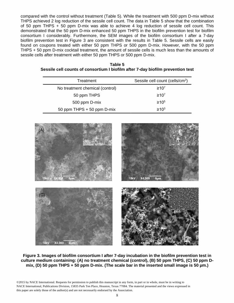
compared with the control without treatment (Table 5). While the treatment with 500 ppm D-mix without THPS achieved 2 log reduction of the sessile cell count. The data in Table 5 show that the combination of 50 ppm THPS + 50 ppm D-mix was able to achieve 4 log reduction of sessile cell count. This demonstrated that the 50 ppm D-mix enhanced 50 ppm THPS in the biofilm prevention test for biofilm consortium I considerably. Furthermore, the SEM images of the biofilm consortium I after a 7-day biofilm prevention test in Figure 3 are consistent with the results in Table 5. Sessile cells are easily found on coupons treated with either 50 ppm THPS or 500 ppm D-mix. However, with the 50 ppm THPS + 50 ppm D-mix cocktail treatment, the amount of sessile cells is much less than the amounts of sessile cells after treatment with either 50 ppm THPS or 500 ppm D-mix.
Table 5 Sessile cell counts of consortium I biofilm after 7-day biofilm prevention test
Treatment Sessile cell count (cells/cm2)
No treatment chemical (control) ≥107
50 ppm THPS ≥107
500 ppm D-mix ≥105
50 ppm THPS + 50 ppm D-mix ≥103
Figure 3. Images of biofilm consortium I after 7-day incubation in the biofilm prevention test in culture medium containing: (A) no treatment chemical (control), (B) 50 ppm THPS, (C) 50 ppm D-
mix, (D) 50 ppm THPS + 50 ppm D-mix. (The scale bar in the inserted small image is 50 µm.)
(D)
(A)
(C)
(B)
8
©2015 by NACE International. Requests for permission to publish this manuscript in any form, in part or in whole, must be in writing to NACE International, Publications Division, 15835 Park Ten Place, Houston, Texas 77084. The material presented and the views expressed in this paper are solely those of the author(s) and are not necessarily endorsed by the Association.

Similar efficacies were obtained in the biofilm removal test for biofilm consortium I. The data provided in Table 6 illustrate that the separate dosages of 50 ppm THPS and 500 ppm D-mix had limited effects on the mature biofilm consortium I. It was found that 50 ppm THPS achieved no reduction of the sessile cell count and 500 ppm D-mix achieved only 2 log reduction of the sessile cell count. While the cocktail of 50 ppm THPS + 50 ppm D-mix provided a 4 log reduction of the sessile cell count during the 3-hour biofilm study and the SEM images support the reduction of the sessile cells (Figure 4). Figure 4 shows that sessile cells are present in the mature biofilm consortium I after treatment with either 50 ppm THPS or 100 ppm D-mix.
Table 6 Sessile cell counts of consortium I after 3-hour biofilm removal test in a dish
Treatment Sessile cell count (cells/cm2)
No treatment chemical (control) ≥107
50 ppm THPS ≥107
500 ppm D-mix ≥105
50 ppm THPS + 50 ppm D-mix ≥103
Figure 4. Images of biofilm consortium I after 3-hour treatment in a dish containing PBS buffer and: (A) no treatment chemical (control), (B) 50 pm THPS, (C) 100 ppm D-mix, (E) 50 ppm THPS
+ 50 ppm D-mix, in the biofilm removal test. (The scale bar in the inserted small images is 50 µm.)
(A)
(D)
(B)
(C)
9
©2015 by NACE International. Requests for permission to publish this manuscript in any form, in part or in whole, must be in writing to NACE International, Publications Division, 15835 Park Ten Place, Houston, Texas 77084. The material presented and the views expressed in this paper are solely those of the author(s) and are not necessarily endorsed by the Association.

During the evaluation of the biofilm consortium II, similar dosage responses were obtained. It was found that 50 ppm D-mix enhanced 50 ppm THPS in both biofilm prevention and biofilm removal tests. Comparable to the mitigation of consortium I, 50 ppm THPS alone had no effect on the sessile cell count in both biofilm prevention and biofilm removal tests for biofilm consortium II compared with the untreated control (Tables 7 and 8). The cocktail of 50 ppm THPS + 50 ppm D-mix achieved 3 log reduction of the sessile cell count in both biofilm prevention and biofilm removal tests for biofilm consortium II. In Figures 5 and 6, sessile cells are easily found on the following coupons: the control coupon (no treatment), the coupon treated with 50 ppm THPS, and the coupon treated with 500 ppm D-mix separately. Although sessile cells are still noticeable on the surface of coupons treated with 50 ppm THPS + 50 ppm D-mix, they are less abundant.
Table 7 Sessile cell counts of Consortium II after 7-day biofilm prevention test
Treatment Sessile cell count (cells/cm2)
No treatment chemical (control) ≥107
50 ppm THPS ≥107
500 ppm D-mix ≥106
50 ppm THPS + 50 ppm D-mix ≥104
Table 8
Sessile cell counts of Consortium II after 3 hour biofilm removal test in a dish
Treatment Sessile cell count (cells/cm2)
No treatment chemical (control) ≥107
50 ppm THPS ≥107
500 ppm D-mix ≥106
50 ppm THPS + 50 ppm D-mix ≥104
10
©2015 by NACE International. Requests for permission to publish this manuscript in any form, in part or in whole, must be in writing to NACE International, Publications Division, 15835 Park Ten Place, Houston, Texas 77084. The material presented and the views expressed in this paper are solely those of the author(s) and are not necessarily endorsed by the Association.

Figure 5. Images of biofilm Consortium II after 7-day incubation in the biofilm prevention test with: (A) no treatment chemical (control), (B) 50 pm THPS, (C) 100 ppm D-mix (D) 50 ppm THPS + 50 ppm D-mix in biofilm prevention test. (The scale bar in the inserted small images is 50 µm.)
(E)
(C) (D)
(B)
11
©2015 by NACE International. Requests for permission to publish this manuscript in any form, in part or in whole, must be in writing to NACE International, Publications Division, 15835 Park Ten Place, Houston, Texas 77084. The material presented and the views expressed in this paper are solely those of the author(s) and are not necessarily endorsed by the Association.

Figure 6. Images of biofilm Consortium II after 3-hour treatment in a dish containing PBS buffer and: (A) no treatment chemical (control), (B) 100 pm THPS, (C) 100 ppm D-mix, (D) 50 ppm THPS
+ 50 ppm D-mix in biofilm removal test. (The scale bar in the inserted small images is 50 µm.) In general, the cocktail of 50 ppm THPS + 50 ppm D-mix achieved better efficacy (1 log more reduction) for biofilm consortium I than for biofilm consortium II, suggesting that biofilm consortium II might be more recalcitrant. It is also possible that a different D-amino acid mixture may work better for biofilm consortium II. Although, the data indicate that the individual D-amino acid such as D-tyr did not enhance the efficacy of THPS in the mitigation of the biofilm consortia, the D-amino acid mixture showed considerable enhancement against the same biofilm consortia, even with lower concentrations of each D-amino acids with the exception of D-tyr (Table 4). This suggested a synergic effect among different D-amino acids in the mixture.
CONCLUSIONS
This lab work showed that 100 ppm D-methionine and 1 ppm D-tyrosine individually did not enhance 50 ppm THPS in the mitigation of two field collected biofilm consortia on C1018 (UNS G10180) coupon surfaces although they worked very well for the pure-strain D. vulgaris biofilm. This evaluation demonstrated that an equimolar mixture of D-met, D-tyr, D-leu, and D-trp enhanced THPS in the mitigation of the biofilm consortia. The mixture was tested to enhance 50 ppm THPS against the biofilm consortia. It was found that the cocktail achieved 4 log reduction of the sessile cell counts in both the biofilm prevention and biofilm removal tests for biofilm consortium I. While the same mixture achieved 3 log reduction for the biofilm consortium II. These results suggested a synergistic effect of the D-amino acid mixture with THPS that enhanced the mitigation of the sessile community compared with individual D-amino acids. Further work of testing different choices and proportions of D-amino acids with THPS in the mitigation of different biofilm consortia are under way.
(A)
(C) (D)
(B)
12
©2015 by NACE International. Requests for permission to publish this manuscript in any form, in part or in whole, must be in writing to NACE International, Publications Division, 15835 Park Ten Place, Houston, Texas 77084. The material presented and the views expressed in this paper are solely those of the author(s) and are not necessarily endorsed by the Association.

ACKNOWLEDGEMENTS We gratefully acknowledge the financial support from the US DOT-PHMSA CAAP program, TOTAL, and SABIC.
REFERENCES
1. H.A. Gaines, “Bacterial activity as a corrosion induced in the soil,” J. Engineer. Ind. Chem. 2 (1910): pp. 128–130.
2. G.A. Jacobson, “Corrosion at Prudhoe Bay: A lesson on the line,” Mater. Performance 46 (2007): pp. 26–34.
3. S. Bhat, V.K. Sharma, S. Thomas, P.F. Anto, and S.K. Singh, “8-in Pipeline from group gathering station to central tank farm,” Mater. Performance 50 (2011): pp. 50–53.
4. D. Hinkson, C. Wheeler, and C. Oney, “MIC in a CO2 gathering line? A field case study of microbiologically influenced corrosion,” Corrosion/2013 paper no. C2013-0002276 (Houston, TX: NACE, 2013).
5. T. Skovhus and R. Eckert, “Practical aspects of MIC detection, monitoring and management in the oil and gas industry,” Corrosion/2014 paper no. C2014-3920 (Houston, TX: NACE, 2014).
6. C.A.H. Von Wolzogen Kuehr and L.S. Van der Vlugt, “The graphitization of cast iron as an electrochemical process in anaerobic soils,” Water 18 (1934): pp. 147–165.
7. T. Gu, K. Zhao, and S. Nesic, “A practical mechanistic model for MIC based on a Biocatalytic Cathodic Sulfate Reduction (BCSR) theory,” Corrosion/2009 paper no. 09390 (Houston, TX: NACE, 2009).
8. D. Xu, Y. Li, F. Song, and T. Gu, “Laboratory investigation of microbiologically influenced corrosion of C1018 carbon steel by nitrate reducing bacterium Bacillus licheniformis,” Corros. Sci. 77 (2013): pp. 385–390.
9. D. Xu and T. Gu, “Carbon source starvation triggered more aggressive corrosion against carbon steel by the Desulfovibrio vulgaris biofilm,” Int. Biodeter. & Biodegr. 91 (2014): pp. 74–81.
10. T. Gu, “Can acid producing bacteria be responsible for very fast MIC pitting,” Corrosion/2012 paper no. C2012-0001214 (Houston, TX: NACE, 2012).
11. P. Zhang, D. Xu, Y. Li, K. Yang, and T. Gu, “Electron mediators accelerate the microbiologically influenced corrosion of 304 stainless steel by the Desulfovibrio vulgaris biofilm,” Bioelectroch. 101 (2015): pp. 14–21.
12. T.-F.C. Mah and G.A. O’Toole, “Mechanisms of biofilm resistance to antimicrobial agents,” Trends Microbiol. 9 (2001): pp. 34–39.
13. P.S. Stewart and W. Costerton, “Antibiotic resistance of bacteria in biofilms,” The Lancet 358 (2001): pp. 135–138.
13
©2015 by NACE International. Requests for permission to publish this manuscript in any form, in part or in whole, must be in writing to NACE International, Publications Division, 15835 Park Ten Place, Houston, Texas 77084. The material presented and the views expressed in this paper are solely those of the author(s) and are not necessarily endorsed by the Association.

14. E. Tuomanen, D.T. Durack, and A. Tomasz, “Antibiotic tolerance among clinical isolates of bacteria,” Antimicrob. Agents Chemother. 30 (1986): pp. 521–527.
15. K. Lewis, “Riddle of biofilm resistance,” Antimicrob. Agents Chemother. 45 (2001): pp. 999–1007. 16. H.A. Videla, “Manual of biocorrosion,” (CRC, 1996). 17. J.C. Nickel, I. Ruseska, J.B. Wright, and J.W. Costerton, “Tobramycin resistance of Pseudomonas aeruginosa cells growing as a biofilm on urinary catheter material,” Antimicrob. Agents Chemother. 27 (1985): pp. 619–624. 18. I. Vance and D.R. Thrasher, “Reservoir souring: mechanisms and prevention,” in Ollivier, B. and Magot, M., (eds), Petroleum Microbiology, Washington D.C., ASM Press, 2005, pp 123–142. 19. H.A. Videla, “Prevention and control of biocorrosion,” Int. Biodeter. & Biodegr. 49 (2002): pp. 259–270.
20. J. Tiratsoo, “The ultimate guide to unpiggable pipelines,” Pipelines International (2013).
21. US EPA, “THPS Biocides: A new class of antimicrobial chemistry. 1997 Designing Greener Chemicals Award.” United States Environmental Protection Agency 1997.
22. S.P. Denyer, “Mechanisms of action of antibacterial biocides,” Int. Biodeter. & Biodegr. 36 (1995): pp. 227–245.
23. E.A. Greene, V. Brunelle, G.E. Jenneman, and G. Voordouw, “Synergistic Inhibition of microbial sulfide production by combinations of the metabolic inhibitor nitrite and biocides,” Appl. Environ. Microbiol. 72 (2006): pp. 7897–7901.
24. A.D. Russell, “Antibiotic and biocide resistance in bacteria: Introduction,” J. of Appl. Microbiol. 92 (2002): pp. 1S–3S.
25. B. Ballantyne and S. Jordan, “Biocides,” in Marrs, T. and Ballantyne, B., (eds), Pesticide Toxicology and International Regulation. West Sussex, John Wiley & Sons Inc., 2004, pp 365–409.
26. D. Xu, Y. Li, and T. Gu, “D-methionine as a biofilm dispersal signaling molecule enhanced tetrakis hydroxymethyl phosphonium sulfate mitigation of Desulfovibrio vulgaris biofilm and biocorrosion pitting,” Mater. Corros. 65 (2013): pp. 837-845.
27. D. Xu, Y. Li, and T. Gu, “A synergistic D-tyrosine and tetrakis hydroxymethyl phosphonium sulfate biocide combination for the mitigation of an SRB biofilm,” World J. Microb. Biot. 28 (2012): pp. 3067–3074.
28. D. Xu, J. Wen, W. Fu, T. Gu, and I.I. Raad, “D-amino acids for the enhancement of a binary biocide cocktail consisting of THPS and EDDS against an SRB biofilm,” World J. Microb. Biot. 28 (2012): pp. 1641–1646.
29. R. Konno, H. Bruckner, A. D’Aniello, G.H. Fisher, and N. Fujii, Eds., “D-amino acids practical methods and protocols: analytical methods for D-amino acids,” (Nova Science Pub. Inc., 2009).
14
©2015 by NACE International. Requests for permission to publish this manuscript in any form, in part or in whole, must be in writing to NACE International, Publications Division, 15835 Park Ten Place, Houston, Texas 77084. The material presented and the views expressed in this paper are solely those of the author(s) and are not necessarily endorsed by the Association.

30. H. Lam, D.-C. Oh, F. Cava, C.N. Takacs, J. Clardy, M.A. de Pedro, and M.K. Waldor, “D-amino acids govern stationary phase cell wall remodeling in bacteria,” Science 325 (2009): pp. 1552–1555.
31. I. Kolodkin-Gal, D. Romero, S. Cao, J. Clardy, R. Kolter, and R. Losick, “D-amino acids trigger biofilm disassembly,” Science 328 (2010): pp. 627–629.
32. H. Xu and Y. Liu, “D-amino acid mitigated membrane biofouling and promoted biofilm detachment,” J. Membrane Sci. 376 (2011): pp. 266–274.
33. F. Cava, H. Lam, M. de Pedro, and M. Waldor, “Emerging knowledge of regulatory roles of D-amino acids in bacteria,” Cell. Mol. Life Sci. 68 (2011): pp. 817–831.
34. S.A. Leiman, J.M. May, M.D. Lebar, D. Kahne, R. Kolter, and R. Losick, “D-amino acids indirectly inhibit biofilm formation in Bacillus subtilis by interfering with protein synthesis,” J. Bacteriol. 195 (2013): pp. 5391–5395.
15
©2015 by NACE International. Requests for permission to publish this manuscript in any form, in part or in whole, must be in writing to NACE International, Publications Division, 15835 Park Ten Place, Houston, Texas 77084. The material presented and the views expressed in this paper are solely those of the author(s) and are not necessarily endorsed by the Association.


![OTMguide Screens v3.ppt [Read-Only]...HTML Document, 790 bytes com.au Paint (RFU) in duding Con s umables Refinish GU C] N/S,F GU GU GU C] NSF GU C] N/S,F GU Paint Onh Hours Repair](https://img.pdfslide.net/doc/110x75/5e823631d11dde0c3b540dc3/otmguide-screens-v3ppt-read-only-html-document-790-bytes-comau-paint-rfu.jpg)


![jL/]Gb|gu/ gu/kflnsf :yfgLo /fhkq](https://img.pdfslide.net/doc/110x75/6271c26f6eef2f252a0b912c/jlgbgu-gukflnsf-yfglo-fhkq.jpg)
