Embed Size (px)
Citation preview
Paraesophageal Hernia
Dmitry Oleynikov, MDa,*, Jennifer M. Jolley, MDb
KEYWORDS
� Hiatal � Paraesophageal � Nissen fundoplication � Hernia � Laparoscopic
KEY POINTS
� A paraesophageal hernia is a common diagnosis with surgery as the mainstay oftreatment.
� Accurate arrangement of ports for triangulation of the working space is important.
� The key steps in paraesophageal hernia repair are reduction of the hernia sac, completedissection of both crura and the gastroesophageal junction, reapproximation of the hiatus,and esophageal lengthening to achieve at least 3 cm of intra-abdominal esophagus.
� On-lay mesh with tension-free reapproximation of the hiatus.
� Anti-reflux procedure is appropriate to restore lower esophageal sphincter (LES)competency.
INTRODUCTION
Hiatal hernias were first described by Henry Ingersoll Bowditch in Boston in 1853 andthen further classified into 3 types by the Swedish radiologist, Ake Akerlund, in1926.1,2 In general, a hiatal hernia is characterized by enlargement of the space be-tween the diaphragmatic crura, allowing the stomach and other abdominal viscerato protrude into the mediastinum. The cause of hiatal defects is related to increasedintra-abdominal pressure causing a transdiaphragmatic pressure gradient betweenthe thoracic and abdominal cavities at the gastroesophageal junction (GEJ).3 Thispressure gradient results in weakening of the phrenoesophageal membrane andwidening of the diaphragmatic hiatus aperture. Conditions that are associated withincreased intra-abdominal pressure are those linked with all abdominal wall hernias,including obesity, pregnancy, chronic constipation, and chronic obstructive pulmo-nary disease with chronic cough. There has even been a potential genetic componentdiscovered in the development of hiatal hernias. Specific familial clusters across gen-erations have been identified, indicating a possible autosomal dominant mode of
a Center for Advanced Surgical Technology, University of Nebraska Medical Center, 986245Nebraska Medical Center, Omaha, NE 68198-6245, USA; b Department of Surgery, Universityof Nebraska Medical Center, 986245 Nebraska Medical Center, Omaha, NE 68198-6245, USA* Corresponding author.E-mail address: [email protected]
Surg Clin N Am 95 (2015) 555–565http://dx.doi.org/10.1016/j.suc.2015.02.008 surgical.theclinics.com0039-6109/15/$ – see front matter � 2015 Elsevier Inc. All rights reserved.
Abbreviations
EGD EsophagogastroduodenoscopyGEJ Gastroesophageal junctionGERD Gastroesophageal reflux diseaseGI GastrointestinalLES Lower esophageal sphincterLUQ Left upper quadrantPEH Paraesophageal hernia
Oleynikov & Jolley556
inheritance. Certain evidence has linked a collagen-encoding COL3A1 gene and analtered collagen-remodeling mechanism in the formation of hiatal hernias.4 This linkindicates that there may be both genetic and acquired factors that contribute to thedevelopment of hiatal hernias.Although hiatal hernias were originally only classified into 3 types, the current clas-
sification scheme defines 4 types of hiatal or paraesophageal hernias (PEHs).1 Thesetypes are listed next:
Type 1: Sliding hernia, the GEJ migrates into the thoraxType 2: True PEH or rolling hernia, the herniation of the gastric fundus through aweakness in the phrenoesophageal membrane, the GEJ remains in the normalanatomic location
Type 3: Combination of types 1 and 2, the herniation of the GEJ and stomach intothe chest (occasionally they can be larger and are sometimes termed giant PEHs)
Type 4: Includes other intra-abdominal viscera, such as colon, small bowel, omen-tum, or spleen along with the stomach migrating into the chest
Type 1, or sliding hiatal hernias, are the most common type and account for approx-imately 95% of hiatal hernias. The other 3 types combine to make up the remaining 5%of hiatal hernias.1 All can be approached with similar preoperative and operative stra-tegies when patients are symptomatic.
CLINICAL PRESENTATION
Although the true prevalence of these hernias is difficult to determine because they areoften asymptomatic or poorly defined, more recent epidemiologic studies have shownthem to be more common than previously recognized in the Western population.4
Typical patients are female and elderly, more commonly in or beyond their sixthdecade of life. They may present with vague symptoms of intermittent epigastricpain and postprandial fullness. Sliding hiatal hernias are most commonly associatedwith gastroesophageal reflux disease (GERD). Large hiatal defects tend to presentwith symptoms of progressive intolerance to solids/liquids with regurgitation, nausea,and vomiting. These defects can also present with symptoms related to the space theyoccupy, such as chest pain and respiratory problems caused by lung compression oraspiration. These respiratory issues may include shortness of breath, asthma, andbronchitis. Other unpredictable symptoms that may be revealed with a thorough his-tory include hoarseness, cough, laryngitis, and pharyngitis.4 An unusual cause ofgastrointestinal (GI) bleeding and iron deficiency anemia is Cameron lesions relatedto hiatal hernias. These lesions are linear gastric ulcers or erosions located on thegastric mucosal folds at the diaphragmatic impression of large hiatal hernias.5
Cameron lesions are prevalent in 5% of patients with a hiatal hernia discovered on up-per endoscopy, and the risk of one existing increases with hernia size.5 More acutecomplications of PEHs are mechanical problems, such as gastric obstruction,
Paraesophageal Hernia 557
volvulus, incarceration, and strangulation, which require urgent surgical intervention.These problems may be seen in patients who present with obstructive symptoms ofnew-onset dysphagia, chest pain, and early satiety. There is a well-known triad,Borchardt triad, associated with gastric volvulus that one should look for in patients:epigastric pain, inability to vomit, and failure to pass a nasogastric tube into the stom-ach.2 Some long-term effects of hiatal hernias are the development of severe refluxwith associated erosive esophagitis, Barrett esophagus, and an increased risk of sub-sequent esophageal cancer.4 All symptomatic patients are recommended to undergoPEH repair if deemed to be a good surgical candidate.
PREOPERATIVE EVALUATION
Hiatal hernias may be discovered incidentally on lateral chest radiographs as a retro-cardiac bubble or during the work-up of unexplained upper GI symptoms, cardiac, orrespiratory symptoms. The evaluation of these patients should follow a standard pro-tocol in all instances including a complete history and physical. The history mayreveal symptoms that were not previously apparent. The physical examination ofthese patients is usually unremarkable unless they are having acute complicationsrelated to the hiatal hernia. The best diagnostic study to determine the presenceand size of the hernia is an upper GI barium study. It can also demonstrate associ-ated esophageal motility dysfunction or stricture/stenosis related to long-standingGERD.4 Esophagogastroduodenoscopy (EGD) is essential to evaluating esophageallength and the mucosa of the herniated stomach for any other abnormal pathology,such as ulcers, esophagitis, gastritis, Barrett esophagus, or neoplasms. EGDs shouldbe performed on all patients who are being evaluated for PEH repair in order to betterunderstand the important anatomic landmarks specific to each patient. Esophagealmanometry is also essential to assess esophageal motility and lower esophagealsphincter (LES) characteristics (pressure, relaxation, and location), which may alterthe operative approach with regard to the choice of fundoplication performed.Although not essential in the preoperative evaluation, an esophageal pH study canhelp to provide a quantitative analysis of reflux episodes by correlating pH with a pa-tient’s subjective complaints of reflux.3 Often, because these patients are elderly,they have significant comorbidities that require further evaluation, specifically with re-gard to the assessment of cardiac and respiratory status. With the help of anes-thesia, cardiology, and pulmonology preoperative consultations, the necessaryadditional studies (such as cardiac stress tests or pulmonary function tests) are ob-tained. All of these examinations help to determine whether or not patients are suit-able surgical candidates.
TREATMENT
Most patients who are found to be symptomatic from a hiatal hernia will have experi-enced little relief for their upper GI symptoms with over-the-counter antacids, hista-mine receptor antagonists, or proton pump inhibitors. Although it is good tocontinue these medications to help control symptoms, the definitive management ofPEHs is surgery. Historically PEHs were treated with a thoracotomy or laparotomy;but the laparoscopic transabdominal approach has become the gold standard forrepair of all hiatal hernias. The first laparoscopic PEH repair was reported in 1991and continues to evolve with respect to variations in technique, including removal orreduction of the hernia sac, the use of mesh to reinforce the cruroplasty, and whetheror not to incorporate an antireflux procedure. Despite these controversies in practice,certain basic tenants exist in laparoscopic hiatal hernia repair: reduction of the hernia
Oleynikov & Jolley558
sac and its contents, complete dissection of both crura and the GEJ, tension-freereapproximation of the hiatus, and adequate mobilization of the esophagus to achieveat least 3 cm of intra-abdominal esophagus.3
LAPAROSCOPIC PARAESOPHAGEAL HERNIA REPAIR PROCEDURE
1. Proper and secure patient placementa. Patients are placed in a supine position with both arms tucked and appropriately
padded in order to minimize neurologic injury.b. Placement of a Foley catheter is important for accurate measurement of urine
output, as the procedure may be lengthy.c. The author uses a footboard at the base of the bed and a belt strap at the waist
for steep reverse Trendelenburg positioning.d. The video monitor is located at the head of the bed so that both operators can
watch one screen.e. An orogastric tube is inserted before beginning the operation for stomach
decompression.f. Each patient receives preoperative antibiotics and chemical and mechanical
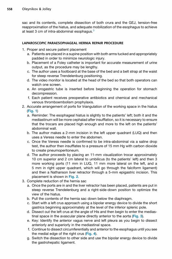
venous thromboembolism prophylaxis.2. Accurate arrangement of ports for triangulation of the working space in the hiatus
(Fig. 1)a. Reminder: The esophageal hiatus is slightly to the patients’ left; both it and the
mediastinumwill be more cephalad after insufflation, so it is necessary to ensurethat the trocars are placed high enough and more to the left on the patients’abdominal wall.
b. The author makes a 2-mm incision in the left upper quadrant (LUQ) and thenuses a Veress needle to enter the abdomen.
c. Once the Veress needle is confirmed to be intra-abdominal via a saline droptest, the author then insufflates to a pressure of 15 mm Hg with carbon dioxideto create pneumoperitoneum.
d. The author proceeds by placing an 11-mm visualization trocar approximately10 cm superior and 2 cm lateral to umbilicus (to the patients’ left) and then 3more working ports (11 mm in LUQ, 11 mm more lateral on the left, and a5 mm in right upper quadrant, which will go through the falciform ligament)and then a Nathanson liver retractor through a 5-mm epigastric incision. Thisplacement is shown in Fig. 2.
3. Complete reduction of the hernia saca. Once the ports are in and the liver retractor has been placed, patients are put in
steep reverse Trendelenburg and a right-side-down position to optimize theview of the hiatus.
b. Pull the contents of the hernia sac down below the diaphragm.c. Start with a left crus approach using a bipolar energy device to divide the short
gastrics beginning approximately at the level of the inferior splenic pole.d. Dissect out the left crus at the angle of His and then begin to enter the medias-
tinal space in the avascular plane directly anterior to the aorta (Fig. 3).e. Key: Identify the anterior vagus nerve and left pleura as you begin to dissect
anteriorly and superiorly in the mediastinal space.f. Continue to dissect circumferentially and anterior to the esophagus until you see
the medial edge of the right crus (Fig. 4).g. Switch the dissection to other side and use the bipolar energy device to divide
the gastrohepatic ligament.
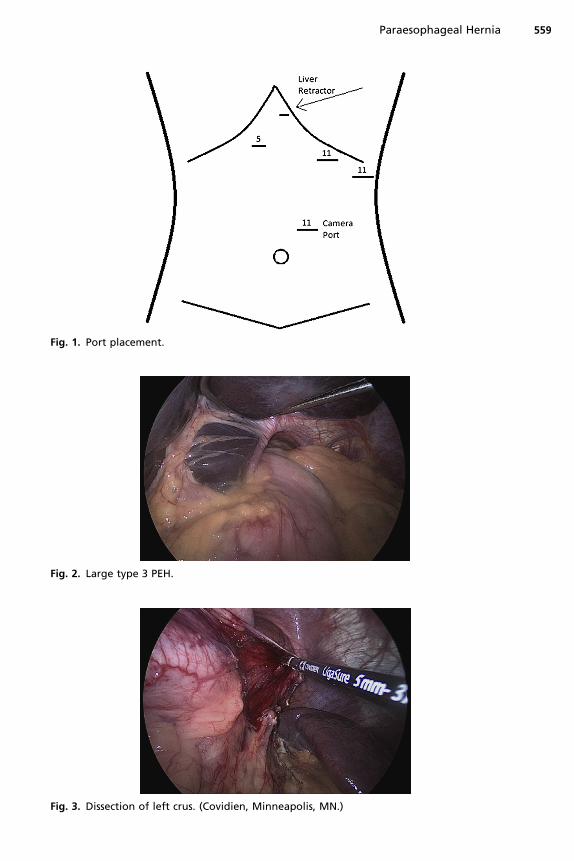
Fig. 1. Port placement.
Fig. 2. Large type 3 PEH.
Fig. 3. Dissection of left crus. (Covidien, Minneapolis, MN.)
Paraesophageal Hernia 559
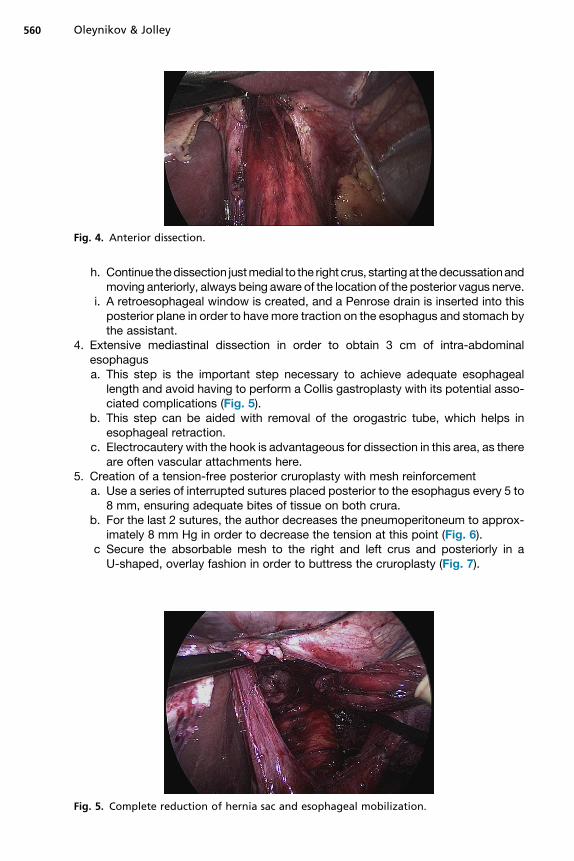
Fig. 4. Anterior dissection.
Oleynikov & Jolley560
h. Continue thedissection justmedial to the right crus, startingat thedecussationandmoving anteriorly, always being aware of the location of the posterior vagus nerve.
i. A retroesophageal window is created, and a Penrose drain is inserted into thisposterior plane in order to havemore traction on the esophagus and stomach bythe assistant.
4. Extensive mediastinal dissection in order to obtain 3 cm of intra-abdominalesophagusa. This step is the important step necessary to achieve adequate esophageal
length and avoid having to perform a Collis gastroplasty with its potential asso-ciated complications (Fig. 5).
b. This step can be aided with removal of the orogastric tube, which helps inesophageal retraction.
c. Electrocautery with the hook is advantageous for dissection in this area, as thereare often vascular attachments here.
5. Creation of a tension-free posterior cruroplasty with mesh reinforcementa. Use a series of interrupted sutures placed posterior to the esophagus every 5 to
8 mm, ensuring adequate bites of tissue on both crura.b. For the last 2 sutures, the author decreases the pneumoperitoneum to approx-
imately 8 mm Hg in order to decrease the tension at this point (Fig. 6).c Secure the absorbable mesh to the right and left crus and posteriorly in a
U-shaped, overlay fashion in order to buttress the cruroplasty (Fig. 7).
Fig. 5. Complete reduction of hernia sac and esophageal mobilization.
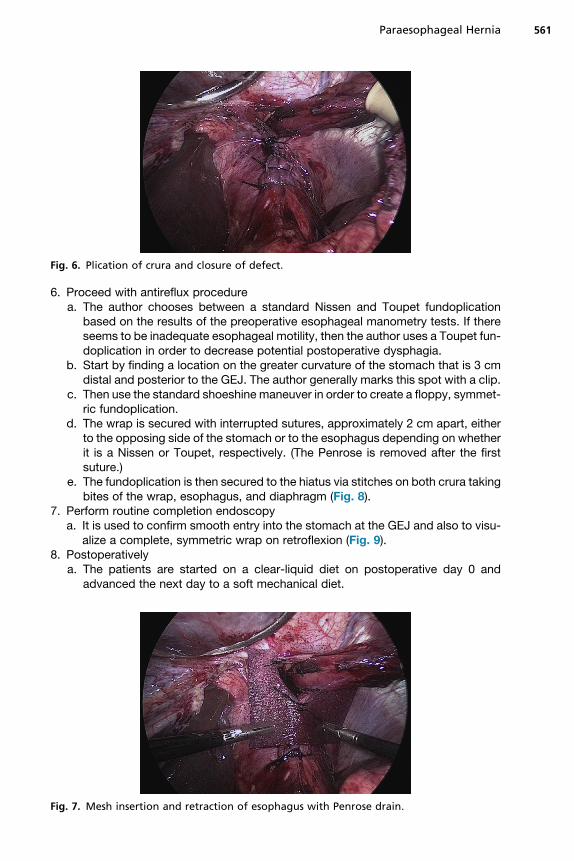
Fig. 6. Plication of crura and closure of defect.
Paraesophageal Hernia 561
6. Proceed with antireflux procedurea. The author chooses between a standard Nissen and Toupet fundoplication
based on the results of the preoperative esophageal manometry tests. If thereseems to be inadequate esophageal motility, then the author uses a Toupet fun-doplication in order to decrease potential postoperative dysphagia.
b. Start by finding a location on the greater curvature of the stomach that is 3 cmdistal and posterior to the GEJ. The author generally marks this spot with a clip.
c. Then use the standard shoeshine maneuver in order to create a floppy, symmet-ric fundoplication.
d. The wrap is secured with interrupted sutures, approximately 2 cm apart, eitherto the opposing side of the stomach or to the esophagus depending on whetherit is a Nissen or Toupet, respectively. (The Penrose is removed after the firstsuture.)
e. The fundoplication is then secured to the hiatus via stitches on both crura takingbites of the wrap, esophagus, and diaphragm (Fig. 8).
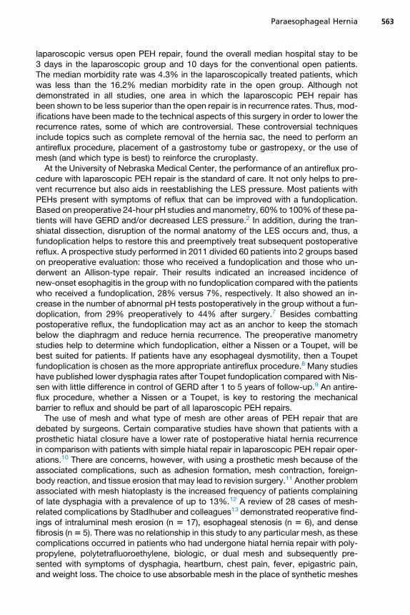
7. Perform routine completion endoscopya. It is used to confirm smooth entry into the stomach at the GEJ and also to visu-
alize a complete, symmetric wrap on retroflexion (Fig. 9).8. Postoperatively
a. The patients are started on a clear-liquid diet on postoperative day 0 andadvanced the next day to a soft mechanical diet.
Fig. 7. Mesh insertion and retraction of esophagus with Penrose drain.
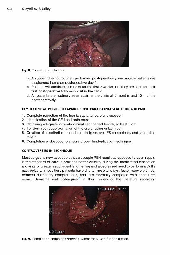
Fig. 8. Toupet fundoplication.
Oleynikov & Jolley562
b. An upper GI is not routinely performed postoperatively, and usually patients aredischarged home on postoperative day 1.
c. Patients will continue a soft diet for the first 2 weeks until they are seen for theirfirst postoperative follow-up visit in the clinic.
d. All patients are routinely seen again in the clinic at 6 months and 12 monthspostoperatively.
KEY TECHNICAL POINTS IN LAPAROSCOPIC PARAESOPHAGEAL HERNIA REPAIR
1. Complete reduction of the hernia sac after careful dissection2. Identification of the GEJ and both crura3. Obtaining adequate intra-abdominal esophageal length, at least 3 cm4. Tension-free reapproximation of the crura, using onlay mesh5. Creation of an antireflux procedure to help restore LES competency and secure the
repair6. Completion endoscopy to ensure proper fundoplication technique
CONTROVERSIES IN TECHNIQUE
Most surgeons now accept that laparoscopic PEH repair, as opposed to open repair,is the standard of care. It provides better visibility during the mediastinal dissectionallowing for greater esophageal lengthening and a decreased need to perform a Collisgastroplasty. In addition, patients have shorter hospital stays, faster recovery times,reduced pulmonary complications, and less morbidity compared with open PEHrepair. Draaisma and colleagues,6 in their review of the literature regarding
Fig. 9. Completion endoscopy showing symmetric Nissen fundoplication.
Paraesophageal Hernia 563
laparoscopic versus open PEH repair, found the overall median hospital stay to be3 days in the laparoscopic group and 10 days for the conventional open patients.The median morbidity rate was 4.3% in the laparoscopically treated patients, whichwas less than the 16.2% median morbidity rate in the open group. Although notdemonstrated in all studies, one area in which the laparoscopic PEH repair hasbeen shown to be less superior than the open repair is in recurrence rates. Thus, mod-ifications have been made to the technical aspects of this surgery in order to lower therecurrence rates, some of which are controversial. These controversial techniquesinclude topics such as complete removal of the hernia sac, the need to perform anantireflux procedure, placement of a gastrostomy tube or gastropexy, or the use ofmesh (and which type is best) to reinforce the cruroplasty.At the University of Nebraska Medical Center, the performance of an antireflux pro-
cedure with laparoscopic PEH repair is the standard of care. It not only helps to pre-vent recurrence but also aids in reestablishing the LES pressure. Most patients withPEHs present with symptoms of reflux that can be improved with a fundoplication.Based on preoperative 24-hour pH studies andmanometry, 60% to 100%of these pa-tients will have GERD and/or decreased LES pressure.2 In addition, during the tran-shiatal dissection, disruption of the normal anatomy of the LES occurs and, thus, afundoplication helps to restore this and preemptively treat subsequent postoperativereflux. A prospective study performed in 2011 divided 60 patients into 2 groups basedon preoperative evaluation: those who received a fundoplication and those who un-derwent an Allison-type repair. Their results indicated an increased incidence ofnew-onset esophagitis in the group with no fundoplication compared with the patientswho received a fundoplication, 28% versus 7%, respectively. It also showed an in-crease in the number of abnormal pH tests postoperatively in the group without a fun-doplication, from 29% preoperatively to 44% after surgery.7 Besides combattingpostoperative reflux, the fundoplication may act as an anchor to keep the stomachbelow the diaphragm and reduce hernia recurrence. The preoperative manometrystudies help to determine which fundoplication, either a Nissen or a Toupet, will bebest suited for patients. If patients have any esophageal dysmotility, then a Toupetfundoplication is chosen as the more appropriate antireflux procedure.8 Many studieshave published lower dysphagia rates after Toupet fundoplication compared with Nis-sen with little difference in control of GERD after 1 to 5 years of follow-up.9 An antire-flux procedure, whether a Nissen or a Toupet, is key to restoring the mechanicalbarrier to reflux and should be part of all laparoscopic PEH repairs.The use of mesh and what type of mesh are other areas of PEH repair that are
debated by surgeons. Certain comparative studies have shown that patients with aprosthetic hiatal closure have a lower rate of postoperative hiatal hernia recurrencein comparison with patients with simple hiatal repair in laparoscopic PEH repair oper-ations.10 There are concerns, however, with using a prosthetic mesh because of theassociated complications, such as adhesion formation, mesh contraction, foreign-body reaction, and tissue erosion that may lead to revision surgery.11 Another problemassociated with mesh hiatoplasty is the increased frequency of patients complainingof late dysphagia with a prevalence of up to 13%.12 A review of 28 cases of mesh-related complications by Stadlhuber and colleagues13 demonstrated reoperative find-ings of intraluminal mesh erosion (n 5 17), esophageal stenosis (n 5 6), and densefibrosis (n5 5). There was no relationship in this study to any particular mesh, as thesecomplications occurred in patients who had undergone hiatal hernia repair with poly-propylene, polytetrafluoroethylene, biologic, or dual mesh and subsequently pre-sented with symptoms of dysphagia, heartburn, chest pain, fever, epigastric pain,and weight loss. The choice to use absorbable mesh in the place of synthetic meshes
Oleynikov & Jolley564
is to minimize some of these morbidities. In the literature review by Antoniou and col-leagues,12 porcine dermal collagen mesh was shown by several studies to havedecreased adhesion formation and fibrosis and enhanced neovascularization, whichdemonstrate the advantages of biologic implants over synthetic meshes. Oelschlagerand colleagues14 investigated the use of porcine small intestinal submucosa as a bio-logic mesh to supplement laparoscopic PEH repair in a multicenter, prospective trialconsisting of 108 patients who were randomized to repair with or without this onlay,keyhole-shaped biologic mesh. After 6 months, 90% of the study group underwentupper gastrointestinal series testing; the results indicated a significant reduction inrecurrence rates in the mesh patient population versus the nonmesh group (9%compared with 24%, respectively) without any mesh-related complications.12,14
One study by Schmidt and colleagues15 compared mesh repair (onlay biologic) versusprimary suture repair among 70 patients with small hiatal defects less than 5 cm. At1 year after surgery, the patients were studied with a barium swallow and/or EGD toassess for recurrence. For the suture cruroplasty group, 5 of 32 patients (16%) demon-strated recurrence, whereas none of the 38 patients that had crural reinforcement withabsorbable mesh recurred (P 5 .017). Lee and colleagues16 studied the results of us-ing human acellular dermal matrix mesh in laparoscopic hiatal hernia repair in 52 pa-tients after 1 year and demonstrated a recurrence rate of only 3.8% and no mesh-related complications. An absorbable mesh cruroplasty helps to decrease the earlyrecurrence rate of PEHs after laparoscopic repair without increasing the complicationrate. Further studies are necessary to help determine the long-term recurrence ratesand their significance after laparoscopic PEH repair with absorbable mesh.
SUMMARY
The treatment of PEHs is challenging. They tend to occur in patients in their 60s and 70swith multiple medical problems and a variety of associated symptoms. Detailed preop-erative evaluation is crucial to determining a safe and effective strategy for repair in theoperating room. Laparoscopic PEH repair has shown to be advantageous comparedwith conventional open repairwith regard to hospital stay, recovery time, anddecreasedcomplications. Although some results indicate there are higher recurrence rates in lapa-roscopic PEH repair, the clinical significance of these recurrences has not yet beendetermined. In order to maximize the efficacy of this procedure, modifications haveemerged, such as performing a fundoplication and using an absorbable mesh onlayto reinforce thecruroplasty.Althoughmoreprospective, randomizedstudiesareneededto support the superior results of these surgical adjuncts, laparoscopic PEH repair withan antireflux procedure and absorbable mesh should be the current standard of care.
REFERENCES
1. Arafat FO, Teitelbaum EN, Hungness ES. Modern treatment of paraesophagealhernia: preoperative evaluation and technique for laparoscopic repair. SurgLaparosc Endosc Percutan Tech 2012;22(4):297–303.
2. Auyang E, Oelschlager B. Laparoscopic Paraesophageal Hernia Repair. In:Swanstrom L, Soper N, editors. Mastery of Endoscopic and Laparoscopic Sur-gery, Chapter 23. Philadelphia: Lippincott Williams & Wilkins; 2013. p. 222–30.
3. Oleynikov D, Ranade A, Parcells J, et al. Paraesophageal Hernias and NissenFundoplication. In: Oleynikov D, Bills ND, editors. Robotic Surgery for the GeneralSurgeon, Chapter 6. Hauppauge (NY): Nova Science Publishers; 2014. p. 65–74.
Paraesophageal Hernia 565
4. Tiwari M, Tsang A, Reynoso J, et al. Options for large hiatal defects. In:Murayama KM, Chand B, Kothari S, et al, editors. Evidence-Based Approachto Minimally Invasive Surgery. Woodbury (CT): Cine-Med, Inc; 2011. p. 47–56.
5. Maganty K, Smith RL. Cameron lesions: unusual cause of gastrointestinalbleeding and anemia. Digestion 2008;77(3–4):214–7.
6. Draaisma WA, Gooszen HG, Tournoij E, et al. Controversies in paraesophagealhernia repair: a review of literature. Surg Endosc 2005;19(10):1300–8.
7. DeMeester SR. Laparoscopic paraesophageal hernia repair: critical steps andadjunct techniques to minimize recurrence. Surg Laparosc Endosc PercutanTech 2013;23(5):429–35.
8. Soper NJ, Teitelbaum EN. Laparoscopic paraesophageal hernia repair: currentcontroversies. Surg Laparosc Endosc Percutan Tech 2013;23(5):442–5.
9. Stefanidis D, Hope WW, Kohn GP, et al. Guidelines for surgical treatment ofgastroesophageal reflux disease. Surg Endosc 2010;24(11):2647–69.
10. Granderath FA, Carlson MA, Champion JK, et al. Prosthetic closure of the esoph-ageal hiatus in large hiatal hernia repair and laparoscopic antireflux surgery. SurgEndosc 2006;20(3):367–79.
11. Davis SS Jr. Current controversies in paraesophageal hernia repair. Surg ClinNorth Am 2008;88(5):959–78, vi.
12. Antoniou SA, Pointner R, Granderath FA. Hiatal hernia repair with the use of bio-logic meshes: a literature review. Surg Laparosc Endosc Percutan Tech 2011;21(1):1–9.
13. Stadlhuber RJ, Sherif AE, Mittal SK, et al. Mesh complications after prostheticreinforcement of hiatal closure: a 28-case series. Surg Endosc 2009;23(6):1219–26.
14. Oelschlager BK, Barreca M, Chang L, et al. The use of small intestine submucosain the repair of paraesophageal hernias: initial observations of a new technique.Am J Surg 2003;186(1):4–8.
15. Schmidt E, Shaligram A, Reynoso JF, et al. Hiatal hernia repair with biologic meshreinforcement reduces recurrence rate in small hiatal hernias. Dis Esophagus2014;27(1):13–7.
16. Lee YK, James E, Bochkarev V, et al. Long-term outcome of cruroplasty reinforce-ment with human acellular dermal matrix in large paraesophageal hiatal hernia.J Gastrointest Surg 2008;12(5):811–5.