Embed Size (px)
Citation preview

Parallelizable Strategy for the Estimation of the 3D Structure ofBiological Macromolecules
Claudia Caudai∗, Monica Zoppe†, Emanuele Salerno∗, Ivan Merelli‡ and Anna Tonazzini∗∗CNR, Institute of Information Science and Technologies, Via Moruzzi, 1, 56124 Pisa, Italy
Email: [email protected]†CNR, Institute of Clinical Physiology, Via Moruzzi, 1, 56124 Pisa, Italy
‡CNR, Institute of Biomedical Technologies, Via Fratelli Cervi, 93, 20090 Segrate (MI), Italy
Abstract—We present a parallelizzable, multilevel algorithmfor the study of three-dimensional structure of biologicalmacromolecules, applied to two fundamental topics: the 3Dreconstruction of Chromatin and the elaboration of motion ofproteins. For Chromatin, starting from contact data obtainedthrough Chromosome Conformation Capture techniques, ourmethod first subdivides the data matrix in biologically relevantblocks, and then treats them separately, at several levels,depending on the initial data resolution. The result is a familyof configurations for the entire fiber, each one compatiblewith both experimental data and prior knowledge aboutspecific genomes. For Proteins, the method is conceived as asolution for the problem of identifying motion and alternativeconformations to the deposited structures. The algorithm, usingquaternions, processes the main chain and the aminoacid sidechains independently; it then exploits a Monte Carlo methodfor selection of biologically acceptable conformations, based onenergy evaluation, and finally returns a family of conformationsand of trajectories at single atom resolution.
Keywords-Parallel Computing; Macromolecules; Hierarchi-cal Reconstruction; Chromatin; Protein Motion.
I. INTRODUCTION
The need to observe the structure of biological macro-molecules at multiple resolution levels emerged in recentyears, following major advances in investigating the three-dimensional structure of proteins and nucleic acids. Aprimary example of this approach is represented by thestudy of chromatin, which resulted in the identification ofTADs (Topologically Associating Domains) [1], portions ofchromatin with many internal and few external interactions,described as fairly isolated structures within the fibre. For the3D reconstruction of chromatin structure using ChromosomeConformation Capture and other HI-C data [2]–[4] it istherefore possible and convenient to deal in parallel withthe TAD structures and then reconstruct and investigate thestructure of the chromatin in its entirety, adopting a coarse-grained approach that follows the most natural division.Another important topic in biology is the recognition of thedynamic nature of protein activity. Because at this time itis impossible to observe protein movements experimentally,it is relevant to speed up the methods for the analysisof their structural and conformational changes. Treating inparallel autonomous or partially independent sub-structures
is a smart strategy for the analysis of three-dimensionalbiological structures. For example, in the case of proteins,the subdivision of the chain in amino acids provides thesmallest units for computation.This approach leads to consider biological phenomena atmultiple levels of resolution. Organic structures are oftenfractal, which is why coarse-grained methods are successfulin biology. Even better than coarse-grained methods, multi-level methods can be used to observe biological structuresat different resolutions. The organization of chromatin intoTADs, for example, leads to overcome the two-dimensionalmodel, and to the description of DNA as a pattern of nestedstructures in which TADs and sub-TADs form a complexhierarchy. [5].Following this approach, we propose an iterative and mul-tilevel parallelizable algorithm for the investigation of thethree-dimensional structure of macromolecules.
II. METHOD
The method we propose, whose code flow is illustratedin Algorithm 1, is a parallelizable and multilevel algorithm.The molecular chain is modelled as a bead-chain,maintaining the biological order of the sub-structures.The algorithm always starts from the highest possibleresolution, named INPUTCHAIN in Algorithm 1, andsubdivides the global structure into sub-structures trying torespect as much as possible the natural subdivisions alreadypresent. Sub-structures are processed in parallel, lookingfor configurations compatible with a-priori constraints andwith low potential energy. The molecular chain is thenrecomposed and evaluated from a structural and energeticpoint of view. If successive levels of resolution exist, thechain is modelled with coarse-grained techniques and thealgorithm repeats, as illustrated in Algorithm 1. At theend of the reconstruction a final configuration is proposed,named OUTPUTCHAIN in Algorithm 1. The coordinatesof all the sub-structures are recovered, up to the highestresolution, so as to provide a tool to explore macromoleculesin their entirety at multiple levels of resolution.
Algorithm 1 Code FlowInput: inputchain
The input is the molecular chain at the highest resolution1: nlevel= 0
Initialisation at level=0:2: inputchain=C3: nchains=[len(C)]4: while nchains[nlevel]>1 do5: subchains=dividechain(nlevel,C)
C is a string and subchains is an array of strings6: nchains[nlevel]=len(subchains)7: nchains.append(nchains[nlevel])
Parallel Part:8: for i in range(nchains[nlevel]) do9: Ci =annealing(subchains[i])
10: end forIncrease level:
11: nlevel=nlevel+112: end while
Compute coordinates of the highest resolution chain:13: outputchain=compose(Ci)14: return outputchain
Figure 1. Heatmap of contact frequency matrix of Chromosome 16 at100kbp resolution. Multilevel subdivision in diagonal blocks was performedby our algorithm. Note the repeating pattern in the zoom of a block.
III. EXPERIMENTS
Our method is suited for both the 3D reconstruction ofchromatin, starting from HI-C data, and for the elaborationof Molecular Dynamics of proteins [6], as shown below.
A. 3D Chromatin Conformation
To obtain 3D Chromatin conformations consistent withinformation stored in contact frequency matrices derivedfrom HI-C experiments [7], we introduce a chromatinmodel consisting of a chain of consecutive and partiallypenetrable beads whose properties (bead size, elasticity,curvature, etc.) are suitably constrained. Our algorithm[8] automatically divides the contact matrix in diagonal
blocks, dividing the chain into sub-chains corresponding toTADs and sub-TADs. The method is parallel and iterative:starting from the highest resolution level, the algorithmelaborates the smallest sub-chains in parallel, using aMonte Carlo method [9]; it then re-iterates on the chain atsuccessive resolution, in which conformations obtained inthe previous level are modeled as beads with coarse-grainmethods. Perturbations and movements of the chains areperfomed by using quaternions, a mathematical devicemore efficient than Euler matrices in managing rotations,as fully explained in [10]. In the algorithm the data-fitfuncion rewards the proximity of beads with high contactfrequencies and avoids deep interpenetartions. The processis repeated for every resolution level up to the lowest one.At the end the whole chain is modelled and the structurecan be investigated at the highest possible resolution.Starting from a contact matrix, several final conformationsare produced, in order to sample the space of possibleconformations fitting with initial contact information. Afundamental step in the analysis and processing of datafrom HI-C experiments is the division in blocks of thecontact matrix. This subdivision is based on an intrinsicproperty of DNA, which can be conceptually subdivided intocontiguous domains, called TADs, consisting of compactspatial modules. This pattern suggests that the spatialorganization of DNA follows a hierarchical model. In Figure1 a heat-map of the contact matrix at 100kbp resolutionof chormosome 16, obtained from freely available HI-Cdata of Rao [11] is represented; diagonal blocks with highcontact frequency can be easily distinguished by eye; azoom of one of these blocks shows that this pattern ismultilevel. The TAD identification step is crucial in 3DChromatin reconstruction and can be performed followingdifferent principles; our block detection method is basedon the algorithm of [12], and leads to a nested seriesof blocks, reflecting the hierarchical nature of Chromatincompaction in cellular nuclei [13]. As an example, in Figure2 we report some final configurations of Chromosome 16,obtained starting form contact matrices binned at differentresolutions. The higher the resolution, the higher the sizeof the contact matrix and consequently the compuationalcomplexity of the system. In the most complex scenario, thecontact matrix can be divided into numerous small blocks,and at several levels. These are computed in parallel,witheach sub-chain computed independently, in order to reducecalculation times, as illustrated in Table I. By computingevery sub-chain in parallel with different processors,calculation times decrease fast, because on one side thereis no exchange of information within the sub-chains ofeach level, and on the other side most of the CPU timeis used for sub-chains calculations. Treating sub-chains inseries the total elapsed time is the sum of elapsed timesof every sub-chain of every resolution level. By contrast,computing sub-chains in parallel (in different processors),
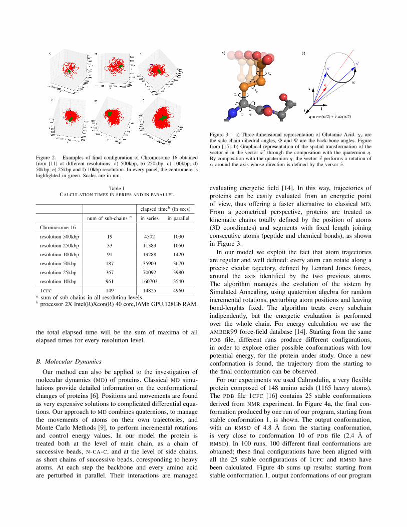
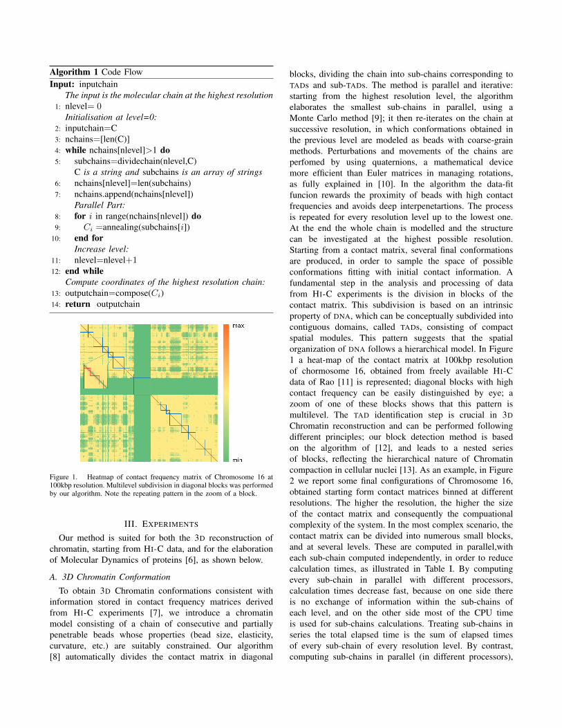
Figure 2. Examples of final configuration of Chromosome 16 obtainedfrom [11] at different resolutions: a) 500kbp, b) 250kbp, c) 100kbp, d)50kbp, e) 25kbp and f) 10kbp resolution. In every panel, the centromere ishighlighted in green. Scales are in nm.
Table ICALCULATION TIMES IN SERIES AND IN PARALLEL
elapsed timeb (in secs)
num of sub-chains a in series in parallel
Chromosome 16
resolution 500kbp 19 4502 1030
resolution 250kbp 33 11389 1050
resolution 100kbp 91 19288 1420
resolution 50kbp 187 35903 3670
resolution 25kbp 367 70092 3980
resolution 10kbp 961 160703 3540
1CFC 149 14825 4960a sum of sub-chains in all resolution levels.b processor 2X Intel(R)Xeon(R) 40 core,16Mb GPU,128Gb RAM.
the total elapsed time will be the sum of maxima of allelapsed times for every resolution level.
B. Molecular Dynamics
Our method can also be applied to the investigation ofmolecular dynamics (MD) of proteins. Classical MD simu-lations provide detailed information on the conformationalchanges of proteins [6]. Positions and movements are foundas very expensive solutions to complicated differential equa-tions. Our approach to MD combines quaternions, to managethe movements of atoms on their own trajectories, andMonte Carlo Methods [9], to perform incremental rotationsand control energy values. In our model the protein istreated both at the level of main chain, as a chain ofsuccessive beads, N-CA-C, and at the level of side chains,as short chains of successive beads, coresponding to heavyatoms. At each step the backbone and every amino acidare perturbed in parallel. Their interactions are managed
Figure 3. a) Three-dimensional representation of Glutamic Acid. χi arethe side chain dihedral angles, Φ and Ψ are the back-bone angles. Figurefrom [15]. b) Graphical representation of the spatial transformation of thevector ~x in the vector ~x′ through the composition with the quaternion q.By composition with the quaternion q, the vector ~x performs a rotation ofα around the axis whose direction is defined by the versor v.
evaluating energetic field [14]. In this way, trajectories ofproteins can be easily evaluated from an energetic pointof view, thus offering a faster alternative to classical MD.From a geometrical perspective, proteins are treated askinematic chains totally defined by the position of atoms(3D coordinates) and segments with fixed length joiningconsecutive atoms (peptide and chemical bonds), as shownin Figure 3.
In our model we exploit the fact that atom trajectoriesare regular and well defined: every atom can rotate along aprecise cicular tajectory, defined by Lennard Jones forces,around the axis identified by the two previous atoms.The algorithm manages the evolution of the sistem bySimulated Annealing, using quaternion algebra for randomincremental rotations, perturbing atom positions and leavingbond-lenghts fixed. The algorithm treats every subchainindipendently, but the energetic evaluation is performedover the whole chain. For energy calculation we use theAMBER99 force-field database [14]. Starting from the samePDB file, different runs produce different configurations,in order to explore other possible conformations with lowpotential energy, for the protein under study. Once a newconformation is found, the trajectory from the starting tothe final conformation can be observed.
For our experiments we used Calmodulin, a very flexibleprotein composed of 148 amino acids (1165 heavy atoms).The PDB file 1CFC [16] contains 25 stable conformationsderived from NMR experiment. In Figure 4a, the final con-formation produced by one run of our program, starting fromstable conformation 1, is shown. The output conformation,with an RMSD of 4.8 A from the starting conformation,is very close to conformation 10 of PDB file (2,4 A ofRMSD). In 100 runs, 100 different final conformations areobtained; these final configurations have been aligned withall the 25 stable configurations of 1CFC and RMSD havebeen calculated. Figure 4b sums up results: starting fromstable conformation 1, output conformations of our program

Figure 4. a) Structures of Calmodulin: in yellow the conformation 1 ofthe PDB file 1CFC, used as input of our program; in red one of the outputconfigurations; in light violet the conformation 10 of the PDB file 1CFC,very similar to the output configuration. b) Pie-plot of the correspondenceof the outputs of 100 runs with stable conformations of Calmodulin foundin PDB file 1CFC.
result close (RMSD between 2.6 and 3.4 A) to 8 stableconformations present in the 1CFC PDB file.
Treating every aminoacid and back-bone in parallelwith different processors, calculation times decrease, asshown in Table I. Reducing calculation times in MD ischallenging and we propose a smart and fast approach to theinvestigation of the possible trajectories and conformationchanges of proteins.
IV. CONCLUSION
In conclusion, we present here an algorithmic method tocompute some of the most challenging and interesting as-pects of biology: chromatin structure and protein dynamics.The methods, despite the obvious differences related to thenature of peptides and nucleic acids, have some featuresin common, including the use of quaternions to introducerotations, and, most prominent, the possibility of computingsub-structures in an independent, and hence parallelizable,way. For DNA the smallest units are the smallest TADsthat can be identified from the contact matrix; these canbe so small, that a second, third and higher order of TADmight be necessary for the 3D reconstruction of the completesequence.For proteins the smallest units are the single aminoacids,each of which is characterized by its specific set of parame-ters, derived from the study of known protein structures. Themassive parallelization introduced by the method, allowsfor a significant reduction in the computation time for eachprocess under study, making it possible to produce a largenumber of solutions, as it is common (and necessary) inbiological studies. At the same time, splitting the problemsinto smaller units does not preclude the incorporation offurther information at the successive levels, as demonstratedin the study of human Chromosome 16.
ACKNOWLEDGMENT
This work has been partially supported by the ItalianFlagship Project InterOmics, WP01-ISTI, and by ISTI-CNR,through scientific agreement with ITB-CNR, Milan.
REFERENCES
[1] J. R. Dixon et al., “Topological domains in mammaliangenomes identified by analysis of chromatin interactions,”Nature, vol. 485, pp. 376–380, 2012.
[2] J. Dekker, K. Rippe, M. Dekker, and N. Kleckner, “Capturingchromosome conformation,” Science, vol. 295, pp. 1306–1311, 2002.
[3] J. Fraser et al., “Chromatin conformation signatures of cellu-lar differentiation,” Genome Biology, vol. 10, p. 37, 2009.
[4] S. Wang, J. Xu, and J. Zeng, “Inferential modeling of 3dchromatin structure,” Nucl. Ac. Res., vol. 43, p. e54, 2015.
[5] M. Forcato, C. Nicoletti, P. Koustav, C. M. Livi, F. Ferrari,and S. Bicciato, “Comparison of computational methods forhi-c data analysis,” Nature Methods, vol. 14, p. 679685, 2017.
[6] J. D. Hirst, D. R. Glowacki, and M. Baaden, “Molecularsimulations and visualization: introduction and overview,”Faraday Discuss, vol. 169, pp. 9–22, 2014.
[7] E. Lieberman-Aiden et al., “Comprehensive mapping of long-range interactions reveals folding principles of the humangenome,” Science, vol. 326, pp. 289–293, 2009.
[8] C. Caudai, E. Salerno, M. Zoppe, and A. Tonazzini, “Inferring3d chromatin structure using a multiscale approach based onquaternions,” BMC Bioinformatics, vol. 16, p. 234, 2015.
[9] M. Rousseau, J. Fraser, M. A. Ferraiuolo, J. Dostie, andM. Blanchette, “Three-dimensional modeling of chromatinstructure from interaction frequency data using markov chainmonte carlo sampling,” BMC Bioinf., vol. 12, pp. 414–429,2011.
[10] C. F. Karney, “Quaternions in molecular modeling,” J. Mol.Graph. Model., vol. 25, pp. 595–604, 2007.
[11] S. S. Rao et al., Cell, vol. 159, p. 16651680, 2014.
[12] B. R. Lajoie, J. Dekker, and N. Kaplan, “The hitchhiker’sguide ti hi-c analysis: Practical guidelines,” Methods, vol. 72,pp. 65–75, 2015.
[13] C. Caudai, E. Salerno, M. Zoppe, and A. Tonazzini, “Estima-tion of the spatial chromatin structure based on a multireso-lution bead-chain model,” IEEE/ACM TBCC, 2018, in press,doi: 10.1109/TCBB.2018.2791439.
[14] W. D. Cornell et al., “A second generation force field for thesimulation of proteins, nucleic acids, and organic molecules,”J Am Chem Soc, vol. 118(9), pp. 2309–2309, 1996.
[15] T. Harder et al., “Beyond rotamers: a generative, probabilisticmodel of side chains in proteins,” BMC Bioinformatics,vol. 11, pp. 306–319, 2010.
[16] H. Kuboniwa, N. Tjandra, S. Grzesiek, H. Ren, C. B. Klee,and A. Bax, “Solution structure of calciumfree calmodulin,”Nat Struct Biol, vol. 2(9), pp. 768–776, 1995.