Embed Size (px)
Citation preview
Brief Report
Vol. 30, No. 4, 2018 503
Received June 16, 2017, Revised September 8, 2017, Accepted for publication September 18, 2017
Corresponding author: Je-Ho Mun, Department of Dermatology, Seoul National University College of Medicine, 101 Daehak-ro, Jongno-gu, Seoul 03080, Korea. Tel: 82-2-2072-1996, Fax: 82-2-742-7344, E-mail: [email protected]: https://orcid.org/0000-0002-0734-2850
This is an Open Access article distributed under the terms of the Creative Commons Attribution Non-Commercial License (http://creativecommons.org/ licenses/by-nc/4.0) which permits unrestricted non-commercial use, distribution, and reproduction in any medium, provided the original work is properly cited.
Copyright © The Korean Dermatological Association and The Korean Society for Investigative Dermatology
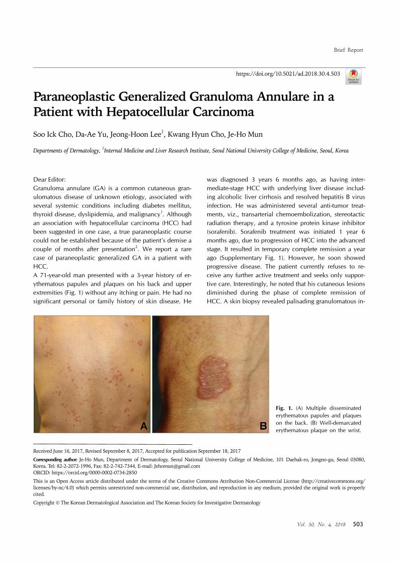
Fig. 1. (A) Multiple disseminated erythematous papules and plaques on the back. (B) Well-demarcated erythematous plaque on the wrist.
https://doi.org/10.5021/ad.2018.30.4.503
Paraneoplastic Generalized Granuloma Annulare in a Patient with Hepatocellular Carcinoma
Soo Ick Cho, Da-Ae Yu, Jeong-Hoon Lee1, Kwang Hyun Cho, Je-Ho Mun
Departments of Dermatology, 1Internal Medicine and Liver Research Institute, Seoul National University College of Medicine, Seoul, Korea
Dear Editor:Granuloma annulare (GA) is a common cutaneous gran-ulomatous disease of unknown etiology, associated with several systemic conditions including diabetes mellitus, thyroid disease, dyslipidemia, and malignancy1. Although an association with hepatocellular carcinoma (HCC) had been suggested in one case, a true paraneoplastic course could not be established because of the patient’s demise a couple of months after presentation2. We report a rare case of paraneoplastic generalized GA in a patient with HCC.A 71-year-old man presented with a 3-year history of er-ythematous papules and plaques on his back and upper extremities (Fig. 1) without any itching or pain. He had no significant personal or family history of skin disease. He
was diagnosed 3 years 6 months ago, as having inter-mediate-stage HCC with underlying liver disease includ-ing alcoholic liver cirrhosis and resolved hepatitis B virus infection. He was administered several anti-tumor treat-ments, viz., transarterial chemoembolization, stereotactic radiation therapy, and a tyrosine protein kinase inhibitor (sorafenib). Sorafenib treatment was initiated 1 year 6 months ago, due to progression of HCC into the advanced stage. It resulted in temporary complete remission a year ago (Supplementary Fig. 1). However, he soon showed progressive disease. The patient currently refuses to re-ceive any further active treatment and seeks only suppor-tive care. Interestingly, he noted that his cutaneous lesions diminished during the phase of complete remission of HCC. A skin biopsy revealed palisading granulomatous in-

Brief Report
504 Ann Dermatol
Fig. 2. (A) Skin biopsy showing normal epidermis with palisaded granulomatous inflammation in the upper dermis (H&E, ×40). (B) Palisading histiocytes surrounding collagen bundles (H&E, ×100).
flammation with histiocytic infiltration surrounding colla-gen bundles in the dermis (Fig. 2). Based on these find-ings, the patient was diagnosed as having generalized GA and was subsequently treated with 0.25% topical pre-dnicarbate ointment.GA is a benign granulomatous skin disease that usually presents as annular skin-colored to erythematous papules localized to the dorsal aspects of the hands or feet. There are four clinical variants: localized, generalized, subcuta-neous and perforating. According to a multicenter study analyzed 54 patients with generalized GA, generalized GA usually occurred on the trunk and extremities. These can be differentiated from the localized type in that they were observed in an older age group, had widespread dis-tribution, and were refractory to therapy3. Several inves-tigators have suggested a relationship between GA and other systemic diseases, most commonly diabetes mellitus. Additionally, GA is reported to be associated with malignancies, including hematopoietic cancers and adenocarcinomas; however, an association between GA and liver disease has hardly been reported1,4. Mestre et al.2 reported a case of generalized GA in a patient with HCC, although the authors failed to establish a temporal relationship between GA and the malignancy owing to the patient’s demise. The case of Mestre et al.2 showed mixed papules and annular plaques, similar to the present case. The paraneoplastic variant of GA shows a poor response to conventional GA treatment, although it resolves sponta-neously after removal of the underlying neoplasm5. The possible mechanism of paraneoplastic GA includes tumor antigens triggering an immunological response, which re-sults in a granulomatous reaction2,5.In conclusion, we report the second case of generalized GA in a patient with HCC, in whom GA demonstrated a paraneoplastic course, as lesions diminished during the temporary phase of remission of the HCC treated with
sorafenib. Therefore, the cutaneous lesions were presum-ably caused by a paraneoplastic syndrome from an in-ternal malignancy. In elderly patients demonstrating gen-eralized GA, dermatologists must consider underlying ma-lignancy including HCC.
SUPPLEMENTARY MATERIALS
Supplementary data can be found via http://anndermatol. org/src/sm/ad-30-503-s001.pdf.
CONFLICT OF INTEREST
The authors have nothing to disclose.
REFERENCES
1. Thornsberry LA, English JC 3rd. Etiology, diagnosis, and
therapeutic management of granuloma annulare: an update.
Am J Clin Dermatol 2013;14:279-290. 2. Mestre T, Rodrigues AM, Cardoso J. Disseminated granuloma
annulare and hepatocellular carcinoma: association or
coincidence? BMJ Case Rep 2014. doi: 10.1136/bcr-2014- 205883.
3. Yun JH, Lee JY, Kim MK, Seo YJ, Kim MH, Cho KH, et al.
Clinical and pathological features of generalized granuloma annulare with their correlation: a retrospective multicenter
study in Korea. Ann Dermatol 2009;21:113-119.
4. Alsahafi M, AlJasser MI, Kalia S, Yang HM, Ramji A. Chronic hepatitis with liver granulomas in a patient with granuloma
annulare: a case report and review of the literature. Case
Rep Gastrointest Med 2017;2017:8768529. 5. Chiu ML, Tang MB. Generalized granuloma annulare
associated with gastrointestinal stromal tumour: case report
and review of clinical features and management. Clin Exp Dermatol 2008;33:469-471.


![Perforating granuloma annulare in children: A case reportPerforating granuloma annulare. Int J Dermatol 36: 340-348. [Crossref] 4. Ratnavel RC, Norris PG (1995) Perforating granuloma](https://img.pdfslide.net/doc/110x75/608f693f0f920b09c84ee530/perforating-granuloma-annulare-in-children-a-case-report-perforating-granuloma.jpg)